Embed Size (px)
Citation preview
Neuron
NeuroResource
A Systematic Nomenclature for the Insect BrainKei Ito,1,14,* Kazunori Shinomiya,1,14,15 Masayoshi Ito,1 J. Douglas Armstrong,2 George Boyan,3 Volker Hartenstein,4
Steffen Harzsch,5 Martin Heisenberg,6 Uwe Homberg,7 Arnim Jenett,8,16 Haig Keshishian,9 Linda L. Restifo,10,17
Wolfgang Rossler,11 Julie H. Simpson,8 Nicholas J. Strausfeld,10 Roland Strauss,12 and Leslie B. Vosshall13; Insect BrainName Working Group1Institute of Molecular and Cellular Biosciences, The University of Tokyo, Bunkyo-ku, Tokyo 113-0032, Japan2School of Informatics, University of Edinburgh, Edinburgh, EH8 9AB, UK3Division of Neurobiology, Ludwig-Maximilians-University of Munich, D-82152 Martinsried, Germany4Department of Molecular, Cell and Developmental Biology, University of California, Los Angeles, Los Angeles, CA 90095, USA5Zoological Institute and Museum, University of Greifswald, D-17498 Greifswald, Germany6Rudolph-Virchow-Center, Julius-Maximilians-University of Wurzburg, D-97080 Wurzburg, Germany7Department of Biology, Animal Physiology, Philipps-University of Marburg, D-35032 Marburg, Germany8HHMI Janelia Farm Research Campus, Ashburn, VA 20147, USA9Department of Molecular, Cellular and Developmental Biology, Yale University, New Haven, CT 06511 USA10Department of Neuroscience and Center for Insect Science, University of Arizona, Tucson, AZ 85721, USA11Behavioral Physiology and Sociobiology, Biocenter, Julius-Maximilians-University of Wurzburg, D-97074 Wurzburg, Germany12Department of Zoology, Johannes-Gutenberg-University of Mainz, D-55099 Mainz, Germany13Laboratory of Neurogenetics and Behavior, HHMI-The Rockefeller University, New York, NY 10065, USA14These two authors contributed equally to the generation of the data and maps15Present address: Department of Psychology and Neuroscience, Dalhousie University, Halifax, NS B3H 4R2, Canada16Present address: Institute de Neu robiologie Alfred Fessard, Centre National de la Rechere Scientifique, 91198 Gif sur Yvette, France17Present address: Department of Neurology, Arizona Health Sciences Center, Tucson, AZ 85724, USA
*Correspondence: [email protected]
http://dx.doi.org/10.1016/j.neuron.2013.12.017
SUMMARY
Despite the importance of the insect nervous systemfor functional and developmental neuroscience,descriptions of insect brains have suffered from alack of uniform nomenclature. Ambiguous definitionsof brain regions and fiber bundles have contributedto the variation of names used to describe the samestructure. The lack of clearly determined neuropilboundaries has made it difficult to document preciselocations of neuronal projections for connectomicsstudy. To address such issues, a consortiumof neurobiologists studying arthropod brains, theInsect Brain Name Working Group, has establishedthe present hierarchical nomenclature system, usingthe brain of Drosophila melanogaster as the refer-ence framework, while taking the brains of othertaxa into careful consideration for maximum consis-tency and expandability. The following summarizesthe consortium’s nomenclature system and high-lights examples of existing ambiguities and remediesfor them. This nomenclature is intended to serve asa standard of reference for the study of the brain ofDrosophila and other insects.
INTRODUCTION
Thanks to their size, their relevance to medicine and agriculture,
and their use for the development of novel molecular genetic
techniques, insect brains have provided important insights in
neuroscience research (Burne et al., 2011). Like those of verte-
brates, the brains of insects consist of various regions that
synergistically cooperate to achieve computational tasks. These
regions are given names that facilitate descriptions of neural cir-
cuits, the projections of specific neurons, and the distribution of
molecules associated with neural functions. Existing nomencla-
tures for describing insect brains have, however, suffered from
the following crucial deficiencies: (1) comparable brain regions
have been given different names depending on the species
and the researcher studying them, (2) the same terms and words
have been used to refer to different structures, (3) the boundaries
of many brain regions have not been defined clearly, and (4)
some volumes of the brain have no established names and
thus, by definition, have no defined boundaries.
As in other sciences, neuroscience requires systematic and
consistent nomenclature. Descriptions of brain and central ner-
vous system are no exception, and many demand cartographic
discipline. Linguistic inconsistencies detract from published
studies or, worse, provide descriptions that are intelligible to
only a few. Historically, insect neuroscience has focused on
just a few well-known brain regions, such as those loosely
defined as the ‘‘optic lobes,’’ ‘‘antennal lobes,’’ ‘‘mushroom
bodies,’’ and ‘‘central complex.’’ However, comprehensive ana-
lyses of the development and connections of what are now
recognized as elaborate and complex brains assume greater
importance as whole-brain network analyses and behavioral ge-
netics promise to link organization to function (Cachero et al.,
2010; Yu et al., 2010; Chiang et al., 2011; Ito et al., 2013; Yu
et al., 2013). A systematic and consistent nomenclature that
can be applied across insect brains in general is a prerequisite
for such studies. Researchers of avian brains have faced similar
problems and have solved them by agreeing to a unified
Neuron 81, 755–765, February 19, 2014 ª2014 Elsevier Inc. 755
Neuron
Systematic Insect Brain Nomenclature
nomenclature (Jarvis et al., 2005). The present paper describes
the results of a comparable nomenclature effort by a broad
representation of neuroscientists working on the central nervous
systems of insects and related arthropods.
RESULTS
Organization of the Working GroupIn 2007, requests from two independent bodies associated with
the Howard Hughes Medical Institute (HHMI) and the National
Institutes of Health (NIH) resulted in the formation of a working
group that discussed proposals for a systematic nomenclature
of the insect brain. The Insect Neuroanatomy Meeting, held
in March 2007 at the HHMI Janelia Farm Research Campus in
Virginia, USA, brought together insect neuroscientists to discuss
ongoing studies and problems in mapping neurons and their
networks. Thismeeting acknowledged that a systematic nomen-
clature was urgently required, and an initial working group of
seven individuals was formed. In the same year, the NIH Neuro-
science Blueprint for Neuroscience Research, headed by Daniel
Gardner and coordinated with the Society for Neuroscience
Neuroinformatics Committee, invited a group of several neurobi-
ologists working on the Drosophila brain to discuss a blueprint
for a systematic nomenclature.
These two groups subsequently merged to form the Insect
Brain Name Working Group. However, because its members
mostly worked on the Drosophila nervous system, invitations
to join the working group were extended to scientists studying
the brains of other insects (e.g., locust, cockroach, moth, and
honey bee) as well as crustaceans, the sister group of insects.
The aim of the working group was expanded to evolve a nomen-
clature that could be used across arthropod species. The fly
Drosophila melanogaster is the most commonly used species
in insect neurobiology and has an acknowledged impact in
neuroscience research in the broadest sense. It is a proving
ground for developing a range of genetics-based techniques
for structural and functional analyses, and it serves as a test
bed for the development of methods and algorithms for
brain connectomics. For these reasons, the group agreed to
use the brain of Drosophila melanogaster as the basis taxon for
developing a nomenclature system. However, because it was
recognized that the brains of different insect species are a
consequence of more than 400 million years of divergent
evolution, other taxa had to be taken into consideration if the
nomenclature would apply across Insecta and reflect, as far as
possible, a ground pattern organization that is common to all.
Due to the working group’s geographical dispersion, discus-
sions were carried out via an online mailing list initiated in early
2007. In addition to the NIH Neuroscience Blueprint meeting
that was held in New York in 2008, the group twice held 2-day
face-to-face meetings in 2008 and 2010 at the Janelia Farm
Research Campus. Online discussions throughout the process
further fine-tuned the present nomenclature. During the entire
time course of this project, the developing nomenclature was
presented at international meetings attended by a broad repre-
sentation of arthropod neuroscientists. This enabled opinions
and comments from the wider community. Such events included
the Janelia Farm Insect Neuroanatomy Meeting in 2008; the
756 Neuron 81, 755–765, February 19, 2014 ª2014 Elsevier Inc.
Janelia Farm Meetings held between 2008 and 2011 on the
mushroom body, visual system, brain evolution, and central
complex; the Neurofly European Drosophila neurobiology meet-
ing in 2008 (Wurzburg) and in 2010 (Manchester); the Gordon
Research Conference of Neuroethology in 2008 (Oxford); the
European Symposium for Insect Taste and Olfaction (ESITO) in
2009 (Sardinia) and in 2011 (St. Petersburg); the Cold Spring
Harbor Laboratory (CSHL) Banbury Conference on Evolution in
2009, Neuronal Circuits Meeting in 2010, and Neurobiology of
Drosophila Course in 2012 (Cold Spring Harbor); the Japan
Drosophila Research Conference in 2009 (Kakegawa) and
Molecular Ethology Workshop in 2010 (Tokyo); the EMBO/ESF
Minibrain Symposium in 2010 (Sant Feliu de Guixols); the
EvoDevo Meeting in 2010 (Paris); the Max Planck Institute’s
Neuro-EvoMeeting in 2010 (Jena), the German Science Founda-
tion’s (DFG) focus working group meeting on ‘‘Metazoan Deep
Phylogeny’’ in 2010 (Munich); the NCBS Maggot Meeting in
2010 (Bangalore); Fly Group Meetings at Edinburgh, London,
Leicester, and Cambridge in 2011; the Cold Spring Harbor Asia
Arthropod Neuroscience Meeting in 2012 (Suzhou); the SICSA/
INCF Workshop on Atlas Informatics in 2012 (Madison); and
the Nervous System of Drosophila melanogaster conference
in 2013 (Freiburg). Throughout, the proposed nomenclature
received strong support from the research community and
benefited from many constructive suggestions that were incor-
porated into the nomenclature proposal.
Preparation of Brain Samples as a FrameworkEstablishment of a systematic nomenclature requires two steps:
(1) a clear definition of the objects to be named and (2) finding the
most appropriate names for them. To develop a nomenclature
for the insect brain, its synapse-rich neuropils—where neuronal
processes contact and form synaptic connections—needed to
be distinguished as clearly defined volumes. However, except
for a few neuropils, such as the antennal and optic lobes, mush-
room bodies, and those of the central complex, many other brain
regions have historically been referred to as ‘‘unstructured’’ or
‘‘diffuse,’’ with different regions distinguished only by approxi-
mate cartographic terms such as, for example, ‘‘superior lateral’’
in the case of the protocerebrum (Strausfeld, 1976). Though
boundaries between neuropil volumes had been provisionally
established using neighbor-defining landmarks (Otsuna and
Ito, 2006), those boundaries did not necessarily correspond to
the organization of underlying neural architectures.
To establish distinct boundaries throughout the brain, we
first prepared confocal as well as paraffin serial section images
that visualize various aspects of neurons and glia. These pro-
vided an initial framework for analysis. Simultaneous pan-
neuronal expression of cytoplasmic dsRed (Verkhusha et al.,
2001), synaptic vesicle-targeted n-syb-GFP (Estes et al.,
2000), and Rdl-type GABA-receptor-HA fusion protein (San-
chez-Soriano et al., 2005) driven by the elav-GAL4 driver (Lin
and Goodman, 1994) were used to visualize neuronal fibers
and the distribution of presynaptic and postsynaptic sites in
the same brain sample (Figure 1A). Reduced-silver-stained
paraffin sections (Blest, 1961) have traditionally been used for
constructing brain atlases and were useful here for resolving
the fibrous architecture (Figure 1B). The anti-Bruchpilot
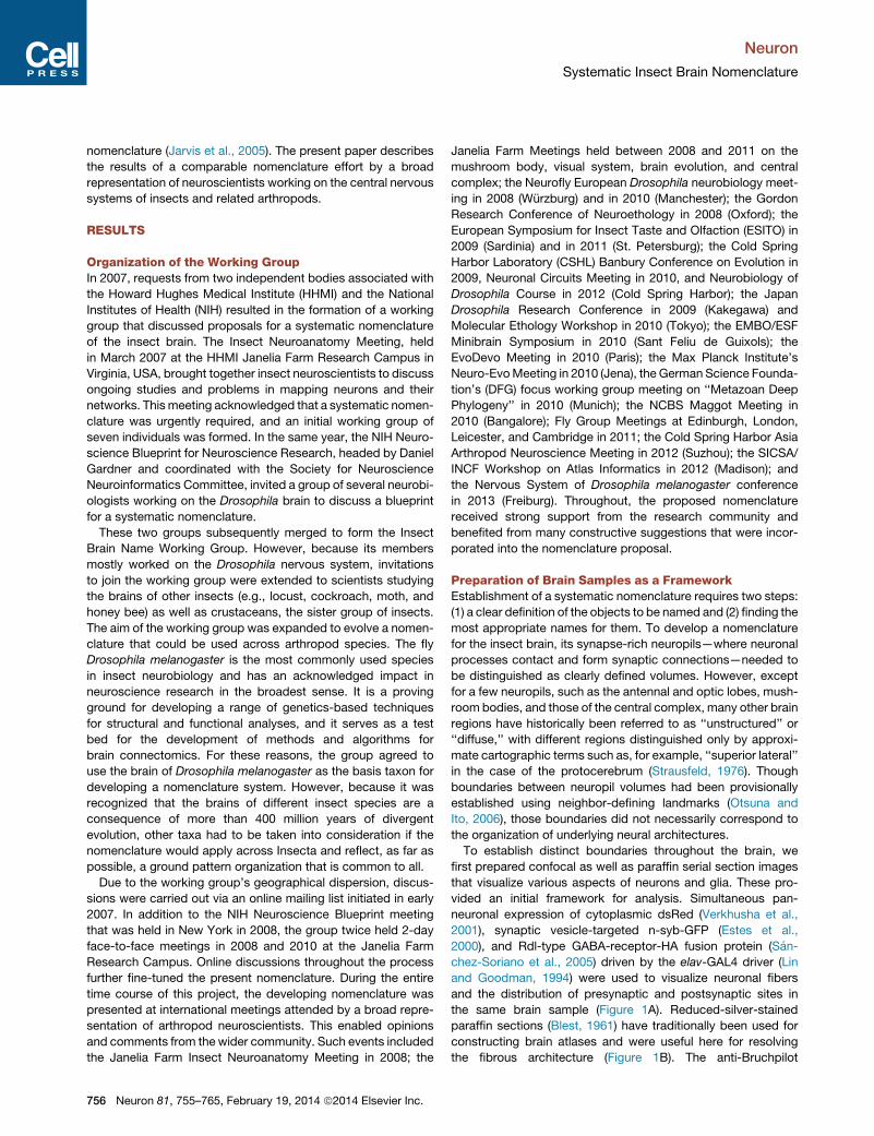
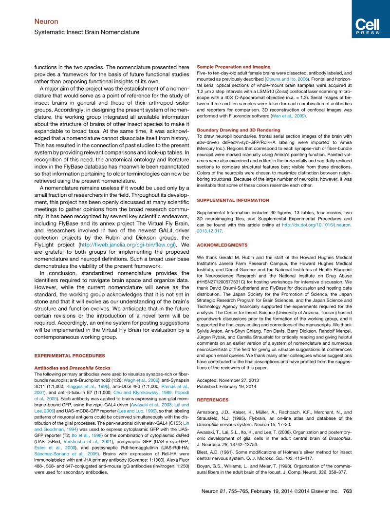
A E
FB
CC
DD
Figure 1. Sections of the Insect Brain Visualized with Various Markers
Frontal sections of the same region of the Drosophila brain are shown. Yellow and white characters denote the names of the synapse-rich neuropils and fiber
bundles, respectively.
(A) Cytoplasmic dsRed (red), synaptic-vesicles-targeted n-syb-GFP (green), and GABA-receptor-targeted Rdl-HA (blue) expressed by the pan-neuronal elav-
GAL4 expression driver, with outlines of neuropil boundaries superimposed.
(B) Silver stain (paraffin section) to visualize neural fibers.
(C) Anti-Bruchpilot nc82 antibody to visualize the density of synapses.
(D) Cytoplasmic GFP expressed by the pan-glial repo-GAL4 expression driver.
(E) Anti-b-tublin antibody to visualize fibrous structures of neurons and glia.
(F) Anti-Discs-large antibody to label membranes of neurons. (E) and (F) are superimposed with the repo-GAL4-driven GFP signal (orange) to visualize glial
processes. Bar indicates 50 mm.
Neuron
Systematic Insect Brain Nomenclature
antibody (nc82), which visualizes neuropils according to the
density of an active-zone-specific protein (Wagh et al., 2006),
helped to distinguish synapse-rich neuropils, because regions
occupied by neuronal axons, cell body fibers, and glial pro-
cesses are left unlabeled (Figure 1C). GFP expressed by the
pan-glial repo-GAL4 driver (Lai and Lee, 2006) visualizes glial
cells and their processes, which form bounding sheaths
surrounding many (but not all) neuropils (Figure 1D). Anti-
b-tubulin antibody was used to visualize fibrous structures of
both neurons and glial cells (Chu and Klymkowsky, 1989; Po-
podi et al., 2005) (Figure 1E), and anti-discs large
(DLG) antibody was used to label membranes of neuronal cell
bodies, neuronal fibers, and synapses by detecting DLG
proteins required for septate junction structure (Parnas
et al., 2001) (Figure 1F). Serial section images obtained
with these labeling methods are available via the Brain
Explorer function of the Flybrain Neuron Database (http://ndb.
flybrain.org).
To achieve maximum consistency with previous and ongoing
studies, we took into account the known projection patterns
of various types of neurons, including those of single identified
neurons and the trajectories of their fiber bundles (e.g., Chiang
et al., 2011; Crittenden et al., 1998; Fischbach and Dittrich,
1989; Hanesch et al., 1989; Otsuna and Ito, 2006; Shinomiya
et al., 2011; Strausfeld, 1976; Stocker et al., 1990; Tanaka
et al., 2008, 2012; Yang et al., 1995; Young and Armstrong,
2010; Yu et al., 2010), the distribution of glial processes (Awasaki
et al., 2008; Younossi-Hartenstein et al., 2003), the projections of
Neuron 81, 755–765, February 19, 2014 ª2014 Elsevier Inc. 757
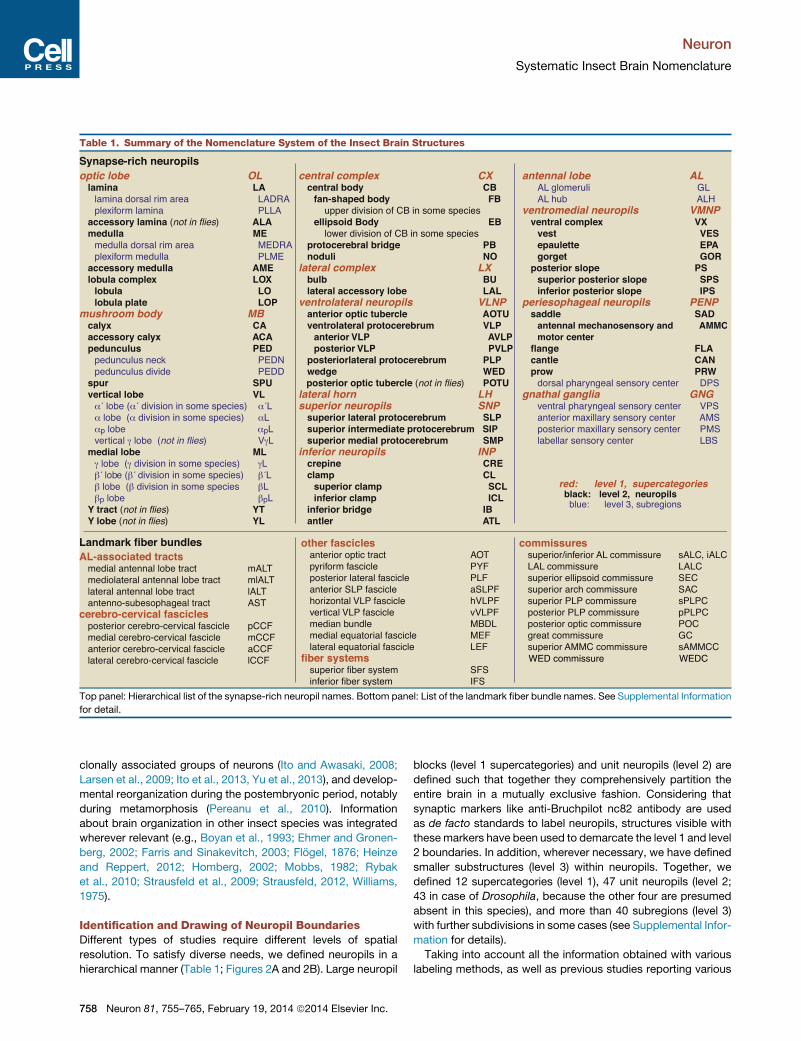
Table 1. Summary of the Nomenclature System of the Insect Brain Structures
Top panel: Hierarchical list of the synapse-rich neuropil names. Bottom panel: List of the landmark fiber bundle names. See Supplemental Information
for detail.
Neuron
Systematic Insect Brain Nomenclature
clonally associated groups of neurons (Ito and Awasaki, 2008;
Larsen et al., 2009; Ito et al., 2013, Yu et al., 2013), and develop-
mental reorganization during the postembryonic period, notably
during metamorphosis (Pereanu et al., 2010). Information
about brain organization in other insect species was integrated
wherever relevant (e.g., Boyan et al., 1993; Ehmer and Gronen-
berg, 2002; Farris and Sinakevitch, 2003; Flogel, 1876; Heinze
and Reppert, 2012; Homberg, 2002; Mobbs, 1982; Rybak
et al., 2010; Strausfeld et al., 2009; Strausfeld, 2012, Williams,
1975).
Identification and Drawing of Neuropil BoundariesDifferent types of studies require different levels of spatial
resolution. To satisfy diverse needs, we defined neuropils in a
hierarchical manner (Table 1; Figures 2A and 2B). Large neuropil
758 Neuron 81, 755–765, February 19, 2014 ª2014 Elsevier Inc.
blocks (level 1 supercategories) and unit neuropils (level 2) are
defined such that together they comprehensively partition the
entire brain in a mutually exclusive fashion. Considering that
synaptic markers like anti-Bruchpilot nc82 antibody are used
as de facto standards to label neuropils, structures visible with
thesemarkers have been used to demarcate the level 1 and level
2 boundaries. In addition, wherever necessary, we have defined
smaller substructures (level 3) within neuropils. Together, we
defined 12 supercategories (level 1), 47 unit neuropils (level 2;
43 in case of Drosophila, because the other four are presumed
absent in this species), and more than 40 subregions (level 3)
with further subdivisions in some cases (see Supplemental Infor-
mation for details).
Taking into account all the information obtained with various
labeling methods, as well as previous studies reporting various
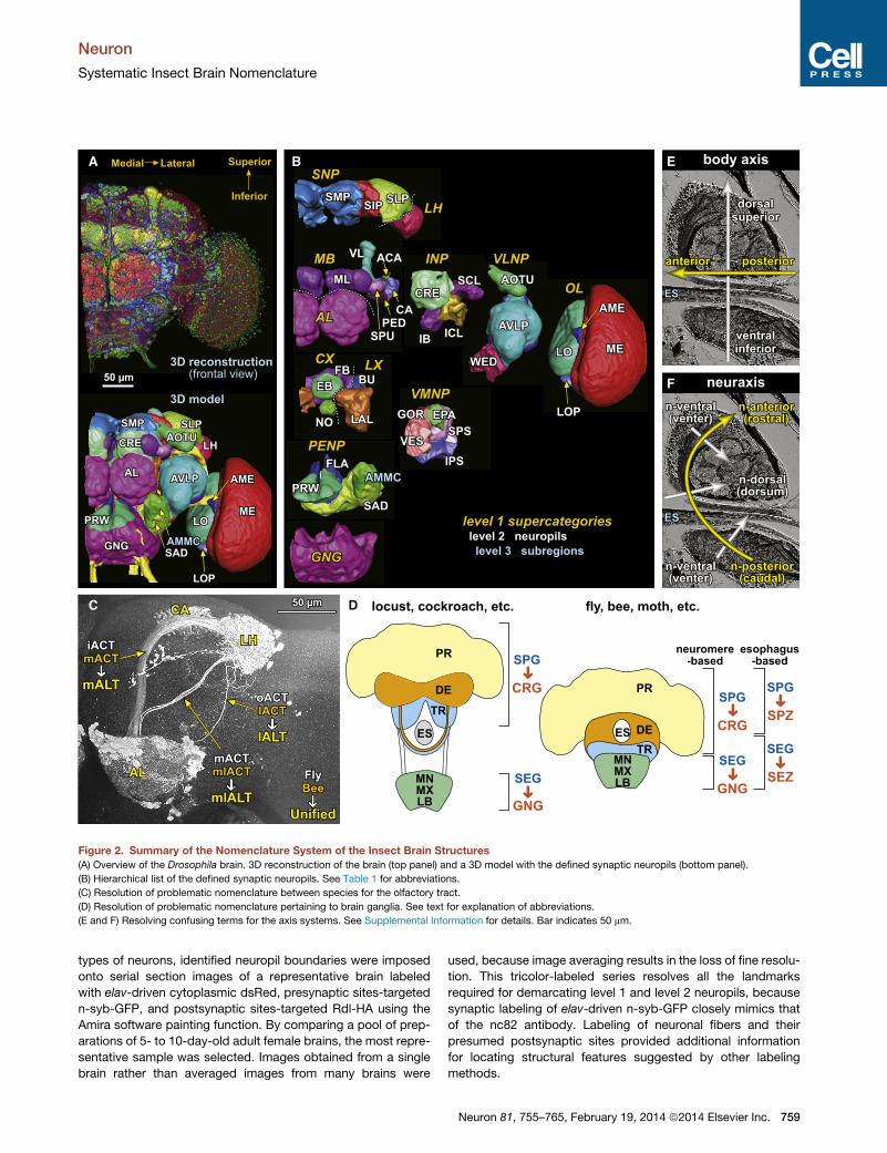
A B
C D
E
F
Figure 2. Summary of the Nomenclature System of the Insect Brain Structures
(A) Overview of the Drosophila brain. 3D reconstruction of the brain (top panel) and a 3D model with the defined synaptic neuropils (bottom panel).
(B) Hierarchical list of the defined synaptic neuropils. See Table 1 for abbreviations.
(C) Resolution of problematic nomenclature between species for the olfactory tract.
(D) Resolution of problematic nomenclature pertaining to brain ganglia. See text for explanation of abbreviations.
(E and F) Resolving confusing terms for the axis systems. See Supplemental Information for details. Bar indicates 50 mm.
Neuron
Systematic Insect Brain Nomenclature
types of neurons, identified neuropil boundaries were imposed
onto serial section images of a representative brain labeled
with elav-driven cytoplasmic dsRed, presynaptic sites-targeted
n-syb-GFP, and postsynaptic sites-targeted Rdl-HA using the
Amira software painting function. By comparing a pool of prep-
arations of 5- to 10-day-old adult female brains, the most repre-
sentative sample was selected. Images obtained from a single
brain rather than averaged images from many brains were
used, because image averaging results in the loss of fine resolu-
tion. This tricolor-labeled series resolves all the landmarks
required for demarcating level 1 and level 2 neuropils, because
synaptic labeling of elav-driven n-syb-GFP closely mimics that
of the nc82 antibody. Labeling of neuronal fibers and their
presumed postsynaptic sites provided additional information
for locating structural features suggested by other labeling
methods.
Neuron 81, 755–765, February 19, 2014 ª2014 Elsevier Inc. 759
Neuron
Systematic Insect Brain Nomenclature
Application of Boundaries to Other SpecimensTo meet the diverse needs of researchers, the Supplementary
Information demonstrate several ways for understanding and
synthesizing information about neuropils: (1) the 84-page Sup-
plemental Information provides detailed overviews of neuropils
and landmark fiber bundles, together with how to name and
abbreviate them, as well as discussion about conflicting terms
and solutions; (2) serial section movies (Movies S1, S2, S3, and
S4) provide dynamic images of synaptic labeling and maps of
neuropils and fiber bundles; (3) Movies S5 and S6 provide inter-
active clickable 3D maps, with which users can visualize any
combination of defined neuropils (with Movies S5 and S6) and
fiber bundles (withMovie S5) from any desired angle. This assists
understanding of the spatial relationships among neuropils.
With these guidelines and tools, researchers will be able to
locate neuropils in their own specimens of interest when these
are counterlabeled with synaptic markers such as nc82.
In addition to these documents and movies, raw confocal
image data files showing identified neuropils and underlying syn-
aptic labeling are downloadable via FlyBase (http://flybase.org).
They will be useful for spatially matching the provided neuropil
images with the images of other brain samples using 3D registra-
tion software such as Computational Morphometry ToolKit (Jef-
feris et al., 2007) or BrainAligner (Peng et al., 2011). Researchers
can either use the provided image file as a target template onto
which their own image data would bemorphed and registered or
morph the provided image file to any target template used for
their own particular study. For example, Virtual Fly Brain
(http://www.virtualflybrain.org/), an annex project of FlyBase,
has registered the images of over 10,000 single-neuron data
by Chiang et al. (2011) onto the template neuropils described
by us so that the projection patterns of these neurons can be
understood in the framework of the current nomenclature. In
addition, the official anatomy ontology and the existing anatom-
ical literature index in FlyBase has been updated and reanno-
tated with this new nomenclature via the Virtual Fly Brain project.
Assignments of Names to the Identified StructuresArriving at a systematic nomenclature required extensive discus-
sions about suitable names for neuropils. We have retained
classic terminology whenever possible. But when several
different names have been historically used to refer to an iden-
tical structure, or when a single name has historically been
used to refer to different structures in different insect species,
or by different researchers, the working group identified names
that resolved such ambiguities (see below). Many regions of
the insect brain had no established names at all. For those neuro-
pils, the group had to devise names. For the sake of brevity and
the convenience of electronic text searching, as well as for
minimizing acronyms, we determined simple, unique names
that are suggestive of the shapes or relative positions of neuro-
pils. This follows established conventions for naming genes
and mutants, as well as the history of naming vertebrate and
invertebrate brain structures according to shapes, such as
‘‘Ammon’s horn,’’ ‘‘olive,’’ ‘‘mushroom,’’ ‘‘fan-shaped,’’ etc.
Crucially, names associated with neural functions are specif-
ically avoided, because future studies are likely to reveal yet-
unknown functions relating to neuropils. In addition, acknowl-
760 Neuron 81, 755–765, February 19, 2014 ª2014 Elsevier Inc.
edging that some names may be needed that reflect the relative
positions of structures, we include lists of alternative position-
based synonyms (see Supplemental Information).
Developmental and evolutionary studies agree that three
segmental neuromeres compose the supraesophageal ganglia
of insects (Hirth et al., 2003; Kumar et al., 2009). However,
because the outgrowth of processes contributing to the adult
brain obscures its embryonically defined segments, identifying
neuromere boundaries in the mature brain is technically difficult.
Considering the long history of debate on this issue, we have
generally avoided neuromere-related terms. The resultant
nomenclature system is neutral; it does not depend on the
segmental nature of the arthropod brain.
Unlike in the vertebrate brain, most neuronal cell bodies are
located in the cell body rind that encloses the brain’s neuropil
mass. The locality of a patch or cluster of neuronal cell bodies
is most easily given by referring to the name of the next nearest
(often adjacent) neuropils. Names describing the location of
patches and clusters in the cell body rind are provided in Supple-
mental Information.
The insect brain also features abundant bundles of neuronal
fibers (neurites) connecting different neuropils. We have defined
and named the most prominent fiber bundles as well as those
that form useful landmarks for determining the boundaries of
synapse-rich neuropils. To make the terminology systematic,
we defined ‘‘fascicles’’ and ‘‘tracts’’ as connecting two different
brain regions ipsilaterally and ‘‘commissures’’ as connecting two
regions contralaterally. Thus, a few fiber bundles historically
referred to as ‘‘tracts’’ have been renamed as ‘‘commissures’’
when they connect neuropils in both brain hemispheres.
Establishment of AbbreviationsIn publications, the names of brain structures are generally
abbreviated. A systematic nomenclature would not serve well if
abbreviations are not controlled. Toward this end, the working
group established systematic abbreviations for all the provided
nomenclature. As with naming genes, unique combinations of
a few characters are preferred. Whenever possible, we kept
existing abbreviations used in previous literature, but if a single
abbreviation has been used to refer to more than one structure,
we assigned a unique abbreviation for each structure to avoid
ambiguity.
In addition, if we modified the definition of a neuropil so that it
significantly departed from a previous description, we proposed
an alternative abbreviation to distinguish the old and new terms.
For example, the acronym ‘‘slpr’’ was once used to denote the
superior lateral protocerebrum. Because its boundaries have
been shifted in this documentation (see Supplemental Informa-
tion for details), we employed the new abbreviation ‘‘SLP’’ to
distinguish new definition from the previous one.
For the last character of any neuropil name, the letters C, T,
and F are generally avoided because elsewhere in the docu-
ment these refer to, respectively, ‘‘commissures,’’ ‘‘tracts’’ and
‘‘fascicles.’’
Resolving Ambiguities of TerminologyAmajor task of theworking groupwas to resolve ambiguities and
confusion in existing terminology. We reviewed the names for
Neuron
Systematic Insect Brain Nomenclature
synapse-rich neuropils and fiber bundles used in previous litera-
ture and retained published terminology whenever possible so
that past accounts do not lose their relevance with respect to
present and future descriptions. However, there were over thirty
instances where terms required clarification and revision. These
are provided in Supplemental Information.
Different names that refer to an identical structure, or vice
versa, cause immense confusion when discussing the results
of past studies. In such cases, the working group decided on
one name alone that was the least confusing and which best
denoted the relevant structure. For example, the terms ‘‘ventral
body,’’ used in descriptions of dipteran brains, and ‘‘lateral
accessory lobe,’’ used in descriptions of the brains of locusts
and moths, refer to homologous structures. Because the sec-
ond term is used more often in studies suggesting possible
roles of this neuropil, we excluded the first term and opted
for ‘‘lateral accessory lobe.’’ Likewise, two terms, ‘‘lateral
horn’’ and ‘‘lateral protocerebrum,’’ both refer to the same sec-
ondary olfactory center. However, because the latter term
refers also to the lateral part of the protocerebrum in general,
it was decided that only the first term would unambiguously
refer to an olfactory center, irrespective of the species-specific
shape of the protocerebrum.
The literature abounds with terms used without any clear defi-
nition. For example, the terms ‘‘central body’’ and ‘‘central com-
plex’’ refer to combinations of neuropils in the central part of
the brain without specifying which neuropils are, or should be,
included. After examining the context in which these terms
have been used in the past, we defined the former to mean the
combination of the fan-shaped body and the ellipsoid body
and the latter to mean the combination of these two neuropils
and two closely interconnected regions: the protocerebral
bridge and noduli. In addition, the bulb and the lateral accessory
lobe, both neuropils closely associated with the central complex,
are collectively defined as the ‘‘lateral complex.’’
In certain cases, a single name was used for different struc-
tures. To avoid confusion, it was decided to adopt entirely new
terms for those structures, particularly if adopting just one of
them would make comparisons of future accounts with already
published ones difficult. For example, one of several ascending
axon bundles from the antennal lobe was termed mACT, mean-
ing the ‘‘middle’’ antennocerebral tract (ACT) seen in flies and
moths (Stocker et al., 1990). Studies of honey bee brains used
the same term to refer to the ‘‘medial’’ ACT, which corresponds
to the ‘‘inner’’ ACT (iACT) in cockroaches and flies (Figure 2C)
(Mobbs, 1982; Malun et al., 1993). Such inconsistencies make
it impossible to compare the trajectories of antennal lobe projec-
tion neurons across species. Considering that the anatomical
arrangement of the three pathways is better described as being
medial, mediolateral, and lateral rather than inner, middle, and
outer, we have adopted the convention used for the honey bee
brain. To distinguish clearly the new unified terminology from
previous ones, we selected the novel term ALT (antennal lobe
tract) instead of the double-barreled ACT (antenno-cerebral
tract). ALT best describes the tract with regard to its origin.
Likewise, structures referred to as the ‘‘lateral triangle’’ were
not identical between flies and locusts. To remedy this, it
was decided to employ the novel term ‘‘bulb’’ to refer to this
structure, based on its characteristic organization of clustered
microglomeruli.
It was also essential to resolve nomenclature for those ganglia
that contribute to the brain (Figure 2D). Developmental and
evolutionary evidence demonstrates that the insect (and mala-
costracan) brain comprises six neuromeres (Scholtz and Edge-
combe, 2006): the protocerebrum (PR); deutocerebrum (DE);
tritocerebrum (TR); and the mandibular (MN), maxillary (MX),
and labial (LB) neuromeres. The terms supraesophageal and
subesophageal ganglia (SPG and SEG) have been used, respec-
tively, to denote the PR, DE, and TR separately from theMN,MX,
and LB. However, SPG and SEG have also been used to gener-
ally refer to brain tissue above and below the level of the esoph-
agus. The two employments of SPG and SEG are not, and
cannot be, synonymous, however, because developmental
studies show that the esophagus penetrates the deutocerebral
neuromere during embryogenesis (Boyan et al., 2003). Two
organizations can be found in the resulting adult. In many Hemi-
metabola, such as locusts and cockroaches, deutocerebral
and tritocerebral neuropils below the esophagus are reduced
to thin commissures, resulting in a clear distinction of three
neuromeres above and three below the esophagus (Figure 2D,
left). On the contrary, in many Holometabola, such as flies,
bees, and moths, the deutocerebrum and tritocerebrum lie
around and below the level of the esophagus. Thus, the terms
SPG and SEG have contradictory meanings (Figure 2D, right).
To resolve this, we here employ the historical (and robust)
terms ‘‘cerebral ganglia’’ (CRG) to replace the term ‘‘supraeso-
phageal ganglion’’ (SPG) and ‘‘gnathal ganglia’’ (GNG) to replace
the term ‘‘subesophageal ganglion’’ (SEG) for neuromere-based
definitions that are independent of the location of the esophagus
(Haeckel, 1896; Snodgrass, 1956). To generally refer to those
parts above and below the esophagus, the word ‘‘ganglion’’ is
avoided, because an arbitrary boundary might not match that
of a neuromere and because of variation across species. After
debating alternative terms, the group chose ‘‘supraesophageal
zone’’ (SPZ) and ‘‘subesophageal zone’’ (SEZ), respectively, to
refer to brain tissue above and below the level of the esophagus.
Introduction of new terms enables clear distinction of the
studies that follow old or new nomenclature: literature that
uses conventional but double-barreled terms like the ACT, lateral
triangle, or SPG/SEG can be distinguished easily from studies
that employ the new systematic nomenclature, which use terms
like ALT, bulb, or CRG/GNG and SPZ/SEZ.
It was also important to avoid terms that can be applied to
descriptions of the arthropod brain with terms used for describ-
ing the vertebrate brain. For example, the accumulation of neural
cell bodies that cover the surface of the insect brain’s neuropils
has often been referred to as the ‘‘cortex.’’ This term is inappro-
priate as it historically refers to sheet-like processing centers of
the vertebrate forebrain. Unlike vertebrates, insect neurons
rarely have synapses on their cell bodies, and therefore, neural
computation seldom (if ever) occurs at that level. To avoid
misleading implications of ‘‘cortex,’’ we adopted the alternative
term ‘‘cell body rind,’’ which has a historical precedent (Straus-
feld, 1976). The central volume of the insect brain, excluding
the laterally extending optic lobes (the primary visual centers),
is often referred to as the ‘‘central brain’’ or ‘‘midbrain.’’ To avoid
Neuron 81, 755–765, February 19, 2014 ª2014 Elsevier Inc. 761
Neuron
Systematic Insect Brain Nomenclature
confusion with the vertebrate midbrain, with its segmental
implication, we have chosen the former term, ‘‘central brain.’’
We have also tried to avoid terms that could be confused with
current Drosophila gene and allele names.
Divergent spelling of the English language can pollute nomen-
clature. For example, US English simplifies the original spelling
of ‘‘oesophagus’’ to ‘‘esophagus,’’ a difference that has led to in-
consistencies in abbreviating that part of the brain beneath
the esophagus as either the SOG or the SEG (SEZ in the new
nomenclature). Considering that most journals and coordinated
ontologies follow US English spelling, it was decided to suggest
the term ‘‘subesophageal’’ irrespective of the spelling policy of
each journal.
To summarize, the working group identified and discussed
37 controversial issues relevant to unifying brain nomenclature.
Resolution of these is provided in Supplemental Information.
Terms for Coordinate AxesThe working group also focused on the important issue of
defining which axes of reference should be best used in descrip-
tions of brain. Two systems previously employed are the body
axes, determined by the longitudinal axis of the body (Figure 2E),
or the neuraxis, which reflects the alignments of segmental neu-
romeres (Figure 2F). The two axes correspond in the thorax and
abdomen, but because the adult brain undergoes a rotation of
about 90�, the neuraxis is almost perpendicular to the body
axis. Confusion has often occurred because the same directional
terms (anterior/posterior and dorsal/ventral) have been used in-
discriminately. We reached the consensus of adding the prefix
‘‘n-’’ to indicate directions that explicitly are based on the neu-
raxis. For example, in flies, the antennal lobes are directed
forwards and are therefore anterior with regard to the body
axis; however they are n-ventral with respect to the neuraxis.
The prefix ‘‘b-’’ can be added to indicate a body axis descriptor
explicitly (e.g., b-anterior). In addition, the terms ‘‘dorsum’’ and
‘‘venter’’ (n-dorsal/n-ventral) and ‘‘rostral’’ and ‘‘caudal’’ (n-ante-
rior/n-posterior) are used specifically for describing locations
according to the neuraxis, and ‘‘superior’’ and ‘‘inferior’’ are
used specifically to indicate dorsal and ventral locations accord-
ing to the body axis.
DISCUSSION
Many previous accounts have identified and named insect
brain regions and their connections. Such studies have been
useful guides for the present system of nomenclature, even
though some of the terms used had to be abandoned in the
development of this nomenclature. We acknowledge such
earlier efforts, which include printed atlases of ganglia or
brains (e.g., Power, 1943; Strausfeld, 1976; Tyrer and Gregory,
1982; Brandt et al., 2005; Otsuna and Ito, 2006; Kurylas
et al., 2008) and web-based maps and databases, such as
the ‘‘Flybrain’’ database (Armstrong et al., 1995) and other
web-based databases that serve current research programs
(Chiang et al., 2011; Shinomiya et al., 2011; Jenett et al.,
2012, Milyaev et al., 2012). Each has provided invaluable infor-
mation and guidance. In the present project, the Insect Brain
Name Working Group has addressed two major concerns in
762 Neuron 81, 755–765, February 19, 2014 ª2014 Elsevier Inc.
order to facilitate communication among diverse researchers
working in insect neuroscience: (A) establishing a common
framework to describe the brain’s structural organization and
(B) resolving inconsistency in nomenclature. A controlled
nomenclature enables a common language and thus ease
of communication among trainees and researchers alike,
including those who are not yet familiar with insect neurosci-
ence. The strategy of devising a standardized nomenclature
through consensus has been employed most recently by a
consortium of vertebrate neuroscientists for defining and
naming regions of the avian brain (Jarvis et al., 2005), resulting
in that nomenclature’s universal acceptance. Considering the
increasing importance of insect studies in neuroscience
research, the present system of nomenclature should be help-
ful not only for those using Drosophila but also those working
with other species. And, it is hoped, a controlled terminology
will assist interdisciplinary collaborations between vertebrate
and insect neuroscientists.
The common framework devised here for documenting brain
organization demanded seven essential features. (1) Integrity:
the framework had to include the entire brain relating all its parts
to comparable spatial resolution. (2) Unambiguity: the bound-
aries of each brain region had to be clearly defined using land-
marks that enable consistent identification across individuals.
(3) Neutrality: the framework had to be detached from function,
because rapidly progressing studies are likely to identify unex-
pected functions for neuropils. (4) Expandability: the framework
had to be applicable to brains of other insects, and even crusta-
cean species, with minimum alterations. (5) Consistency: com-
parisons with previous nomenclature had to be provided for
the ease of transition to new terminologies. (6) Universality: a
system of terms designed for broad use by the community. (7)
Flexibility: a framework that permits its own evolution and
adaptive integration of novel findings.
The nomenclature proposed here is comprehensive: 47 brain
regions identified in this project comprise the entire brain. Vol-
umes previously referred to as diffuse neuropils, which may
occupy as large as 90% of the central brain, are here denoted
by clearly defined names and boundaries. Without such
notation, it is impossible to adequately describe locations and
projections of neurons within such volumes. The integrity
and unambiguity of the current nomenclature obviates such
problems and provides essential support of research aimed at
elucidating the organization, function, and development of
neural circuits in not only a few well-known regions but also in
the entire insect brain.
Generally, functional studies should not neglect underlying
neural organization, and structural studies should consider
possible functions. However, this has its risks: mushroom
bodies, once thought to serve only olfactory learning due to their
connections with the antennal lobes, have since been accorded
a variety of other attributes. The working group has been keenly
aware that implying functional associations would disadvantage
a nomenclature designed for annotating brain architecture.
Crucially, divorcing structure from function is essential if we
consider that insect brains have undergone divergent evolution.
A center identifiable in, for example, Drosophila that is also
present in an aquatic beetle may support entirely different
Neuron
Systematic Insect Brain Nomenclature
functions in the two species. The nomenclature presented here
provides a framework for the basis of future functional studies
rather than proposing functional insights of its own.
A major aim of the project was the establishment of a nomen-
clature that would serve as a point of reference for the study of
insect brains in general and those of their arthropod sister
groups. Accordingly, in designing the present system of nomen-
clature, the working group integrated all available information
about the structure of brains of other insect species to make it
expandable to broad taxa. At the same time, it was acknowl-
edged that a nomenclature cannot dissociate itself from history.
This has resulted in the connection of past studies to the present
system by providing relevant comparisons and look-up tables. In
recognition of this need, the anatomical ontology and literature
index in the FlyBase database has meanwhile been reannotated
so that information pertaining to older terminologies can now be
retrieved using the present nomenclature.
A nomenclature remains useless if it would be used only by a
small fraction of researchers in the field. Throughout its develop-
ment, this project has been openly discussed at many scientific
meetings to gather opinions from the broad research commu-
nity. It has been recognized by several key scientific endeavors,
including FlyBase and its annex project The Virtual Fly Brain,
and researchers involved in two of the newest GAL4 driver
collection projects by the Rubin and Dickson groups, the
FlyLight project (http://flweb.janelia.org/cgi-bin/flew.cgi). We
are grateful to both groups for implementing the proposed
nomenclature and neuropil definitions. Such a broad user base
demonstrates the viability of the present framework.
In conclusion, standardized nomenclature provides the
identifiers required to navigate brain space and organize data.
However, while the current nomenclature will serve as the
standard, the working group acknowledges that it is not set in
stone and that it will evolve as our understanding of the brain’s
structure and function evolves. We anticipate that in the future
certain revisions or the introduction of a novel term will be
required. Accordingly, an online system for posting suggestions
will be implemented in the Virtual Fly Brain for evaluation by a
contemporaneous working group.
EXPERIMENTAL PROCEDURES
Antibodies and Drosophila Stocks
The following primary antibodies were used to visualize synapse-rich or fiber-
bundle neuropils: anti-Bruchpilot nc82 (1:20; Wagh et al., 2006), anti-Synapsin
3C11 (1:1,000; Klagges et al., 1996), anti-DLG 4F3 (1:1,000; Parnas et al.,
2001), and anti-b-tubulin E7 (1:1,000; Chu and Klymkowsky, 1989; Popodi
et al., 2005). Each antibody was applied to brains expressing pan-glial mem-
brane-bound GFP, using the repo-GAL4 driver (Awasaki et al., 2008; Lai and
Lee, 2006) and UAS-mCD8-GFP reporter (Lee and Luo, 1999), so that labeling
patterns of neuronal antigens could be observed simultaneously with the dis-
tribution of the glial processes. The pan-neuronal driver elav-GAL4 (C155; Lin
and Goodman, 1994) was used to express cytoplasmic GFP with the UAS-
GFP reporter (T2; Ito et al., 1998) or the combination of cytoplasmic dsRed
(UAS-DsRed; Verkhusha et al., 2001), presynaptic GFP (UAS-n-syb-GFP;
Estes et al., 2000), and postsynaptic Rdl-hemagglutinin (UAS-Rdl-HA;
Sanchez-Soriano et al., 2005). Brains with expression of Rdl-HA were
immunolabeled with anti-HA primary antibody (Covance; 1:1000). Alexa Fluor
488-, 568- and 647-conjugated anti-mouse IgG antibodies (Invitrogen; 1:250)
were used for secondary antibodies.
Sample Preparation and Imaging
Five- to ten-day-old adult female brains were dissected, antibody labeled, and
mounted as previously described (Otsuna and Ito, 2006). Frontal and horizon-
tal serial optical sections of whole-mount brain samples were acquired at
1.2 mm z step intervals with a LSM510 (Zeiss) confocal laser scanning micro-
scope with a 403 C-Apochromat objective (n.a. = 1.2). Serial images of be-
tween three and ten samples were taken for each combination of antibodies
and reporters for comparison. 3D reconstruction of confocal images was
performed with Fluorender software (Wan et al., 2009).
Boundary Drawing and 3D Rendering
To draw neuropil boundaries, frontal serial section images of the brain with
elav-driven dsRed/n-syb-GFP/Rdl-HA labeling were imported to Amira
(Mercury Inc.). Regions that correspond to each synapse-rich or fiber-bundle
neuropil were marked manually using Amira’s painting function. Painted vol-
umes were also examined and edited in the horizontally and sagittally resliced
sections to compare structural features best visible from these directions.
Colors of the neuropils were chosen to maximize distinction between neigh-
boring structures. Because of the large number of neuropils, however, it was
inevitable that some of these colors resemble each other.
SUPPLEMENTAL INFORMATION
Supplemental Information includes 30 figures, 13 tables, four movies, two
3D neuroimaging files, and Supplemental Experimental Procedures and
can be found with this article online at http://dx.doi.org/10.1016/j.neuron.
2013.12.017.
ACKNOWLEDGMENTS
We thank Gerald M. Rubin and the staff of the Howard Hughes Medical
Institute’s Janelia Farm Research Campus, the Howard Hughes Medical
Institute, and Daniel Gardner and the National Institutes of Health Blueprint
for Neuroscience Research and the National Institute on Drug Abuse
(HHSN271200577531C) for hosting workshops for intensive discussion. We
thank David Osumi-Sutherland and FlyBase for discussion and hosting data
distribution. The Japan Society for the Promotion of Science, the Japan
Strategic Research Program for Brain Sciences, and the Japan Science and
Technology Agency financially supported the experiments required for the
analysis. The Center for Insect Science (University of Arizona, Tucson) hosted
groundwork discussions prior to the formation of the working group, and it
supported the final copy editing and corrections of the manuscripts. We thank
Sylvia Anton, Ann-Shyn Chiang, Ron Davis, Barry Dickson, Randolf Menzel,
Jurgen Rybak, and Camilla Strausfeld for critically reading and giving helpful
comments on an earlier version of a system of nomenclature and numerous
neuroscientists of the field for giving us valuable suggestions at conferences
and upon email queries. We thank many other colleagues whose suggestions
have contributed to the final descriptions and have profited from the sugges-
tions of the reviewers of this paper.
Accepted: November 27, 2013
Published: February 19, 2014
REFERENCES
Armstrong, J.D., Kaiser, K., Muller, A., Fischbach, K.F., Merchant, N., and
Strausfeld, N.J. (1995). Flybrain, an on-line atlas and database of the
Drosophila nervous system. Neuron 15, 17–20.
Awasaki, T., Lai, S.L., Ito, K., and Lee, T. (2008). Organization and postembry-
onic development of glial cells in the adult central brain of Drosophila.
J. Neurosci. 28, 13742–13753.
Blest, A.D. (1961). Some modifications of Holmes’s silver method for insect
central nervous system. Q. J. Microsc. Sci. 102, 413–417.
Boyan, G.S., Williams, L., and Meier, T. (1993). Organization of the commis-
sural fibers in the adult brain of the locust. J. Comp. Neurol. 332, 358–377.
Neuron 81, 755–765, February 19, 2014 ª2014 Elsevier Inc. 763
Neuron
Systematic Insect Brain Nomenclature
Boyan, G., Reichert, H., and Hirth, F. (2003). Commissure formation in the
embryonic insect brain. Arthropod Struct. Dev. 32, 61–77.
Brandt, R., Rohlfing, T., Rybak, J., Krofczik, S., Maye, A., Westerhoff, M.,
Hege, H.C., and Menzel, R. (2005). Three-dimensional average-shape atlas
of the honeybee brain and its applications. J. Comp. Neurol. 492, 1–19.
Burne, T., Scott, E., van Swinderen, B., Hilliard, M., Reinhard, J., Claudianos,
C., Eyles, D., and McGrath, J. (2011). Big ideas for small brains: what can psy-
chiatry learn from worms, flies, bees and fish? Mol. Psychiatry 16, 7–16.
Cachero, S., Ostrovsky, A.D., Yu, J.Y., Dickson, B.J., and Jefferis, G.S. (2010).
Sexual dimorphism in the fly brain. Curr. Biol. 20, 1589–1601.
Chiang, A.S., Lin, C.Y., Chuang, C.C., Chang, H.M., Hsieh, C.H., Yeh, C.W.,
Shih, C.T., Wu, J.J., Wang, G.T., Chen, Y.C., et al. (2011). Three-dimensional
reconstruction of brain-wide wiring networks in Drosophila at single-cell reso-
lution. Curr. Biol. 21, 1–11.
Chu, D.T., and Klymkowsky, M.W. (1989). The appearance of acetylated
alpha-tubulin during early development and cellular differentiation in
Xenopus. Dev. Biol. 136, 104–117.
Crittenden, J.R., Skoulakis, E.M., Han, K.A., Kalderon, D., and Davis, R.L.
(1998). Tripartite mushroom body architecture revealed by antigenic markers.
Learn. Mem. 5, 38–51.
Ehmer, B., and Gronenberg, W. (2002). Segregation of visual input to the
mushroom bodies in the honeybee (Apis mellifera). J. Comp. Neurol. 451,
362–373.
Estes, P.S., Ho, G.L., Narayanan, R., and Ramaswami, M. (2000). Synaptic
localization and restricted diffusion of a Drosophila neuronal synaptobre-
vin—green fluorescent protein chimera in vivo. J. Neurogenet. 13, 233–255.
Farris, S.M., and Sinakevitch, I. (2003). Development and evolution of the
insect mushroom bodies: towards the understanding of conserved develop-
mental mechanisms in a higher brain center. Arthropod Struct. Dev. 32,
79–101.
Fischbach, K.F., and Dittrich, A.P.M. (1989). The optic lobe of Drosophila
melanogaster. - I. A Golgi analysis of wild-type structure. Cell Tissue Res.
258, 441–475.
Flogel, J.H.L. (1876). Uber den feineren Bau des Arthropodengehirns. Tagebl.
Versaml. Deutscher Naturforsch. Arzte 49, 115–120.
Haeckel, E. (1896). Systematische Phylogenie. Zweiter Theil: Systematische
Phylogenie der wirbellosen Thiere (Invertebrata). (Berlin: Reimer).
Hanesch, U., Fischbach, K.F., and Heisenberg, M. (1989). Neuronal architec-
ture of the central complex in Drosophila melanogaster. Cell Tissue Res. 257,
343–366.
Heinze, S., and Reppert, S.M. (2012). Anatomical basis of sun compass navi-
gation I: the general layout of the monarch butterfly brain. J. Comp. Neurol.
520, 1599–1628.
Hirth, F., Kammermeier, L., Frei, E., Walldorf, U., Noll, M., and Reichert, H.
(2003). An urbilaterian origin of the tripartite brain: developmental genetic
insights from Drosophila. Development 130, 2365–2373.
Homberg, U. (2002). Neurotransmitters and neuropeptides in the brain of the
locust. Microsc. Res. Tech. 56, 189–209.
Ito, K., and Awasaki, T. (2008). Clonal unit architecture of the adult fly brain.
Adv. Exp. Med. Biol. 628, 137–158.
Ito, K., Suzuki, K., Estes, P., Ramaswami, M., Yamamoto, D., and Strausfeld,
N.J. (1998). The organization of extrinsic neurons and their implications in the
functional roles of the mushroom bodies in Drosophila melanogaster Meigen.
Learn. Mem. 5, 52–77.
Ito, M., Masuda, N., Shinomiya, K., Endo, K., and Ito, K. (2013). Systematic
analysis of neural projections reveals clonal composition of the Drosophila
brain. Curr. Biol. 23, 644–655.
Jarvis, E.D., Gunturkun, O., Bruce, L., Csillag, A., Karten, H., Kuenzel, W.,
Medina, L., Paxinos, G., Perkel, D.J., Shimizu, T., et al.; Avian Brain
Nomenclature Consortium (2005). Avian brains and a new understanding of
vertebrate brain evolution. Nat. Rev. Neurosci. 6, 151–159.
764 Neuron 81, 755–765, February 19, 2014 ª2014 Elsevier Inc.
Jefferis, G.S., Potter, C.J., Chan, A.M., Marin, E.C., Rohlfing, T., Maurer, C.R.,
Jr., and Luo, L. (2007). Comprehensive maps of Drosophila higher olfactory
centers: spatially segregated fruit and pheromone representation. Cell 128,
1187–1203.
Jenett, A., Rubin, G.M., Ngo, T.T., Shepherd, D., Murphy, C., Dionne, H.,
Pfeiffer, B.D., Cavallaro, A., Hall, D., Jeter, J., et al. (2012). A GAL4-driver
line resource for Drosophila neurobiology. Cell Rep. 2, 991–1001.
Klagges, B.R., Heimbeck, G., Godenschwege, T.A., Hofbauer, A., Pflugfelder,
G.O., Reifegerste, R., Reisch, D., Schaupp, M., Buchner, S., and Buchner, E.
(1996). Invertebrate synapsins: a single gene codes for several isoforms in
Drosophila. J. Neurosci. 16, 3154–3165.
Kumar, A., Fung, S., Lichtneckert, R., Reichert, H., and Hartenstein, V. (2009).
Arborization pattern of Engrailed-positive neural lineages reveal neuromere
boundaries in the Drosophila brain neuropil. J. Comp. Neurol. 517, 87–104.
Kurylas, A.E., Rohlfing, T., Krofczik, S., Jenett, A., and Homberg, U. (2008).
Standardized atlas of the brain of the desert locust, Schistocerca gregaria.
Cell Tissue Res. 333, 125–145.
Lai, S.L., and Lee, T. (2006). Genetic mosaic with dual binary transcriptional
systems in Drosophila. Nat. Neurosci. 9, 703–709.
Larsen, C., Shy, D., Spindler, S.R., Fung, S., Pereanu, W., Younossi-
Hartenstein, A., and Hartenstein, V. (2009). Patterns of growth, axonal exten-
sion and axonal arborization of neuronal lineages in the developing
Drosophila brain. Dev. Biol. 335, 289–304.
Lee, T., and Luo, L. (1999). Mosaic analysis with a repressible cell marker for
studies of gene function in neuronal morphogenesis. Neuron 22, 451–461.
Lin, D.M., and Goodman, C.S. (1994). Ectopic and increased expression of
Fasciclin II alters motoneuron growth cone guidance. Neuron 13, 507–523.
Malun, D., Waldow, U., Kraus, D., and Boeckh, J. (1993). Connections
between the deutocerebrum and the protocerebrum, and neuroanatomy of
several classes of deutocerebral projection neurons in the brain of male
Periplaneta americana. J. Comp. Neurol. 329, 143–162.
Milyaev, N., Osumi-Sutherland, D., Reeve, S., Burton, N., Baldock, R.A., and
Armstrong, J.D. (2012). The Virtual Fly Brain browser and query interface.
Bioinformatics 28, 411–415.
Mobbs, P.G. (1982). The brain of the honeybee Apis mellifera. I. The connec-
tions and spatial organizations of the mushroom bodies. Philos. Trans. R.
Soc. B. 298, 309–345.
Otsuna, H., and Ito, K. (2006). Systematic analysis of the visual projection
neurons of Drosophila melanogaster. I. Lobula-specific pathways. J. Comp.
Neurol. 497, 928–958.
Parnas, D., Haghighi, A.P., Fetter, R.D., Kim, S.W., and Goodman, C.S. (2001).
Regulation of postsynaptic structure and protein localization by the Rho-type
guanine nucleotide exchange factor dPix. Neuron 32, 415–424.
Peng, H., Chung, P., Long, F., Qu, L., Jenett, A., Seeds, A.M., Myers, E.W., and
Simpson, J.H. (2011). BrainAligner: 3D registration atlases of Drosophila
brains. Nat. Methods 8, 493–500.
Pereanu, W., Kumar, A., Jennett, A., Reichert, H., and Hartenstein, V. (2010).
Development-based compartmentalization of the Drosophila central brain.
J. Comp. Neurol. 518, 2996–3023.
Popodi, E.M., Hoyle, H.D., Turner, F.R., and Raff, E.C. (2005). The proximal
region of the beta-tubulin C-terminal tail is sufficient for axoneme assembly.
Cell Motil. Cytoskeleton 62, 48–64.
Power, M.E. (1943). The brain of Drosophila. J. Morphol. 72, 517–559.
Rybak, J., Kuß, A., Lamecker, H., Zachow, S., Hege, H.C., Lienhard, M.,
Singer, J., Neubert, K., andMenzel, R. (2010). The digital bee brain: integrating
and managing neurons in a common 3D reference system. Front. Syst.
Neurosci. 4, 30.
Sanchez-Soriano, N., Bottenberg, W., Fiala, A., Haessler, U., Kerassoviti, A.,
Knust, E., Lohr, R., and Prokop, A. (2005). Are dendrites in Drosophila homol-
ogous to vertebrate dendrites? Dev. Biol. 288, 126–138.
Neuron
Systematic Insect Brain Nomenclature
Scholtz, G., and Edgecombe, G.D. (2006). The evolution of arthropod heads:
reconciling morphological, developmental and palaeontological evidence.
Dev. Genes Evol. 216, 395–415.
Shinomiya, K., Matsuda, K., Oishi, T., Otsuna, H., and Ito, K. (2011). Flybrain
neuron database: a comprehensive database system of the Drosophila brain
neurons. J. Comp. Neurol. 519, 807–833.
Snodgrass, R.E. (1956). Anatomy of the Honey Bee. (Ithaca: Comstock Pub.
Associates).
Stocker, R.F., Lienhard, M.C., Borst, A., and Fischbach, K.F. (1990). Neuronal
architecture of the antennal lobe in Drosophila melanogaster. Cell Tissue Res.
262, 9–34.
Strausfeld, N.J. (1976). Atlas of an Insect Brain. (Berlin, Heidelberg, New York,
Tokyo: Springer-Verlag).
Strausfeld, N.J. (2012). Arthropod Brains: Evolution, Functional Elegance, and
Historical Significance. (Cambridge: Harvard University Press).
Strausfeld, N.J., Sinakevitch, I., Brown, S.M., and Farris, S.M. (2009). Ground
plan of the insect mushroom body: functional and evolutionary implications.
J. Comp. Neurol. 513, 265–291.
Tanaka, N.K., Tanimoto, H., and Ito, K. (2008). Neuronal assemblies of the
Drosophila mushroom body. J. Comp. Neurol. 508, 711–755.
Tanaka, N.K., Endo, K., and Ito, K. (2012). Organization of antennal lobe-asso-
ciated neurons in adult Drosophila melanogaster brain. J. Comp. Neurol. 520,
4067–4130.
Tyrer, N.M., and Gregory, G.E. (1982). A guide to the neuroanatomy of locust
subesophageal and thoracic ganglia. Philos. Trans. R. Soc. B. 297, 91–123.
Verkhusha, V.V., Otsuna, H., Awasaki, T., Oda, H., Tsukita, S., and Ito, K.
(2001). An enhancedmutant of red fluorescent protein DsRed for double label-
ing and developmental timer of neural fiber bundle formation. J. Biol. Chem.
276, 29621–29624.
Wagh, D.A., Rasse, T.M., Asan, E., Hofbauer, A., Schwenkert, I., Durrbeck, H.,
Buchner, S., Dabauvalle, M.C., Schmidt, M., Qin, G., et al. (2006). Bruchpilot, a
protein with homology to ELKS/CAST, is required for structural integrity and
function of synaptic active zones in Drosophila. Neuron 49, 833–844.
Wan, Y., Otsuna, H., Chien, C.-B., and Hansen, C. (2009). An interactive visu-
alization tool for multi-channel confocal microscopy data in neurobiology
research. IEEE Trans. Vis. Comput. Graph. 15, 1489–1496.
Williams, J.L.D. (1975). Anatomical studies of the insect central nervous sys-
tem: a ground-plan of the midbrain and an introduction to the central complex
in the locust, Schistocerca gregaria (Orthoptera). J. Zool. 176, 67–86.
Yang, M.Y., Armstrong, J.D., Vilinsky, I., Strausfeld, N.J., and Kaiser, K. (1995).
Subdivision of the Drosophilamushroom bodies by enhancer-trap expression
patterns. Neuron 15, 45–54.
Young, J.M., and Armstrong, J.D. (2010). Building the central complex in
Drosophila: the generation and development of distinct neural subsets.
J. Comp. Neurol. 518, 1525–1541.
Younossi-Hartenstein, A., Salvaterra, P.M., and Hartenstein, V. (2003). Early
development of the Drosophila brain: IV. Larval neuropile compartments
defined by glial septa. J. Comp. Neurol. 455, 435–450.
Yu, J.Y., Kanai, M.I., Demir, E., Jefferis, G.S., and Dickson, B.J. (2010). Cellular
organization of the neural circuit that drives Drosophila courtship behavior.
Curr. Biol. 20, 1602–1614.
Yu, H.-H., Awasaki, T., Schroeder, M.D., Long, F., Yang, J.S., He, Y., Ding, P.,
Kao, J.-C., Wu, G.Y., Peng, H., et al. (2013). Clonal development and organi-
zation of the adult Drosophila central brain. Curr. Biol. 23, 633–643.
Neuron 81, 755–765, February 19, 2014 ª2014 Elsevier Inc. 765