Embed Size (px)
Citation preview

Follow these principles to encourage disease prevention and healthful
living among your patients.
Supported by an unrestricted grant from Bausch + LombContent by
A Supplement to
WELLNESS ESSENTIALS
FOR CLINICAL PRACTICE 2nd edition2
001_owns1019_fc.indd 3001_owns1019_fc.indd 3 10/14/19 12:40 PM10/14/19 12:40 PM

2 REVIEW OF OPTOMETRY • OCTOBER 15, 2019
B O A R D O F D I R E C T O R S
Stuart Richer, OD, PhD, President Dr. Richer is director of ocular preventive medicine at the Lovell Federal Health Care Center and associate or adjunct professor at several institutions. He was principal investigator of the Veterans Lutein Antioxidant Supplementation Trial and the Zeaxanthin and Visual Function Study.
Julie Poteet, OD, CNS, Vice PresidentDr. Poteet is in private practice and research with an emphasis on nutritional, environmental and biochemical aspects of chronic health problems. Her passion has been using scientifi cally based targeted nutritional therapies to address underlying systemic imbalances and disease.
Dennis Ruskin, OD, Secretary Dr. Ruskin has been an infl uential presence throughout his four-decade career. He has served or advised government committees, the Canadian Examiners of Optometry, the Toronto College of Optometrists, the Ontario Association of Optometrists and numerous ophthalmic corporations.
Susan Summerton, OD, CNS, Fellowship Committee ChairDr. Summerton, a private practitioner from Naples, Florida, is also an adjunct professor of nutrition at Hodges University. She is a certifi ed nutrition specialist, a member of the American College of Nutrition and a diplomate of the American Clinical Board of Nutrition.
Dorothy L. Hitchmoth OD, ABO, ABCMO Diplomate Dr. Hitchmoth, an award-winning professor, lecturer and educator, recently retired from the VA after 22 years of service. She is a consultant for Carl Zeiss Meditec and the University of Massachusetts medical school, and serves on many medical and scientifi c advisory boards.
Be a Part of the VanguardFocus on ocular wellness before disease sets in.From the board of the Ocular Wellness & Nutrition Society
The Nuts and Bolts of NutrientsEyes and bodies need a balanced diet rich in vitamins and minerals. Here’s how you can recommend healthy eating habits and appropriate supplementation. By Stuart Richer, OD, PhD
The Virtues of Vitamin CThis substance is critical to eye health and function. Here’s why it should be an important part of your arsenal.By Thomas E. Levy, MD, JD
Influencing Myopia: Science and Clinical PracticeIn search of dietary and lifestyle changes that could alter the course of childhood refractive status and quality of life.By Karan R. Gregg Aggarwala, OD (NIH Equiv), PhD, and Stuart P. Richer, OD, PhD
4
8
14
18
C O N T E N T S
8
21
WELLNESS ESSENTIALS
FOR CLINICAL PRACTICE
002_ns1019_TOC v2.indd 2002_ns1019_TOC v2.indd 2 10/14/19 11:02 AM10/14/19 11:02 AM
3REVIEW OF OPTOMETRY • OCTOBER 15, 2019
Karan R. “Gregg” Aggarwala, OD (NIH Equiv.), PhDDr. Aggarwala trained at the All-India Institute of Medical Sciences and SUNY College of Optometry, with emphasis on chromatic stimuli for ocular accommodation. Following his MS and PhD degrees and an NEI-awarded post-doctoral fellowship in behavioral neuroscience, he studied nutrition, public health, vision therapy, and behavior modifi cation.
Pinakin Davey OD, PhDDr. Davey is a tenured professor at Western University of Health Sciences College of Optometry, with particular interest and expertise in glaucoma. He has authored over 50 international publications and given over 200 conference and invited presentations. He is an active researcher in ocular and vision sciences for both the NIH and industry.
Kerry Gelb, ODA graduate of SUNY College of Optometry, with a doctorate from Illinois College of Optometry, Dr. Gelb is in private practice in New York and New Jersey. He has also volunteered in the OneSight program, helping to restore and preserve vision for people in need internationally.
Lisa Renzi Hammond, PhDDr. Renzi-Hammond specializes in neurological and visual development, as well as risk for acquired neurological diseases across the lifespan. She founded the Human Biofactors Laboratory, which investigates ways in which nutrition can impact various visual and cognitive functions.
Steven G. Pratt, MDThe author of fi ve ‘Superfoods’ books, including two bestsellers, Dr. Pratt is a clinical assistant professor of ophthalmology at the University of California, San Diego. He has been trained in holistic medicine and holds a degree from the American Board of Holistic Medicine.
E X E C U T I V E D I R E C T O R
Kari ClineMs. Cline has diverse experience in community education, corporate fundraising and non-profi t board governance. She has worked for nearly a decade in non-profi t leadership, lobbying and event management. Her background is in environmental chemistry and laboratory quality assurance.
Carotenoids: Front to Back Ocular ProtectionAdding the marine newcomer, astaxanthin, to your plant food–based diet can fill in the ocular health gaps left by lutein and zeaxanthin. By Stuart Richer, OD, PhD, Dorothy Hitchmoth, OD, and Lisa Renzi-Hammond, PhD
Uncover the Mechanisms of Macular PigmentTo determine a retina-friendly regimen for your patient, you need to see and track the work of the carotenoids in action.By Pinakin Gunvant Davey, OD, PhD
Gut Instinct: Why the Microbiome MattersIt can be considered an additional human organ, rivaling the liver in the number of biochemical reactions in which it participates.By Julie Poteet, OD, CNS
Put Wellness on the MenuLearn how the right dietary choices can improve patients’ outcomes—and lives.By Steven G. Pratt, MD, Stuart Richer, OD, PhD, Dennis Ruskin, OD, and Kerry Gelb, OD
Two Big Controversies in Ocular NutritionAMD supplements and omega-3s are both still in the hot seat.By Stuart Richer, OD, PhD, and Dennis Ruskin, OD
21
26
30
34
42
34
002_ns1019_TOC v2.indd 3002_ns1019_TOC v2.indd 3 10/14/19 11:02 AM10/14/19 11:02 AM

4 REVIEW OF OPTOMETRY • OCTOBER 15, 2019
All day long, you see patients and strive to do the best you can for them in the limited time you have. For those
with vision or eye health concerns, you diligently explain treatment options, monitoring schedules and referral paths if necessary. For healthy patients, how-ever, you probably just send them on their way with a reminder about their next exam in a year’s time.
Both of these groups, but especially the latter, need a little more attention, if you can spare it. The best way to ‘treat’ disease is to avoid it in the first place.
In an age of ever-increasing com-peting priorities, what could be of greater importance than optimizing the ocular and visual welfare of your patients? While eye care professionals are exceptionally busy today, it’s critical to remember that preventive medicine and the ongoing repair and maintenance of ocular tissue should be at the core of primary eye care practice. Taking steps to monitor patients’ eye and vision health early and frequently, and educate patients about key wellness and nutritional strategies, are essential steps in helping prevent degenerative diseases and pathology, prompted not only by the aging process but also by unhealthy lifestyle choices.
PRIMARY EYE CARE LAGS BEHINDA 2019 study in The Lancet showed that, globally, one-fifth of all deaths, or 11 million mortalities, were associated with poor diet.1 In the US, 63% of all food consumed has been found to be processed, with only 12% of food com-
ing from plant sources; the remaining 25% comes from animal sources.2 At OWNS, we believe that minimizing processed, manufactured food and exchanging it with whole, organic food is the single most important factor in improving ocular and overall health through reducing inflammation—the core component of all chronic disease. At the same time, researchers have found that higher fish and nut con-sumption is associated with a lower risk of AMD progression among sub-jects in certain settings.3
Unfortunately, optometry and medi-cal schools are not evolving to reflect a changing wellness landscape. Their philosophies and curricula continue
to center upon symptoms-based medicine rather than addressing root causes. OWNS is committed to raising the awareness of practicing health providers to counteract this outdated approach to eye care and medicine. The mission of OWNS is “to provide leadership, education, advice, and guidance to eye care and other health care professionals and consumers regarding the role of lifestyle choices and nutritional support as it relates to vision and eye health. The OWNS supports evidence-based analysis concerning nutritional influences on eyes and systemic disease.”
As our mission spells out, the society supports evi-denced-based information. And we disseminate this sci-entific guidance in a variety of ways to help health care providers make the best decisions for their patients, with the goal of better patient outcomes and elevated doctor-patient relationships.
Be a Part of the Vanguard
Focus on ocular wellness before disease sets in.
FROM THE BOARD OF THE OCULAR WELLNESS & NUTRITION SOCIETY
MESSAGE FROM OWNS
Diabetic retinopathy is just one condition optometrists are vital players in managing—and, ideally, preventing.
Photo
: Ste
ven F
erru
cci, OD
004_ns1019_intro.indd 4004_ns1019_intro.indd 4 10/14/19 11:04 AM10/14/19 11:04 AM
5REVIEW OF OPTOMETRY • OCTOBER 15, 2019
Q&A With a CNS Optometrist
Neda Gioia, OD, an independent optom-etry practice owner and student in the OWNS Certified Nutritionist Specialist (CNS) program, shares her experiences going through the CNS program, and mak-ing nutrition and health a major focus for the last five years of her life.
Can you explain how nutrition and well-ness became so important for you in general
and how it’s intersected with your optometric life?
I always thought of myself as healthy, but I was exposed to something that was very unhealthy, which devolved into a neurological condition. That led me to seek other avenues of trying to treat myself. So I was ready to start imple-menting nutrition and functional medicine into my own life. And that drive surpassed the desire of just trying to learn something for knowledge; it became personal.
As an optometrist who deals with primary care, I thought it would be a loss for my patients if I didn’t expose them to what I’ve learned. That’s when I started researching how I could bring the areas of wellness, nutrition and functional medicine into optom-etry vs. having it be a completely separate entity.
You are working your way through the OWNS CNS program clinical hours under the supervision of another CNS mentor. What has your experience been like with the CNS program? Did you learn anything that surprised you, going through the program?
My experience with the CNS program has been wonderful in the sense that it’s so organized, and the courses have been so enlight-ening and so in-depth. The program really put me on a different level of understanding about the core concepts of nutrition.
There were surprises throughout the program. My biggest surprise was with the gut microbiome. Many companies are trying to tap into research findings about the microbiome to treat a lot of chronic health conditions, and even eye conditions are being studied.
How do you think the CNS program will benefit your patients and practice into the future?
Any patient who walks in the door of an optometry office where the doctor has exposure to nutritional education such as from the CNS program is in better standing. That patient’s likely not get-ting this information anywhere else in their medical consultations. Therefore, we’re going to stand out as an impactful subspecialty of the medical world that can introduce these patients to a differ-ent way of treating their health concerns. This is medicine of the future, and we have an opportunity to help lead.
You also completed the first module for The Institute for Functional Medicine recently. What is driving you to seek additional credential opportunities in the areas of nutrition and functional medicine rela-tive to your optometric practice?
When I was investigating different programs for nutrition, The Institute of Functional Medicine always popped up. The program
really gears toward medical doctors. And when I first applied, the admissions staff rejected me because I was an optometrist. I wrote them a long letter making my case for why I should be admitted. Nothing happened, and a year later I found out that the Ocular Wellness & Nutrition Society had gotten them to accept qualify-ing optometrists. I reapplied and was accepted, so I finished the main module on implementation. That is why I decided to go even further—to learn how to implement functional medicine in my practice.
You have noted the unique nature of your office. Can you explain what you mean, relative to the discipline of human nutrition and functional medicine in optometric practice?
We currently are in a launch phase. I always wanted to have an optometry practice with a nutrition program weaved in. And that’s what I decided to do with this office—not have nutrition just as a side program, but actually as a highlight. In addition to protocols for patients who want to go through my nutrition program, I have laboratories that I’m working with to do special diagnostic tests, and I implemented an antioxidant tester.
I also have a standby coach to help me complete the programs for patients. If you want to do a full program, it takes a lot of time between the lab work and creating protocols for the patient, which you have to keep adjusting. We’re going to have all the primary care services that a primary care optometry office has, but for the patients who have chronic conditions—which unfortunately now is more the norm—they can see that we offer wellness advice and would like to help them with their issues. If they are comfortable with that, maybe they’ll want to seek more help in the world of nutrition.
My other motivation for having a dual functioning practice model was to figure out how to put it front and center as a busi-ness model. With the time that you spend to do all of this, col-leagues are going to ask why, and if you’re not able to show them how to incorporate nutrition into the practice, they will have a hard time deciding to go forward with a similar program. That’s why I want to prove the model. I would like to create a way to help counsel other health care providers on how to do it in the future, too.
Do you think that more practices around the country will be seek-ing to become credentialed in the areas of nutrition and functional medicine relative to your optometric practice in the future?
Absolutely. Today’s patients are asking more questions. They want to know, ‘What other options do I have? What can I do?’ And they’re used to answers like, ‘Well, it is what it is.’ But that’s not how we should be treating our patients. We should empower them, and we should walk side by side with them to try to find answers, or help them in other avenues that intersect. Younger providers are also asking questions. They are much more exposed to the concept of health change and wellness; that philosophy is now out there in the media. I think, going forward, they want change, too. And the CNS program provides that change.
Dr. Gioia, owner of Integrative Vision, Shrewsbury, NJ
004_ns1019_intro.indd 5004_ns1019_intro.indd 5 10/14/19 11:05 AM10/14/19 11:05 AM
6 REVIEW OF OPTOMETRY • OCTOBER 15, 2019
GET INVOLVED—AND EVOLVEDWellness efforts can range from a simple, quick conversa-tion with an AMD or prediabetes patient about lifestyle modifications to a full-blown commitment to embracing the principles of preventive medicine with additional for-mal training. In short, there’s a way forward for everyone. Below are some of the things you can do to adopt a well-ness mindset in your practice.
1. Join OWNS. Optometrists who join the society set themselves on a path to learn fundamental and advanced
concepts in wellness, build a network of like-minded col-leagues to seek out as mentors and begin to apply these principles in their practices. This is a vital first step.
2. Earn an OWNS Fellowship. To further its mission, OWNS offers a fellowship program for candidates seek-ing to be credentialed at the highest level of professional competence. Individuals have up to five years to complete the process. Retroactive nutrition-related hours for the past three years can be applied toward the total education requirement. The Fellowship Committee works with can-didates to develop materials that demonstrate eligibility to sit for the oral exam. The qualification process is designed to help candidates develop as professionals and successfully become fellows through a four-step process that includes a validated application, attainment of 150 CE hours, com-pleted written documentation and a passing oral exam. Learn more about this esteemed designation here: www.ocularnutritionsociety.org/fellowship-program.
3. Become a Certified Nutrition Specialist. Recently, OWNS, in collaboration with the University of Western States, developed an online course suite enabling qualified optometrists to sit for the Certified Nutrition Specialist (CNS) Examination. The CNS certificate is held by clinical nutritionists, physicians and other health professionals with a specialty in nutrition. It is the only non-dietetics credential and examination widely respected in state nutrition licen-sure laws. Thus, optometric CNS certification monetizes ocular health promotion, for your private pay and some insurance carriers. It is a model for the future of eye care that OWNS hopes will be adopted by the AOA.
BE A PART OF THE VANGUARD
MESSAGE FROM OWNS
The CNS CurriculumOptometrists who pursue the Certified Nutrition Specialist pro-gram will undergo the following courses:• MSN 6200 Nutritional Biochemistry (offered Spring and Fall)• MSN 6101 Evidence-Based Nutrition (offered Summer and
Winter)• MSN 6305 Whole Food Nutrition and Supplementation
(offered Summer and Winter)• MSN 6204 Gastrointestinal Imbalances (offered Spring and
Fall)• MSN 7215 Cardiovascular Disease and Metabolic Imbalances
(offered Summer and Winter)• MSN 6300 Detoxification and Biotransformation Pathways
and Imbalances (offered Summer and Winter)Totals: 18 quarter-credits (4.5 quarter-credits biochemistry, 13.5 quarter-credits nutrition).Tuition, determined by the UWS Board of Trustees, is $473 per credit. OWNS members receive a 15% discount. See www.ocular-nutritionsociety.org/become-a-cns for more.
OWNS Liaisons at Optometry Colleges
School Location Faculty Advisor Faculty email
Illinois College of Optometry Chicago, IL Stuart Richer, OD, PhD [email protected]
Indiana School of Optometry Bloomington, IN Julie Torbit, OD [email protected]
Midwestern University Downers Grove, IL Diyana Ivanova, OD [email protected]
Nova Southeastern University Ft. Lauderdale, FL Lori Vollmer, OD [email protected]
New England College of Optometry Boston, MA Diane Russo, OD [email protected]
Pacific University College of Optometry Forest Grove, ORJames Kundart, OD Dina Erickson, OD
[email protected] [email protected]
Southern College of Optometry Memphis, TN Taylor Kiser, OD [email protected]
SUNY College of Optometry New York, NY Jerry Rapp, PhD [email protected]
University of Alabama at Birmingham Birmingham, AL Kim Duong, OD [email protected]
University of Houston College of Optometry Houston, TX Bruce Onofrey, OD, RPh [email protected]
University of Missouri at St. Louis St. Louis, MO Mary Beth Rhomberg, OD [email protected]
Western University College of Optometry Pomona, CA Pinakin G. Davey, OD, PhD [email protected]
004_ns1019_intro.indd 6004_ns1019_intro.indd 6 10/14/19 11:05 AM10/14/19 11:05 AM
7REVIEW OF OPTOMETRY • OCTOBER 15, 2019
4. Join the Academy’s SIG. Other educational efforts in nutrition, conducted by many members of OWNS, happen through the American Academy of Optometry’s Nutrition, Disease Prevention & Wellness special interest group (SIG). The mission of the SIG is to promote excellent patient care with lifestyle and nutritional support for prevention and management of eye diseases and related systemic disorders through professional education, scientific investigation and multidisciplinary collaboration. The group strives to foster camaraderie and men-torship for students, doctors and researchers inter-ested in nutrition science and wellness as it relates to eye care.
5. Get connected locally with an OWNS liaison. Optometrists currently in practice and those students now joining the ranks can reach out to an OWNS representative at their local college of optometry. The society is in the process of appointing an OWNS liaison at each of the colleges to serve as a resource. Go to the society’s main website for information and updates: www.ocularnutritionsociety.org.
THE FUTURE OF WELLNESSIt is the intention of our society to create self-funded, sustainable preventive ocular health and wellness clinics at optometry colleges. We envision each clinic to consist of three divisions:
(1) Ocular/vision/systemic evaluation, includ-ing macular pigment optical density, three-channel color vision and systemic antioxidant status.
(2) An educational center with finger blood spot/saliva/urine biomarker self-assessment.
(3) An apothecary of nutritional and pharma-ceutical offerings.
Actionable testing would include, for example, predictive epigenetic biomarkers such as genetic testing for AMD, celiac and hemochromatosis diseases (for more, see the article by Russel Jaffe, MD, PhD, in the 2018 edition of this supplement at www.reviewofoptometry.com/publications/well-ness2018). Home testing would include, for exam-ple, 25-OH vitamin D liver reserve status, RBC EPA/DHA cellular membrane essential fatty acid status, early morning urine pH magnesium and potassium status, high-sensitivity (hs)-C-reactive protein (CRP) inflammatory status and other broad biomarkers of health. In addition, devices to measure macular pigment optic density and skin carotenoid concentrations would be available.
This is one half of our dream; the other half is creden-tialing as many optometrists with the CNS designation as
there are practicing ODs. We hope you consider becoming a part of OWNS and stepping into our bright wellness vision of the future. ❍
1. GBD 2017 Diet Collaborators. Health effects of dietary risks in 195 countries, 1990-2017:
a systematic analysis for the Global Burden of Disease Study 2017. Lancet. 2019 May
11;393(10184):1958-72.
2. USDA Economic Research Service. U.S. Food Consumption as a % of Calories. 2009. Available at:
https://www.healthyschoolfood.org/docs/color_pie_chart.pdf. (last accessed September 3, 2019).
3. Seddon JM, Cote J, Rosner B. Progression of age-related macular degeneration: association with
dietary fat, transunsaturated fat, nuts, and fish intake. Arch Ophthalmol. 2003;121(12):1728-37.
OWNS on the MoveIt’s an exciting time to be a member of OWNS. Here are some of the latest goings-on:
• Julie Poteet, OD, CNS, vice president of OWNS, is now overseeing our Weekly Wellness Spotlight email to members, which offers relevant informa-tion from the preventative medicine landscape around the country and beyond.
• Our fall 2018 Pre-American Academy of Optometry meeting in San Antonio featured Barry Tan, PhD, founder of American River Nutrition and one of the world’s foremost experts on vitamin E. Dr. Tan discussed the abil-ity of vitamin E isomers (tocotrienols) to mitigate chronic conditions, includ-ing ocular disease.
• Our 12th Annual Ocular Wellness and Nutrition Meeting was held at the University of Missouri, St. Louis College of Optometry April 13 to 14, and featured spirited lecture topics such as energy medicine and environmental influences on myopia. Keynote speaker Dan Winter, an international energy medicine expert, was featured via a video link from Paris. Also presenting was Don Mutti, OD, PhD, Janis Eells, PhD, Julie Dekinder, OD, and Stuart Richer, OD, PhD To view these presentations, go to www.ocularnutritionsociety.org/meetings.
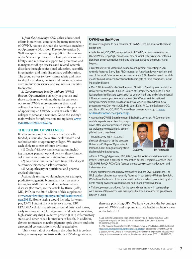
• As retiring OWNS Board member Elizabeth J. Johnson, PhD, one of the world’s experts in carotenoids, steps down after years of dedicated service, we welcome two new highly accom-plished board members:
—Pinakin Davey, PhD, OD, FAAO, director of research at the Western University College of Optometry in Pomona, Calif., brings a strong statis-tical medicine background.
—Karan R “Gregg” Aggarwala, PhD, FAAO, a nutritionist and vision scientist at InVite Health, and a protégé of researcher-author Benjamin Clarence Lane, OD, MPH, FAAO, FCOVD, is focused on eye care research, education and instrumentation.
• Many optometry schools now have active student OWNS chapters. The UAB student chapter was recently featured in our Weekly Wellness Spotlight. We believe the future of the society will be bolstered and promoted by stu-dents raising awareness about ocular health and overall wellness.
• This supplement, produced for the second year in a row in partnership with Review of Optometry, was made possible by an unrestricted grant from Bausch + Lomb.
Dr. AggarwalaDr. Davey
004_ns1019_intro.indd 7004_ns1019_intro.indd 7 10/14/19 11:05 AM10/14/19 11:05 AM

REVIEW OF OPTOMETRY • OCTOBER 15, 20198
Most Americans don’t eat enough vegetables, fruits and other nutrient-rich foods, given the cur-
rent rate of obesity—40%, as last reported by the CDC.1 Not only do poor food choices and high-calorie, low-nutrient alternatives literally weigh on Americans, they can also be harmful to the eyes, vision, cognition and other critical systems in the body.
It’s been said that vitamin defi-ciency is indistinguishable from radiation damage when examin-ing cultured cells in a petri dish. Vitamin and mineral deficiency is boosting the incidence of age-related diseases, including a number of ocular diseases such as age-related macular degeneration (AMD) and diabetic retinopathy.
Our patients can improve their wellness by making proper vitamin and mineral intake a goal rather than an afterthought in their lives. But how can you help them make the right choices? With dozens of nutrients playing a part in human health—each with specific mechanisms and dietary sources—keeping up with the relevant effects can be intimidating even for doc-tors. This guide highlights a few key concepts for each, out of a vast body of nutritional knowledge that all doc-tors would do well to understand more deeply.
VITAMIN A This vital substance supports all five senses, with particular relevance to olfaction, hear-ing and night vision. Three dietary forms exist: the pre-formed molecules retinol and retinyl ester, plus carotenoid (e.g., beta carotene) precur-sors to vitamin A. All forms of vitamin A are solubilized in the intestinal lumen and absorbed by duodenal muco-sal cells.
In the retina, vitamin A is converted to retinol, oxidized to retinal and then to retinoic acid. Given its importance to ocular health, vitamin A was included in both the Age-Related Eye Disease Study (AREDS) and AREDS2 supplements.
However, the Blue Mountains Eye study found that elevated beta carotene intake was associated with an increased risk of AMD.2
B VITAMINS This entire family can provide significant benefits and should be prescribed ideally within a comprehensive high-potency, multivitamin-mineral supplement. Here is a quick look at all eight members of the family:
The Nuts and Bolts of Nutrients
Eyes and bodies need a balanced diet rich in vitamins and minerals. Here’s how you can recommend healthy eating habits and appropriate supplementation.
BY STUART RICHER, OD, PhD
VITAMINS & MINERALS
This patient with intermediate AMD is at risk for progression and is a viable candidate for dietary supplementation use to increase carotenoid levels.
Photo
: Julie
Pote
et, O
D
008_ns1019_nutrition.indd 8008_ns1019_nutrition.indd 8 10/14/19 11:07 AM10/14/19 11:07 AM
REVIEW OF OPTOMETRY • OCTOBER 15, 2019 9
Vitamin B1 (thiamin). This is part of the pyruvate dehydroxynase system responsible for converting carbo-hydrates into glucose, as well as breaking down fats and proteins. It helps produce the neurotransmitter acetyl-choline, stimulates the production of red blood cells and relieves the effects of alcoholic cirrhosis, infections and hyperthyroidism. Thiamin protects nerves, preventing the degeneration of myelin sheath coverings that manifests as neuropathy in patients suffering from alcohol and/or uncontrolled diabetes.
This energy-producing vitamin is dramatically reduced in our American diet high in refined sugars, alcohol, cof-fee, tea and drugs that block its absorption. Benfo-thia-mine, fat-soluble thiamin, is an excellent supplement for our patients suffering from neuropathy, cardiomyopathy and Alzheimer’s disease.
Vitamin B2 (riboflavin). As part of the cellular elec-tron transport chain, riboflavin is another B vitamin necessary for energy production. It helps convert car-bohydrates to sugar, processes amino acids and fats, and fuels myriad cellular functions. Riboflavin is also a cofactor for GSH reductase—a major intracellular anti-oxidant.
Though riboflavin is a retinal photosensitizer in high doses, more common ocular deficiency symptoms are non-specific and subtle, such as conjunctival injection, photosensitivity and dry eye.
Vitamin B3 (niacin). This aids in digestive system function—converting proteins, fats and carbs into energy—supports properly functioning muscles and nerves, and promotes a ‘glow’ to the skin.
Those who are deficient (e.g., patients with pellagra disease) have weak muscles, digestive problems and skin irritation. In high doses, niacin has been used to treat schizophrenia.
Niacin comes in three forms, with different properties: • Nicotinic acid reduces high blood LDL cholesterol. • Inositol hexaphosphate (IP6) is useful against cardio-
vascular disease and lowering blood pressure.• Niacinamide boosts mitochondrial function (and
hence energy) that is reduced in aging and neurodegen-erative diseases such as Alzheimer’s and Parkinson’s.
Vitamin B5 (pantothenic acid). This vitamin aids in producing neurotransmitters and steroids, enhances immunity and liver detoxification and assists in the extraction of fats, proteins and other vital nutrients from food.
The most common and irritating symptoms of vitamin B5 deficiency are burning foot syndrome, in which a person experiences a lack of feeling in their feet, accom-panied by intense inflammatory pain, chronic fatigue and weakness.
Vitamin B6 (pyridoxamine). This is a functional cofactor in a number of enzymatic systems involving pro-teins. The close association between pyridoxamine and enzymes assists in proper functioning of the nervous sys-tem. Deficiencies can affect cognition, ambulation, carpel tunnel, multiple sclerosis, immunity (lessening arthritis) and dermatologic issues of the skin and hair.
Vitamin B7 (biotin). A catalyst for controlling a num-ber of metabolic reactions that provide energy from fats, proteins and carbohydrates, biotin is an essential compo-nent for maintaining skin, nail and hair health. Patients experiencing dry scalp, dandruff or hair loss might be suffering from biotin deficiency.
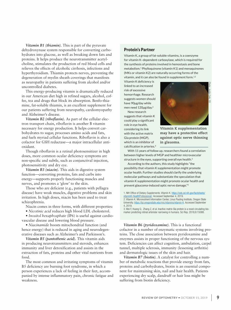
Protein’s PartnerVitamin K, a group of fat-soluble vitamins, is a coenzyme
for vitamin K–dependent carboxylase, which is required for
the synthesis of proteins involved in hemostasis and bone
metabolism.1 Phylloquinone (vitamin K1) and menaquinones
(MKs or vitamin K2) are naturally occurring forms of the
vitamin, and it can also be found in supplement form.1,2
Vitamin K deficiency is
linked to an increased
risk of excessive
hemorrhage. Research
suggests women should
have 90μg/day while
men need 120μg/day.2
New research
suggests that vitamin K
could play a significant
role in eye health,
considering its link
with the active matrix
Gla protein (MGP),
which is an inhibitor of
calcification in arteries.3
With 11 years of follow up, researchers found a correlation
between higher levels of MGP and healthier microvascular
structure in the eyes, supporting overall eye health.3
According to the authors, this study highlights “the
possibility that vitamin K supplementation might promote
ocular health. Further studies should clarify the underlying
molecular pathways and substantiate the speculation that
vitamin K supplementation might promote ocular health and
prevent glaucoma-induced optic nerve damage.”3
1. NIH Office of Dietary Supplements. Vitamin K. https://ods.od.nih.gov/factsheets/
vitaminK-HealthProfessional/. Accessed September 4, 2019.
2. Vitamin K. Micronutrient Information Center, Linus Pauling Institute, Oregon State
University. https://lpi.oregonstate.edu/mic/vitamins/vitamin-K. Accessed September
4, 2019.
3. Wei F, Huang Q, Zhang Z, et al. Inactive matrix Gla protein is a novel circulating bio-
marker predicting retinal arteriolar narrowing in humans. Sci Rep. 2018;8:15088.
Vitamin K supplementation may have a protective effect against optic nerve thinning in glauoma.
Photo
: Justin
Cole
, OD
008_ns1019_nutrition.indd 9008_ns1019_nutrition.indd 9 10/14/19 11:07 AM10/14/19 11:07 AM

REVIEW OF OPTOMETRY • OCTOBER 15, 201910
Vitamin B9 (folate). The natural form of B9 is essen-tial for DNA creation, preventing mutations and the growth of new cells. Specifically, folate plays a role in building new red blood cells and stimulating peripheral end organ blood flow. This means that organ systems, including the eyes and brain, are well-oxygenated and working at full capacity.
The health benefits of vitamin B9 include prevention of heart disorders, stroke, cancer and neural tube defects during early pregnancy. Folate also helps provide relief from mental and emotional disorders.
Folic acid, the synthetic version of vitamin B9, has historically been used as a supplement. US grain products and some manufactured foods are fortified with it. Food folate (i.e., green leafy vegetables) or 5-methyltetrahy-drofolate (5-MTHF) is far superior as it’s absorbed and metabolized in the digestive system, avoiding any unde-sirable partially metabolized or non-metabolized folic acid.
The folic acid found in most supplements is not metab-olized in the digestive system; rather, it moves to the liver, where multiple enzymatic reactions generate an active form, often resulting in high undesirable levels of non-metabolized serum folic acid and serum homocysteine.
Studies show that unmetabolized folic acid may have undesirable effects on the body, such as an increased cancer risk, masking of B12 deficiency or accelerated car-diovascular and ophthalmic vascular disease. Where pos-sible, choose supplements with food folate or preformed 5-MTHF.
Vitamin B12 (cobalamin). This is cleaved from protein during digestion and is dependent upon gastric intrin-sic factor and parietal cell hydrochloric acid. As with food folate, cobalamin assists in cell maintenance, par-ticularly red blood cells, and DNA formation. Vitamin B12 provides relief for patients with pernicious anemia, megaloblastic anemia and sickle cell anemia, and supple-mentation often resolves symptoms of fatigue and neuri-tis (e.g., tingling, numbness), even when blood levels are adequate
Vitamin B12 deficiency is rampant in Americans for several reasons: (1) hydrochloric acid secretion dimin-ishes as we age (achlorhydria); (2) from chronic over-use of proton-pump inhibitors to treat acid reflux disease; (3) by many individuals experiencing subclinical H. pylori infection, which interferes with healthy functioning acid secreting parietal cells; and (4) from the use of the diabe-tes drug metformin in some individuals.
VITAMIN C This is the major extracellular antioxidant that sets the redox potential of cells. It is found at 10x to 30x serum
concentration in every ocular tissue and protects blood vessels. It assists collagen formation, wound healing (including that of the cornea and retina), neurotransmit-ter synthesis, drug detoxification and more.
Deficiency results in slow healing, frequent infections, low platelets (typical of the elderly), bleeding gums, loose teeth, retinal microaneurysms and cataracts. Vitamin C reduces dermal bruising and thinning skin, important to those on blood thinners.
Virtually no one except supplement users maintain adequate vitamin C levels due to rapid excretion of this water-soluble vitamin. Maintaining a therapeutic dose for optimal blood concentration requires the synergism of polyphenols, serial dosing (i.e., grazing on plant food) or supplementation with liposomal C.
See, “The Virtues of Vitamin C,” p. 14, for a detailed discussion of this important antioxidant’s role in ocular health.
VITAMIN D Some would argue vitamin D is a hormone. Vitamin D modulates the absorption of calcium and phosphorous from the small intestine, as well as more than 1,000 genes. It plays a seminal role in the top three killers: can-cer, cardiovascular disease and Alzheimer’s.
There are two forms: vitamin D2 (the less potent, plant-based ergo-calciferol) and vitamin D3 (i.e., fish liver cholcalcefirol). Vitamin D3 converts into 1,25-hydroxy-cholcalciferol, the most potent endogenous steroid hormone. The most abundant source of vitamin D is sunlight.
Optometrists should aim for a vitamin D status between 50ng/ml and 80ng/ml in our patients by cal-culating the required dose of D3 based on lab results. (Remember, 1,25-hydroxy-cholcalciferol serum liver reserve status varies by ethnicity, which is why it is criti-cal to do this lab test to calculate the proper dose.)
While vital for everyone, optimal vitamin D levels are crucial for patients facing recalcitrant uveitis/retinitis, multiple sclerosis, herpes simplex and zoster reactivation, decreasing neovascularization in macular degeneration and patients with or at risk for diabetes and multiple sys-temic cancers.
Vitamin D repletion typically decreases excessive anti-VEGF treatments in housebound elderly patients. Vita-min D status also plays a role in lowering systolic blood pressure and the degree of arteriolar sclerotic retinopa-thy, arcus senilis and cardiovascular plaque.
Some 73% of Americans are insufficient or deficient in vitamin D. Its status is lower in those with higher melanin counts (i.e., darker skin) due to the pigment’s interference with sun absorption. Vitamin D deficiency
THE NUTS AND BOLTS OF NUTRIENTS
VITAMINS & MINERALS
008_ns1019_nutrition.indd 10008_ns1019_nutrition.indd 10 10/14/19 11:07 AM10/14/19 11:07 AM

REVIEW OF OPTOMETRY • OCTOBER 15, 2019 11
is also common in older people, those living in northern latitudes and patients prescribed chronic use of proton-pump inhibitors.
VITAMIN E ISOMERSThis nutrient is composed of eight isomers: four tocoph-erols and four tocotrienols (alpha, beta, gamma, delta). Only one isomer of vitamin E (alpha tocopherol) was employed in the AREDS and AREDS2 studies, an obvi-ous criticism.
Vitamin E tocotrienols are potent antioxidants in competition with the tocopherols. The best sources of gamma and delta tocotrienol (ideal for protection against cardiovascular disease, cancer and diabetes) derive from annatto beans. Tocotrienols increase tear production, retard cataract formation and reduce propensity for dia-betic retinopathy and angiogenesis.
ESSENTIAL MINERALSIn contrast to vitamins, which are large molecules, miner-als are atoms and ions—right off the periodic chart. Your body cannot make these; fortunately, the earth is a one-stop shop for everything your cells need. However, there has been a decline in soil nutrient levels within the last 50 years, with only a few minerals artificially re-introduced into the soil.
Calcium is key for muscle, heart and digestive cell messaging systems and is a constituent of bones and teeth. Adequate levels also support blood clotting, but calcium can be problematic when supplemented in excess (especially without balanced magnesium), where it accu-mulates in blood vessels, the mitral valve and soft tissue.
Chloride is a systemic electrolyte that maintains fluid and electrolyte balance, which is needed for production of hydrochloric acid for digestion.
Chromium is associated with insulin function and is required for the release of energy from glucose. Along with vitamin B3, its absence in the diet results in insulin resistance.
Copper is necessary for the absorption and use of iron, supports formation of hemoglobin and several enzymes, and is a cofactor of many enzymes, including cytochrome C oxidase. Labile copper is a strong divalent oxidizing mineral modulated by the concentration of binding liver ceruloplasmin as well as zinc status. In Wilson’s disease, pathognomonic corneal deposits are found.
Excess copper is common in the US population due to the ubiquity of copper plumbing, use of unfiltered tap water, low ceruloplasmin from subclinical liver disease and lack of dietary zinc. Thus, high-quality adult multivi-tamin-mineral formulas do not contain copper. For those who need it, almonds are a great dietary source. AMD
eyes and Alzheimer’s brains are over-mineralized with this divalent mineral.
Iodine is a component of thyroid hormones and helps regulate growth, development and metabolic rate. Dietary deficiency is the result of fluoridated water in two-thirds of US jurisdictions, brominated US wheat products (i.e., dough fortified with potassium bromate) and unfiltered chlorinated water.
Iron, a divalent metal, is part of the protein hemoglo-bin molecule that carries oxygen throughout the body. Different intake requirements are based on age (younger: important for growth) and gender (premenopausal: important due to blood loss).
Iron is typically not included in high-quality adult mul-tivitamins, as in excess it rapidly accelerates cardiovas-cular disease and oculovascular disease. While one-third of the US population has non-alcoholic fatty liver disease and excess stored iron, other patients have anemia of chronic inflammation, infection and malignancy where the body sequesters free and reactive oxidizing iron. AMD eyes are over-mineralized with divalent iron.
Magnesium (MG) is the center atom of chlorophyll in dark green leafy vegetables and is the fourth most abundant mineral in the body. MG is involved in more than 300 biochemical reactions and supports bone min-eralization, protein building, muscular contraction, nerve impulse transmission, immunity and mitochondrial ade-nosine triphosphate energy production. This mineral is deficient, due to modern soil depletion and lack of intake of leafy vegetables in the American diet. In the past few decades, MG intake has dropped by 50% while the need
The tocotrienol isomers of vitamin E have antioxidant effects that can lower a patient’s risk of diabetic retinopathy and angiogenesis.
Photo
: Thom
as A
. Wong, O
D
008_ns1019_nutrition.indd 11008_ns1019_nutrition.indd 11 10/14/19 11:07 AM10/14/19 11:07 AM
REVIEW OF OPTOMETRY • OCTOBER 15, 201912
for magnesium has increased by 50%. Magnesium protects against cardiovascular disease
and diabetes and, along with potassium, naturally regu-lates blood pressure. An overly acidic first morning urine pH reflects a poor magnesium (and potassium) status.
It is important to encourage supplementation or weekly Epsom salt baths/footbaths in addition to a multivitamin for those taking a calcium supplement and those with MG lab values at 50% of ‘normal’ or below. Magnesium deficiency has been linked to retinopa-thy, neuropathy, foot ulcerations, acephalic migraines, twitching eyelids (ocular myokymia), cold fingers (Rayn-aud’s phenomenon) and low-tension glaucoma.
Manganese, a cofactor for superoxide dismutase, is the principal antioxidant enzyme in mitochondria. Several enzymes activated by manganese contribute to the metab-olism of carbohydrates, amino acids and cholesterol.
Molybdenum is a cofactor for the oxidases xanthine, aldehyde and sulfite, all of which facilitate cellular pro-cesses. A low molybdenum level is why some individuals are sensitive to sulfites in food and wine.
Phosphorus is required for the formation of cells, bones and teeth, and it maintains acid-base balance, digestion detoxification and sex drive. A phosphorous deficiency can lead to weak muscles, joints and bones as well as low stamina and even cognitive dysfunction.
Potassium maintains fluid and electrolyte balance, cell integrity, muscle contraction and nerve impulse transmission. Patients with kidney disease should avoid over-supplementation from food (e.g., vegetable juice, bananas). High serum potassium is a mortality risk, as it can stop the heart.
Selenium is a cofactor essential to the activity of anti-oxidant enzymes like glutathione peroxidase and works with vitamin E to protect cells from oxidation. It also helps protect patients against cancer and AMD, and converts T4 to biologically active T3 within the thyroid gland. Selenium is found in Brazil nuts and sulfur-rich foods such as garlic. It inhibits viral replication. Higher doses (e.g., 200mcg seleno-methionine) can typically be found in high-quality vitamins.
Sodium maintains fluid and electrolyte balance, and supports muscle contraction and nerve impulse transmis-sions. Along with chloride, it is needed for production of hydrochloric acid.
Zinc is a cofactor of many enzymes and a trans-porter of vitamin A. It is involved in the production of genetic material, proteins, sperm, immune factors and fetal development. It’s also necessary for taste percep-tion, smell and wound healing. In the eye, zinc is most concentrated in the retinal pigment epithelium, but also found in most other ocular tissues.
SUPPLEMENTS Even those who pride themselves on regularly eating healthy may not be getting optimal amounts of certain vitamins and nutrients. Certainly, increasing environmen-tal toxins and blue light exposure aren’t helping.
As a result, we should all supplement our daily food intake with specific vitamins and nutrients. Specific to eye care, ocular supplements can help safeguard against progressive damage from age-related diseases such as AMD and cataracts, and preserve and enhance vision and all-around health. Eye care providers most often recommend AREDS supplements for ocular health, although both formulations are accompanied by contro-versy (see “Two Big Controversies in Ocular Nutrition,” p. 41).
AREDS & AREDS2These two trials, sponsored by the National Eye Institute (NEI), show benefits to using certain supplements to slow the effects of AMD.
Participants in AREDS were given one of four treat-ments: (1) zinc alone, (2) antioxidants alone, (3) a combination of antioxidants and zinc, or (4) placebo. Researchers evaluated supplementation of vitamin C (500mg), vitamin E (400IU), beta-carotene (15mg), zinc in the form of zinc oxide (80mg) and copper as cupric oxide (2mg).
The researchers concluded that individuals at high risk of progressing to advanced AMD stages reduced their risk by about 25% when treated with a high-dose combi-nation of vitamin C, vitamin E, beta-carotene and zinc.5 Within the high-risk group, the supplements reduced the risk of vision loss by about 19%.
For study participants who had no or early AMD, the supplements didn’t provide an apparent benefit. They also had no significant effect on the development or progression of cataracts. High-dose zinc, however, was associated with elevated risk of genitourinary disease and beta-carotene with lung cancer risk in smokers.
AREDS2, a five-year study, was designed to test whether the original AREDS formulation could be improved by adding omega-3 fatty acids, lutein and zea-xanthin, removing beta-carotene and reducing zinc.6,7 Researchers chose to add the new carotenoids in hopes of forestalling the risk of lung cancer found in the origi-nal AREDS for smokers. The study also examined how different combinations of the supplements performed.
Participants took one of four AREDS formulations daily for five years: the original AREDS formula, AREDS with no beta-carotene, AREDS with low zinc (25mg), or AREDS with no beta-carotene and low zinc. All partici-pants also took one of four additional supplements or
THE NUTS AND BOLTS OF NUTRIENTS
VITAMINS & MINERALS
008_ns1019_nutrition.indd 12008_ns1019_nutrition.indd 12 10/14/19 11:08 AM10/14/19 11:08 AM

REVIEW OF OPTOMETRY • OCTOBER 15, 2019 13
combinations, including lutein/zeaxanthin (10mg/2mg), omega-3 fatty acids (1,000mg), lutein/zeaxanthin and omega-3 fatty acids, or placebo.
The study concluded that, though omega-3 fatty acids had no effect on the formulation, lutein and zeaxanthin appeared to be a safe and effective alternative to beta-carotene. Later, after further analysis, Emily Chew, MD, deputy director of the NEI Division of Epidemiology and Clinical Applications, noted that the study had also revealed that participants with low dietary intake of lutein and zeaxanthin at the start of the study who took an AREDS formulation with the carotenoids were about 25% less likely to develop advanced AMD compared with participants with similar dietary intake who didn’t.8
In addition, Dr. Chew said that long-term use of AREDS supplements appeared to be safe and protective against advanced AMD.8 While zinc was an important component of the AREDS formulation, she said, based on evidence from AREDS2 it was unclear how much zinc was necessary.
There is now an entire body of literature supporting the use of carotenoids for enhanced vision and cognitive function. (For more, see “Carotenoids: Front to Back Ocular Protection,” page 21.)
OMEGA-3sNumerous studies over the years have touted the ocular health benefits of omega-3 supplementation, and many eye care professionals have noted positive outcomes with the supplement for their patients. Still, the DREAM study, published in the May 2018 New England Journal of Medicine, found little or no evidence of a clinically meaningful effect of 3,000mg of a triglyceride-based fish oil for dry eye disease patients compared with a placebo
group taking olive oil.9
In a multicenter trial, 535 patients with moderate to severe dry eye disease were randomized to receive a daily oral dose of either 3,000mg of fish-derived n-3 eicosa-pentaenoic and docosahexaenoic acids or an olive oil placebo.
After one year of supplementation, the mean Ocular Surface Disease Index score change was not significantly different between the omega-3 group (13.9 point reduc-tion) and placebo (12.5 point reduction). There was also no significant difference in conjunctival staining score, corneal staining score, tear break-up time and Schirmer’s test score between the groups.
The findings of the DREAM study initially caused quite a buzz in the optometric and ophthalmic communi-ties, with some critics taking issue with the methodology of the study while others supported the validity of the study. Many vowed to keep using omega-3 supplementa-tion due to conflicting research and their own positive clinical experiences, the preponderance of scientific evi-dence and its broad importance as a positive biomarker in human health.
THE ROLE OF THE ODAs with all new and potentially important research find-ings, it’s up to individual practitioners to review the results, investigate the details of the study and consider their possible limitations, then make educated decisions on how to proceed as clinicians.
Eye care providers need to quiet the noise around them and do what they do best: help heal patients and teach them sustainable ways to prevent disease from taking root in their bodies. Our patients’ eyesight and health are worth it. ❍
1. CDC National Center for Health Statistics. Prevalence of Obesity Among Adults and Youth: United States, 2015–2016. www.cdc.gov/nchs/data/databriefs/db288.pdf. Accessed September 6, 2019. 2. Tan JS, Wang JJ, Flood V, et al. Dietary antioxidants and the long-term incidence of age-related macular degeneration: the Blue Mountains Eye Study. Ophthalmology. 2008;115:334-41.3. Age-Related Eye Disease Study Research Group. A randomized, placebo-controlled, clini-cal trial of high-dose supplementation with vitamins C and E, beta carotene, and zinc for age-related macular degeneration and vision loss: AREDS report no. 8. Arch Ophthalmol. 2001;119(10):1417-36.4. Age-Related Eye Disease Study Research Group. A randomized, placebo-controlled, clinical trial of high-dose supplementation with vitamins C and E and beta carotene for age-related cata-ract and vision loss: AREDS report no. 9. Arch Ophthalmol. 2001;119(10):1439-52.5. National Eye Institute. Antioxidant Vitamins and Zinc Reduce Risk of Vision Loss from Age-Related Macular Degeneration. https://nei.nih.gov/news/pressreleases/101201. Accessed September 6, 2019.6. AREDS2 Research Group. Lutein + zeaxanthin and omega-3 fatty acids for age-related macu-lar degeneration: the Age-Related Eye Disease Study 2 (AREDS2) randomized clinical trial. JAMA. 2013;309(19):2005-15. 7. AREDS2 Research Group. Lutein/zeaxanthin for the treatment of age-related cataract. AREDS2 Randomized Trial Report No. 4. JAMA Ophthalmol. 2013;131(7):843-50.8. National Eye Institute. NIH study provides clarity on supplements for protection against blind-ing eye disease. https://www.nei.nih.gov/news/pressreleases/050513. Accessed Septmeber 6, 2019.9. Dry Eye Assessment and Management Study Research Group. n-3 Fatty Acid Supplementation for the Treatment of Dry Eye Disease. N Engl J Med. 2018;378(18):1681-90.
Ocular surface staining in dry eye is a common target of omega-3 supplementation, but the recent DREAM study has challenged the practice.
Photo
: handra
Mickle
s, OD
, MS
008_ns1019_nutrition.indd 13008_ns1019_nutrition.indd 13 10/14/19 11:09 AM10/14/19 11:09 AM
REVIEW OF OPTOMETRY • OCTOBER 15, 201914
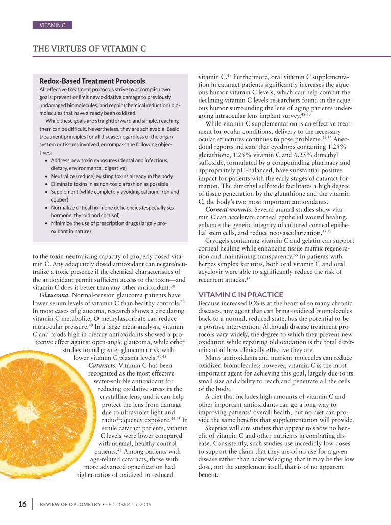
The Virtues of Vitamin C
This substance is critical to eye health and function. Here’s why it should be an important part of your arsenal.
BY THOMAS E. LEVY, MD, JD
VITAMIN C
Increased oxidative stress in and around the eye, as well as elsewhere in the body, is the most consistently iden-tifiable evidence of any ongoing disease process. Anti-oxidants, and vitamin C in particular, can be a crucial
addition to anyone’s nutritional health to stave off such oxidative stress and the subsequent disease processes. For eye health specifically, vitamin C deficiency is characteris-tic of many common eye disorders.
Maintaining a higher intake of vitamin C reduces the incidence of ocular conditions such as cataract, glaucoma and age-related macular degeneration (AMD).1-3 Prospec-tive studies support the notion that increased vitamin C can also help slow disease progression and occasionally even reverse it, depending largely on the chronicity of the condition and the level of antioxidant supplementation.
Antioxidant molecules such as the carotenoids lutein, astaxanthin and zeaxanthin, along with vitamin E, are important in minimizing oxidative stress in the eye. While vitamin C is not mentioned nearly as often as a protective antioxidant in the eye as other antioxidants, it remains the pivotal antioxidant for minimizing ocular oxidative stress, since it is essential for keeping these other antioxidant molecules in the reduced, protective state.4
The soundest clinical approach is to keep the body sup-plied with a broad spectrum of antioxidant protection, with a special focus on high vitamin C blood levels.
An abundance of literature reaches the same conclu-sion: the greater your intake (whether dietary or supple-mental) of vitamin C, the longer you live. Studies show that maintaining higher blood levels of vitamin C results in greater longevity, and the chance of death from any
cause is clearly reduced.5-14 This includes critically ill patients given intravenous vitamin C.15
DEFINING DISEASEBiomolecules—such as proteins, sugars, enzymes, fats, nucleic acid—work best when they are in a chemically reduced state (having a full contingent of electrons). When they lose electrons to a pro-oxidant, they become oxidized and lose some or all of their normal functions and roles in metabolic pathways. Thus, all disease or tis-sue damage is secondary to the degree to which a given array of biomolecules have been oxidized.
Although biomolecules can be subject to oxida-tion anywhere in the body, the lion’s share of oxidative damage takes place inside the affected cells of a given dis-ease. Without oxidative stress inside a cell, it is physiologi-cally normal and does not contribute to any disease process. While many clinical researchers would assert that increased oxi-dation causes disease, it is more accurate to say that increased oxidation (a higher quantity of oxidized biomolecules) is dis-ease.
014_ns1019_vitamins.indd 14014_ns1019_vitamins.indd 14 10/14/19 11:10 AM10/14/19 11:10 AM

REVIEW OF OPTOMETRY • OCTOBER 15, 2019 15
This means that the degree to which biomolecules are oxidized is the entire determining factor as to how advanced a disease or medical condition might be. Addi-tionally, it directly determines the extent and degree of the symptoms that are present. It is also an important factor in determining the efficacy of any given therapeutic protocol.
All oxidizing agents are toxic, and all toxins are oxidiz-ing agents in some fashion. Toxins cause their damage by oxidizing a unique variety and concentration of biomol-ecules based on the access afforded the toxin by its unique chemical structure.
The only way to ameliorate, stabilize, reverse or even clinically cure a given condition is by restoring and maintaining enough oxidized biomolecules to a reduced, normal state with a sufficient influx of antioxidant mol-ecules.16
SUCCESS BEGINS WITH CVitamin C, as the premier antioxidant in the body, is the primary supplier of the fuel—electrons—on which every cell in the body runs. Some of the characteristics that make it the body’s most important antioxidant include its small molecular size; its similarity to glucose (allow-ing insulin to enhance intracellular uptake); its ability to penetrate all cells and tissues, including crossing the blood-brain barrier; its capability to donate two electrons per molecule rather than one; and its ability to regenerate other significant oxidized antioxidants. Additionally, vita-min C has an intermediate, stable form (ascorbyl radical) that can act as a buffer in a given microenvironment.
Vitamin C has also been documented to support the immune system in a number of different ways:17-25
• Enhanced phagocytic function of white blood cells• Enhanced T lymphocyte response, proliferation and
longevity• Enhanced B lymphocyte proliferation and antibody
production • Enhanced immune system comple-
ment activity• Enhanced interferon produc-
tion• Strongly enhanced natural
killer cell activityMore specifically, vitamin
C has a long track record
of con-fer-ring
benefits for any number of
ocular conditions:
Retinal disease. The retina is one of the highest oxy-gen-consuming tissues in the body, with a metabolism that generates large amounts of reactive oxygen species (pro-oxidants).26 As such, the retina is a tissue in need of substantial antioxidant protection to prevent or minimize the evolution of diseases related to oxidation-impaired function.27
In many ways, diabetes is the prototypical disease for demonstrating the negative health impact of increased intracellular oxidative stress (IOS). When vitamin C and magnesium levels sufficiently decline and calcium levels significantly rise inside cells, a metabolic state of elevated IOS always exists.
Part of the reason that intracellular levels of vitamin C are depleted in poorly controlled diabetes patients is that glucose and vitamin C directly compete with each for cel-lular uptake, as they are chemically similar molecules; the uptake of each is further facilitated by insulin, in contrast to passive uptake. The higher the blood glucose levels, the less vitamin C is able to enter the cells.28,29
In proliferative diabetic retinopathy, the degree of increased oxidative stress is massive. Such patients have a ten-fold decrease in vitamin C levels in the vitreous humor, and the degree of macular ischemia further correlates with the degree of vitamin C depletion.30
A study of 479 patients with nonproliferative diabetic retinopathy found that vitamin C given with statins resulted in a reduced complication rate.31 In a secondary randomized study of these patients, researchers noted a stabilization of macular degeneration with improved visual acuity scores with regular supplementation of lutein, zeaxanthin and astaxanthin over a two-year period.32
Even in the absence of diabetes, this disease is related to increased oxidative stress, and research shows vitamin C significantly reduces the oxidative stress in human retinal pigment epithelial cells.33 Supplemental antioxidants can be protective against AMD, and can significantly improve an array of visual function parameters in patients with AMD.34,35
In vitro, vitamin C has also demonstrated a dose-dependent effect in controlling the rate of cell replication in another retinal disease, proliferative vitreoretinopathy.27
Iatrogenic disease. Many toxins, often in the form of prescription medicines with toxic side effects, can cause oxidative damage to the retina and end up causing visual deterioration. As the most significant antioxidant in the body, vitamin C is the premier antitoxin. Acute poison-ings unresponsive to all traditional measures will reliably be resolved by the acute administration of high amounts of vitamin C, as long as clinical deterioration is not too advanced.37 No toxin has ever been shown to be refractory
014_ns1019_vitamins.indd 15014_ns1019_vitamins.indd 15 10/14/19 11:11 AM10/14/19 11:11 AM
REVIEW OF OPTOMETRY • OCTOBER 15, 201916
to the toxin-neutralizing capacity of properly dosed vita-min C. Any adequately dosed antioxidant can negate/neu-tralize a toxic presence if the chemical characteristics of the antioxidant permit sufficient access to the toxin—and vitamin C does it better than any other antioxidant.38
Glaucoma. Normal-tension glaucoma patients have lower serum levels of vitamin C than healthy controls.39 In most cases of glaucoma, research shows a circulating vitamin C metabolite, O-methylascorbate can reduce intraocular pressure.40 In a large meta-analysis, vitamin C and foods high in dietary antioxidants showed a pro-tective effect against open-angle glaucoma, while other
studies found greater glaucoma risk with lower vitamin C plasma levels.41-43
Cataracts. Vitamin C has been recognized as the most effective
water-soluble antioxidant for reducing oxidative stress in the crystalline lens, and it can help protect the lens from damage due to ultraviolet light and radiofrequency exposure.44,45 In senile cataract patients, vitamin C levels were lower compared
with normal, healthy control patients.46 Among patients with
age-related cataracts, those with more advanced opacification had
higher ratios of oxidized to reduced
vitamin C.47 Furthermore, oral vitamin C supplementa-tion in cataract patients significantly increases the aque-ous humor vitamin C levels, which can help combat the declining vitamin C levels researchers found in the aque-ous humor surrounding the lens of aging patients under-going intraocular lens implant survey.48-50
While vitamin C supplementation is an effective treat-ment for ocular conditions, delivery to the necessary ocular structures continues to pose problems.51,52 Anec-dotal reports indicate that eyedrops containing 1.25% glutathione, 1.25% vitamin C and 6.25% dimethyl sulfoxide, formulated by a compounding pharmacy and appropriately pH-balanced, have substantial positive impact for patients with the early stages of cataract for-mation. The dimethyl sulfoxide facilitates a high degree of tissue penetration by the glutathione and the vitamin C, the body’s two most important antioxidants.
Corneal wounds. Several animal studies show vita-min C can accelerate corneal epithelial wound healing, enhance the genetic integrity of cultured corneal epithe-lial stem cells, and reduce neovascularization.53,54
Cryogels containing vitamin C and gelatin can support corneal healing while enhancing tissue matrix regenera-tion and maintaining transparency.55 In patients with herpes simplex keratitis, both oral vitamin C and oral acyclovir were able to significantly reduce the risk of recurrent attacks.56
VITAMIN C IN PRACTICEBecause increased IOS is at the heart of so many chronic diseases, any agent that can bring oxidized biomolecules back to a normal, reduced state, has the potential to be a positive intervention. Although disease treatment pro-tocols vary widely, the degree to which they prevent new oxidation while repairing old oxidation is the total deter-minant of how clinically effective they are.
Many antioxidants and nutrient molecules can reduce oxidized biomolecules; however, vitamin C is the most important agent for achieving this goal, largely due to its small size and ability to reach and penetrate all the cells of the body.
A diet that includes high amounts of vitamin C and other important antioxidants can go a long way to improving patients’ overall health, but no diet can pro-vide the same benefits that supplementation will provide.
Skeptics will cite studies that appear to show no ben-efit of vitamin C and other nutrients in combating dis-ease. Consistently, such studies use incredibly low doses to support the claim that they are of no use for a given disease rather than acknowledging that it may be the low dose, not the supplement itself, that is of no apparent benefit.
Redox-Based Treatment ProtocolsAll effective treatment protocols strive to accomplish two
goals: prevent or limit new oxidative damage to previously
undamaged biomolecules, and repair (chemical reduction) bio-
molecules that have already been oxidized.
While these goals are straightforward and simple, reaching
them can be difficult. Nevertheless, they are achievable. Basic
treatment principles for all disease, regardless of the organ
system or tissues involved, encompass the following objec-
tives:
• Address new toxin exposures (dental and infectious,
dietary, environmental, digestive)
• Neutralize (reduce) existing toxins already in the body
• Eliminate toxins in as non-toxic a fashion as possible
• Supplement (while completely avoiding calcium, iron and
copper)
• Normalize critical hormone deficiencies (especially sex
hormone, thyroid and cortisol)
• Minimize the use of prescription drugs (largely pro-
oxidant in nature)
VITAMIN C
THE VIRTUES OF VITAMIN C
014_ns1019_vitamins.indd 16014_ns1019_vitamins.indd 16 10/14/19 11:11 AM10/14/19 11:11 AM
REVIEW OF OPTOMETRY • OCTOBER 15, 2019 17
For anyone still on the fence about vitamin C supple-mentation, rest assured that they are not prescription drugs and have little to no negative impact on the health, even with a high dosage. Given the amount of research now documenting their widespread benefits and nearly nonexistent toxicity, clinicians should have no fear in promoting their use to prevent or ameliorate disease. ❍
Dr. Levy is a consultant at Riordan Clinic in Wichita, KS.
1. Dherani M, Murthy G, Gupta S, et al. Blood levels of vitamin C, carotenoids and retinol are
inversely associated with cataract in a North Indian population. Invest Ophthalmol Vis Sci.
2008;49(8):3328-35.
2. Ravindran R, Vashist P, Gupta S, et al. Inverse association of vitamin C with cataract in
older people in India. Ophthalmology. 2011;118(10):1958-65.
3. Braakhuis A, Raman R, Vaghefi E. The association between dietary intake of antioxidants
and ocular disease. Diseases. 2017;5(1):pii:E3.
4. Lien E, Hammond B. Nutritional influences on visual development and function. Prog Retin
Eye Res. 2011;30(3):188-203.
5. Losonczy K, Harris T, Havlik R. Vitamin E and vitamin C supplement use and risk of all-
cause and coronary heart disease mortality in older persons: the Established Populations for
Epidemiologic Studies of the Elderly. Am J Clin Nutr. 1996;64(2):190-6.
6. Kromhout D, Bloemberg B, Feskens E, et al. Saturated fat, vitamin C and smoking predict
long-term population all-cause mortality rates in the Seven Countries Study. Int J Epidemiol.
2000;29(2):260-5.
7. Khaw K, Bingham S, Welch A, et al. Relation between plasma ascorbic acid and mortality in
men and women in EPIC-Norfolk prospective study: a prospective population study. European
Prospective Investigation into Cancer and Nutrition. Lancet. 2001;357(9257):657-63.
8. Simon J, Hudes E, Tice J. Relation of serum ascorbic acid to mortality among US adults. J
Amer Coll Nutr. 2001;20(3):255-63.
9. Dashti-Khavidaki S, Talasaz A, Tabeefar H, et al. Plasma vitamin C concentrations in
patients on routine hemodialysis and its relationship to patients’ morbidity and mortality. Int J
Vitam Nutr Res. 2011;81(4):197-203.
10. Ford E, Li C, Cunningham T, Croft J. Associations between antioxidants and all-cause
mortality among US adults with obstructive lung function. Br J Nutr. 2014;112(10):1662-73.
11. Kobylecki C, Afzal S, Davey Smith G, Nordestgaard B. Genetically high plasma vitamin C,
intake of fruit and vegetables, and risk of ischemic heart disease and all-cause mortality: a
Mendelian randomization study. A J Clin Nutr. 2015;101(6):1135-43.
12. Sotomayor C, Eisenga M, Gomes Neto A, et al. Vitamin C depletion and all-cause mortal-
ity in renal transplant recipients. Nutrients. 2017;9(6):568.
13. Jayedi A, Rashidy-Pour A, Parohan M, et al. Dietary antioxidants, circulating antioxidant
concentrations, total antioxidant capacity, and risk of all-cause mortality: a systematic
review and dose-response meta-analysis of prospective observational studies. Adv Nutr.
2018;9(6):701-16.
14. Ma E, Iso H, Yamagishi K, et al. Dietary antioxidant micronutrients and all-cause mor-
tality: the Japan Collaborative Cohort Study for Evaluation of Cancer Risk. J Epidemiol.
2018;28(9):388-96.
15. Wang Y, Lin H, Lin B, Lin J. Effects of different ascorbic acid doses on the mortality of
critically ill patients: a meta-analysis. Ann Intensive Care. 2019;9(1):58.
16. Levy T. Primal Panacea. Henderson, NV: MedFox Publishing; 2011.
17. de la Fuente M, Ferrandez M, Burgos M, et al. Immune function in aged women is
improved by ingestion of vitamins C and E. Can J of Physiol and Pharmacol. 1998;76:373-
80.
18. Siegel B, Morton J. Vitamin C and the immune response. Experientia. 1977;33(3):393-5.
19. Kennes B, Dumont I, Brohee D, et al. Effect of vitamin C supplements on cell-mediated
immunity in old people. Gerontology. 1983;29(5):305-10.
20. Campbell J, Cole M, Bunditrutavorn B, Vella A. Ascorbic acid is a potent inhibitor of vari-
ous forms of T cell apoptosis. Cellular Immunol. 1999;194(1):1-5.
21. Schwager J, Schulze J. Influence of ascorbic acid on the response to mitogens and inter-
leukin production of porcine lymphocytes. Int J Vitam Nutr Res. 1997;67(1):10-6.
22. Feigen G, Smith B, Dix C, et al. Enhancement of antibody production and protection
against systemic anaphylaxis by large doses of vitamin C. Res Commun Chem Pathol Phar-
macol. 1982;38(2):313-33.
23. Johnston C, Kolb W, Haskell B. The effect of vitamin C nutriture on complement compo-
nent C1q concentrations in guinea pig plasma. J Nutr. 1987;117(4):764-8.
24. Karpinska T, Kawecki Z, Kandefer-Szerszen M. The influence of ultraviolet irradiation,
L-ascorbic acid and calcium chloride on the induction of interferon in human embryo fibro-
blasts. Arch Immunol Therap Exp. 1982;30(1-2):33-7.
25. Heuser G, Vojdani A. Enhancement of natural killer cell activity and T and B cell function
by buffered vitamin C in patients exposed to toxic chemicals: the role of protein kinase-C.
Immunopharmacol Immunotoxicol. 1997;19(3):291-312.
26. Jarrett S, Boulton M. Consequences of oxidative stress in age-related macular degenera-
tion. Mol Aspects Med. 2012;33(4):399-417.
27. Tsang S, Sharma T. Drug-induced retinal toxicity. Adv Exp Med Biol. 2018;1085:227-32.
28. Moser U, Weber F. Uptake of ascorbic acid by human granulocytes. Int J Vitam Nutr Res.
1984;54(1):47-53.
29. Cunningham J. The glucose/insulin system and vitamin C: implications in insulin-depen-
dent diabetes mellitus. J Am Coll Nutr. 1998;17(2):105-8.
30. Park S, Ghim W, Oh S, et al. Association of vitreous vitamin C depletion with diabetic
macular ischemia in proliferative diabetic retinopathy. PLoS One. 2019;14(6):e0218433.
31. Gurreri A, Pazzaglia A, Schiavi C. Role of statins and ascorbic acid in the natural history
of diabetic retinopathy: a new, affordable therapy? Ophthalmic Surg Lasers Imaging Retina.
2019;50(5):S23-7.
32. Piermarocchi S, Saviano S, Parisi V, et al. Carotenoids in Age-related Maculopathy Italian
Study (CARMIS): two-year results of a randomized study. Eur J Ophthalmol. 2012;22(2):216-
25.
33. Yin J, Thomas F, Lang J, Chaum E. Modulation of oxidative stress responses in the
human retinal pigment epithelium following treatment with vitamin C. J Cell Physiol.
2011;226(8):2025-32.
34. Akuffo K, Beatty S, Peto T, et al. The impact of supplemental antioxidants on visual func-
tion in nonadvanced age-related macular degeneration: a head-to-head randomized clinical
trial. Invest Ophthalmol Vis Sci. 2017;58(12):5347-60.
35. Raimundo M, Mira F, Cachulo M, et al. Adherence to a Mediterranean diet, lifestyle and
age-related macular degeneration: the Coimbra Eye Study—report 3. Acta Ophthalmol.
2018;96(8):e926-32.
36. Heckelen A, Hermel M, Kondring B, Schrage N. Ascorbic acid reversibly inhibits prolifera-
tion of retinal pigment epithelial cells. Acta Ophthalmol Scand. 2004;82(5):564-8.
37. Levy T. Curing the Incurable: Vitamin C, Infectious Diseases, and Toxins. Henderson, NV:
MedFox Publishing; 2002.
38. Levy T. Curing the incurable. In: Vitamin C, Infectious Diseases, and Toxins. Henderson,
NV: MedFox Publishing; 2002.
39. Yuki K, Murat D, Kimura I, et al. Reduced-serum vitamin C and increased uric acid levels
in normal-tension glaucoma. Graefes Arch Clin Exp Ophthalmol. 2010;248(2):243-8.
40. Hysi P, Khawaja A, Menni C, et al. Ascorbic acid metabolites are involved in intraocular
pressure control in the general population. Redox Biol. 2019;20:349-53.
41. Ramdas W, Schouten J, Webers C. The effect of vitamins on glaucoma: a systematic
review and meta-analysis. Nutrients. 2018;10:359.
42. Zanon-Moreno V, Ciancotti-Olivares L, Asencio J, et al. Association between a SLC23A2
gene variation, plasma vitamin C levels, and risk of glaucoma in a Mediterranean population.
Mol Vis. 2011;17:2997-3004.
43. Zanon-Moreno V, Ortega-Azorin C, Asensio-Marquez E, et al. A multi-locus genetic risk
score for primary open-angle glaucoma (POAG) variants is associated with POAG risk in a
Mediterranean population: inverse correlations with plasma vitamin C and E concentrations.
Int J Mol Sci. 2017;18(11):2302.
44. Ishikawa Y, Hashizume K, Kishimoto S, et al. Effect of vitamin C depletion on UVR-B
induced cataract in SMP30/GNL knockout mice. Exp Eye Res. 2012;94(1):85-9.
45. Jelodar G, Akbari A, Nazifi S. The prophylactic effect of vitamin C on oxidative stress
indexes in rat eyes following exposure to radiofrequency wave generated by a BTS antenna
model. Int J Radiat Biol. 2013;89(2):128-31.
46. Jalal D, Koorosh F, Fereidoun H. Comparative study of plasma ascorbic acid levels in
senile cataract patients and in normal individuals. Curr Eye Res. 2009;34(2):118-122.
47. Kisic B, Miric D, Zoric L, et al. Antioxidant capacity of lenses with age-related cataract.
Oxid Med Cell Longev. 2012;2012:467130.
48. Iqbal Z, Midgley J, Watson D, et al. Effect of oral administration of vitamin C on human
aqueous humor ascorbate concentration. Acta Pharmacol Sin. 1999;20(10):879-83.
49. Hah Y, Chung H, Sontakke S, et al. Ascorbic acid concentrations in aqueous humor after
systemic vitamin C supplementation in patients with cataract: pilot study. BMC Ophthalmol.
2017;17(1):121.
50. Canadananovic V, Latinovic S, Barisic S, et al. Age-related changes of vitamin C levels in
aqueous humor. Vojnosanitet Pregl. 2015;72(9):823-6.
51. Umapathy A, Donaldson P, Lim J. Antioxidant delivery pathways in the anterior eye.
Biomed Res Int. 2013;2013:207250.
52. Abdelkader H, Alany R, Pierscionek B. Age-related cataract and drug therapy: oppor-
tunities and challenges for topical antioxidant delivery to the lens. J Pharm Pharmacol.
2015;67(4):537-50.
53. Chen J, Lan J, Liu D, et al. Ascorbic acid promotes the stemness of corneal epithelial
stem/progenitor cells and accelerates epithelial wound healing in the cornea. Stem Cells
Transl Med. 2017;6(5):1356-65.
54. Lee M, Chung S. Treatment of corneal neovascularization by topical application of ascor-
bic acid in the rabbit model. Cornea. 2012;31(10):1165-9.
55. Luo L, Lai J, Chou S, et al. Development of gelatin/ascorbic acid cryogels for potential
use in corneal stromal tissue engineering. Acta Biomater. 2018;65:123-36.
56. Kim G, Yoo W, Park M, et al. Clinical features of herpes simplex keratitis in a Korean
tertiary referral center: efficacy of oral antiviral and ascorbic acid on recurrence. Korean J
Ophthalmol. 2018;32(5):353-60.
014_ns1019_vitamins.indd 17014_ns1019_vitamins.indd 17 10/14/19 11:12 AM10/14/19 11:12 AM

REVIEW OF OPTOMETRY • OCTOBER 15, 201918
Myopia is perhaps the most common diagnosis we optometrists encounter each and every day. Its prevalence is quite high in all age groups—estimated between 22% and 30%
of the world population, and more than 50% in many industrialized countries—with significant progression in habitual computer users that extends over a decade beyond early adulthood.1
Because corrective lens wear is readily available and widely successful in counteracting myopic refractive error, clinicians may lack a sense of urgency about the need to intervene. But it is important to note that the burden of myopic macular degeneration and associated blindness is expected to increase significantly, fueled by a rise in myopia’s global prevalence in children from 312 million in 2015 to 324 million in 2025.2
In short, myopia is more than just a refractive prob-lem. It is an ocular health risk as well.
We at the OWNS are interested in a broad continuing dialogue concerning environmental factors involved in reducing the burden of myopia. More importantly, we wish to contribute to improved clinical practice.
INFLUENCE OF ACCOMMODATIVE LAG Most busy practitioners likely haven’t thought very deeply about the causes of myopia since optometry school, which may be expected as competing clinical responsibilities take precedence. Let’s review the mecha-nisms of myopia so we might be better attuned to the modifiable risk factors within our reach.
Though it’s a source of some debate, sustained hyper-opic defocus is likely behind the etiology, onset and progression of myopia. Those espousing this view believe
that a sustained lag of accommodation during continu-ous near work creates a chromatic signal encoded by retinal neurons and transformed into a neural endocrine signal for growth processes in the retina, choroid and sclera.3,4 The optical stimuli at the retina that specify focus (e.g., comparison of luminance contrast between cone types enabled by longitudinal chromatic aberration) to control accommodation have also been demonstrated to drive eye growth mechanisms.3-5
Although the developing eye can attain emmetropia or compensate for lens-induced defocus without accom-modation, the accuracy of accommodation is thought to be important for the process of emmetropization.4 It is believed that the eye alters its growth to minimize
Influencing Myopia: Science and Clinical Practice
In search of dietary and lifestyle changes that could alter the course of childhood refractive status and quality of life.
BY KARAN R. GREGG AGGARWALA, OD (NIH EQUIV), PhD, AND STUART P. RICHER, OD, PhD
MYOPIA
Ortho-K lenses allow patients to enjoy correction-free vision until they reapply them at bedtime, as corneal reshaping occurs overnight.
018_ns1019_myopia.indd 18018_ns1019_myopia.indd 18 10/14/19 11:13 AM10/14/19 11:13 AM

REVIEW OF OPTOMETRY • OCTOBER 15, 2019 19
the time-averaged blurring of the retinal image, which depends upon refractive state, accuracy of accommoda-tion and proportion of time spent at each viewing dis-tance.6,7
Authors of a Singapore study on children ages seven to nine suggest an association of myopia with number of books read per week and lament that factors such as attention level or patterns of temporal interruption (e.g., brief periods of distance viewing) are important but rarely studied.8 The study also noted the three-year change in axial length of the children at baseline was high and greater in those who were younger, females and those with a parental history of myopia.
Peripheral retinal cues can direct eye growth, with or without involvement of the foveal region in primates, but their role in humans has been difficult to demon-strate.9-11
Other important physiological variables that may con-tribute to the onset and progression of myopia include ciliary muscle fatigue, intraocular pressure, scleral dis-tensibility and scleral remodeling.
AMBIENT LIGHT FACTORSA recent study in rainbow trout demonstrated that ocu-lar elongation is inhibited when the spectral composition of white light is dominated by short wavelength blue light or is lacking the red component.12 An environment that deprives the eye of blue light stimulation—either from low color temperature tungsten bulbs, dim illumi-nation, spectral filtering by spectacles or reduced sun-light exposure—will tend to make the eye vulnerable to myopia at low temporal flicker frequencies.13
Without blue light, the compensatory changes in choroidal thickness (moving the retina anteriorly) are insufficient to counter the associated elongation of the eye, and long wavelength light is known to induce a myopic shift. It is speculated that the myopia-inhibiting effect of blue light may be related to preferential stimulation of the ‘ON’ retinal pathway.
In closed environments, shifting the dominant wavelength of ambient white light to the short wavelength blue or violet region (360nm to 400nm) reduces ocu-lar elongation.14 Commercially available “cold white” LED bulbs (approximately 6500K color temperature, 7000 to 10,000 lumens) used at a distance of about two meters, may provide adequate yet safe lev-els of blue light.13
The effect of the short wavelength (i.e.,
near UV, violet and blue) component of white light on retarding the development of myopia is diminished if the light intensity is low. A wider spectral bandwidth of illumination induces wavelength-dependent defocus and improves the accuracy of dynamic accommodation. Integration of multiple visual cues from the retina plays a critical role in determining the direction of refractive change. Recent work also highlights the role of illumina-tion (spectrum, intensity, temporal frequency), circadian rhythm disruption and light-induced changes in sex hor-mones.15
ENVIRONMENTAL FACTORS AND INTERVENTIONSWhile genetics were once at the forefront of thought on why people develop myopia, this philosophy has shifted to include environmental factors, such as vitamin D levels and limited time spent outdoors.16,17 Ethnic differ-ences in myopia and the effects of parental myopia now seem likely to be explained, in part, by environmental influences.16
A 2007 study looked at whether parental history of myopia and children’s outdoor activity levels could predict juvenile-onset myopia. Researchers found lower amounts of recreation and outdoor activity increased the odds of becoming myopic in children with two myopic parents—more than in children with either no or one myopic parent.17
Neural biomarkers of myopia have recently been identified, suggesting that high myopia is associated with abnormalities in several parts of the brain involved in visuomotor integration, the sensorimotor network and the limbic system.
The global prevalence of myopia has grown by 66% in the past three decades, and it has been estimated that nearly half of the world’s population will be myopic by 2050.1,2
018_ns1019_myopia.indd 19018_ns1019_myopia.indd 19 10/14/19 11:14 AM10/14/19 11:14 AM
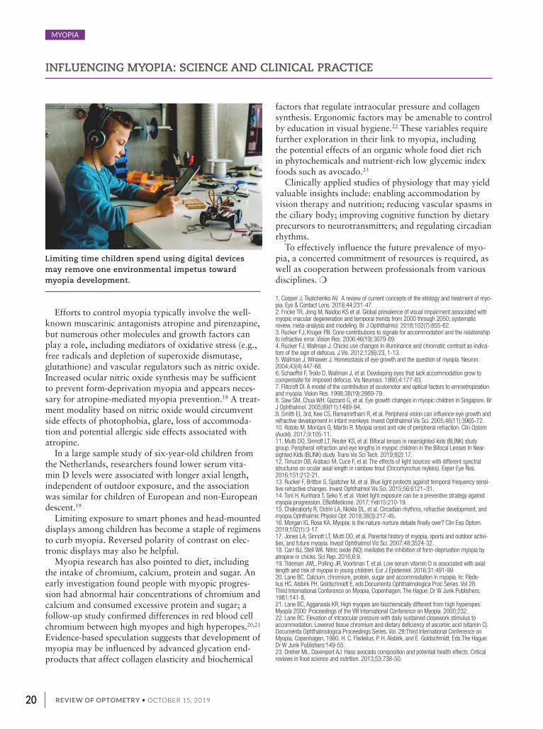
REVIEW OF OPTOMETRY • OCTOBER 15, 201920
Efforts to control myopia typically involve the well-known muscarinic antagonists atropine and pirenzapine, but numerous other molecules and growth factors can play a role, including mediators of oxidative stress (e.g., free radicals and depletion of superoxide dismutase, glutathione) and vascular regulators such as nitric oxide. Increased ocular nitric oxide synthesis may be sufficient to prevent form-deprivation myopia and appears neces-sary for atropine-mediated myopia prevention.18 A treat-ment modality based on nitric oxide would circumvent side effects of photophobia, glare, loss of accommoda-tion and potential allergic side effects associated with atropine.
In a large sample study of six-year-old children from the Netherlands, researchers found lower serum vita-min D levels were associated with longer axial length, independent of outdoor exposure, and the association was similar for children of European and non-European descent.19
Limiting exposure to smart phones and head-mounted displays among children has become a staple of regimens to curb myopia. Reversed polarity of contrast on elec-tronic displays may also be helpful.
Myopia research has also pointed to diet, including the intake of chromium, calcium, protein and sugar. An early investigation found people with myopic progres-sion had abnormal hair concentrations of chromium and calcium and consumed excessive protein and sugar; a follow-up study confirmed differences in red blood cell chromium between high myopes and high hyperopes.20,21 Evidence-based speculation suggests that development of myopia may be influenced by advanced glycation end-products that affect collagen elasticity and biochemical
factors that regulate intraocular pressure and collagen synthesis. Ergonomic factors may be amenable to control by education in visual hygiene.22 These variables require further exploration in their link to myopia, including the potential effects of an organic whole food diet rich in phytochemicals and nutrient-rich low glycemic index foods such as avocado.23
Clinically applied studies of physiology that may yield valuable insights include: enabling accommodation by vision therapy and nutrition; reducing vascular spasms in the ciliary body; improving cognitive function by dietary precursors to neurotransmitters; and regulating circadian rhythms.
To effectively influence the future prevalence of myo-pia, a concerted commitment of resources is required, as well as cooperation between professionals from various disciplines. ❍
1. Cooper J, Tkatchenko AV. A review of current concepts of the etiology and treatment of myo-pia. Eye & Contact Lens. 2018;44:231-47.2. Fricke TR, Jong M, Naidoo KS et al. Global prevalence of visual impairment associated with myopic macular degeneration and temporal trends from 2000 through 2050: systematic review, meta-analysis and modeling. Br J Opththalmol. 2018;102(7):855-62.3. Rucker FJ, Kruger PB. Cone contributions to signals for accommodation and the relationship to refractive error. Vision Res. 2006;46(19):3079-89.4. Rucker FJ, Wallman J. Chicks use changes in illuminance and chromatic contrast as indica-tors of the sign of defocus. J Vis. 2012;12(6):23, 1-13.5. Wallman J, Winawer J. Homeostasis of eye growth and the question of myopia. Neuron. 2004;43(4):447-68. 6. Schaeffel F, Troilo D, Wallman J, et al. Developing eyes that lack accommodation grow to compensate for imposed defocus. Vis Neurosci. 1990;4:177-83.7. Flitcroft DI. A model of the contribution of oculomotor and optical factors to emmetropization and myopia. Vision Res. 1998;38(19):2869-79.8. Saw SM, Chua WH, Gazzard G, et al. Eye growth changes in myopic children in Singapore. Br J Ophthalmol. 2005;89(11):1489-94.9. Smith EL 3rd, Kee CS, Ramamirtham R, et al. Peripheral vision can influence eye growth and refractive development in infant monkeys. Invest Ophthalmol Vis Sci. 2005;46(11):3965-72.10. Rotolo M, Montani G, Martin R. Myopia onset and role of peripheral refraction. Clin Optom (Auckl). 2017;9:105-11. 11. Mutti DO, Sinnott LT, Reuter KS, et al. Bifocal lenses in nearsighted kids (BLINK) study group. Peripheral refraction and eye lengths in myopic children in the Bifocal Lenses In Near-sighted Kids (BLINK) study. Trans Vis Sci Tech. 2019;8(2):17. 12. Timucin OB, Arabaci M, Cuce F, et al. The effects of light sources with different spectral structures on ocular axial length in rainbow trout (Oncorhynchus mykiss). Exper Eye Res. 2016;151:212-21.13. Rucker F, Britton S, Spatcher M, et al. Blue light protects against temporal frequency sensi-tive refractive changes. Invest Ophthalmol Vis Sci. 2015;56:6121–31.14. Torii H, Kurihara T, Seko Y, et al. Violet light exposure can be a preventive strategy against myopia progression. EBioMedicine. 2017; Feb15:210-19.15. Chakraborty R, Ostrin LA, Nickla DL, et al. Circadian rhythms, refractive development, and myopia.Ophthalmic Physiol Opt. 2018;38(3):217-45. 16. Morgan IG, Rose KA. Myopia: is the nature-nurture debate finally over? Clin Exp Optom. 2019;102(1):3-17. 17. Jones LA, Sinnott LT, Mutti DO, et al. Parental history of myopia, sports and outdoor activi-ties, and future myopia. Invest Ophthalmol Vis Sci. 2007;48:3524-32.18. Carr BJ, Stell WK. Nitric oxide (NO) mediates the inhibition of form-deprivation myopia by atropine in chicks. Sci Rep. 2016;6:9.19. Tideman JWL, Polling JR, Voortman T, et al. Low serum vitamin D is associated with axial length and risk of myopia in young children. Eur J Epidemiol. 2016;31:491-99.20. Lane BC. Calcium, chromium, protein, sugar and accommodation in myopia. In: Flede-lius HC, Alsbirk PH, Goldschmidt E, eds.Documenta Ophthalmologica Proc Series. Vol 28. Third International Conference on Myopia, Copenhagen, The Hague: Dr W Junk Publishers; 1981:141-8.21. Lane BC, Aggarwala KR. High myopes are biochemically different from high hyperopes. Myopia 2000: Proceedings of the VIII International Conference on Myopia. 2000;232.22. Lane BC. Elevation of intraocular pressure with daily sustained closework stimulus to accommodation: Lowered tissue chromium and dietary deficiency of ascorbic acid (vitamin C). Documenta Ophthalmologica Proceedings Series. Vol. 28:Third International Conference on Myopia, Copenhagen, 1980. H. C. Fledelius, P. H. Alsbirk, and E. Goldschmidt, Eds.The Hague: Dr W Junk Publishers:149-55.23. Dreher ML, Davenport AJ. Hass avocado composition and potential health effects. Critical reviews in food science and nutrition. 2013;53:738-50.
MYOPIA
INFLUENCING MYOPIA: SCIENCE AND CLINICAL PRACTICE
Limiting time children spend using digital devices may remove one environmental impetus toward myopia development.
018_ns1019_myopia.indd 20018_ns1019_myopia.indd 20 10/14/19 11:14 AM10/14/19 11:14 AM

REVIEW OF OPTOMETRY • OCTOBER 15, 2019 21
Dietary lutein, zeaxanthin and mesozeaxanthin (an isomer of lutein) are important for maintaining a healthy retina.
They aggregate in the macula more so than anywhere else in the body and compose the macular pigment (MP).1,2 In the retina, they reduce the risk for age-related macular degenera-tion (AMD), as well as improve visual function.3,4 The macular carotenoids protect photoreceptors and the retinal pigment epithelium (RPE) by serv-ing as active antioxidants, absorbing short-wave light capable of damaging retinal tissue and by reducing inflam-mation.5
A less well-known carotenoid called astaxanthin, found in red algae, salmon and krill oil, complements plant-based carotenoids, supporting many functions the carotenoids don’t (Table 1). While the three carotenoids help protect the retina and filter blue light, astaxanthin primarily supports the front of the eye and has been shown to have anti-tumor, anti-diabetic, anti-inflamma-tory and reactive oxygen species scavenging properties in humans. Animal models also show astaxanthin has protective effects on mitochondria and retinal ganglion cells.6-9 Astaxanthin does not convert to vitamin A, so, unlike high doses of beta carotene, in this respect it is non-toxic if taken orally.10 Human studies strongly sup-port astaxanthin’s consideration as a complementary dietary supplement.
1st LINE OF DEFENSE: GREEN LEAFY VEGGIES AND FRUITSLutein, zeaxanthin and mesozeaxan-thin are all antioxidants and free-rad-ical scavengers that benefit the retina by filtering light before it reaches the photoreceptors and RPE, which staves off damage.5 This is crucial for several disease processes and serves to enhance both visual and cognitive function:
AMD. Several studies have linked carotenoids with AMD risk (Figure 1).3,11,12 Large cohort studies suggest
that individuals with lower dietary levels of lutein and zeaxanthin are at increased risk for developing AMD.11-13 Other studies suggest that higher MP optical density (MPOD), which represents longer-term lutein and zeaxanthin dietary intakes, is related to reduced risk for AMD.14
Building macular pigment is important. The Central Retinal Enrichment Supplementation Trial 2 found patients with non-advanced AMD who took antioxidant AREDS2 supplementation with or without mesozea-xanthin had significant increases in MP and improved contrast sensitivity.15 AREDS2 suggests that antioxidant supplementation with lutein and zeaxanthin can reduce risk of progression to late-stage AMD.
Beyond prevention, research also suggests lutein and zeaxanthin may improve responses to standard treat-ments for wet AMD.
Carotenoids: Front to Back Ocular Protection
Adding the marine newcomer, astaxanthin, to your plant food–based diet can fill in the ocular health gaps left by lutein and zeaxanthin.
BY STUART RICHER, OD, PhD, DOROTHY HITCHMOTH, OD, AND LISA RENZI-HAMMOND, PhD
CAROTENOIDS
FIG. 1. Abundant evidence shows that AMD risk is elevated in patients deficient in lutein and zeaxanthin.
Photo
: Natio
nal E
ye In
stitite/N
IH
021_ns1019_carotenoids.indd 21021_ns1019_carotenoids.indd 21 10/14/19 11:15 AM10/14/19 11:15 AM

REVIEW OF OPTOMETRY • OCTOBER 15, 201922
CAROTENOIDS: FRONT TO BACK OCULAR PROTECTION
CAROTENOIDS
A recent trial found oral supplementation with zeaxan-thin in addition to triple therapy (photodynamic therapy plus intravitreal administration of bevacizumab and dexa-methasone) led to improved visual function, with 27% of eyes gaining ≥15 letters compared with 9% in eyes treated with triple therapy alone. Adding oral zeaxanthin also led to a 74% reduced incidence of subsequent neovascular AMD in fellow eyes compared with eyes treated with triple therapy alone.14 Supplementation may also lengthen the time between treatment cycles.15
REAR GUARD: SALMON AND KRILL OILAstaxanthin resides within krill and is what gives flamin-gos and salmon their distinct coloring. It’s also found in shrimp, lobster and other orange-red marine life. In cells, it’s mostly found in mitochondrial membranes, and has a unique chemical structure that allows it to fully span and protect cellular membranes with potent antioxidant and anti-inflammatory properties. It is believed to cross both the blood-retinal and blood-brain barriers.
Astaxanthin may help protect the RPE mitochondria, reduction of which is linked with AMD. The carotenoid may also ward off the increased mitochondrial DNA (mtDNA) lesions and reduced mtDNA repair capacity also associated with AMD.16,17 Astaxanthin increases mitochondrial biogenesis and upregulates both the genes involved in energy production and cytochrome c, which is a component of the mitochondrial electron transport chain and a major PGC-1α-inducible protein.18-20
Astaxanthin is a considerably stronger antioxidant than zeaxanthin, canthaxanthin, lutein, B-carotene and alpha-tocopherol.21 Such a protective role is crucial for several
anterior segment conditions, including: Accommodation and eye strain. Astaxanthin can help
to improve accommodative speed, accommodative ampli-tude, near point of focus and depth perception (Figure 2). These are all important to help focus and re-focus strained eyes, reduce eye fatigue and assist in visual recovery from screen time. One study shows a 21.7% increase in accom-modative amplitude after four weeks of taking astaxan-thin at 5mg/day.22
Astaxanthin also promotes circulation and nourishment of anterior and posterior segment eye tissue while allevi-ating oxidative stress. Specifically, astaxanthin increases ciliary muscle recovery and endurance, improves capillary blood flow and other rheology parameters and inhibits inflammation via the NF-kB pathway.
Dry eye. Astaxanthin plays a unique role in dry eye by providing antioxidant protection for the ocular surface and anti-inflammatory action to support lacrimation, reduce subjective symptoms and promote upregulation of ocular hydration aquaporin channels (Figure 3).
One study followed 22 patients between the ages of 45 and 65 who took astaxanthin 6mg/day and found those
Pre-AMD Astaxanthin Pearls• Protects mitochondria from oxidative stress.
• Reduces UVB-induced oxidative stress.
• Promotes mitochondrial biogenesis.
• Promotes higher mitochondrial energy output.
• Protects lipids from oxidation.
• Promotes retinal blood flow.
• Provides anti-inflammatory properties (NFkB).
Ciliary muscle fatigue causes asthenopia or accommodation failure
Suspensory ligamentsConjunctiva
Sclera
Choroid
Retina
Fovea
Optic nerve
Blind spot
Tendon of rectus muscleCiliary body (containing ciliary muscle)
Posterior chamber
Iris
Cornea
Pupil
Anterior chamber (containing aqueous humor)
Cause of Eye Fatigue
Poor blood flow cannot support highly metabolically active organ
Eye inflammation,e.g. visible swelling, sensations of heat and pain redness
gaments
Sclera
Cho
Ret
O
Blind spot
y (containing
mber
bereous
Astaxanthin
Increases ciliary muscle recovery and endurance
Improves capillary blood flow and blood rheology
Inhibits inflammation via NF-kB pathway
Source
: Asta
Real
FIG. 2. Potential mechanisms by which astaxanthin can help to improve accommodative and vascular function.
021_ns1019_carotenoids.indd 22021_ns1019_carotenoids.indd 22 10/14/19 11:16 AM10/14/19 11:16 AM
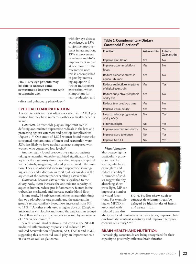
REVIEW OF OPTOMETRY • OCTOBER 15, 2019 23
with dry eye disease experienced a 15% subjective improve-ment in lacrimation, 19% improvement in redness and 46% improvement in pain at one month.23 The researchers note this is accomplished in part by increas-ing aquaporin 5 (water transporter) expression, which is important for tear production and
saliva and pulmonary physiology.24
EYE HEALTH AND NUTRITION The carotenoids are most often associated with AMD pre-vention but they have numerous other eye health benefits as well.
Cataracts. Carotenoids play an important role in defusing accumulated superoxide radicals in the lens and protecting against cataracts and post-op complications (Figure 4).25 One study of 1,802 women found those who consumed high amounts of lutein and zeaxanthin were 32% less likely to have nuclear cataract compared with women who consumed low levels.26
Another study found preoperative cataract patients taking astaxanthin 6mg/day exhibited significantly lower aqueous flare intensity three days after surgery compared with controls, suggesting reduced post-surgical inflamma-tion. They also observed increased superoxide scaveng-ing activity and a decrease in total hydroperoxides in the aqueous of the cataract patients taking astaxanthin.27
Glaucoma. Because astaxanthin is localized to the ciliary body, it can increase the antioxidant capacity of aqueous humor, reduce pro-inflammatory factors in the trabecular meshwork and increase ocular blood flow.
In one study, 36 subjects took either astaxanthin 6mg/day or a placebo for one month, and the astaxanthin group’s retinal capillary blood flow increased from 9% to 11%.28 Another study used a higher dose of 12mg/day astaxanthin vs. placebo and found the astaxanthin group’s blood flow velocity at the macula increased by an average of 15% in one month.29
Several animal studies show a reduction in the NF-KB mediated inflammatory response and reduced LPS-induced accumulation of protein, NO, TNF-α and PGE2, suggesting this carotenoid could play an importance role in uveitis as well as glaucoma.
Visual function. Short-wave light is particularly prone to intraocular scatter, which can cause glare and reduce visibility.26 A number of stud-ies suggest that by absorbing short-wave light, MP can improve a number of visual func-tions. For example, higher MPOD is associated with reduced glare dis-ability, reduced photostress recovery times, improved het-erochromatic contrast sensitivity and improved temporal contrast sensitivity.4,30-32
BRAIN HEALTH AND NUTRITION Increasingly, carotenoids are being recognized for their capacity to positively influence brain function.
Table 1. Complementary Dietary Carotenoid Functions53
Function Astaxanthin Lutein/Zeaxanthin
Improve circulation Yes No
Improve accommodation/
focus
Yes No
Reduce oxidative stress in
aqueous humor
Yes No
Reduce subjective symptoms
of digital eye strain
Yes Yes
Reduce subjective symptoms
of dry eye
Yes No
Reduce tear break-up time Yes No
Improve visual acuity Yes Yes
Help to reduce progression
of dry AMD
No Yes
Filter blue light No Yes
Improve contrast sensitivity No Yes
Improve glare tolerance No Yes
Improve MPOD No Yes
FIG. 3. Dry eye patients may be able to achieve some symptomatic improvement with astaxantin use.
Photo
: Cece
lia K
oettin
g, O
D
FIG. 4. Studies show nuclear cataract development can be delayed by high intake of lutein and zeaxanthin.
Photo
: Jacq
uelin
e T
heis, O
D
021_ns1019_carotenoids.indd 23021_ns1019_carotenoids.indd 23 10/14/19 11:16 AM10/14/19 11:16 AM
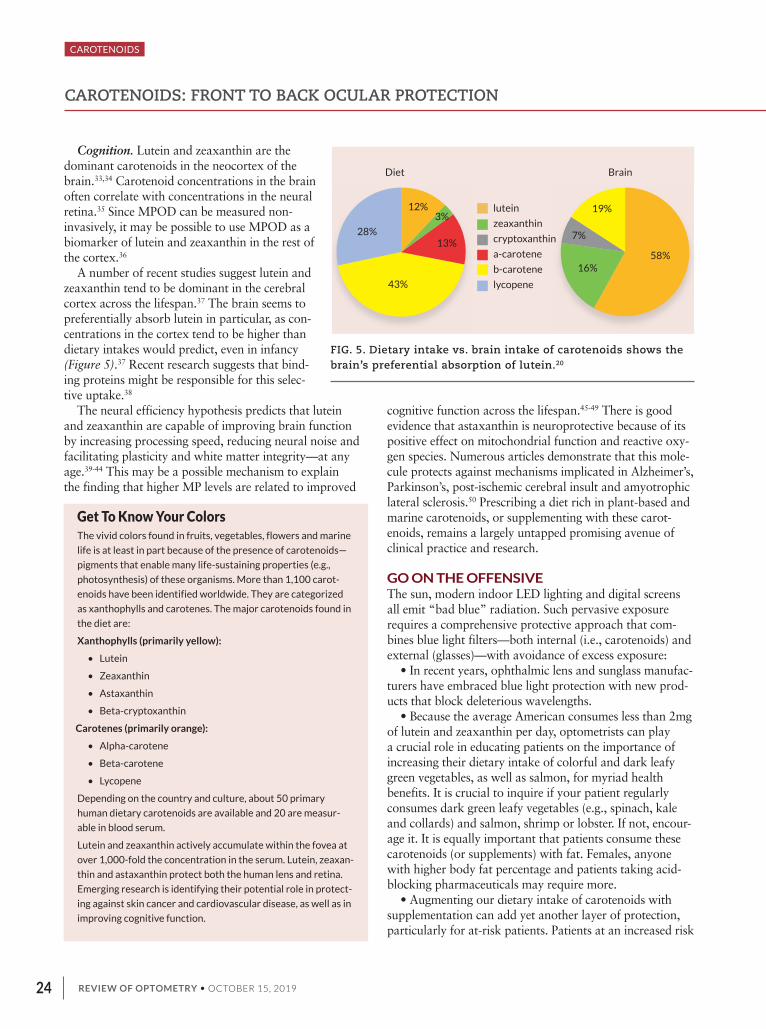
REVIEW OF OPTOMETRY • OCTOBER 15, 201924
Cognition. Lutein and zeaxanthin are the dominant carotenoids in the neocortex of the brain.33,34 Carotenoid concentrations in the brain often correlate with concentrations in the neural retina.35 Since MPOD can be measured non-invasively, it may be possible to use MPOD as a biomarker of lutein and zeaxanthin in the rest of the cortex.36
A number of recent studies suggest lutein and zeaxanthin tend to be dominant in the cerebral cortex across the lifespan.37 The brain seems to preferentially absorb lutein in particular, as con-centrations in the cortex tend to be higher than dietary intakes would predict, even in infancy (Figure 5).37 Recent research suggests that bind-ing proteins might be responsible for this selec-tive uptake.38
The neural efficiency hypothesis predicts that lutein and zeaxanthin are capable of improving brain function by increasing processing speed, reducing neural noise and facilitating plasticity and white matter integrity—at any age.39-44 This may be a possible mechanism to explain the finding that higher MP levels are related to improved
cognitive function across the lifespan.45-49 There is good evidence that astaxanthin is neuroprotective because of its positive effect on mitochondrial function and reactive oxy-gen species. Numerous articles demonstrate that this mole-cule protects against mechanisms implicated in Alzheimer’s, Parkinson’s, post-ischemic cerebral insult and amyotrophic lateral sclerosis.50 Prescribing a diet rich in plant-based and marine carotenoids, or supplementing with these carot-enoids, remains a largely untapped promising avenue of clinical practice and research.
GO ON THE OFFENSIVEThe sun, modern indoor LED lighting and digital screens all emit “bad blue” radiation. Such pervasive exposure requires a comprehensive protective approach that com-bines blue light filters—both internal (i.e., carotenoids) and external (glasses)—with avoidance of excess exposure:
• In recent years, ophthalmic lens and sunglass manufac-turers have embraced blue light protection with new prod-ucts that block deleterious wavelengths.
• Because the average American consumes less than 2mg of lutein and zeaxanthin per day, optometrists can play a crucial role in educating patients on the importance of increasing their dietary intake of colorful and dark leafy green vegetables, as well as salmon, for myriad health benefits. It is crucial to inquire if your patient regularly consumes dark green leafy vegetables (e.g., spinach, kale and collards) and salmon, shrimp or lobster. If not, encour-age it. It is equally important that patients consume these carotenoids (or supplements) with fat. Females, anyone with higher body fat percentage and patients taking acid-blocking pharmaceuticals may require more.
• Augmenting our dietary intake of carotenoids with supplementation can add yet another layer of protection, particularly for at-risk patients. Patients at an increased risk
FIG. 5. Dietary intake vs. brain intake of carotenoids shows the brain’s preferential absorption of lutein.20
28%
43%
13%
12%
Diet Brain
58%
19%3%
7%
16%
lutein
zeaxanthin
cryptoxanthin
a-carotene
b-carotene
lycopene
CAROTENOIDS: FRONT TO BACK OCULAR PROTECTION
CAROTENOIDS
Get To Know Your ColorsThe vivid colors found in fruits, vegetables, flowers and marine
life is at least in part because of the presence of carotenoids—
pigments that enable many life-sustaining properties (e.g.,
photosynthesis) of these organisms. More than 1,100 carot-
enoids have been identified worldwide. They are categorized
as xanthophylls and carotenes. The major carotenoids found in
the diet are:
Xanthophylls (primarily yellow):
• Lutein
• Zeaxanthin
• Astaxanthin
• Beta-cryptoxanthin
Carotenes (primarily orange):
• Alpha-carotene
• Beta-carotene
• Lycopene
Depending on the country and culture, about 50 primary
human dietary carotenoids are available and 20 are measur-
able in blood serum.
Lutein and zeaxanthin actively accumulate within the fovea at
over 1,000-fold the concentration in the serum. Lutein, zeaxan-
thin and astaxanthin protect both the human lens and retina.
Emerging research is identifying their potential role in protect-
ing against skin cancer and cardiovascular disease, as well as in
improving cognitive function.
021_ns1019_carotenoids.indd 24021_ns1019_carotenoids.indd 24 10/14/19 11:16 AM10/14/19 11:16 AM

REVIEW OF OPTOMETRY • OCTOBER 15, 2019 25
of AMD should consider both nutrition and supplementa-tion with 4mg to 10mg of zeaxanthin and 6mg to 20mg of lutein. The exact amount needed for ‘repigmentation’ of the fovea will depend upon various patient characteristics such as gender, omega-3 index and baseline MPOD.49,51 Research suggests 4mg to 6mg of astaxanthin—the amount found in 1.5 servings of sockeye salmon or three servings of Coho salmon—are important for eye health.
• Digital device manufacturers have been retooling their screens to minimize blue light emissions. And each of us has the most potent reducer of digital exposure always within reach: the “off switch.”
Despite a dearth of clinical evidence, it seems prudent to prescribe blue light protection until the evidence accu-mulates otherwise. Children born in the last 10 years are exposed to direct blue light in ways that prior generations have not experienced. Precaution is prudent when it comes to protecting young patients in the context of the growing numbers of cases of AMD in younger patients.52 ❍
1. Bone RA, Landrum JT, Tarsis SL. Preliminary identification of the human macular pigment. Vision
Res. 1985;25(11):1531-1535.
2. Snodderly DM, Handelman GJ, Adler AJ. Distribution of individual macular pigment carotenoids in
central retina of macaque and squirrel monkeys. Invest Ophthalmol Vis Sci. 1991;32(2):268-279.
3. Ribaya-Mercado JD, Blumberg JB. Lutein and zeaxanthin and their potential roles in disease pre-
vention. J Am Coll Nutr. 2004;23(6 Suppl):567S-587S.
4. Stringham JM, Hammond BR. Macular pigment and visual performance under glare conditions.
Optom Vis Sci. 2008;85(2):82-88.
5. Yang JH, Basinger SF, Gross RL, Wu SM. Blue light-induced generation of reactive oxygen spe-
cies in photoreceptor ellipsoids requires mitochondrial electron transport. Invest Ophthalmol Vis Sci.
2003;44(3):1312-9.
6. Kobayashi M. In vivo antioxidant role of astaxanthin under oxidative stress in the green alga Hae-
matococcus pluvialis. Applied Microbiology and Biotechnology. 2000;54(4):550-55.
7. Chew BP, Park JS, Wong MW, Wong TS. A comparison of the anticancer activities of dietary beta-
carotene, canthaxanthin and astaxanthin in mice in vivo. Anticancer Research. 1999;19(3A):1849-
53.
8. Ikeuchi M, Koyama T, Takahashi J, Yazawa K. Effects of astaxanthin in obese mice fed a high-fat
diet. Bioscience, Biotechnology, and Biochemistry. 2007;71(4):893-99.
9. Pashkow FJ, Watumull DG, Campbell CL. Astaxanthin: a novel potential treatment for oxidative
stress and inflammation in cardiovascular disease. Am J Cardiol. 2008;101(10A):58D-68D.
10. Cooper RDG, Davis JB, Leftwick AP, et al. (1975). Carotenoids and related compounds. XXXII.
Synthesis of astaxanthin, hoenicoxanthin, hydroxyechinenone, and the corresponding diosphenols. J
Chem Soc Perkin Trans. 1975;1(21):2195-2204.
11. Evans JR, Lawrenson JG. Antioxidant vitamin and mineral supplements for slowing the progres-
sion of age-related macular degeneration. Cochrane Database Syst Rev. July 2017.
12. Seddon JM, Ajani UA, Sperduto RD, et al. Dietary carotenoids, vitamins A, C, and E, and
advanced age-related macular degeneration. Eye Disease Case-Control Study Group. JAMA.
1994;272(18):1413-1420.
13. Moeller SM, Parekh N, Tinker L, et al. Associations between intermediate age-related macular
degeneration and lutein and zeaxanthin in the Carotenoids in Age-Related Eye Disease Study
(CAREDS). Arch Ophthalmol. 2006;124(8):1151.
14. Renzi LM, Johnson EJ. Lutein and age-related ocular disorders in the older adult: A review. J Nutr
Elder. 2007;26(3-4).
15. Akuffo KO, Beatty S, Peto T, et al. The impact of supplemental antioxidants on visual function in
nonadvanced age-related macular degeneration: a head-to-head randomized clinical trial. Invest
Ophthalmol Vis Sci. 2017;58(12):5347-60.
16. Feher J, Kovacs I, Artico M, et al. Mitochondrial alterations of retinal pigment epithelium in age-
related macular degeneration. Neurobiol Aging. 2006;27(7):983-93.
17. Lin H, Xu H, Liang F-Q, et al. Mitochondrial DNA damage and repair in RPE associated with aging
and age-related macular degeneration. Invest Ophthalmol Vis Sci. 2011;52(6):3521-29.
18. Liu J, Sun Z, Gerken H, et al. Chlorella zofingiensis as an alternative microalgal producer of astax-
anthin: biology and industrial potential. 2014;12(6):3487-515.
19. Naito Y, Uchiyama K, Mizushima K, et al. Microarray profiling of gene expression patterns in
glomerular cells of astaxanthin-treated diabetic mice: a nutrigenomic approach. Int J Mol Med.
2006;18:685-95.
20. Kanazashi M, Tanaka M, Murakami S, et al. Amelioration of capillary regression and atrophy of
the soleus muscle in hindlimb unloaded rats by astaxanthin supplementation and intermittent loading.
Exp. Physiol. 2014;99:1065-77.
21. Miki W. Biological functions and activities of animal carotenoids. Pure Appl Chem.
1991;63(1):141-46.
22. Nagaki Y, Hayasaka S, Yamada T, et al. Effects of astaxanthin on accommodation, critical
flicker fusion, and pattern visual evoked potential in visual display terminal workers. J Trad Med,
2002;19(5):170-73.
23. Kajita M, Tsukahara H, Kato M. The effects of a dietary supplement containing astaxanthin on
the accommodation function of the eye in middle-aged and older people. Med Consult N Remed.
2009;46(3):89-93.
24. Kuraji M, Matsuno T, Satoh T. Astaxanthin affects oxidative stress and hyposalivation in aging
mice. J Clin Biochem Nutr. 2016;59(2):79-85.
25. Weikel KA, Garber C, Baburins A, Taylor A. nutritional modulation of cataract. Nutr Rev.
2014;72(1):30-47.
26. Wooten BR, Hammond BR. Macular pigment: influences on visual acuity and visibility. Prog Retin
Eye Res. 2002;21:225-240.
27. Hashimoto H, Arai K, Hayashi S, et al. The effect of astaxanthin on vascular endothelial growth
factor (VEGF) levels and peroxidation reactions in the aqueous humor. J Clin Biochem Nutr.
2016;59(1):10-5.
28. Nagaki Y, Mihara M, Takahashi T, et al. The effect of astaxanthin on retinal capillary blood flow in
normal volunteers. J Clin Thera Med. 2005;21(5).
29. Saito M, Yoshida K, Saito W, et al. Astaxanthin increases choroidal blood fow velocity. Graefes
Arch Clin Exp Ophthalmol. 2012;250:239-45
30. Hammond BR, Fletcher LM, Roos F, et al. A double-blind, placebo-controlled study on the effects
of lutein and zeaxanthin on photostress recovery, glare disability, and chromatic contrast. Invest Oph-
thalmol Vis Sci. 2014;55(12):8583-89.
31. Renzi LM, Hammond Jr. BR. The relation between the macular carotenoids, lutein and zeaxanthin,
and temporal vision. Ophthalmic Physiol Opt. 2010;30(4).
32. Renzi LM, Hammond BR. The effect of macular pigment on heterochromatic luminance contrast.
Exp Eye Res. 2010;91(6):896-900.
33. Johnson EJ, Vishwanathan R, Johnson MA, et al. Relationship between serum and brain carot-
enoids, a-tocopherol, and retinol concentrations and cognitive performance in the oldest old from the
Georgia centenarian study. J Aging Res. 2013;2013:951786.
34. Craft N, Dorey CK. Carotenoid, tocopherol, and retinal concentrations in elderly human brain. J
Nutr Heal Aging. 2004;8(3):156-162.
35. Vishwanathan R, Schalch W, Johnson EJ. Macular pigment carotenoids in the retina and occipital
cortex are related in humans. Nutr Neurosci. March 2015.
36. Wooten BR, Hammond BR, Land RI, Snodderly DM. A practical method for measuring macular
pigment optical density. Investig Ophthalmol Vis Sci. 1999;40(11):2481-2489.
37. Vishwanathan R, Kuchan MJ, Sen S, Johnson EJ. Lutein and preterm infants with decreased
concentrations of brain carotenoids. J Pediatr Gastroenterol Nutr. 2014;59;659-65.
38. Tanprasertsuk J, Li B, Bernstein PS, et al. Relationship between concentrations of lutein and
StARD3 among pediatric and geriatric human brain tissue. PLoS One. 2016;11(5):e0155488.
39. Hammond BR, Wooten BR. CFF thresholds: relation to macular pigment optical density. Ophthal-
mic Physiol Opt. 2005;25(4):315-319.
40. Bovier ER, Renzi LM, Hammond BR. A double-blind, placebo-controlled study on the effects of
lutein and zeaxanthin on neural processing speed and efficiency. PLoS One. 2014;9(9):e108178.
41. Walk AM, Khan NA, Barnett SM, et al. From neuro-pigments to neural efficiency: The relationship
between retinal carotenoids and behavioral and neuroelectric indices of cognitive control in child-
hood. Int J Psychophysiol. 2017;118:1-8.
42. Mewborn CM, Terry DP, Renzi-Hammond LM, et al. Relation of retinal and serum lutein and
zeaxanthin to white matter integrity in older adults: a diffusion tensor imaging study. Arch Clin Neuro-
psychol. November 2017:1-14.
43. Lindbergh CA, Renzi-Hammond LM, Hammond BR, et al. Lutein and zeaxanthin influence brain
function in older adults: a randomized controlled trial. J Int Neuropsychol Soc. 2017 July:1-14.
44. Walk AM, Edwards CG, Baumgartner NW, et al. The Role of Retinal Carotenoids and Age on
Neuroelectric Indices of Attentional Control among Early to Middle-Aged Adults. Front Aging Neurosci.
2017;9:183.
45. Renzi-Hammond L, Bovier E, Fletcher L, et al. Effects of a lutein and zeaxanthin intervention on
cognitive function: a randomized, double-Masked, placebo-controlled trial of younger healthy adults.
Nutrients. 2017;9(11):1246.
46. Johnson E, Vishwanathan R, Mohn E, et al. Avocado consumption increases neural lutein and
improves cognitive function. FASEB J. 2015;29(Supp 1):32.8.
47. Hammond BR, Miller LS, Bello MO, et al. Effects of lutein/zeaxanthin supplementation on the cog-
nitive function of community dwelling older adults: A randomized, double-masked, placebo-controlled
trial. Front Aging Neurosci. 2017;9:254.
48. Barnett SM, Khan NA, Walk AM, et al. Macular pigment optical density is positively associated
with academic performance among preadolescent children. Nutr Neurosci. May 2017:1-9.
49. Renzi LM, Dengler MJ, Puente A, et al. Relationships between macular pigment optical density
and cognitive function in unimpaired and mildly cognitively impaired older adults. Neurobiol Aging.
2014;35(7).
50. Galasso C, Orefice I, Pellone P, et al. On the neuroprotective role of astaxanthin: new perspec-
tives?. Mar Drugs. 2018;16(8):247.
51. Richer SP, Stiles W, Graham-Hoffman K, et al. Randomized, double-blind, placebo-controlled
study of zeaxanthin and visual function in patients with atrophic age-related macular degeneration:
the Zeaxanthin and Visual Function Study FDA IND #78, 973. Optometry. 2011;82(11):667-80.
52. Höhn R, Kottler U, Peto T, et al. The Ophthalmic Branch of the Gutenberg Health Study: study
design, cohort profile and self-reported diseases. PLoS ONE. 2015;10(3):e0120476.
53. AstraReal. Health Benefits. astarealusa.com/health-benefits. Accessed October 4, 2019.
021_ns1019_carotenoids.indd 25021_ns1019_carotenoids.indd 25 10/14/19 11:17 AM10/14/19 11:17 AM

REVIEW OF OPTOMETRY • OCTOBER 15, 201926
Of the numerous carotenoids found in nature and the 20 or so that can be detected in serum, just three—lutein, zeaxanthin and mesozea-xanthin—deposit in the retina, specifically the
macula.1,2 These three contain hydroxyl groups at the end of each molecule, which makes them biochemically dis-tinct from the other carotenoids. The collective term for them is xanthophylls.1,2 They give yellow coloration to the fovea.3,4
The density of the xanthophyll pigment in the retina potentially serves as a biomarker in healthy and disease states. Clinical measurement of macular pigment optical density (MPOD) helps the clinician steer the course of at-risk patients by revealing vulnerabilities that might war-rant behavioral changes in food intake and augmenting by oral supplementation of various carotenoids.
Xanthophylls are at higher concentration at the center of the macula, in the axons of the photoreceptors and inner plexiform layers of the retina.1,3,5 Their concentra-tion decreases 100-fold when moved few millimeters to the periphery, where lutein is more prevalent than zea-xanthin 2.4:1.1,6 This difference in concentration of both pigments correlates with the rod-cone ratio.1,6 Mesozea-xanthin, believed to be a biochemical conversion of lutein, is a carotenoid present across the macula.1,7,8
Various factors influence MP levels, including the age of the patient, dietary intake and other health factors. This article explores the process of pigment deposition and how monitoring it can become a viable clinical tool to improve care.
FACTORS CONTRIBUTING TO MPOD Macular carotenoids work to maintain the health of the retina, and thus visual performance.3,4,9 With the excep-tion of mesozeaxanthin, they are typically acquired through dietary intake of food such as vegetables, spin-ach, corn and egg yolks.1,7,9 Certain dietary habits, like greater consumption of green leafy vegetables among the Chinese, yield a relatively higher dietary intake of lutein and zeaxanthin compared to individuals in the West.10-12
Several studies have investigated associations and cor-relations between MPOD and social factors such as sex, age, body mass index (BMI) and iris color. Although the findings are not overwhelmingly unanimous, males in specific age groups overall have higher MPOD than females.13,14 The difference has been variously docu-mented as being in the range of 13% to 38%, or perhaps none at all.3 This finding could be due to the carotenoid-lipid transport system, which is hormonally controlled, as well as the influence of steroid hormones.3,15
There appears to be decline in MPOD values with increase in age, particularly in individuals 60 years and older.13 There’s also an inverse relationship between BMI and MPOD such that those individuals with higher BMI tend to have lower levels of MPOD.13,16 This may in part be because carotenoids are stored in fat. An individual with higher body fat content may be storing more carot-enoids in their body fat, and hence depriving their macula region of pigment. This could also serve as an explana-tion of why females have a lower MPOD level than males; women are known to have a higher percentage of
Uncover the Mechanisms of Macular Pigment To determine a retina-friendly regimen for your patient, you need to see and
track the work of the carotenoids in action.
BY PINAKIN GUNVANT DAVEY, OD, PhD
MACULAR PIGMENT
026_ns1019_macular.indd 26026_ns1019_macular.indd 26 10/14/19 11:18 AM10/14/19 11:18 AM
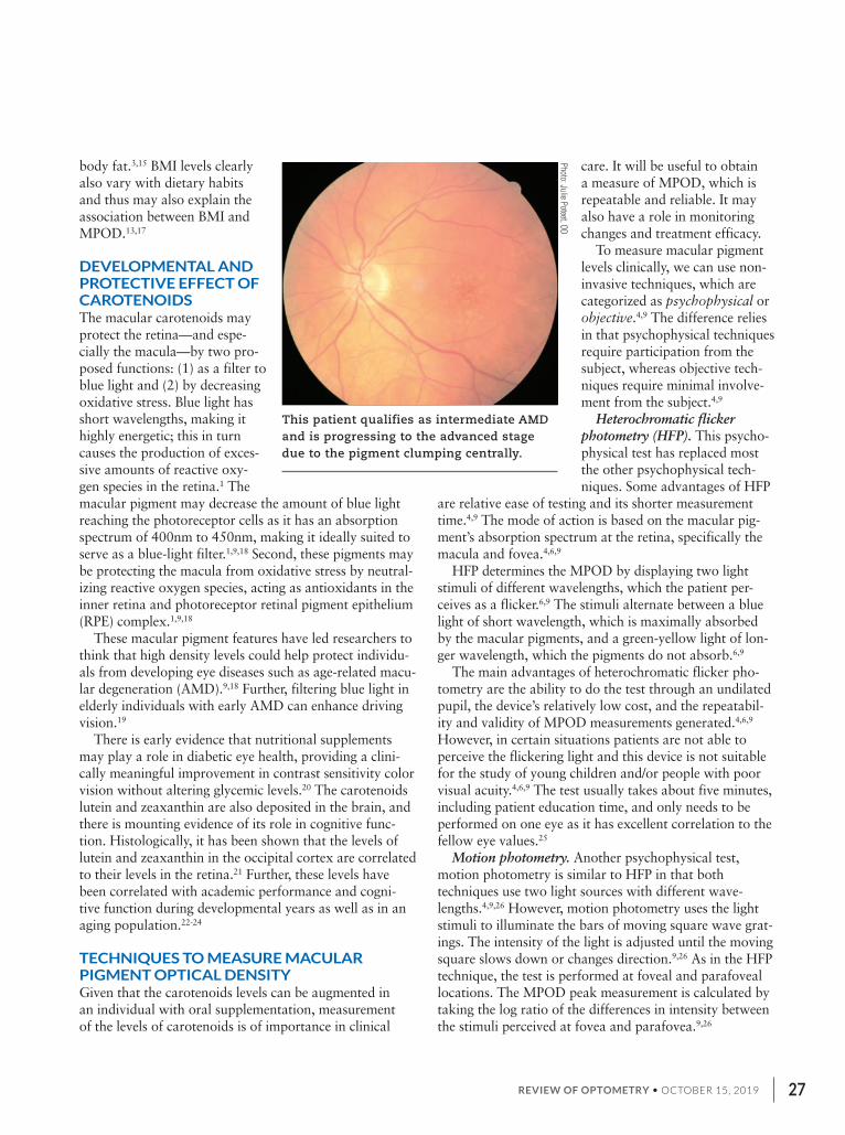
REVIEW OF OPTOMETRY • OCTOBER 15, 2019 27
body fat.3,15 BMI levels clearly also vary with dietary habits and thus may also explain the association between BMI and MPOD.13,17
DEVELOPMENTAL AND PROTECTIVE EFFECT OF CAROTENOIDSThe macular carotenoids may protect the retina—and espe-cially the macula—by two pro-posed functions: (1) as a filter to blue light and (2) by decreasing oxidative stress. Blue light has short wavelengths, making it highly energetic; this in turn causes the production of exces-sive amounts of reactive oxy-gen species in the retina.1 The macular pigment may decrease the amount of blue light reaching the photoreceptor cells as it has an absorption spectrum of 400nm to 450nm, making it ideally suited to serve as a blue-light filter.1,9,18 Second, these pigments may be protecting the macula from oxidative stress by neutral-izing reactive oxygen species, acting as antioxidants in the inner retina and photoreceptor retinal pigment epithelium (RPE) complex.1,9,18
These macular pigment features have led researchers to think that high density levels could help protect individu-als from developing eye diseases such as age-related macu-lar degeneration (AMD).9,18 Further, filtering blue light in elderly individuals with early AMD can enhance driving vision.19
There is early evidence that nutritional supplements may play a role in diabetic eye health, providing a clini-cally meaningful improvement in contrast sensitivity color vision without altering glycemic levels.20 The carotenoids lutein and zeaxanthin are also deposited in the brain, and there is mounting evidence of its role in cognitive func-tion. Histologically, it has been shown that the levels of lutein and zeaxanthin in the occipital cortex are correlated to their levels in the retina.21 Further, these levels have been correlated with academic performance and cogni-tive function during developmental years as well as in an aging population.22-24
TECHNIQUES TO MEASURE MACULAR PIGMENT OPTICAL DENSITYGiven that the carotenoids levels can be augmented in an individual with oral supplementation, measurement of the levels of carotenoids is of importance in clinical
care. It will be useful to obtain a measure of MPOD, which is repeatable and reliable. It may also have a role in monitoring changes and treatment efficacy.
To measure macular pigment levels clinically, we can use non-invasive techniques, which are categorized as psychophysical or objective.4,9 The difference relies in that psychophysical techniques require participation from the subject, whereas objective tech-niques require minimal involve-ment from the subject.4,9
Heterochromatic flicker photometry (HFP). This psycho-physical test has replaced most the other psychophysical tech-niques. Some advantages of HFP
are relative ease of testing and its shorter measurement time.4,9 The mode of action is based on the macular pig-ment’s absorption spectrum at the retina, specifically the macula and fovea.4,6,9
HFP determines the MPOD by displaying two light stimuli of different wavelengths, which the patient per-ceives as a flicker.6,9 The stimuli alternate between a blue light of short wavelength, which is maximally absorbed by the macular pigments, and a green-yellow light of lon-ger wavelength, which the pigments do not absorb.6,9
The main advantages of heterochromatic flicker pho-tometry are the ability to do the test through an undilated pupil, the device’s relatively low cost, and the repeatabil-ity and validity of MPOD measurements generated.4,6,9 However, in certain situations patients are not able to perceive the flickering light and this device is not suitable for the study of young children and/or people with poor visual acuity.4,6,9 The test usually takes about five minutes, including patient education time, and only needs to be performed on one eye as it has excellent correlation to the fellow eye values.25
Motion photometry. Another psychophysical test, motion photometry is similar to HFP in that both techniques use two light sources with different wave-lengths.4,9,26 However, motion photometry uses the light stimuli to illuminate the bars of moving square wave grat-ings. The intensity of the light is adjusted until the moving square slows down or changes direction.9,26 As in the HFP technique, the test is performed at foveal and parafoveal locations. The MPOD peak measurement is calculated by taking the log ratio of the differences in intensity between the stimuli perceived at fovea and parafovea.9,26
This patient qualifies as intermediate AMD and is progressing to the advanced stage due to the pigment clumping centrally.
Photo: Julie P
oteet, OD
026_ns1019_macular.indd 27026_ns1019_macular.indd 27 10/14/19 11:18 AM10/14/19 11:18 AM

REVIEW OF OPTOMETRY • OCTOBER 15, 201928
Now, let’s turn our attention to three objective tech-niques.
Fundus reflectometry. This measures MPOD on the basis of light reflected from the retina and the cho-roid.9,27,28 It uses light of two wavelengths: one absorbed by the macular pigments (in the blue spectrum) and another that is not absorbed. The device performs two analyses. First, it compares the reflection of light at the central and peripheral regions.9 The second method is based on spectral analysis, which involves analyzing the spectrum of the reflected light from a specific region of the retina.4,9
Unlike HFP, fundus reflectometry is an objective method of measurement and thus may obtain MPOD estimates in pediatric and special needs populations.9 It has also proven to be repeatable, and estimates of MPOD can be obtained in short duration.4,9,27,28
Recently, a near-commercial prototype (ZeaVision) was evaluated in our lab and preliminary results of its repeatability and reproducibility were reported.28 The major advantage of fundus reflectometry using a spec-tral analysis is that it can identify the individual levels of lutein and zeaxanthin in the retina, along with MPOD values and lens optical density. Our team also found the MPOD measurements more repeatable than the psycho-physical technique and they can be obtained without dilation. However, if accurate measures of lutein and zeaxanthin are desired, pupillary dilation is preferred; precise alignment and signal strength from the retina may influence its values.
Fundus autofluorescence (FAF). This is another objec-
tive technique to measure MPOD in vivo. It is based on the intrinsic fluorescence or autofluroescence of lipofus-cin, a waste product that accumulates with age on the RPE.4,9,29 Lipofuscin is the main fluorophore in auto-fluroescence and will fluoresce when excited with light wavelengths between 400nm and 590nm. The absorption spectrum of lipofuscin is similar to that of the macular pigments (400nm to 540nm).4,9,29
Macular pigments are located in the axons of the photoreceptors and inner plexiform layers of the retina, which is anterior to the location of lipofuscin in the RPE. The lipofuscin fluorescence will be less pronounced in eyes with higher macular pigment density and greater in eyes with decreased macular pigmentation.9 MPOD values are calculated by measuring the difference between autofluroescence emitted from the fovea and parafo-vea.9,29 A recent report showed that fundus autofluo-rescence is strongly associated with macular pigment levels.30
Raman spectroscopy. This new technique is cur-rently being developed for MPOD measurement and is not yet clinically available.9 When a monochromatic light is directed through any molecule, it will result in two types of light scattering: elastic and inelastic.9 The inelastic scattering causes a shift in the wavelength of the incident light, known as Raman shift, which is molecule-specific.9,31 When the incident wavelength is similar to the absorption spectrum of the molecule, there is an enhancement of the Raman shift, thus allowing for the molecule to be identified.9
Raman spectroscopy can be used to identify macular pigments because these substances exhibit five orders of magnitude of resonance enhance-ment upon excite-ment by 488nm of argon laser light.4,9
This technique has gained interest from researchers, as it is the only technique that mea-sures the pigments themselves instead of measuring pig-ments indirectly by analyzing other structures.31 One of the main advantages of this
MACULAR PIGMENT
UNCOVER THE MECHANISMS OF MACULAR PIGMENT
Images: K
erry Gelb, O
D
The QuantifEye MPOD measurement instrument shows MPOD improvement after supplementation with a daily carotenoid formula.
026_ns1019_macular.indd 28026_ns1019_macular.indd 28 10/14/19 11:18 AM10/14/19 11:18 AM

REVIEW OF OPTOMETRY • OCTOBER 15, 2019 29
technique is that it is sensitive and specific for macular carotenoids and can be used in subjects with low visual acuity.9,32 Some disadvantages are its need for pupillary dilation and the use of highly specialized and expensive devices.9,32
PHILOSOPHY OR PHYSIOLOGY?We are what we eat, goes the saying. In an ideal world, our diet is our medicine and there will be no need for nutraceuticals to supplement or augment our poor diets. We live far from such an ideal, however, and the consumption and the role of nutraceuticals in managing chronic disease is indeed increasing. Just like the man-aging of diabetes patients without HbA1c measurement cannot be imagined, I am of the opinion that the use of xanthophyll supplements without baseline measurement of MPOD makes no sense.
There is significant evidence about the validity of the aforementioned multiple techniques to measure MPOD. Low levels have shown association with different dis-ease states, and there is evidence that MPOD values can change with oral supplementation. However, due to the multifactorial nature of disease, the efficacy of carotenoid supplementation is still to be unequivocally proven. Further research is indeed needed to establish treatment regimens and protocols of carotenoid supplementation when and how to use MPOD values clinically to identify and monitor disease progression. However, we can be certain that the need to measure MPOD clinically is not in dispute. ❍
Acknowledgements: The author wishes to thank Drs. Katia Suarez-Berumen and Dennis Gierhart for assistance in literature search and interesting discussions.
Conflict of Interest statement: Dr. Davey is a researcher and consultant for Optovue, ZeaVision and Guardion Health Sciences.
1. Bernstein PS, Delori FC, Richer S, et al. The value of measurement of macular carotenoid
pigment optical densities and distributions in age-related macular degeneration and other reti-
nal disorders. Vision Res. 2010;50(7):716-28.
2. Gruszecki WI, Sielewiesiuk J. Orientation of xanthophylls in phosphatidylcholine multibilayers.
Biochimica Et Biophysica Acta. 1990;1023(3):405-12.
3. Raman R, Rajan R, Biswas S, et al. Macular pigment pptical density in a South Indian popula-
tion. Invest Ophthalmol Vis Sci. 2011;52(11):7910-6.
4. Leung IYF. Macular pigment: new clinical methods of detection and the role of carotenoids in
age-related macular degeneration. Optometry. 2008;79(5):266-72.
5. de Kinkelder R, van der Veen RLP, Verbaak FD, et al. Macular pigment optical density
measurements: evaluation of a device using heterochromatic flicker photometry. Eye (Lond).
2011;25(1):105-12.
6. Bartlett H, Howells O, Eperjesi E. The role of macular pigment assessment in clinical practice:
a review. Clin Exp Optom. 2010 Sep;93(5):300-8.
7. Bone RA, Landrum JT, Hime GW, et al. Stereochemistry of the human macular carotenoids.
Invest Ophthalmol Vis Sci. 1993;34(6):2033-40.
8. Bone RA, Landrum JT, Mayne ST, et al. Macular pigment in donor eyes with and without
AMD: A case-control study. Invest Ophthalmol Vis Sci. 2001;42(1):235-40.
9. Howells O, Eperjesi F, Bartlett H. Measuring macular pigment optical density in vivo: a review
of techniques. Graefes Arch Clin Exp Ophthalmol. 2011 Mar;249(3):315-47. doi: 10.1007/
s00417-010-1577-5
10. Yu J, Johnson EJ, Shang F, et al. Measurement of macular pigment optical density in a
healthy Chinese population sample. Invest Ophthalmol Vis Sci. 2012;53(4):2106-11.
11. Wang G, Brun TA, Geissler CA, et al. Vitamin A and carotenoid status in rural China. The
British Journal of Nutrition. 1996;76(6):809-20.
12. Zhou H, Thao X, Johnson EJ, et al. Serum carotenoids and risk of age-related macular
degeneration in a Chinese population sample. Invest Ophthalmol Vis Sci. 2011;52(7):4338-44.
13. Celentano JC, Burke JD, Hammond BR. In vivo assessment of retinal carotenoids: Macular
pigment detection techniques and their impact on monitoring pigment status. J Nutr. 2002
Mar;132(3):535S-539S.
14. Hammond BR Jr., Curran-Celentano J, Judd S, et al. Sex differences in macular pigment
optical density: relation to plasma carotenoid concentrations and dietary patterns. Vision
Research. 1996;36(13):2001-2012.
15. Broekmans WMR, Berendschot TT, Klöpping-Ketelaars IA, et al. Macular pigment density
in relation to serum and adipose tissue concentrations of lutein and serum concentrations of
zeaxanthin. American Journal of Clinical Nutrition. 2002;76(3):595-603.
16. Hammond BR, Ciulla TA, Snodderly DM. Macular pigment density is reduced in obese sub-
jects. Invest Ophthalmol Vis Sci. 2002;43(1):47-50.
17. Wolf-Schnurrbusch UEK, Roosli N, Weyermann E, et al. Ethnic differences in macular pig-
ment density and distribution. Invest Ophthalmol Vis Sci. 2007;48:3783-87.
18. Howells O, Eperjesi F, Bartlett H. Improving the repeatability of heterochromatic flicker
photometry for measurement of macular pigment optical density. Graefes Arch Clin Exp Oph-
thalmol. 2013;251(3):871-80.
19. Richer S, Park DW, Epstein R, et al. Macular re-pigmentation enhances driving vision in
elderly adult males with macular degeneration. J Clin Exp Ophthalmol. 2012;3:217.
20. Chous AP, Richer SP, Gerson JD, et al. The diabetes visual function supplement study. Br J
Ophthalmol 2016;100:227-34.
21. Vishwanathan R, Schalch W, Johnson E. Macular pigment carotenoids in the retina and
occipital cortex are related in humans J Nutr Neurosci. 2016;19(3):95-101.
22. Renzi-Hammond LM, Bovier ER, Fletcher LM, et al. Effects of a lutein and zeaxanthin inter-
vention on cognitive function: a randomized, double-masked, placebo-controlled trial of younger
healthy adults. Nutrients. 2017;9:1246.
23. Barnett SM, Khan NA, Walk AM, et al. Macular pigment optical density is positively associ-
ated with academic performance among preadolescent children. Nutritional Neuroscience.
2017;21(9):632-40.
24. Hammond BR Jr., Miller LS, Bello MO, Lindbergh CA, et al. Effects of lutein/zeaxanthin
supplementation on the cognitive function of community dwelling older adults: a randomized,
double-masked, placebo-controlled trial. Front. Aging Neurosci. 2017;9:254.
25. Davey PG, Alvarez SD, Lee JY Macular pigment optical density: repeatability, intereye
correlation, and effect of ocular dominance. Clin Ophthalmol. 2016 Aug 29;10:1671-8. doi:
10.2147/OPTH.S111708. eCollection 2016.
26. Moreland JD. Macular pigment assessment by motion photometry. Arch Biochem Biophys.
2004;430(2):143-8.
27. Berendschot T, van Norren D. Objective determination of the macular pigment optical den-
sity using fundus reflectance spectroscopy. Arch Biochem Biophys. 2004430(2):149-55.
28. Davey PG, Ngo A, Cross J, Gierhart DL. Macular pigment reflectometry: development and
evaluation of a novel clinical device for rapid objective assessment of the macular carotenoids.
Proc. 2019;10858(Ophthalmic Technologies XXIX).
29. Delori FC, Goger DG, Hammond BR, et al. Macular pigment density measured by autofluo-
rescence spectrometry: comparison with reflectometry and heterochromatic flicker photometry.
J Opt Soc Am A Opt Image Sci Vis. 2001;18(6):1212-30.
30. Sauer L, Andersen KM, Li B, et al. Fluorescence lifetime imaging ophthalmoscopy (FLIO) of
macular pigment. Invest Ophthalmol Vis Sci. 2018;59:3094-3103.
31. Bernstein PS, Yoshida MD, Katz NB, et al. Raman detection of macular carotenoid pigments
in intact human retina. Invest Ophthalmol Vis Sci. 1998;39(11):2003-11.
32. Gellermann W, Ermakov IV, Ermakova MR, et al. In vivo resonant Raman measurement of
macular carotenoid pigments in the young and the aging human retina. J Opt Soc Am A Opt
Image Sci Vis. 2002;19(6):1172-86.
Images: K
erry Gelb, O
D
FAF imaging can help reveal macular degeneration due to the accumulation of lipofuscin.
026_ns1019_macular.indd 29026_ns1019_macular.indd 29 10/14/19 11:19 AM10/14/19 11:19 AM

REVIEW OF OPTOMETRY • OCTOBER 15, 201930
One cannot open a magazine these days—whether it be a scientific journal or News-week—without seeing something on the microbiome or probiotics. The hype sur-
rounding the explosion of microbiome research, driven by Internet gurus and corporations aiming to make a profit, far exceeds the scientific validation of many of the claims. Although we are just at the tip of the iceberg in our understanding of the impact that our commensal microbes have on us, we now know some important details about how our lifetime partners, the trillions of bugs that live in and on us, affect us.
GO WITH YOUR GUTOf all the parts of the microbiome—from the ocular to the oral—the gut microbiome is by far the most researched, and we know that it orchestrates human metabolism, immunity and gene expression. Researchers have also mapped out the role it plays in any number of systemic functions, including the pro-duction of certain B and K vitamins, neurotransmitters, and short-chain fatty acids important for brain health and in maintaining the integrity of the gut lining. The gastrointestinal (GI) microbiome also modulates the immune system by shifting T-helper cell balance towards Th1, resulting in decreased production of IgE and eosinophils, dampened hypersensitiv-
ity reactions and intestinal inflammation, greater oral tolerance and prevention of atopic diseases. It facilitates xenobiotic metabolism, important for the absorption and proper functioning of phytoestrogens, lignans, flavonoids and some medicinal herbs.
Most importantly, a healthy microbiome provides pro-tection against colonization of the intestinal tract with potentially pathogenic bacteria afforded by the intestinal
flora. A healthy microflora plays a vital role in weight maintenance and energy homeostasis, preventing obe-sity by its increased capacity to har-vest energy from the diet.
The foods we choose impact our microbiome and dictate the epigenetic expression of immune system func-tion (Figure 1). Since almost every ocular disorder involves an inflam-matory component, it is our duty to educate patients on proper nutrition because we must change the diet to change the microbes.
THE CONJUNCTIVAL MICROBIOTAThe microbial community of the ocu-lar surface is more diverse than that of the skin but contains a significantly lower quantity than other bodily sur-faces. This disparity is thought to be due to the antibacterial effect of tear film components.
In contact lens wearers, however, the ocular microflora is less diverse (more akin to that of the skin) and contains
Gut Instinct: Why the Microbiome Matters
It can be considered an additional human organ, rivaling the liver in the number of biochemical reactions in which it participates.
BY JULIE POTEET, OD, CNS
MICROBIOTA
TLR4Activation
Omega-3Omega-6
Saturated Fats
Artificial Sweetners
Sugar
Red Meat
Phytonutrients
Gluten
Salt
IL-7IL-1, IL-6, TNF
Gut inflammation
DybiosisLeaky Gut &
Systemic LPS
Myles IA. Fast food fever: reviewing the impacts of the western diet on immunity. Nutrion Jour-nal. 2014;13:61. doi:10.1186/1475-2891-13-61.
Down-regulation of Immune Responsiveness
Infection/CancerSusceptibility
ReducedTreg Cells
SystemicInflammation
Allergy &Autoimmunity
FIG. 1. In recent years, scientists have discovered that the gut microbiome orchestrates human metabolism, immunity and gene expression.
030_ns1019_microbiome.indd 30030_ns1019_microbiome.indd 30 10/14/19 11:24 AM10/14/19 11:24 AM
REVIEW OF OPTOMETRY • OCTOBER 15, 2019 31
more gram-negative species. Researchers are still trying to find out if the increased risk of eye infections in contact lens wearers is related to lenses contaminated with bacte-ria from the finger or if lenses exert selective pressures on the eye’s bacterial community in favor of skin bacteria.
Aside from acute ocular surface infections, the colonic microflora’s influence on ocular health is, in general terms, far more important to ocular disease than the conjunctiva’s due to the colonic microflora’s ability to modulate the immune system and create inflammation and autoimmunity elsewhere in the body.
CONSEQUENCES OF DISRUPTION Dysbiosis is the growth of potentially pathogenic micro-organisms over their beneficial counterparts or altera-tions in the metabolic activities of the flora. Although different subtypes of dysbiosis exist based on location, research shows dysbiosis of the GI tract, where the majority of our microbiome microorganisms reside, has the greatest impact on immune system dysfunction and chronic and degenerative diseases.
Any number of outside forces can lead to a change in the intestinal flora:
Probiotic Myths Busted Myth 1: Research conducted on one probiotic strain can be accu-rately extrapolated to other strains within the same species.
Truth: To assess the quality of the bacteria in a given supple-ment or probiotic food, you need to know not only the species of bacteria but also the strain because different probiotic strains have different actions. We use different strains of bacteria for different therapeutic applications. As an example, researchers compared two Lactobacillus strains in the treatment of viral gastroenteritis: L. casei subspecies casei GG (LGG) and L. casei supspecies rhamnosus (lactophilus).1 The specific strain made a significant difference in the duration of diarrhea: 1.8 days in the LGG group compared with 2.8 days in the lactophilus group. On day two, only 19% of patients in the LGG group had diarrhea vs. 64% of subjects in the lactophilus group. The researchers also noted that rotavirus-specific IgA levels were significantly higher in the LGG group during the convalescent period, showing supe-rior efficacy of one strain of L. casei over another.
This is important, as patients can waste time and money on probiotics not knowing the strain included in the product may not be effective for their condition. One study found strain 299v of Lactobacillus plantarum significantly decreased abdominal pain severity, frequency and bloating in patients with irritable bowel syndrome (IBS).2 However, another study noted a similar strain, Lactobacillus plantarum MF1298, significantly worsened IBS symptoms—so much so that patients preferred the placebo.3
Myth 2: Don’t use probiotics during antibiotic treatment, as antibiotics with kill all the probiotics.
Truth: Research shows that concurrent administration of pro-biotics with antibiotics not only significantly decreases antibiot-ic-related side effects, but also attenuates antibiotic-associated damage to the gut microbiota.4 It is good clinical practice to prescribe over-the-counter Lactobacillus rhamnosus GG—a widely available probiotic and one of the most well-researched strains—concomitantly to prevent damage to the gut microbi-ome from antibiotic use.
Myth 3: Probiotics are best taken on an empty stomach.Truth: Research clearly shows improved bacterial survival
when probiotics are taken with a meal (preferably a larger meal such as dinner). Dairy-, fiber- or grain-based meals optimize sur-vival through the upper GI tract.5,6
Myth 4: Probiotic strains ingested orally will permanently colo-nize the GI tract and you can re-colonize with probiotic supplements.
Truth: No exogenously supplied probiotic strain permanently colonizes the human GI tract. Once you stop taking a certain strain, it will eventually die off. You cannot re-colonize perma-nently by taking probiotic supplements for a limited time.
Myth 5: Infants should only be supplemented with strains of Bifidobacterium infantis.
Truth: Babies need special baby probiotics. By week one of age in vaginally-delivered, breastfed infants, the flora is domi-nated by Bifidobacterium spp. and Ruminococcaceae spp. but a multitude of other species are present as well.7-9
Myth 6: Giving probiotics in supplement form (i.e., capsule, pow-der or tablet) is superior to food forms (i.e., yogurt).
Truth: Yogurt often contains greater numbers of viable bacte-ria. In one study, researchers found 100 million bacteria provid-ed in a dairy base led to a greater number of live bacteria in the colon than 10 billion provided in a capsule.10 Numerous clinical trials show that medicinal yogurts have therapeutic effects such as decreased rates of vaginal candidiasis and decreased abdomi-nal adiposity and body weight.11,12
1. Majamaa H, Isolauri E, Saxelin M, Vesikari T. Lactic acid bacteria in the treatment of acute rotavirus gastroenteritis. J Pediatr Gastroenterol Nutr. 1995;20(3):333-8.2. Ducrotté P, Sawant P, Jayanthi V. Clinical trial: Lactobacillus plantarum 299v (DSM 9843) improves symptoms of irritable bowel syndrome. World J Gastroenterol. 2012;18(30):4012-18.3. Ligaarden SC, Axelsson L, Naterstad K, et al. A candidate probiotic with unfavourable effects in subjects with irritable bowel syndrome: a randomised controlled trial. BMC Gastroenterol. 2010 Feb;10:16.4. Szajewska H, Ruszczy ski M, Radzikowski A. Probiotics in the prevention of antibiotic-associated diarrhea in children: a meta-analysis of randomized controlled trials. J Pediatr. 2006;149(3):367-72.5. Kailasapathy K, Chin J. Survival and therapeutic potential of probiotic organisms with refer-ence to Lactobacillus acidophilus and Bifidobacterium spp. Immunol Cell Biol. 2000;78(1):80-8.6. Laurens-Hattingh A, Viljoen BC. Yogurt as probiotic carrier food. Internat Dairy J. 2001;11(1-2):1-17.7. Morelli L. Postnatal development of intestinal microflora as influenced by infant nutrition. J Nutr. 2008;138(9):1791S–95S.8. Martin R, Langa S, Reviriego C, et al. Human milk is a source of lactic acid bacteria for the infant gut. J Pediatr. 2003;143:754-58.9. Albesharat R, Ehrmann MA, Korakli M, et al. Phenotypic and genotypic analyses of lactic acid bacteria in local fermented food, breast milk and faeces of mothers and their babies. Syst Appl Microbiol. 2011;34(2):148-55. 10. Siitonen, S.,Vapaatalo, H. & Salminen, S. Colonization of the human gastrointestinal tract by probiotic bacteria (Lactobacillus GG). Nutrition Today. 1996;31:5s-9s.11. Hilton, E., H. D. Isenberg, et al. Ingestion of yogurt containing Lactobacillus acidophilus as prophylaxis for candidal vaginitis. Annals of Internal Medicine. 119;116(5):353-57.12. Kadooka Y, Sato M, et al. Regulation of abdominal adiposity by probiotics (Lactobacillus gasseri SBT2055) in adults with obese tendencies in a randomized controlled trial. Eur J Clin Nutr. 2010;64(6):636-43.
030_ns1019_microbiome.indd 31030_ns1019_microbiome.indd 31 10/14/19 11:24 AM10/14/19 11:24 AM

REVIEW OF OPTOMETRY • OCTOBER 15, 201932
Antibiotics. Of all the factors that can impact the gut microflora, antibiotics have the greatest detrimen-tal effect. Research using culturing techniques suggests quantitative changes could last up to 40 days after one round of antibiotics. Metabolic derangements can last up to 18 months. New research using more sensitive molec-ular analysis techniques (RNA studies) has revealed the presence of antibiotic-resistant microorganisms for up to four years post-treatment.1,2
Alterations can last significantly longer than pre-viously believed: 18 to 24 months after the use of clindamycin and four years after triple therapy for Heli-cobacter pylori. Some organisms never recover.
Stress. This induces changes to the GI tract’s motility and secretions. In addition, increases in the circulation of the stress hormone norepinephrine acts a growth inducer to potentially pathogenic members of the microflora.
Caesarean sections. Research suggests babies born via C-section have a different microbiome. One study found bifidobacteria was present in 56.6% of vaginally delivered infants at day three and in 0% of C-section infants.3 This disturbance in the gut microbiome of C-section babies can last for at least six months.4
Birth location. Where a baby is born can also impact the microbiome. Research suggests full-term infants who were born vaginally at home and were breastfed exclusively have the highest numbers of bifidobacteria and the lowest numbers of Clostridium difficile and Escherichia coli—the most beneficial gut microbiota.5
Formula feeding. Babies fed formula are more often colonized by E. coli, C. difficile and Bacteroidia fragilis compared with exclu-sively breastfed infants.5,6 Counts of these microorganisms are also significantly higher. There is also the presence of different bacterial species.
Diet. Sulphate and sulphite compounds are often added to foods (e.g., dried fruits
and vegetables, shellfish, packaged fruit juices, baked goods, most junk foods, white bread and the majority of fermented alcoholic beverages) as preservatives. These cause the growth of sulphate-reducing bacteria, which increases hydrogen sulphide production and causes intes-tinal hyperpermeability and the inhibition of colonocyte metabolism.
High-protein diets can be particularly destructive to the GI microbiome. Protein is fermented by members of the microbiota (putrefaction), producing potentially harmful metabolites (Figure 2).
The more protein consumed, the more indoles, phe-nols, hydrogen sulphide and ammonia produced.7,8 Phenol and indole production may be attenuated by an increased fiber intake, which decreases colonic pH due to short-chain fatty acid production.7 The production and toxicity of colonic ammonia can be attenuated through prebiotic consumption.9,10
High-protein diets also induce changes to the eco-system such as decreases in fecal concentrations of bifidobacteria—research shows a 50% decline on a high-protein, low-carb diet in just four weeks.11 This dietary choice also decreases the concentrations of butyrate-producing species. All of these changes can be attenuated through prebiotic consumption.
High-fat diets are associated with increases in the ratio of gram-negative to gram-positive bacteria, which causes an increased proportion of lipopolysaccharide-containing microbiota in the gut and a greater pool of luminal endo-toxin.12 Meals high in fat increase endotoxin absorption wherein postprandial endotoxemia causes low-grade
Prescribing PearlsBefore recommending any probiotic supplement, make sure you know the strains it contains. Ensure these strains exhibit the desirable criteria (based on published research, not just the manufacturer’s word). Avoid products that do not detail their strain(s). Choose the correct strain to treat the clinical scenario at hand, as each probiotic strain should be viewed as a unique therapeutic agent with specific actions and applications. Lastly, you cannot extrapolate research results from one strain to another, even if they are within the same species.
High Protein Diet
SCFA and other organic
acids
CH4 CO2
H2
Proteins
Peptides
Amino acids
Uncertainphysiolicsignificance
Mutagen/cellular poison
Neurotransmitters/mutagen precursors
Products of protein putrefaction in the colon (Macfarlane & Macfarlane, 2012)
Co-carcinogens
Cellular toxins
BCFA
Phenols
Indols
Ammonia
Amines
HS
Thiols
MICROBIOTA
FIG. 2. These diets can create a significant GI microbiome imbalance.
GUT INSTINCT: WHY THE MICROBIOME MATTERS
030_ns1019_microbiome.indd 32030_ns1019_microbiome.indd 32 10/14/19 11:24 AM10/14/19 11:24 AM
REVIEW OF OPTOMETRY • OCTOBER 15, 2019 33
systemic inflammation.13 The higher the fat content, the higher the serum level of endotoxins. Saturated fat appears to further enhance endotoxin absorption, while omega-3 fatty acids and concurrent consumption of 30g of fiber attenuate postprandial endotoxemia.14,15
Diets high in refined carbohydrates can disrupt the GI microbiome in the following ways:
• Slow intestinal transit time, increasing exposure to potentially toxic bowel contents
• Alter the colonic gas metabolism, which can lead to changes in the species composition of the flora
• Decrease short-chain fatty acid production • Increase colonic pHDiets low in fiber increase fermentation of the pro-
tective layer of mucin and peptides/proteins, due to the limitation in food sources (fiber/resistant starch), which compromises the mucosal defense and increases produc-tion of toxic putrefaction products. This causes direct contact between colonic cells and bacterial products and antigens, leading to inflammation and increased mucosal permeability.
Sucralose consumption, often in the form of artificial sweeteners, causes significant changes in the microflora, including reductions in beneficial bacteria such as bifido-bacteria and lactobacilli and reductions in total anaerobes and aerobes. It also causes increased fecal pH.16
GETTING BACK ON TRACKPrebiotics, probiotics, colonic foods and antimicrobials can all help combat dysbiosis. Prebiotics and probiotics can be consumed in supplement form and they are being added to commercial foods to promote a healthy digestive track. There are many different types of prebiotics and probiotics and they both work synergistically to promote the growth of beneficial flora.
Prebiotics are complex carbohydrates—oligosaccha-rides—that are not digestible. This means they can survive the rough journey through the digestive tract, making it safely into the colon. Prebiotics selectively stimulate the growth of beneficial microorganisms.
Xylooligosaccharides are a preferred form of oligosac-charide manufacturers often use because they can target good bacteria using a lower dosage than other prebiotics. In fact, doses as low as 1.5 to 2 grams can modify the gut microbiome significantly, resulting in an increase of good bacteria and a decrease of the bad.19 Prebiotics include foods such as breast milk (infants), fruits and vegetables, tea, chocolate, fermented foods and those rich in polyphe-nols and vitamin D.
Probiotics are live, mostly gram-positive, bacteria (e.g., Bifidobacteriaum, Lactobacillus, Lactococcus, Pediococcus and other non-pathogenic strains of E. coli). These gener-
ally promote intestinal barrier integrity, prevent bacterial translocation in the gut and reduce inflammatory response.
Colonic foods serve as substrates for the endogenous colonic bacteria, thus indirectly providing the host with energy, metabolic substrates and essential micronutrients.17 Examples of colonic foods include carrots, cocoa, green tea, brown rice and prebiotic foods such as onions, garlic, chicory root, barley, bananas, leeks and apples.
Fecal microbiota transplantation, also known as fecal bacteriotherapy or fecal infusion, an emerging method to treat dysbiosis, uses “the principle of engrafting the microbiota from healthy donors into a patient recipient to re-introduce or re-establish a stable environment that influences both the endogenous microbes and the host.”18
Dysbiosis is implicated in eye and systemic disease asso-ciated with vision loss and other morbidity. Treatments that can help restore human microbiota homeostasis can be simple and have been shown to support improved out-comes. Optometrists should understand these treatments and prescribe them accordingly. ❍
1. Jernberg C, Löfmark S, Edlund C, Jansson JK. Long-term impacts of antibiotic exposure on the human intestinal microbiota. Microbiology. 2010;156(Pt 11):3216-23. 2. Cotten CM, Benjamin DK Jr, Smith PB, et al. Empirical antibiotic therapy for suspected early-onset bacterial sepsis. Pediatrics. 2012 Oct;130(4):e1052-3. 3. Biasucci G, Rubini M, Riboni S, et al. Mode of delivery affects the bacterial community in the newborn gut. Early Hum Dev. 2010;86(suppl 1):13-15.4. Grönlund MM, Salminen S, Mykkänen H, et al. Development of intestinal bacterial enzymes in infants--relationship to mode of delivery and type of feeding. APMIS. 1999;107(7):655-60.5. Penders J, Thijs C, Vink C, et al. Factors influencing the composition of the intestinal micro-biota in early infancy. Pediatrics. 2006;118(2):511-21.6. Morelli L. Postnatal development of intestinal microflora as influenced by infant nutrition. J Nutr. 2008;138(9):1791S–95S.7. MacFarlane S, MacFarlane GT. Proteolysis and amino acid fermentation. In: Gibson GR, MacFarlane GT, eds. Human Colonic Bacteria: Role in Nutrition, Physiology and Pathology. Boca Raton: CRC Press; 1995.8. Magee EA, Richardson CJ, Hughes R, Cummings JH. Contribution of dietary protein to sul-fide production in the large intestine: an in vitro and a controlled feeding study in humans. Am J Clin Nutr. 2000 Dec;72(6):1488-94.9. Geboes KP, De Hertogh G, et al. The influence of inulin on the absorption of nitro-gen and the production of metabolites of protein fermentation in the colon. Br J Nutr. 2006;96(6):1078-86.10. Weber F Jr. Lactulose and combination therapy of hepatic encephalopathy: The role of the intestinal microflora. Digest. Dis. 1996;14:53-63. 11. Duncan SH, Belenguer A, Holtrop G, et al. Reduced dietary intake of carbohydrates by obese subjects results in decreased concentrations of butyrate and butyrate-producing bacte-ria in feces. Appl Environ Microbiol. 2007 Feb;73(4):1073-8. 12. Cani PD, Bibiloni R, Knauf C, et al. Changes in gut microbiota control metabolic endotox-emia-induced inflammation in high-fat diet-induced obesity and diabetes in mice. Diabetes. 2008;57(6):1470-81.13. Laugerette F, Vors C, Geloen A, et al. Emulsified lipids increase endotoxemia: possible role in early postprandial low-grade inflammation. J Nutrition Biochem. 2011;22(1):53-59.14. Mani V, Hollis JH, Gabler NK. Dietary oil composition differentially modulates intestinal endotoxin transport and postprandial endotoxemia. Nutr Metab. 2013;10(1):6.15. Ghanim H, Abuaysheh S, Sia CL, Korzeniewski K, Chaudhuri A, Fernandez-Real J, Dan-dona P. Increase in plasma endotoxin concentrations and the expression of Toll-like receptors and suppressor of cytokine signaling-3 in mononuclear cells after a high-fat, high-carbohy-drate meal. Diabetes Care. 2009;32(12):2281-7.16. Abou-Donia MB, El-Masry EM, Abdel-Rahman AA, et al. Splenda alters gut microflora and increases intestinal p-glycoprotein and cytochrome p-450 in male rats. J Toxicol Environ Health A. 2008;71:1415-29.17. Hawrelak JA, Myers SP. The causes of intestinal dysbiosis: a review. Altern Med Rev. 20064;9:180-97.18. DeGruttola AK, Low D, Mizoguchi A, Mizoguchi E. Current understanding of dysbiosis in Disease in human and animal models. Inflamm Bowel Dis. 2016;22(5):1137-50.19. Sydney Finegold PM, Finegold SM, Zhaoping Li A, et al. Linking the chemistry and physics of food with health and nutrition Xylooligosaccharide increases bifidobacteria but not lactoba-cilli in human gut microbiota. Food Funct. 2014;5:403-614.
030_ns1019_microbiome.indd 33030_ns1019_microbiome.indd 33 10/14/19 11:24 AM10/14/19 11:24 AM

REVIEW OF OPTOMETRY • OCTOBER 15, 201934
Most people would agree that nutrition is a cor-nerstone of good health. The right vitamins, minerals and nutrients help to safeguard our vision and senses for a lifetime and aid in
peak brain function. In particular, ‘superfood’ components mitigate many of the changes that occur in our bodies over time. These are rich in antioxidants and anti-inflammato-ries, the vitamins and minerals that keep our hearts and lungs healthy into our senior years, protect our vision and keep our minds sharp. Not only can the right nutritional choices optimize patients’ eye health and vision over the lifecycle, they also help to keep free-radical damage and disease at bay.
Research continues to validate why good nutrition should be an integral part of eye care. This article delves into the relationship between what we eat and how we see.
SUPERFOOD DEFENSE AGAINST DEGENERATIVE & OXIDATIVE PROCESSESIt’s important for patients to load up on superfood nutri-ents to help preserve their eyesight into their senior years. To qualify as a superfood, it has to be readily available, with a significant number of scientific publications verifying the power of the food (specifically, its nutrients) to prevent disease and promote wellness and longevity. Here are some top performers to include in the diet:
Pomegranate juice can stimulate vasodilation, which increases blood flow. One manifestation of age-related macular degeneration (AMD) is reduced blood flow to the eye. Cellular metabolism depends on adequate oxygen and nutrients as well as proper elimination of waste products. Improving blood flow to an organ system helps it stay in
good health. Pomegranates are also phytochemical heavy-weights; these fruits have two to three times the antioxidant power of green tea or red wine and also possess potent anti-inflammatory chemicals.1
Walnuts have a high concentration of omega-3 fats. The fluid and flexible characteristics of omega-3 fats maxi-mize cells’ abilities to absorb their nutrients and eliminate wastes. In addition, the alpha-lipoic acid (ALA) and other polyphenols in walnuts act as antioxidants to block adverse cellular signals from free-radical exposure that can increase inflammation. Walnuts may even help prevent AMD pro-gression. A study in Archives of Ophthalmology found that individuals who ate more than one serving of nuts a week decreased their risk of AMD progression by more than 50%.2
Put Wellness on the Menu
Learn how the right dietary choices can improve patients’ outcomes—and lives.
BY STEVEN G. PRATT, MD, STUART RICHER, OD, PhD, DENNIS RUSKIN, OD, AND KERRY GELB, OD
DIET
A 10-point Plan to Prevent AMD1. Avoid smoking, weight gain, high glycemic index foods, high
fructose corn syrup, soda and sweetened drinks, artificial sweeteners, dairy, grain-fed meat and trans fats.
2. Eat foods with omega-3s and/or take supplements.3. Embrace an anti-inflammatory or paleo diet.4. Eat foods that contain lutein and zeaxanthin.5. Drink 1.5 liters of water a day with green tea and lemon.6. Include spices such as cinnamon, rosemary, oregano, garlic
powder, Himalayan salt, turmeric (contains curcumin), gin-ger and paprika.
7. Maintain optimal vitamin D3 blood levels.8. Get regular exercise and use stress reduction strategies.9. Take supplements for ocular nutrition.10. Undergo a regular retinal and macular pigment density
exam.
034_ns1019_diet.indd 34034_ns1019_diet.indd 34 10/14/19 11:28 AM10/14/19 11:28 AM
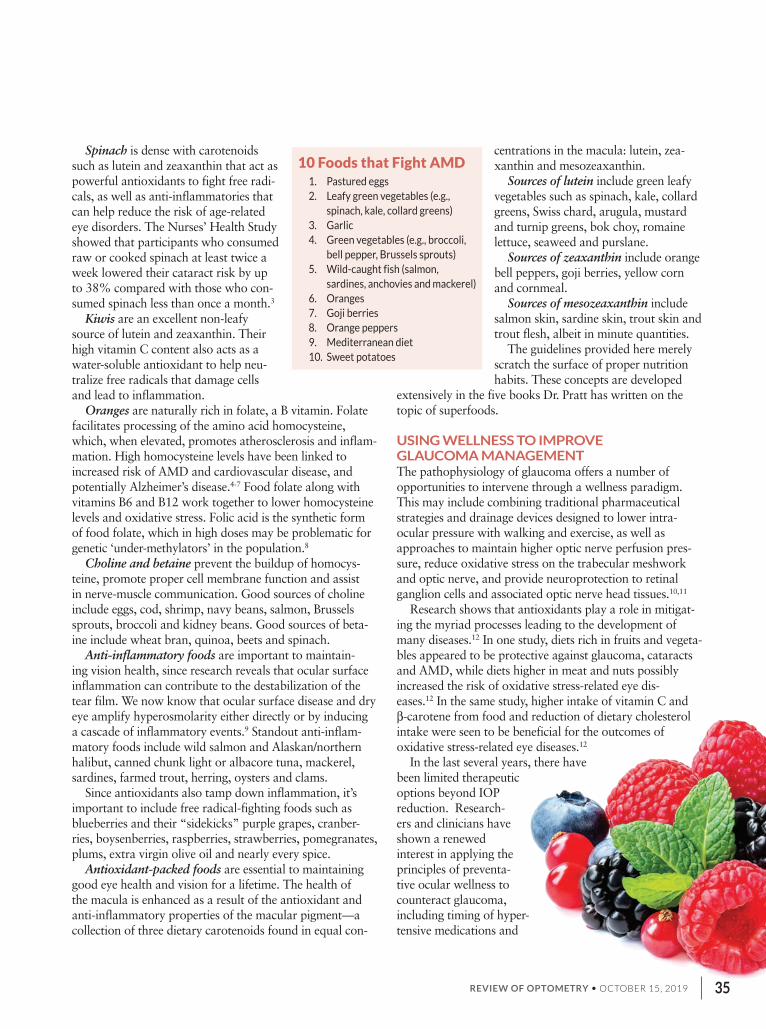
REVIEW OF OPTOMETRY • OCTOBER 15, 2019 35
Spinach is dense with carotenoids such as lutein and zeaxanthin that act as powerful antioxidants to fight free radi-cals, as well as anti-inflammatories that can help reduce the risk of age-related eye disorders. The Nurses’ Health Study showed that participants who consumed raw or cooked spinach at least twice a week lowered their cataract risk by up to 38% compared with those who con-sumed spinach less than once a month.3
Kiwis are an excellent non-leafy source of lutein and zeaxanthin. Their high vitamin C content also acts as a water-soluble antioxidant to help neu-tralize free radicals that damage cells and lead to inflammation.
Oranges are naturally rich in folate, a B vitamin. Folate facilitates processing of the amino acid homocysteine, which, when elevated, promotes atherosclerosis and inflam-mation. High homocysteine levels have been linked to increased risk of AMD and cardiovascular disease, and potentially Alzheimer’s disease.4-7 Food folate along with vitamins B6 and B12 work together to lower homocysteine levels and oxidative stress. Folic acid is the synthetic form of food folate, which in high doses may be problematic for genetic ‘under-methylators’ in the population.8
Choline and betaine prevent the buildup of homocys-teine, promote proper cell membrane function and assist in nerve-muscle communication. Good sources of choline include eggs, cod, shrimp, navy beans, salmon, Brussels sprouts, broccoli and kidney beans. Good sources of beta-ine include wheat bran, quinoa, beets and spinach.
Anti-inflammatory foods are important to maintain-ing vision health, since research reveals that ocular surface inflammation can contribute to the destabilization of the tear film. We now know that ocular surface disease and dry eye amplify hyperosmolarity either directly or by inducing a cascade of inflammatory events.9 Standout anti-inflam-matory foods include wild salmon and Alaskan/northern halibut, canned chunk light or albacore tuna, mackerel, sardines, farmed trout, herring, oysters and clams.
Since antioxidants also tamp down inflammation, it’s important to include free radical-fighting foods such as blueberries and their “sidekicks” purple grapes, cranber-ries, boysenberries, raspberries, strawberries, pomegranates, plums, extra virgin olive oil and nearly every spice.
Antioxidant-packed foods are essential to maintaining good eye health and vision for a lifetime. The health of the macula is enhanced as a result of the antioxidant and anti-inflammatory properties of the macular pigment—a collection of three dietary carotenoids found in equal con-
centrations in the macula: lutein, zea-xanthin and mesozeaxanthin.
Sources of lutein include green leafy vegetables such as spinach, kale, collard greens, Swiss chard, arugula, mustard and turnip greens, bok choy, romaine lettuce, seaweed and purslane.
Sources of zeaxanthin include orange bell peppers, goji berries, yellow corn and cornmeal.
Sources of mesozeaxanthin include salmon skin, sardine skin, trout skin and trout flesh, albeit in minute quantities.
The guidelines provided here merely scratch the surface of proper nutrition habits. These concepts are developed
extensively in the five books Dr. Pratt has written on the topic of superfoods.
USING WELLNESS TO IMPROVE GLAUCOMA MANAGEMENT The pathophysiology of glaucoma offers a number of opportunities to intervene through a wellness paradigm. This may include combining traditional pharmaceutical strategies and drainage devices designed to lower intra-ocular pressure with walking and exercise, as well as approaches to maintain higher optic nerve perfusion pres-sure, reduce oxidative stress on the trabecular meshwork and optic nerve, and provide neuroprotection to retinal ganglion cells and associated optic nerve head tissues.10,11
Research shows that antioxidants play a role in mitigat-ing the myriad processes leading to the development of many diseases.12 In one study, diets rich in fruits and vegeta-bles appeared to be protective against glaucoma, cataracts and AMD, while diets higher in meat and nuts possibly increased the risk of oxidative stress-related eye dis-eases.12 In the same study, higher intake of vitamin C and β-carotene from food and reduction of dietary cholesterol intake were seen to be beneficial for the outcomes of oxidative stress-related eye diseases.12
In the last several years, there have been limited therapeutic options beyond IOP reduction. Research-ers and clinicians have shown a renewed interest in applying the principles of preventa-tive ocular wellness to counteract glaucoma, including timing of hyper-tensive medications and
10 Foods that Fight AMD1. Pastured eggs2. Leafy green vegetables (e.g.,
spinach, kale, collard greens)3. Garlic4. Green vegetables (e.g., broccoli,
bell pepper, Brussels sprouts)5. Wild-caught fish (salmon,
sardines, anchovies and mackerel)6. Oranges7. Goji berries8. Orange peppers9. Mediterranean diet10. Sweet potatoes
034_ns1019_diet.indd 35034_ns1019_diet.indd 35 10/14/19 11:28 AM10/14/19 11:28 AM
REVIEW OF OPTOMETRY • OCTOBER 15, 201936
addressing sleep apnea and excessive sleeping. This holistic approach to disease, along with DNA genetic markers and personalized epigenetic medicine, should play a much greater role in fighting the disease in the future.13
VITAMIN C’S IMPACT ON IOP A seminal 2019 study combined metabolomic and genetic data to identify metabolic processes that modulate IOP in healthy populations.14 It found that O-methylascorbate, a circulating vitamin C metabolite, had a significant IOP-lowering effect, consistent with existing knowledge of the antihypertensive and antioxidative role of ascorbate compounds.14 The naturally occurring metabolite is mini-mally cytotoxic and has a strong reductive capacity against photo-oxidative stress, which might play a role in trabecu-lar meshwork degradation due to aging.
Bioflavonoids—highly hydroxylated polyphenols—extend the half-life of ascorbic acid and appear to play a role in lowering IOP. One commercial product, Mirtogenol,
contains two of these substances (80mg bilberry/Vaccinium myrtillus L and 40mg pycnogenol or French maritime bark extract/Pinus pinaster). Mirtogenol was found to lower IOP on its own and in combination with traditional glaucoma drops, in two small studies.15,16 A second product, Formula 216, is a form of vitamin C that can be taken once per day yet doubles the serum molar concentration of vitamin C.
NUTRIENTS AIDING OPTIC NERVE PERFUSION In the Maracaibo Aging Study, extreme reductions in nocturnal systolic and diastolic blood pressure (>20% compared with daytime blood pressure) in a Hispanic population were significant risk factors for glaucomatous damage, with dramatic odds ratios of 19.78 and 5.55, respectively.17
Virtually all of the B vitamins align with the goal of physiologically enhancing optic nerve perfusion and mitochondrial-targeted ophthalmic nutrients. Vitamin B1 (thiamin) is necessary for glucose metabolism; it plays a key
DIET
PUT WELLNESS ON THE MENU
AMD is the leading cause of irreversible vision loss in developed nations and is anticipated to affect 196 million people worldwide by 2020, yet the etiology of this disease remains unknown.1 We believe nutrition plays a pivotal role in helping patients find new ways to treat this disease.
AMD & NutritionIn 2017, a transformative paper was published on the origin of AMD and its ties to nutrition and wellness.1 The authors, who had sought to estimate AMD prevalence, noted that between the years 1851 and 1930, AMD was a medical rarity worldwide.1 Its preva-lence rose modestly in the 1930s in the US and UK and reached epidemic proportions in the US by 1975.1 Simultaneously, between 1880 and 2009, processed, nutrient-deficient foods gradually sup-planted and displaced whole, unprocessed, nutrient-dense foods in developed nations.1 By 2009, 63% of the American diet was made up of nutrient-deficient foods in the form of refined white flour, added sugars, vegetable oils and artificially created trans-fats.1
Correlative data in 25 nations showed that increasing sugar and polyunsaturated oil consumption was associated with new onset or rising prevalence of AMD, generally within 30 to 40 years of the start of greater processed food consumption.1 The study support-ed its hypothesis that processed, nutrient-deficient foods were the primary and proximate cause of AMD.1 The authors added that the study also indicated macular degeneration was entirely prevent-able through dietary strategies and avoidance of processed foods, and the research had implications for patients with early and inter-mediate AMD.1
AMD DiagnosisWith this in mind, we believe that AMD technology for early detection should include dark adaptation, macular pigment opti-
cal density, ForeseeHome’s Preferential Hyperacuity Perimetry technology, SD-OCT and genetic testing. Once a patient is deter-mined to be at risk, prevention strategies are essential to decrease the disease burden. It’s important to note that AMD is not only an ocular disease, but also a systemic vascular disease and can be associated with cardiovascular disease.2 SD-OCT angiography can provide valuable information concerning the state of the chorio-capillaris.
AMD Prevention Our goal should be to prevent AMD from developing in the first place. Omega-3 fatty acids, according to many studies, can decrease the risk of macular degeneration. A meta-analysis study from Archives of Ophthalmology showed that a high dietary intake of omega-3 fatty acids was associated with a 38% reduction in the risk of late macular degeneration.3 Many reputable companies provide high-quality omega-3 supplements, and a quick and inex-pensive red blood cell omega-3 index test can indicate whether or not a patient is replete.
Many quality ocular supplements also are now available to decrease AMD risk. It’s important to use supplements that are well researched. Selling supplements in the office is an ideal way to ensure that patients are getting a high-quality product with evidence-based medicine behind it.
1. Knobbe CA, Stojanoska M. The ‘displacing foods of modern commerce’ are the primary
and proximate cause of age-related macular degeneration: A unifying singular hypothesis.
Med Hypotheses. 2017 Nov;109:184-98.
2. Wu J, Uchino M, Sastry SM, Schaumberg DA, et al. Age-related macular degeneration
and the incidence of cardiovascular disease: a systematic review and meta-analysis. PLoS
ONE. 2014;9(3):e89600.
3. Chong EW, Kreis AJ, Wong TY, et al. Dietary omega-3 fatty acid and fish intake in the
primary prevention of age-related macular degeneration: a systematic review and meta-
analysis. Arch Ophthalmol. 2008;126(6):826-33.
Addressing the Growth of AMD with Nutrition
034_ns1019_diet.indd 36034_ns1019_diet.indd 36 10/14/19 11:28 AM10/14/19 11:28 AM
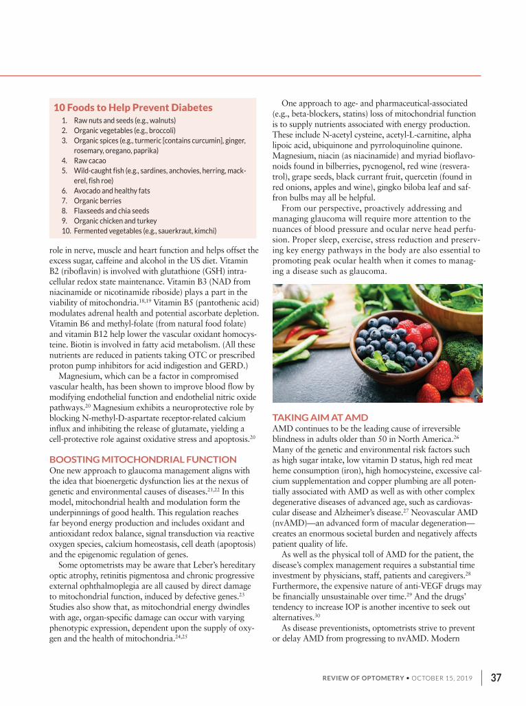
REVIEW OF OPTOMETRY • OCTOBER 15, 2019 37
role in nerve, muscle and heart function and helps offset the excess sugar, caffeine and alcohol in the US diet. Vitamin B2 (riboflavin) is involved with glutathione (GSH) intra-cellular redox state maintenance. Vitamin B3 (NAD from niacinamide or nicotinamide riboside) plays a part in the viability of mitochondria.18,19 Vitamin B5 (pantothenic acid) modulates adrenal health and potential ascorbate depletion. Vitamin B6 and methyl-folate (from natural food folate) and vitamin B12 help lower the vascular oxidant homocys-teine. Biotin is involved in fatty acid metabolism. (All these nutrients are reduced in patients taking OTC or prescribed proton pump inhibitors for acid indigestion and GERD.)
Magnesium, which can be a factor in compromised vascular health, has been shown to improve blood flow by modifying endothelial function and endothelial nitric oxide pathways.20 Magnesium exhibits a neuroprotective role by blocking N-methyl-D-aspartate receptor-related calcium influx and inhibiting the release of glutamate, yielding a cell-protective role against oxidative stress and apoptosis.20
BOOSTING MITOCHONDRIAL FUNCTION One new approach to glaucoma management aligns with the idea that bioenergetic dysfunction lies at the nexus of genetic and environmental causes of diseases.21,22 In this model, mitochondrial health and modulation form the underpinnings of good health. This regulation reaches far beyond energy production and includes oxidant and antioxidant redox balance, signal transduction via reactive oxygen species, calcium homeostasis, cell death (apoptosis) and the epigenomic regulation of genes.
Some optometrists may be aware that Leber’s hereditary optic atrophy, retinitis pigmentosa and chronic progressive external ophthalmoplegia are all caused by direct damage to mitochondrial function, induced by defective genes.23 Studies also show that, as mitochondrial energy dwindles with age, organ-specific damage can occur with varying phenotypic expression, dependent upon the supply of oxy-gen and the health of mitochondria.24,25
One approach to age- and pharmaceutical-associated (e.g., beta-blockers, statins) loss of mitochondrial function is to supply nutrients associated with energy production. These include N-acetyl cysteine, acetyl-L-carnitine, alpha lipoic acid, ubiquinone and pyrroloquinoline quinone. Magnesium, niacin (as niacinamide) and myriad bioflavo-noids found in bilberries, pycnogenol, red wine (resvera-trol), grape seeds, black currant fruit, quercetin (found in red onions, apples and wine), gingko biloba leaf and saf-fron bulbs may all be helpful.
From our perspective, proactively addressing and managing glaucoma will require more attention to the nuances of blood pressure and ocular nerve head perfu-sion. Proper sleep, exercise, stress reduction and preserv-ing key energy pathways in the body are also essential to promoting peak ocular health when it comes to manag-ing a disease such as glaucoma.
TAKING AIM AT AMD AMD continues to be the leading cause of irreversible blindness in adults older than 50 in North America.26 Many of the genetic and environmental risk factors such as high sugar intake, low vitamin D status, high red meat heme consumption (iron), high homocysteine, excessive cal-cium supplementation and copper plumbing are all poten-tially associated with AMD as well as with other complex degenerative diseases of advanced age, such as cardiovas-cular disease and Alzheimer’s disease.27 Neovascular AMD (nvAMD)—an advanced form of macular degeneration—creates an enormous societal burden and negatively affects patient quality of life.
As well as the physical toll of AMD for the patient, the disease’s complex management requires a substantial time investment by physicians, staff, patients and caregivers.28 Furthermore, the expensive nature of anti-VEGF drugs may be financially unsustainable over time.29 And the drugs’ tendency to increase IOP is another incentive to seek out alternatives.30
As disease preventionists, optometrists strive to prevent or delay AMD from progressing to nvAMD. Modern
10 Foods to Help Prevent Diabetes1. Raw nuts and seeds (e.g., walnuts)2. Organic vegetables (e.g., broccoli)3. Organic spices (e.g., turmeric [contains curcumin], ginger,
rosemary, oregano, paprika)4. Raw cacao 5. Wild-caught fish (e.g., sardines, anchovies, herring, mack-
erel, fish roe)6. Avocado and healthy fats7. Organic berries8. Flaxseeds and chia seeds9. Organic chicken and turkey10. Fermented vegetables (e.g., sauerkraut, kimchi)
034_ns1019_diet.indd 37034_ns1019_diet.indd 37 10/14/19 11:28 AM10/14/19 11:28 AM

REVIEW OF OPTOMETRY • OCTOBER 15, 201938
technology enables the detection of early AMD progression so referrals can be made when necessary. However, this is only half of the management story; the other half is having a discussion with high-risk patients about a nutrition and lifestyle plan that is customized to their personalized health
care needs. Minimizing the progression of nvAMD begins with the knowledge that prevention always trumps tertiary care; anti-VEGF treatment should be a patients’ last line of defense, not their first.
PATHOGENESIS OF nvAMD Our understanding of the pathogenesis of AMD has evolved over the last 18 years since the publishing of the Age-Related Eye Disease Study (AREDS 1).31 From a his-torical perspective, the earliest reference to nvAMD is from Junius and Kuhnt, who first described hemorrhagic macu-lar lesions in 1926.32 This disease observation was later termed senile disciform macular degeneration. Initially, no treatment was available for the disease, as photodynamic therapy using verteporfin and other anti-VEGF therapy had not been developed yet.
In the early 1990s, the AREDS 1 randomized clinical study was designed to determine AMD progression rates after participants were assigned to take an antioxidant for-mulation of vitamins or placebo.31 Patients who progressed to advanced AMD were considered to be at the end stage of AMD. Advanced AMD indicates progression to two distinct clinical end-points—nvAMD or geographic atrophy (GA). For convenience, and because both diseases were untreatable at the time of the study’s start, the conditions were lumped together as a common endpoint.
AREDS was formulated specifically for nvAMD and is not effective in a setting of GA. The conclusion that Johanna Seddon reached in a 2016 paper—that the effec-tiveness of antioxidant and zinc supplementation appeared to differ by genotype—was a cautionary note to the scien-tific community that AREDS analysis should not include GA, as it will dilute and obscure meaningful relationships related to nvAMD progression rates.33
Age-related changes that predispose individuals to AMD occur in the posterior segment that includes the photorecep-tors, retinal pigment epithelium (RPE), Bruch’s membrane and choriocapillaris; these alterations potentially cause a lack of homeostasis and dysregulation of the “comple-ment” system, although specific mechanisms have yet to be determined.34 Researchers at the Wilmer Eye Institute found that a combination of impaired complement regulators can result in inadequately controlled complement by the RPE in AMD, which induces RPE damage; drusen are a by-product of complement-mediated inflammatory processes.34 As such, patients who have unregulated complement activation can be at risk for producing angiogenic growth factors lead-ing to nvAMD.
Lifestyle triggers that change the rate of progression to nvAMD include nutritional and epigenetic factors (see, “A 10-point Plan to Prevent AMD”). Dr Richer has also published several peer-reviewed papers concerning the
DIET
PUT WELLNESS ON THE MENU
A New Model of PracticeAs our health care model continues to evolve, optometric and medical practices of the future may look very different. The fol-lowing is a snapshot of the OD and MD practices of three OWNS board members from the East Coast, Midwest and West Coast:
• Kerry Gelb, OD, LensCrafters, Woodbridge, NJ. During con-versations with patients about their conditions, I give patients an option: “Would you like me to teach you how to treat the root cause, or just treat your symptoms?” The response is usually for me to help treat the root cause, with ensuing comments such as, “Why hasn’t my primary doctor spoken to me about this sub-ject?” It’s important to first explain to patients that nutrition and lifestyle medicine are adjuncts of traditional symptoms-based treatments. Patients must also understand that early diagnosis usually translates into better eventual outcomes and can reduce the need for expensive and chronic medication use.
I have created comprehensive nutrition plans for patients with AMD, diabetes and DED. And I advise other optometrists interested in adding a functional medicine component to their practices to create similar plans for common ocular conditions associated with nutrition and lifestyle medicine. Eye care profes-sionals should also reach out to doctors in other specialties who practice preventive functional medicine as a way to build the practice and obtain referrals. In fact, I receive most of my refer-rals from like-minded MDs.
• Steven G. Pratt, MD, La Jolla Laser Vision, La Jolla, Calif. My surgical practice is an extension of my personal experiences with macular degeneration and subsequent nutrition and wellness practices, outlined in my trademarked SuperFoodsRx products. I and my practice colleagues promote natural and active outdoor living. And my website, www.superhealthyliving.com, provides patients with access to free pamphlets and handouts to learn about natural ways to help prevent macular degeneration and migraine headaches, manage dry eye and heal wounds. The site also offers tips about supplements and has a section for pet health, and individuals can purchase my books, such as the latest, SuperFoodsRx for Pregnancy: The Right Choices for a Healthy, Smart, Super Baby.
• Stuart Richer, OD, PhD, Captain James A. Lovell Federal Health Care Facility, North Chicago. I work on behalf of veterans and active duty US Navy military personnel at one of the largest US federal health care centers in the country. In the office, I rely on props (e.g., cans of spinach, sardines, low-salt V8, plastic ber-ries) and single-page educational handouts to directly educate patients. During weekly lectures and department-wide email blasts, I encourage other staff members, residents and optometry and medical students to practice preventive ocular medicine.
034_ns1019_diet.indd 38034_ns1019_diet.indd 38 10/14/19 11:29 AM10/14/19 11:29 AM

REVIEW OF OPTOMETRY • OCTOBER 15, 2019 39
science of epigenetics and the prescription of Longevinex to improve visual function (including dark adaptation) and retinal structure (including stem cell regeneration) in patients into their 90s.27
NUTRIENTS, GENETICS AND PROGRESSIONVarious studies demonstrate a link between diet and pro-gression to AMD. The Eye Risk Consortium study revealed that adherence to the Mediterranean diet was associated with a 41% reduced risk in nvAMD.35 The findings sup-ported the role of a diet laden with nutrient-rich foods such as fruits, vegetables, legumes and fish in the prevention of AMD. In June 2018, Chapman et al. performed a sys-tematic review of significant long-term studies intended to evaluate the role of diet and food intake in AMD.36 Nega-tive drivers contributing to an increase in progression risk to nvAMD of between 56% and 270% included choosing a high dietary glycemic index diet, eating processed meat and consuming a typical Western diet.36 High consumption of vegetables rich in carotenoids and fish containing omega-3 fatty acids was beneficial for those at risk of AMD.36
Though genetics play a role in eye and overall health, wellness or disease is greatly dependent upon desirable or undesirable biological activities interacting with those genet-ics. The CFH gene provides instructions for making protein complement factor H, used to help regulate the body’s immune response. Lechanteur et al. showed that patients who had high-risk CFH risk alleles had a reduced timeline for pathogenesis to nvAMD by 12.2 years.37 Millen et al. reported that blood levels pointing to vitamin D deficiency were associated with a 1.8-fold increase in the odds of hav-ing AMD among women with low-risk CFH, and rose to a seven-fold increase in women with high-risk CFH, sug-gesting a synergistic effect between vitamin D status and complement cascade protein function.38
Another study, CAREDS, reported that diets rich in lutein plus zeaxanthin might protect against intermediate AMD in healthy women younger than 75 years.39
The power of good nutrition is a top priority in reducing the progression to nvAMD. Supplements should be consid-ered as a backup to core nutritional practices to prevent or reduce nvAMD (see, “10 Foods that Fight AMD”).
THE PUBLIC HEALTH CHALLENGENutrition and lifestyle medicine are not new concepts to those of us at the OWNS. Yet, it still surprises us how unfamiliar these ideas are for many of our patients and new graduates and how much interest they express in learning more. A growing body of research is revealing the impor-tance of such a strategy.
A 2019 study in The Lancet showed that, globally, one-fifth of all deaths were associated with poor diets.40 In addition, more than 90% of Americans are deficient in at least one nutrient, based on our estimates from USDA survey findings and CDC Nutrition Report data. A stag-gering 63% of food consumed in the US is processed, with only 12% of food coming from plant sources; the remain-ing 25% comes from animal sources.41 We at the OWNS believe that minimizing processed, manufactured food and exchanging it with whole, organic food is the single most important factor in improving ocular and overall health through reducing inflammation—the core component of chronic disease.
A 2016 report and review of 12 major epidemiological studies as well as the 2010 United States Census popula-tion estimated that more than half of the US population over age 40—about 90 million of 142 million adults—experienced vision problems due to eye diseases such as AMD, cataracts, diabetic retinopathy and glaucoma, in addition to vision impairment, blindness and refractive error.42
Foods to Help Prevent Dry Eye Disease1. Raw nuts and seeds (e.g., walnuts, pine nuts, macadamia
nuts, flaxseed, chia seeds, pumpkin seeds)2. Organic fruits/vegetables (e.g., kale, spinach, Swiss chard,
strawberries, goji berries, acerola cherries, blueberries)3. Organic spices (e.g., turmeric [contains curcumin], ginger,
paprika, rosemary, oregano, pumpkin spice, clove)4. Gamma-linolenic acid foods (e.g., black currant seed oil,
blue-green algae [e.g., spirulina], borage oil, hemp seeds)5. Omega-3 fatty acids (e.g., wild salmon, anchovies, mack-
erel, sardines)6. Avocado7. Organic green tea8. Pastured eggs9. Organic chicken and turkey10. Broccoli and antioxidant-rich cruciferous vegetables (e.g.,
cauliflower, Brussels sprouts, red cabbage, kale, collards, watercress, mustard greens)
034_ns1019_diet.indd 39034_ns1019_diet.indd 39 10/14/19 11:29 AM10/14/19 11:29 AM

REVIEW OF OPTOMETRY • OCTOBER 15, 201940
We at the OWNS strongly believe that AMD and dia-betic macular edema (DME) are heavily associated with poor nutrition and lifestyle choices. And yet, at the same time, traditional medical procedures continue to skyrocket. For example, the number of intravitreal injections (IVIs) for conditions such as AMD and DME are seeing an exponen-tial rise.43 A total of 5.9 million IVIs were performed in the US in 2016.44 This is up dramatically from less than 5,000 IVIs between 1997 and 2001.45
Unfortunately, medical and optometry schools are not evolving to reflect a changing wellness landscape. Their philosophies and curricula continue to center upon symp-toms-based medicine rather than addressing root causes. Optometrists are provided with little to no training in nutrition and lifestyle medicine. Fortunately, the OWNS is cataloging years of research and lectures on ocular nutrition and wellness. And various doctors around the country have played a role in creating valuable ocular nutrition content online for patients. ❍
1. Gil MI, Tomás-Barberán FA, Hess-Pierce B, et al. Antioxidant activity of pomegranate juice and its
relationship with phenolic composition and processing. J Agric Food Chem. 2000 Oct;48(10):4581-9.
2. Seddon JM, Cote J, Rosner B. Progression of age-related macular degeneration: association with
dietary fat, transunsaturated fat, nuts, and fish intake. Arch Ophthalmol. 2003 Dec;121(12):1728-37.
3. Chasan-Taber L, Willett WC, Seddon JM, et al. A prospective study of carotenoid and vitamin A
intakes and risk of cataract extraction in US women. Am J Clin Nutr. 1999 Oct;70(4):509-16.
4. Huang P, Wang F, Sah BK, et al. Homocysteine and the risk of age-related macular degeneration: a
systematic review and meta-analysis. Sci Rep. 2015 Jul 21;5:10585.
5. Ganguly P, Fatima Alam S. Role of homocysteine in the development of cardiovascular disease. Nutr
J. 2015;14:6.
6. Clarke R. B-vitamins and prevention of dementia. Proc Nutr Soc. 2008 Feb;67(1):75-81.
7. Luchsinger JA, Tang MX, Miller J, et al. Relation of higher folate intake to lower risk of Alzheimer
disease in the elderly. Arch Neurol. 2007 Jan;64(1):86-92.
8. Lynch B, Dirty genes: a breakthrough program to treat the root cause of illness and optimize your
health. HarperCollins, New York, NY, Harper One 2018.
9. Nelson JD, Craig, JP, Esen A, et al. TFOS DEWS II Report. Ocul Surf 2017; 2017 July;15(3):269-
650.
10. Coleman AL, Kodjebacheva G. Risk factors for glaucoma needing more attention. Open Ophthalmol
J. 2009 Sep 17;3:38-42.
11. Bhartiya S, Ichpujana P. (Eds.) (2015) Manual of Glaucoma. London, UK:JP medical Ltd.
12. Braakhuis A, Raman R, Vaghefi E. The association between dietary intake of antioxidants and ocu-
lar disease. Diseases. 2017 Jan 30;5(1). pii: E3.
13. Golubnitschaja O. The keyrole of multiomics in the predictive, preventive and personalised medical
approach towards glaucoma management. Klin Monbl Augenheilkd.2018; Feb;235(2):146-150.
14. Hysi PG, Khawaja AP, Menni C, et al. Ascorbic acid metabolites are involved in intraocular pressure
control in the general population. Redox Biol. 2019 Jan;20:349-53.
15. Robert D Steigerwalt, Jr, Gianni Belcaro, Paolo Morazzoni, et al. Mirtogenol® potentiates latano-
prost in lowering intraocular pressure and improves ocular blood flow in asymptomatic subjects. Clin
Ophthalmol. 2010; 4: 471-6.
16. Gizzi C, Torino-Rodriguez P, Belcaro G Hu S, et al, Mirtogenol® supplementation in association with
dorzolamide-timolol or latanoprost improves the retinal microcirculation in asymptomatic patients with
increased ocular pressure. Eur Rev Med Pharmacol Sci. 2017 Oct;21(20):4720-25.
17. Melgarejo JD, Lee JH, Petitto M, et al. Glaucomatous optic neuropathy associated with noc-
turnal dip in blood pressure: findings from the Maracaibo Aging Study. Ophthalmology. 2018
Jun;125(6):807-14.
18. Jung KI, Kim YC, Park CK. Dietary niacin and open-angle glaucoma: the Korean National Health
and Nutrition Examination Survey, Nutrients. 2018 Mar 22;10(4).
19. Williams PA, Harder JM, Foxworth NE, et al. Vitamin B3 modulates mitochondrial vulnerability and
prevents glaucoma in aged mice. Science. 2017 Feb 17;355(6326):756-60.
20. Ekici F, Korkmaz S, Karaca EE, et al. The role of magnesium in the pathogenesis and treatment of
glaucoma. Int Sch Res Notices. 2014;745439.
21. Wallace DC, Mitochondrial genetic medicine. Nat Genet. 2018 Dec;50(12):1642-49.
22. Singh LN, Crowston JG, Lopez Sanchez MIG, et al. Mitochondrial DNA variation and disease sus-
ceptibility in primary open-angle glaucoma. Invest Ophthalmol Vis Sci. 2018 Sep 4;59(11):4598-602.
23. Fraser JA, Biousse V, Newman NJ, et al. The Neuro-ophthalmology of mitochondrial disease. Surv
Ophthalmol. 2010 Jul 8; 55(4): 299-334.
24. Benoist d’Azy C, Pereira B, Chiambaretta F, et al. Oxidative and anti-oxidative stress markers in
chronic glaucoma: a systematic review and meta-analysis. PLoS One 2016 Dec 1;11(12):e0166915.
25. Filina AA, Davydova NG, Endrikhovski SN, et al. Lipoic acid as a means of metabolic therapy of
open angle glaucoma. Vestn Oftamol. 1995 Oct-Dec;111(4):6-8.
26. Pennington KL, DeAngelis MM. Epidemiology of age-related macular degeneration (AMD): associa-
tions with cardiovascular disease phenotypes and lipid factors. Eye Vis (Lond). 2016 Dec 22;3:34.
27. Richer, Ulanski, Popenko, et al. AMD Beyond AREDS II. In: Yannuzzi, eds. Advances in Ophthalmol-
ogy and Optometry 1. Elsevier Press; 2016:335-369.
28. Prenner JL, Halperin LS, Rycroft C, et al. Disease Burden in the treatment of age-related macular
degeneration: findings from a time-and-motion study. Am J Ophthalmol. 2015 Oct;160(4):725-31.e1.
29. Baker-Schena L. Expensive Drugs. American Academy of Ophthalmology. 2017 June. Available at:
https://www.aao.org/eyenet/article/expensive-drugs. (last accessed Aug. 26, 2019).
30. Hoguet A, Chen PP, Junk AK, et al. The effect of anti-vascular endothelial growth factor agents on
intraocular pressure and glaucoma: A Report by the American Academy of Ophthalmology. Ophthal-
mology. 2019 Apr;126(4):611-22.
31. The Age-Related Eye Disease Study Research Group. The Age-Related Eye Disease Study (AREDS):
Design Implications AREDS Report No. 1. Control Clin Trials. 1999 Dec; 20(6): 573-600.
32. Junius P, Kuhnt H. Die scheibenförmige Entartung der Netzhautmitte. Degeneratio maculae luteae
disciformis [The disciform degeneration of the central retina. Disciform Macula Lutea Degeneration]
Berlin: Karger S.; 1926.
33. Seddon JM, Silver RE, Rosner B. Response to AREDS supplements according to genetic fac-
tors: survival analysis approach using the eye as the unit of analysis. Br J Ophthalmol. 2016
Dec;100(12):1731-7.
34. Ebrahimi KB, Fijalkowski N, Cano M, et al. Decreased membrane complement regulators in
the retinal pigmented epithelium contributes to age-related macular degeneration. J Pathol. 2013
Apr;229(5):729-42.
35. Merle BMJ, Colijn JM, Cougnard-Grégoire A, et al. Mediterranean diet and incidence of advanced
age-related macular degeneration: the EYE-RISK Consortium. Ophthalmology. 2019;126(3):381-90.
36. Chapman NA, Jacobs RJ, Braakhuis AJ. Role of diet and food intake in age-related macular
degeneration: a systematic review. Clin Exp Ophthalmol. 2019 Jan;47(1):106-27.
37. Lechanteur YT, van de Camp PL, Smailhodzic D, et al. Association of smoking and CFH and ARMS2
risk variants with younger age at onset of neovascular age-related macular degeneration. JAMA Oph-
thalmol. 2015;133(5):533-41.
38. Millen AE, Meyers KJ, Liu Z, et al. Association between vitamin D status and age-related macular
degeneration by genetic risk. JAMA Ophthalmol. 2015 Oct; 133(10): 1171-9.
39. Moeller SM, Parekh N, Tinker L, et al. Associations between intermediate age-related macular
degeneration and lutein and zeaxanthin in the Carotenoids in Age-Related Eye Disease Study
(CAREDS). Arch Ophthalmol. 2006;124(8):1151.
40. GBD 2017 Diet Collaborators. Health effects of dietary risks in 195 countries, 1990-
2017: a systematic analysis for the Global Burden of Disease Study 2017. Lancet. 2019 May
11;393(10184):1958-72.
41. USDA Economic Research Service. U.S. Food Consumption as a % of Calories. 2009. Available at:
https://www.healthyschoolfood.org/docs/color_pie_chart.pdf. (last accessed September 3, 2019).
42. Prevent Blindness. Vision problems in the U.S.: Methods and sources. 2012b. 2016 July 5. Avail-
able at: http://www.visionproblemsus.org/introduction/methods-and-sources.html. (last accessed Sept.
3, 2019).
43. Grzybowski A, Told R, Sacu S, et al. 2018 Update on Intravitreal Injections: Euretina Expert Consen-
sus Recommendations. Ophthalmologica. 2018;239(4):181-193.
44. Williams GA. IVT Injections: Health Policy Implications. Rev Ophthalmol. 2014 June 5. Available at:
http://www.reviewofophthalmology.com/content/ d/retinal_insider/c/48732. (last accessed September
3, 2019).
45. Ramulu PY, Do DV, Corcoran KJ, et al. Use of retinal procedures in Medicare beneficiaries from
1997 to 2007. Arch Ophthalmol.2010;128:1335-40.
DIET
PUT WELLNESS ON THE MENU
034_ns1019_diet.indd 40034_ns1019_diet.indd 40 10/14/19 11:29 AM10/14/19 11:29 AM
REVIEW OF OPTOMETRY • OCTOBER 15, 2019 41
The idea that proper diet and vitamin supplementa-tion can improve ocular health is a given these days, as research showcases the positive effects certain nutrients can have on conditions such as
age-related macular degeneration (AMD) and cataracts. Despite the progress made by a greater understanding of nutrition and the eye, some fierce debates continue to rage on in the optometric community.
Here’s a look at two red-hot topics in ocular nutrition.
ROUND 1: IS RESPONSE TO AREDS AFFECTED BY GENETICS?When the Age-related Eye Disease Study (AREDS) first published nearly two decades ago, the promise that an oral supplement could reduce the risk of AMD progression was big news.1 The formulation was patented and PreserVision was born. The formulation was shown to reduce the risk of progression by some 25% and it became the standard of care for moderate AMD.
As the genes associated with AMD were discovered over the following decade, researchers asked the question, “Do all patients responded similarly?” This led to eight subse-quent publications.2-9
Four of the publications measured AMD progression to either geographic atrophy (GA) or to choroidal neovas-cularization (CNV).2-5 However, a 2016 study reminded everyone that the AREDS formulation was only shown to be effective against progression to CNV and not against GA.6 The study demonstrated a statistically significant interaction between genetics and the AREDS formulation against progression to CNV.6 Thus, the four publications incorrectly measured progression to GA.2-5 This might be analogous to studying Lucentis (ranibizumab, Genentech)
against GA and CNV when we know that it is only effec-tive against CNV. This mistake clouded the truth and cre-ated intense confusion among practitioners.
The last four research studies all focused on progression to CNV, and all four noted a statistically significant gene
Two Big Controversies in Ocular Nutrition
AMD supplements and omega-3s are both still in the hot seat.
BY STUART RICHER, OD, PHD, AND DENNIS RUSKIN, OD
Riddle of the ZincsZinc, a necessary but otherwise unassuming mineral, was thrust center-stage in the controversy over the AREDS data. Currently, no reliable evidence exists to confirm that 25mg of zinc oxide is safer than 80mg in an AREDS formula. Ananda S. Prasad, MD, the reign-ing authority on zinc therapeutics and supplementation, advises that 45mg of oral zinc can be consumed without creating a zinc/copper imbalance.1 Zinc deficiency is a real concern, with 29% of the US population experiencing inadequate intake (91 million out of a population of 314 million).
However, there is more to this zinc story. Zinc is poorly absorbed, especially the AREDS zinc oxide form, which is insoluble in water. This is often due to a lack of stomach acid. Co-administration of zinc with vitamin B6 increases absorption substantially. Once absorbed, zinc is bound to metallothionein to make sure it doesn’t get out of control, being a metallic mineral. Co-consumption of zinc with selenium releases zinc from metallo-thionein, its binding protein. These nutrient dynamics all influence whether therapy is harmful, has a null effect or is beneficial. Clearly, more research is needed to examine these relationships.
A host of clinical factors that complicate the zinc debate such as age-related hypochlorhydria, concurrent chronic use of over-the-counter and pharmacologic acid blockers, competing iron and cop-per intake as well as H. Pylori infection will all effect zinc bioavail-ability in your patient.
1. Prasad AS. Trace Elements in Human Health & Disease, Volume I. Academic Press:
London 1977.
CONTROVERSIES
041_ns1019_controversies.indd 41041_ns1019_controversies.indd 41 10/14/19 11:30 AM10/14/19 11:30 AM

REVIEW OF OPTOMETRY • OCTOBER 15, 201942
CONTROVERSIES
TWO BIG CONTROVERSIES IN OCULAR NUTRITION
interaction.6-9 In the most recent analysis, the independent authors studied 266 patients and found that patients were four times more likely to have progressed to CNV if they had a specific genotype and took the AREDS/AREDS2 formulation.9
The data now seems clear. The AREDS/AREDS2 formu-lation is a valuable product for most people with moderate AMD. Many patients with certain genotypes can experi-ence a greater than 50% reduction in their risk for pro-gression to CNV. No other formulation has been shown to do that. However, approximately 15% of patients with a specific genotype experience a three- to four-fold increased progression to CNV. Thus begins the era of per-sonalized medicine in AMD for those of us who manage these patients. As practitioners we are now armed with the knowledge necessary to improve patient outcomes, per-haps through improved compliance for those who benefit from the formulation and to limit AREDS/AREDS2 use in those who will not benefit.
ROUND 2: THE DREAM STUDY VS. FISH OIL For more than a decade, many eye care practitioners have enthusiastically recommended omega-3 fatty acid supple-ments to their dry eye patients to alleviate symptoms with-out the double-edged sword of adverse side effects.
But in May 2018, the Dry Eye Assessment and Man-agement (DREAM) study seemed to take the wind out of the sails of fish oil’s effectiveness, since the investigation reported omega-3s offered no benefit over the olive oil placebo.10
This rocked some corners of the profession, especially for those who touted the well-established treatment. While
some backed away from omega 3s after these results, experts cautioned to take a closer look at the study instead of just skimming the headlines.
Fish oil as a treatment for dry eye first came about in a 2005 study.11 It showed women who ate regular amounts of tuna in their diets had lower rates of dry eye symptoms.11
Further proof came along in 2011 in the form of a pilot study that concluded dietary supplementation with omega-3 fatty acids in dry eye showed no significant effect in meibum lipid composition or aqueous tear evaporation rate.12 Yet it also reported the average tear production and tear volume were increased in the omega-3 group.12
The DREAM study was a one-year, double-masked, randomized, multicenter study by the National Eye Insti-tute that looked at the benefits of omega-3 supplements in dry eye patients.10 Patients were allowed to continue their current treatments, which is not the case in most industry-sponsored trials of dry eye treatments.
Eligible participants included those who had signs and symptoms of moderate-to-severe dry eye on two consecu-tive exams performed two weeks apart, dry eye symptoms for at least six months, an Ocular Surface Disease Index (OSDI) score ranging from 25 to 80 at a screening visit and an OSDI score ranging from 21 to 80 at the baseline eligibility visit.10 Patients also had to be willing to continue their current dry eye treatment regimens, be using or want to use artificial tears at least twice daily in the previous two weeks and had other dry eye symptoms.
Basically, the “real world” clinical trial included patients with typical dry eye disease who continued to seek relief of symptoms despite the use of other interventions.
Additionally, patients with a history of thyroid disease, Sjögren’s syndrome, rheumatoid arthritis or inflammatory diseases could be included in the trial if they were other-wise eligible.
The 3,000mg daily dosage of omega-3 fatty acids was the highest dose used to date in clinical trials of fish-derived omega-3 fatty acids.10 The daily placebo was approximately one tablespoon of olive oil, which primarily delivered n-9 oleic acid, a substance usually considered neutral with respect to changes in signs and symptoms of dry eye.10
After 12 months, the study authors did not find signifi-cant differences in mean OSDI or conjunctival staining, corneal staining, tear break-up time and Schirmer’s test between groups.10
Since the DREAM study’s release, some have cried foul over certain aspects of the investigation’s methodology, especially the olive oil placebo, citing its potential thera-peutic effects. Still others took issue with the study’s “real world” approach and the fact patients were allowed to continue taking their medications.
Many moderate AMD patients who take the AREDS or AREDS2 formulation can sigificantly reduce their risk of progression, though a specific genotype increases risk of CNV development in 15% of cases.
Photo: S
teven Ferrucci, OD
041_ns1019_controversies.indd 42041_ns1019_controversies.indd 42 10/14/19 11:30 AM10/14/19 11:30 AM

REVIEW OF OPTOMETRY • OCTOBER 15, 2019 43
The DREAM investigators vigorously defended their choice of the olive oil placebo. They cited the Mediter-ranean diet, which includes an average of 60g of olive oil, and compared it with their olive oil placebo dose (5g, the equivalent of one tablespoon teaspoon) which they coun-tered was too low to have a therapeutic effect.13,14
The researchers continued on the defense by arguing defensively that the anti-inflammatory effects associated with olive oil are attributed mainly to polyphenols not found in the refined olive oil used in the study.15 However, research does show that oleic acid can alter the microbi-ome and reduce dysbiosis, conferring anti-inflammatory effects elsewhere in the body.15 Oleic acid can also counter
the negative impact of saturated fat on the microbiome.16 Others in the profession called out the study’s loose
inclusion and exclusion criteria. The fact that the study allowed moderate-to-severe dye eye patients of different demographics and health profiles made it increasingly dif-ficult to come to a definitive conclusion that all oral ome-gas were ineffective, the opposition argued.
Despite the criticism, the DREAM research team has doubled-down. During a presentation at ARVO’s 2019 meeting, the study’s researchers presented a follow-up investigation that found no significant differences in out-comes between patients who stopped taking the fish oil supplements and those who continued taking them over an additional 12-month period.17 The DREAM investiga-tors said this reaffirmed their original study results of no demonstrable benefit from omega-3 supplements.17
While the research battle rages on, practicing clinicians who have found success with omega-3s will likely take the DREAM study with a grain of salt, and others will believe the study reinforced recommending a gamma linolenic acid along with fish oil. ❍
1. Age-related Eye Disease Study Research Group. A randomized, placebo-controlled, clinical trial of high-dose supplementation with vitamins C and E, beta carotene, and zinc for age-related mac-ular degeneration and vision loss: AREDS report no. 8. Arch Ophthalmol. 2001;119(10):1417-36.2. Awh C, Lane AM, Hawken S, et al. CFH and ARMS2 genetic polymorphisms predict response to antioxidants and zinc in patients with age-related macular degeneration. Ophthalmology. 2013;120(11):2317-23.3. Awh CC, Hawken S, Zanke BW. Treatment response to antioxidants and zinc based on CFH and ARMS2 genetic risk allele number in the Age-Related Eye Disease Study. Ophthalmology. 2015;122(1):162-9.4. Chew EY, Klein ML, Clemons TE, et al. No clinically significant association between cfh and arms2 genotypes and response to nutritional supplements: AREDS Report Number 38. Ophthal-mology. 2014;121(11): 2173-80.5. Assel MJ, Li F, Wang Y, et al. Genetic polymorphisms of CFH and ARMS2 do not predict response to antioxidants and zinc in patients with age-related macular degeneration: inde-pendent statistical evaluations of data from the age-related eye disease study. Ophthalmology. 2018;125(3):391-7.6. Seddon JM, Silver RE, Rosner B. Response to AREDS supplements according to genetic factors: survival analysis approach using the eye as the unit of analysis. Br J Ophthalmol. 2016;100(12):1731-7.7. Vavvas D, Small K, Awh C, et al. CFH and ARMS2 genetic risk determines progression to neo-vascular age-related macular degeneration after antioxidant and zinc supplementation. Proc Natl Acad Sci. 2018;115(4):E696-704.8. Zanke B. Re: Assel et al.: Genetic polymorphisms of CFH and ARMS2 do not predict response to antioxidants and zinc in patients with age-related macular degeneration (Ophthalmology. 2018;125:391-397). 2018;125(5):e34-e35.9. Kaufman S, et al. Interaction of genotypes and AREDS formulation use in the development of neovascular AMD. ASRS Abstract. July 2019. JAMA Ophthalmology. In Press.10. Asbell PA, Maguire MG, Pistilli M. n-3 fatty acid supplementation for the treatment of dry eye disease. N Engl J Med. 2018;378(18):1681-90.11. Miljanovi B, Trivedi KA, Dana MR, et al. The relationship between dietary n-3 and n-6 fatty acids and clinically diagnosed dry eye syndrome in women. Am J Clin Nutr. 2005;82(4):887-93.12. Wojtowicz JC, Butovich I, Uchiyama E, et al. Pilot, prospective, randomized, double-masked, placebo-controlled clinical trial of an omega-3 supplement for dry eye. Cornea. 2011;30(3):308-14.13. Galor A, Asbell PA. DREAM Study: can omega-3 treat dry eye disease? American Academy of Ophthalmology. www.aao.org/interview/dream-study-can-omega-3-treat-dry-eye-disease. Published 2018. Accessed April 17, 2019.14. Gorzynik-Debicka M, Przychodzen P, Cappello F, et al. Potential health benefits of olive oil and plant polyphenols. Int J Mol Sci. 2018;19(3).15. Ruskin D, Poteet J, Richer S. Four great debates in ocular nutrition: wellness essentials for clinical practice. A Supplement to Review of Optometry. 2018:24-25.16. Alcock J, Lin HC. Fatty acids from diet and microbiota regulate energy metabolism. F1000Res. 2015;4(F1000 Faculty Rev):738.17. Hussain M, Shtein RM, Pistilli M, et al. DREAM extension study—comparison of placebo and omega-3 fatty acid supplement groups on OSDI, 4 key signs of DED and adverse events. ARVO 2019. Abstract 6729-B0253.
Eights Threats to CounterHere are major disruptors of modern health that challenge our well-being and drain our vitality if we are sensitive (i.e., immune intolerant) or exposed chronically without offsetting or balancing wellness strategies. However, opinions differ among experts on a number of these, and it can be difficult to effect change in mindset and behavior among some doctors, let alone patients.
• Lectins• Soy products• Broad-spectrum antibiotics• NSAIDs• Gastrointestinal acid blockers • Artificial sweeteners• Hormone disruptors• HerbicidesTwo of these deserve extra attention:Lectins: Steven Gundry’s The Plant Paradox reveals a danger
in the American diet: a toxic protein in plants called lectins.1 These proteins are found in hundreds of common foods, includ-ing ‘nightshade’ vegetables belonging to the 2,000+ species Solanaceae family which includes common vegetables (e.g., eggplant, tomatoes, peppers, potatoes). This is why it is impor-tant to focus on building one’s immune tolerance.
Soy Products: This nutrient can be problematic in two ways. First, soy contains phytoestrogens that mimic estrogen, prompting the body (both female and male) to store fat by leading to disruption of the adrenal and thyroid glands. Thus this ‘toxic superfood’ can increase our body weight, change our body shape and impact our fertility. Second, soy contains goi-trogens—substances that depress our thyroid gland function, making us sluggish, decreasing our concentration and sex drive.
Taken together, 21st century challenges such as lectin and soy sensitivity affect our digestion, immune competence, cogni-tive status, reproductive potential and even increase our sus-ceptibility to cancer, cardiovascular and oculovascular diseases.
1. Gundry SR. The plant paradox: the hidden dangers in “healthy” foods that cause dis-
ease and weight gain. New York, New York: Harper Wave.
041_ns1019_controversies.indd 43041_ns1019_controversies.indd 43 10/14/19 11:31 AM10/14/19 11:31 AM

001_owns1019_fc.indd 2001_owns1019_fc.indd 2 10/14/19 10:27 AM10/14/19 10:27 AM