Embed Size (px)
Citation preview
www.ejpmr.com
Jayanthi et al. European Journal of Pharmaceutical and Medical Research
354
A STUDY ON THE ANATOMICAL CHARACTERISTICS OF ACANTHUS ILLICIFOLIUS
LINN.
P. Santhiavalli1 and G. Jayanthi
2*
1Research and Development Centre, Bharathiar University, Coimbatore.
2PG and Research Department of Botany, Vellalar College for Women (Autonomous), Erode - 638012, Tamilnadu.
Article Received on 12/07/2016 Article Revised on 01/08/2016 Article Accepted on 22/08/2016
INTRODUCTION
Acanthus ilicifolius Linn. (Acanthaceae) is a spiny herb
of the mangrove that is widely distributed in the coastal
areas and it occupy only 5% of the total forest. The
common name of the plant is holy leaved Acanthus
ilicifolius. In tamil it is called as Attumulli and
Kaludaimulli. These are the most hostile environment
with fluctuating tial and saline regime and limited plant
species can survive under such condition. These plants
are most valuable resources and provide economical and
ecological benefits to the coasted people.
It has typical spinose margins on its evergreen leaves and
stipular spines at stem nodes. It is a gregarious, sparingly
branched evergreen shrub, 0.6 – 1.5 meter in height. The
chemical constituents (salts, organic acids,
carbohydrates, hydrocarbons, triterpens, alkaloids,
flavonoids and tannins)[1-3]
.
The plant is used against rheumatism, paralysis, asthma,
skin diseases, boils and wounds[4,5&6,7]
. The present study
describe the anatomical characteristics of Acanthus
ilicifolius.
MATERIALS AND METHODS
The aerial parts of A. ilicifolius were collected from the
coastal areas of Thirumalairayan Pattinam Panchayathu,
Karaikal District, Puducherry State. The plant material
was identified and authenticated taxonomically with the
help of the local floras[8, 9]
and Botanical survey of India,
Southern Regional Centre, Coimbatore, Tamil Nadu. The
herbarium specimen number in BSI is
No.BSI/SRC/5/23/2016/Tech/.632.
Pharmacognostical studies Fresh aerial parts of the study plant were taken for
morphological and histological studies. For the
microscopical studies, leaf and stem were prepared and
stained as per standard procedures [10, 11]
.
Morphological assessment done by physical observation
and measurement of physiognomic features of their fresh
leaf, stem and flower specimens. For the anatomical
studies, the fresh samples were fixed and cross sections
were obtained using a microtome[12]
. The sections were
independently stained with heamatoxyline and safranin.
A light microscope was used to view the slides and
adjusted to finest resolution (40X). Microphotographs
were obtained using a Nikkon digital camera focused
through the microscope eyepiece.
RESULTS
Botanical Description
It is an erect herb whose stems are upto 1.5m which
grow in clumps, little divided and are glabrous (Plate-1).
Leaves are shortly petioled and are oblong or elliptical
whose base is usually spinous, toothed and commonly
solitary. The plant produces a cluster of flowers.
Flowering and fruiting occurs almost throughout the
year, bract and bracteoles present which has 4 sepals,
outer 2 are elliptic rounded, inner 2 are broadly
lanceolate and subacute. The petals are 5 in number, blue
coloured, united, 2 lipped Corolla tube short and
pubescent from within which contains 4 stamens,
didynamous, filaments stout, anthers 1 – lobed, carpels 2,
ovary 2 chambered having 2- ovules in chamber. The
SJIF Impact Factor 3.628
Research Article
ISSN 2394-3211
EJPMR
EUROPEAN JOURNAL OF PHARMACEUTICAL
AND MEDICAL RESEARCH www.ejpmr.com
ejpmr, 2016,3(9), 354-361.
Corresponding Author: Dr. G. Jayanthi
PG and Research Department of Botany, Vellalar College for Women (Autonomous), Erode - 638012, Tamilnadu.
ABSTRACT
Acanthus illicifolius is a mangrove medicinal plant. In this study, the aim was to investigate the anatomical
characteristics of this medicinally important plant species. As a result of the study, it was discovered that the
vascular stem of the midrib is multistranded. The unique type of glandular trichome. The stomata are diacytic and
the venation is densely reticulate. The stem consists of intact epidermis, wide cortex, thin hallow vascular cylinder
and wide pith.
KEY WORDS: Anatomy, medicinal plant, Acanthus ilicifolius, Acanthaceae, mangroves.

www.ejpmr.com
Jayanthi et al. European Journal of Pharmaceutical and Medical Research
355
flowers that develop into pods when the pods ripe, they
explode to propel the seeds up to 2m away.
Microscopic studies
Anatomical studies made on Acanthus ilicifolius showed
dissimilar characteristics of other species of the
Acanthaceae to which this species belongs Metcalfe and
chalk 1957.
Anatomy of the leaf
Midrib: The midrib is cross sectional view appear flat
on the adaxial side and unequally two lobed and thick on
the abaxial side (Plate 2-a). It is 1mm thick and 1.2 mm
wide. The lobes are 300 – 500 µm thick. The vascular
system of the midrib is multistranded. These are three,
independent circular vascular bundles of unequal size
(Plate 2-b) of the three bundles two smaller bundles are
adaxial and lateral in position, the third larger bundle is
abaxial and median (Plate 2-b). All three bundles differ
in their vascular architecture.
The adaxial lateral smaller bundle consists of a bowl
shaped cluster of narrow angular thick walled xylem
elements. On the upper part of the bundle occurs a thick
mass of sclerenchyma cells which extends to the lower
side forming an arc. Phloem is located in small thin layer
of sieve elements on the lower end of the xylem strand
(Plate 2-c).
The adaxial lateral larger bundle is elliptical in outline. It
includes two small groups of circular thick walled
vessels. The vessels are located lateral to each other.
Phloem strands occurs on the lower end of each vascular
bundle. A thick sclerenchyma sheath encloses the entire
vascular bundle (Plate 2-d). The third bundle which is
abaxial and median in position is the largest bundle; it is
circular in outline (Plate 2-e).
It includes a circle of vessels with wide gaps in between
the vessels. The vessels are highly thick walled, circular
and fairly wide. Phloem occurs on the outer part of each
xylem strand. (Plate 2-e) Slerenchymatous bundle sheath
occurs encircling the vascular bundle.
Lamina: The lamina is dorsiventral with smooth abaxial
side and adaxial side. On the adaxial side of the lamina,
there are deep, narrow grooves inside which occurs a
simple, unique type of glandular trichome (Plate 2-f and
g). The lamina is 370 µm thick. The adaxial epidermal
cells of the lamina are thin, tabular in shape and have
thin cuticle. The abaxial epidermis is further thin with
small squarish cells. The mesophyll tissues are
differentiated into adaxial band of two layer of columnar
palisade cells and abaxial zone of small spherical spongy
parenchyma cells which form reticulate partitions for air
– chambers (Plate 2-f).
Leaf-Margin (Plate 2-g): The marginal part of the
lamina is thick, semicircular and measures 350 µm in
thickness. These are a thick circular bundle of fibres with
small, less prominent vascular elements in the centre.
The cells inner of the epidermis are angular, thick walled
and compact “Fig. 3.2”.
Glandular Trichome (Plate 3-a and b): On the adaxial
epidermis of the lamina, these are deep, vertical groves
inside which occurs a short gland. These is large
epidermal cell at the bottom of the groove, on which is
seated the gland (Plate 3-b).
The gland is rectangular, short with thick walls and
dense cytoplasm. The body of the gland is 30 µm thick
and 30 µm in height. One or two circles of epidermal
cells occurs around the groove. The gland appears to
consists of four cells forming a circular outline (Plate 3-c
and d).
Epidermal cells and Stomatal type
In paradermal sections of the lamina the epidermis
appear in surface view. The epidermal cells are
polygonal, thick walled with straight anticlinal walls.
Stomata are densely distributed (Plate 3-e and f). They
are random in orientation. The stomata are diacytic type.
These are two subsidiary cells of unequal or equal size
located on the polar ends of the grand cells; the common
walls of the two subsidiary cells lies at right angles to the
guard cells (Plate 3-g). The guard cells are broadly
elliptical measuring 15 x 20 µm in size.
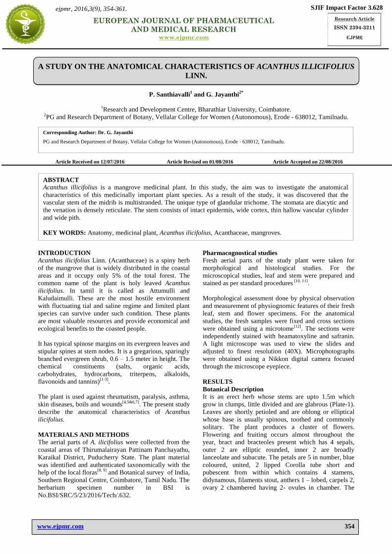
Venation Pattern of the lamina (Plate 4-a & b)
The venation of the lamina is densely reticulate. The
major vein are thick and straight. The lateral veins
become gradually thin ultimately forming wide vein-
islets. The vein islets are mostly elongated and have
thick vein-boundaries. The vein-terminations are
profusely branched forming dendroid outline. The vein-
terminations are thick and straight (Plate 4-b).
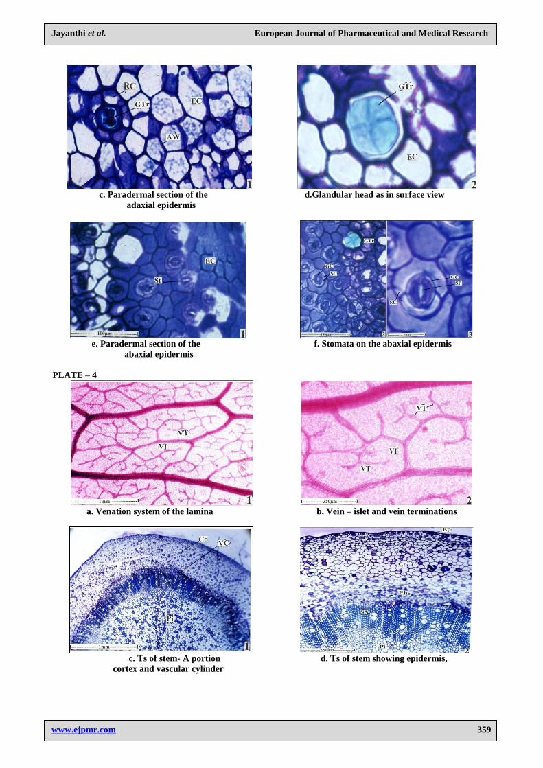
Stem
The stem is circular with smooth-even outline as seen in
cross-sectional view (Plate 4-c). The stem consists of
intact epidermis, wide cortex, thin hallow vascular
cylinder and wide pith. Epidermal layer consists of
small, squarish, thick walled cells with darkly stained
inclusions. The cortex in 500 µm thick. It consists,
angular, thin walled, compact parenchyma cells; many of
the cells have dark cell inclusions (Plate 4-d). The
vascular cylinder is includes outer layer of phloem and
inner xylem. The cylinder is 450 µm thick (Plate 4-d).
The outer zone is the secondary phloem and inner zone is
the secondary xylem. The secondary xylem is wavy
cylinder of unequal thickness. It consists of solitary and
radial multiples two or three vessels which are oblong
elliptical, angular or circular in outline. They have very
thick and lignified secondary walls. The xylem fibers are
in regular compact radial files; they have very thick
lignified walls and narrow lumen xylem rays are fairly
distinct. They are one - cell thick and straight. The ray

www.ejpmr.com
Jayanthi et al. European Journal of Pharmaceutical and Medical Research
356
cells are radially oblong and thick walled (Plate 4-d and
e).
Secondary phloem is composed of several sieve-
elements which diffuse in distribution; the cells are
angular thick walled and compact. Each sieve-tube
member is associated with aprominant companion cell.
Isolated brachysclereids are seen sparsely distributed in
the phloem (Plate 4-f).
Legend for the figures
Plate 1-a: Morphology of Acanthus ilicifolius.
b: An enlarged Inflorescence.
c: Fruiting twig of Acanthus ilicifolius.
Plate 2-a : T.S. of leaf through midrib showing multistranded vascular bundles.
b. Vascular bundles of the midrib-enlarged.
(Ads – Adaxialside; La-Lamina; Lo – Lobe; M.R. – Midrib; Ph – Phloem; VB- Vascular Bundles; X – xylem)
c. Adaxial lateral, small bundle
d. Adaxial lateral larger vascular bundle.
e. Abaxial medium largest vascular bundle
(Ph – phloem; Sc – sclerenchyma; X-xylem)
f. Ts of Lamina.
g. Ts of marginal part of the lamina.
(AbE- Abaxial Epidermis; AdE – Adaxial Epidermis; Gr- Groove ; GTr – Glandular Trichome; LM – Leaf
Margin; LV – Lateral Vein; MB – Marginal Bundle; MT-Mesophyl Tissue; PM – Palisade Mesophyll; SM – Spongy
mesophyll)
Plate 3-a. Ts of lamina showing epidermal groove and glandular trichome.
b. Glandular trichome – enlarge.
(BC – Body cell; Ep – Epidermis; Gr – Groove; Est – Epidermal stalk cell; GTr – Glandular Trichome; PM-
Palisade Mesophyll)
c. Paradermal section of the adaxial epidermis.
d. Glandular head as in surface view
(AW – Anticlinal wall; EC – Epidermal Cell; GTr – Glandular Trichome; RC – Rosette cells).
e. Paradermal section of the abaxial epidermis.
f. Stomata on the abaxial epidermis.
g. A diacytic stomata – enlarged.
(EC: Epidermal Cells; GC – Guard Cells; GTr- Glandural Trichome; SC – Subsidiary Cells; SP – Stomatal Pore; St –
Stomata)
Plate 4-a. Venation system of the lamina
b.. Vein – islet and vein terminations
(VI – Vein Islets; VT. Vein – Terminations)
c. Ts of stem – A portion
d. Ts of stem showing epidermis, cortex and vascular cylinder.
(Co – Cortex; Ep-Epidermis; Pi – Pith; Ph – Phloem; SX – Secondary xylem, VC – Vascular cylinder)
e. Secondary phloem and secondary xylem elements
f. Secondary xylem elements.
(CC – Companion cells; Scl- Sclereides; Sph – Secondary phloem; St – stomata; Sx – Secondary xylem; Ve – Vessels;
XF – Xylem Fibres; XR – Xylem Ray; Pi – Pith)

www.ejpmr.com
Jayanthi et al. European Journal of Pharmaceutical and Medical Research
357
PLATE – 1
a. Morphology of Acanthus ilicifolius
b. An Enlarged Inflorescence c. Fruiting twing
PLATE – 2
a. T.S.of leaf through midrib showing b. Vascular bundles of the
multistranded vascular bundles midrib-enlarged

www.ejpmr.com
Jayanthi et al. European Journal of Pharmaceutical and Medical Research
358
c. Adaxial lateral, small bundle d. Adaxial lateral vascular bundle
e. Adaxial medium larges vascular bundle f. Ts of Lamina
g. Ts of marginal part of the lamina
PLATE – 3
a. Ts of lamina showing epidermal b. Glandular trichome-enlarge
groove and glandular trichome
www.ejpmr.com
Jayanthi et al. European Journal of Pharmaceutical and Medical Research
359
c. Paradermal section of the d.Glandular head as in surface view
adaxial epidermis
e. Paradermal section of the f. Stomata on the abaxial epidermis
abaxial epidermis
PLATE – 4
a. Venation system of the lamina b. Vein – islet and vein terminations
c. Ts of stem- A portion d. Ts of stem showing epidermis,
cortex and vascular cylinder

www.ejpmr.com
Jayanthi et al. European Journal of Pharmaceutical and Medical Research
360
e. Secondary phloem and secondary f. Secondary xylem elements
xylem elements
DISCUSSION
A. ilicifolius was a good remedy for skin diseases
because there was report already documented that this
plant have many pharmacological properties. The present
anatomical study provides characters which would
facilitate quick identification and differentiation of the
drug from alike and other material. The midrib is flat on
the adaxial side and unequally two lobed and thick on the
abaxial side. In lamina portion there are deep narrow
grooves inside which occurs a simple unique type of
glandular trichome “Fig.3.12”. The presence of columnar
palisade cells and reticulate partions for air – chambers
“Fig.3.1” and the stomata are diacytic, the venation is
densely reticulate. The stem consists of intact epidermis,
wide cortex, thin walled vascular cylinder and wide pith,
which are the major diagnostic features noted from the
present study. These features can be used as reliable aid
for detecting adulterations. Similar studies were also
carried out in several other plant species[13-19]
. Generally
leaf margin, type and distribution of glandular trichomes,
epidermal cells and type of stomata are used as
diagnostic characters in microscopical studies of herbal
drugs[19-23]
.
CONCLUSIONS
The current trend of medicinal system of universe is
shifting from synthetic to herbal medicine, so we can say
„come back to Nature‟. Medicinal plants known as
millenaries and highly esteemed all over the world as a
rich source of therapeutic agents for the prevention of
diseases and ailments. Anatomical studies on the leaf and
stem of A. ilicifolius provide diagnostic characters for the
identifications of the original drug.
ACKNOWLEDGEMENT
The authors are thankful and deeply grateful to Dr. P.
Jayaraman (Director, Plant Anatomy Research Centre,
Medicinal Plant Research Unit, Chennai, India) for his
valuable help and suggestions during the course of
investigation.
REFERENCES
1. Kanchanapoom T, Kamel MS, Kasai R, Yamasaki
K, Picheansoontho C, Hiraga Y. Lignan glucosides
from Acanthus ilicifolius. Phytochemistry 2001; 56:
369-372.
2. Kanchanapoom T, Kamel MS, Kasai R,
Picheansoontho C, Hiraga Y, Yamasaki K.
Benzoxazinoid glucosides from Acanthus ilicifolius.
Phytochemistry 2001; 58: 637-640.
3. Wu J, Zhang S, Xiao Q, Li Q, Huang J, Long L, et
al. Phenylethanoid and aliphatic alcohol glycosides
from Acanthus ilicifolius. Phytochemistry 2003; 63:
491-495.
4. Subudhi HN, Choudhary BP & Acharya BC 1992. J
Econ Tax Bot, 16(2): 479-487.
5. Kapil. A., Sharma, S., Wahidulla, S. 1994. Planta
Medica. 60: 187-188.
6. Mani Senthil Kumar KT, Puia Z, Samanta SK, Barik
R, Dutta A, Gorain B, et al. The Gastroprotective
role of Acanthus ilicifolius – A study to unravel the
underlying mechanism of anti-ulcer activity. Sci
Pharm 2012; 80: 701-707.
7. Bandaranayake WM. Traditional and medicinal uses
of mangroves. Mangroves Salt Marshes. 1998; 2:
133-148.
8. Gamble J S and Fischer CEC, Flora of the
Presidency of Madras.London: Ad Lord and Sons
Limited, 1956; I-III.
9. Matthew K M, The Flora of Tamil Nadu Carnatic.
Tamil Nadu: The Rapinat herbarium, St. Joseph‟s
College, 1983.
10. Brain K R and Turner T D, The practical evaluation
of phytopharmaceuticals, Wright-Scientechnica,
Bristol, 1975; 4-9.
11. Kokate C K, Practical Pharmacognosy,
VallabhPrakashan, Delhi, 1999, 4th
Edn, 7-29.
12. Johansen D A,Plant Micro-technique. McGraw- Hill
Publishers, New-York, 1940; 523.
13. Prakash Chandra Gupta and RaoCh
V,Pharmacognostical studies of Cleome viscosa
Linn., Indian J Natural Products and Resources,
2012; 3(4): 527-534.
14. Marija Marin, NebojsaJasnic and
LiaAscensao,Histochemical, Micromorphology and
Ultrastructural investigations in glandular trichomes
of Micromeriat hymifolia, Botanic SERBICA, 2013;
37(1): 49-53.

www.ejpmr.com
Jayanthi et al. European Journal of Pharmaceutical and Medical Research
361
15. Manic Baral, Anatomical and Histological study of
Stem, Root and Leaf of the Medicinal Plant
Amaranthus spinosus Linn., J PharmaSci Tech.,
2013; 2(2): 65-71.
16. Thirumalai D, Paridhavi M and Gowtham M,
Evaluation of physiochemical, pharmacognostical
and phytochemical parameters of Premna
herbacea,Asian J. Pharm Clin Res, 2013; 6(1): 173-
181.
17. Prashanta Kr. Deb, Sanjoy Das,
KaushikNathBhaumik, RajatGhosh,
TarunKantiGhosh and TejendraBhakta,
Pharmacognostic and Preliminary Phytochemical
Investigatins of Neptunia prostrata L.,
JPharmacognosy and Phytochemistry, 2013; 2(3): 5-
11.
18. YunusDogan, Gungor Ay and Ekaterina
Kozuharova, A study on the anatomical
characteristics of Vitex agnuscastus
(Verbenaceae),PhytologiaBalcanica, 2008; 14(1):
97-101.
19. Idu M, Erhabor J O and Odia E A, Morphological
and Anatomical studies of the Leaf and the Stem of
Some Medicinal Plants,Stachytarphetajamaicensis
(L.) Vahl. And S.cayennensis (L.C.Rich.)
Schau,Ethnobotanical Leaflets, 2009; 13: 1417-25.
20. SolerederH,Systematic Anatomy of the
Dicotyledons, Boodle L A and FritichF E (Transl.),
Clarendon Press, Oxford, 1908.
21. ChattawayM M,Crystals in woody tissues, 1956,
Part II, Tropical Woods 104(9): 100–124.
22. Arnott H J and PautardF G E, Development of
raphideidioblasts in Lemna,Am. J. Bot.,1965; 52:
618.
23. MatcalfeC R and Chalk L,Anatomy of the
Dicotyledons. Clarendon Press, Oxford, England,
1979; I and II.