Embed Size (px)
Citation preview

Available online at www.sciencedirect.com
Acta Biomaterialia 5 (2009) 2790–2799
www.elsevier.com/locate/actabiomat
A study on alkaline heat treated Mg–Ca alloy for the controlof the biocorrosion rate
X.N. Gu a, W. Zheng a,b, Y. Cheng c, Y.F. Zheng a,c,*
a State Key Laboratory for Turbulence and Complex System and Department of Advanced Materials and Nanotechnology, College of
Engineering, Peking University, Beijing 100871, Chinab Center for Biomedical Materials and Engineering, Harbin Engineering University, Harbin 150001, China
c Center for Biomedical Materials and Tissue Engineering, Academy for Advanced Interdisciplinary Studies, Peking University, Beijing 100871, China
Received 18 June 2008; received in revised form 14 January 2009; accepted 26 January 2009Available online 7 February 2009
Abstract
To reduce the biocorrosion rate by surface modification, Mg–Ca alloy (1.4 wt.% Ca content) was soaked in three alkaline solutions(Na2HPO4, Na2CO3 and NaHCO3) for 24 h, respectively, and subsequently heat treated at 773 K for 12 h. Scanning electron microscopyand energy-dispersive spectroscopy results revealed that magnesium oxide layers with the thickness of about 13, 9 and 26 lm wereformed on the surfaces of Mg–Ca alloy after the above different alkaline heat treatments. Atomic force microscopy showed that the sur-faces of Mg–Ca alloy samples became rough after three alkaline heat treatments. The in vitro corrosion tests in simulated body fluidindicated that the corrosion rates of Mg–Ca alloy were effectively decreased after alkaline heat treatments, with the following sequence:NaHCO3 heated < Na2HPO4 heated < Na2CO3 heated. The cytotoxicity evaluation revealed that none of the alkaline heat treated Mg–Ca alloy samples induced toxicity to L-929 cells during 7 days culture.� 2009 Acta Materialia Inc. Published by Elsevier Ltd. All rights reserved.
Keywords: Mg–Ca alloy; Surface modification; Alkaline heat treatment; Biocorrosion; Cytotoxicity
1. Introduction
Magnesium alloys have close mechanical properties tonatural bone, perfect biocompatibility and would corrodeat the pH level (7.4–7.6) and in the high chloride environ-ment of physiological systems [1]. These intriguing charac-teristics have attracted great attention in respect ofbiodegradable bone implant applications. With regard tothis point, both the in vitro and in vivo corrosion of mag-nesium alloys have been widely studied [2–7]. Witte et al.[2] compared the corrosion of gravity cast AZ31, AZ91,WE43, LAE442 and SR-PLA96 rods (20 mm length and1.5 mm diameter) in guinea pig femora and indicated that
1742-7061/$ - see front matter � 2009 Acta Materialia Inc. Published by Else
doi:10.1016/j.actbio.2009.01.048
* Corresponding author. Address: Department of Advanced Materialsand Nanotechnology, College of Engineering, Peking University, Beijing100871, China. Tel./fax: +86 10 6276 7411.
E-mail address: [email protected] (Y.F. Zheng).
the median implanted rod area was about 1.29, 1.4, 1.16,1.5 and 1.76 mm2 after 18 weeks’ implantation. Zhanget al. [3] investigated the corrosion of Mg–Mn–Zn rods inrat femora and calculated their corrosion rates accordingto the ratio of the cross-section area of the residual implantto the original one. The results showed that 54% of thesamples had degraded after 18 weeks. And in our previousstudy [7], Mg–Ca alloy implant pins (10 mm in length,2.5 mm in diameter) degraded completely 3 months post-operatively in rabbit.
As an orthopedic biodegradable biomaterial, it is signif-icant that the degradation time of the material shouldmatch the healing or regeneration process of bone [8],which generally proceeds in three phases: an inflammatoryphase, lasting from 3 to 7 days; a reparative phase forstrong healing union, lasting about 3–4 months; and aremodeling phase, which can require months to years forcompletion [9]. It is desirable to have the implanted fixation
vier Ltd. All rights reserved.

X.N. Gu et al. / Acta Biomaterialia 5 (2009) 2790–2799 2791
present for at least 12 weeks [1]. However, the currentlydeveloped bare magnesium alloys degrade earlier than theactual period of the bone healing process. Therefore, thereis a high demand to reduce the corrosion rate to meet therequirement of the synchronization between the implantbiodegradation and the new bone formation by varioussurface treatment techniques.
One surface treatment technique, alkaline heat treat-ment, has been investigated extensively as one of the mostsimple and effective methods for metallic biomaterials[10,11], and preliminary studies using alkaline and alkalineheat treatment techniques on pure magnesium have shownpositive evidence of a reduction in the corrosion rate[12,13]. Our previous study on Mg–Ca alloys with differentCa contents indicated that Mg–Ca alloy with about 1 wt.%Ca content had optimal mechanical and corrosion proper-ties [7]. In this study, Mg–Ca alloy samples with 1.4 wt.%Ca content were explored. The precipitation of Mg2Ca sec-ond phase along the a-Mg grain boundaries in Mg–1.4 wt.% Ca alloy might deteriorate the corrosion resis-tance in comparison to Mg–1 wt.% Ca alloy under the sin-gle phase of a-Mg; therefore it would be more rigorous andeffective to show the reducing effect of various surface mod-ification techniques (with Na2HPO4, Na2CO3 andNaHCO3 alkaline solutions and subsequent heat treat-ment) on the biocorrosion of Mg–Ca alloy samples. Themicrostructure, composition, surface topology and thick-ness of the different alkaline heat treatment coatings werecharacterized, and the anti-corrosion properties and cyto-toxicity were also investigated.
2. Materials and methods
2.1. Preparation of specimens
Commercial pure Mg (99.98%) and Ca (99.95%) metalpowder were melted and cast under a mixed gas atmo-sphere of SF6 and CO2 using a mild steel crucible. AnMg–Ca alloy ingot (1.4 wt.% Ca content) was cut intoplates 10 � 10 � 2 mm3 in size, mechanically polished upto 2000 grit, then ultrasonically cleaned successively in ace-tone, absolute ethanol and distilled water. The resultingMg–Ca alloy samples (denoted as untreated) were sub-jected to alkaline treatments for 24 h (denoted as Na2HPO4
treated, Na2CO3 treated and NaHCO3 treated) followed by12 h heat treatment at 773 K in air (denoted as Na2HPO4
heated, Na2CO3 heated and NaHCO3 heated). All treatedsamples were ultrasonically cleaned in distilled water anddried in air before further characterization.
2.2. Immersion test
The immersion test was carried out in simulated body fluid(SBF; containing NaCl 8.035 g l�1, NaHCO3 0.335 g l�1,KCl 0.225 g l�1, K2HPO4�3H2O 0.231 g l�1, MgCl2�6H2O0.311 g l�1, CaCl2 0.292 g l�1, Na2SO4 0.072 g l�1, Tris(HOCH2)3CNH2 6.228 g l�1; following the preparation pro-
cedure of Ref. [14]) according to ASTM-G31-72 [15]. ThepH value was adjusted to 7.4 ± 0.1 and the temperaturewas kept at 37 ± 0.5 �C using a water bath. After differentimmersion periods, the samples were removed out of the solu-tion, rinsed with distilled water and dried in air. Changes inthe surface morphologies and microstructures of the alkalineheat treated Mg–Ca alloy samples before and after immersionwere characterized by environmental scanning electronmicroscopy (ESEM, AMRAY-1910FE), equipped with anenergy-dispersive spectroscopy (EDS) attachment, and X-ray diffractometry (XRD; Rigaku DMAX 2400). The pHvalue of the solution and the hydrogen evolution rate weremonitored during the immersion test. An average of threemeasurements were taken for each group.
2.3. Electrochemical test
The electrochemical tests were carried out at 37 ± 0.5 �Cin SBF [14] using a corrosion measurement system(CHI660C). A three-electrode cell was used for electro-chemical measurements, the saturated calomel electrode(SCE) as a reference and a platinum electrode as the coun-ter. The open circuit potential (EOCP) was measured as afunction of time. In the potentiodynamic polarization tests,the polarization curve was measured at a scanning rate of1 mV s�1.
2.4. Cytotoxicity evaluation
L-929 cells were cultured in the Dulbecco’s modifiedEagle’s medium (DMEM), 10% fetal bovine serum,100 U ml�1 penicillin and 100 lg ml�1 streptomycin at37 �C in a humidified atmosphere of 5% CO2. Extractionmedium was prepared for 72 h incubation in a humidifiedatmosphere with 5% CO2 at 37 �C according to ISO10993-5:1999 [16] and the extraction medium were storedat 4 �C before experiment. The control groups involvedthe use of DMEM medium as the negative control and0.64% phenol DMEM medium as the positive control.Cells were incubated in 96-well cell culture plates at5 � 103 cells/100 ll of medium in each well and incubatedfor 24 h to allow attachment. The medium was thenreplaced with 100 ll of extracts. After 2, 4 and 7 days incu-bation, 10 ll MTT was added to each well for a 4-h incu-bation before 100 ll of formazan solubilization solutionwas added to each well overnight. The measurement wascarried out spectrophotometrically at 570 nm by Elx-800(bio-Tek instruments).
3. Results
3.1. Characterization of alkaline heat treated Mg–Ca alloy
Fig. 1 presents the XRD patterns of the untreated,Na2HPO4 treated, Na2CO3 treated, NaHCO3 treated,Na2HPO4 heated, Na2CO3 heated and NaHCO3 heatedsamples. Peaks for two new phases – magnesium sodium

Fig. 1. XRD patterns of (a) Na2HPO4 treated and Na2HPO4 heated Mg–Ca alloy samples, (b) Na2CO3 treated and Na2CO3 heated Mg–Ca alloysamples, (c) NaHCO3 treated and NaHCO3 heated Mg–Ca alloy samples.
2792 X.N. Gu et al. / Acta Biomaterialia 5 (2009) 2790–2799
phosphate and calcium phosphate – were recognized afterNa2HPO4 alkaline treatment, and peaks corresponding toMgO were observed after further heat treatment(Fig. 1a). In the case of NaHCO3 treatment, MgCO3 andCaCO3 could be recognized after alkaline treatment besides
the peaks of a-Mg, whereas no MgCO3 peaks could be seenafter further heat treatment (Fig. 1c). For Na2CO3 alkalineheated samples, no peaks of MgCO3 could be found, whichmay be attributed to the thin modified layer on the sub-strate surface (Fig. 1b).
The surface morphologies and chemical compositions ofall experimental samples were examined, and Fig. 2 showsthe SEM surface morphologies of untreated, NaHCO3
treated and NaHCO3 heated samples, the cross-sectionalmorphologies of NaHCO3 heated samples and the corre-sponding EDS (line scanning mode) result as typical exam-ples. The flat surface of the untreated sample was destroyedby the NaHCO3 treatment and covered by rough coatings.After heat treatment, the rough surface of the NaHCO3
treated coatings became relatively compact. However,some micropits could be seen on the surface of Na2CO3
heated sample, and a cauliflower-like surface was formedon then Na2HPO4 heated sample. In addition, theNaHCO3 heated sample showed a relative rougher surfacetopology than the Na2HPO4 heated and Na2CO3 heatedsamples, which could be verified by the surface roughnessobtained by atomic force microscopy, as shown in Table1. The cross-sectional SEM images of all alkaline heatedsamples indicate that the coating layers adhered well totheir Mg–Ca alloy substrates, with the thickness of thecoating layers for the Na2HPO4 heated, Na2CO3 heatedand NaHCO3 heated samples being about 13, 9 and26 lm, respectively. The surface coating compositions ofthe Na2CO3 heated and NaHCO3 heated samples con-tained carbon, oxygen, magnesium and calcium elements,whereas the surface coating of the Na2HPO4 heated sampleconsisted of carbon, oxygen, magnesium, calcium andphosphate.
3.2. Immersion behavior of alkaline heat treated Mg–Ca
alloy in SBF
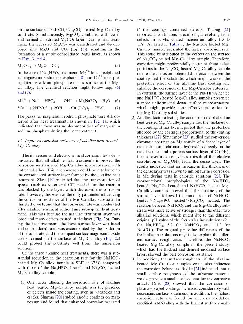
Figs. 3 and 4 show the variation of the pH value of SBFand the hydrogen evolution volume as a function of immer-sion time. It can be seen that the pH values of the solutionscorresponding to the Na2HPO4 heated, Na2CO3 heatedand NaHCO3 heated samples increased more slowly withimmersion time than did the untreated sample in the first200 h. The hydrogen evolution exhibited a behavior similarto the change in pH value. The hydrogen evolution vol-umes of the Na2HPO4 heated, Na2CO3 heated andNaHCO3 heated samples were much smaller than that ofthe untreated sample and the average corrosion ratederived from the curves (Fig. 3) ranked in the followingorder: untreated > Na2CO3 heated > Na2HPO4 heated > -NaHCO3 heated, as listed in Table 1.
Fig. 5 displays the surface morphology of the NaHCO3
heated sample after immersion in SBF at 37 �C for 50 days.It can be seen that the surface of the NaHCO3 heated sam-ple was smooth and compact at low magnification(Fig. 5a). At high magnification (Fig. 5b), the surface pre-sented a porous structure composed of tiny erect flakes.

Fig. 2. SEM micrographs of (a) untreated, (b) NaHCO3 treated and (c) NaHCO3 heated Mg–Ca alloy samples surfaces and (d) cross-sectionalmorphologies and EDS line-scan of NaHCO3 heated Mg–Ca alloy samples.
Table 1Surface roughness and corrosion rate of uncoated and alkaline heattreated Mg–Ca alloy.
Samples RMS vcorr (ml cm�2 h�1)a vcorr (mm year�1)b
Untreated 432.5 nm 0.108 13.27Na2HPO4 heated 1.584 lm 0.029 2.08Na2CO3 heated 1.209 lm 0.036 2.27NaHCO3 heated 2.972 lm 0.020 2.29
a Corrosion rate derived from the hydrogen evolution volume as afunction of immersion time calculated according to Ref. [6].
b Corrosion rate derived from the potentiodynamic polarization curvesaccording to ASTM-G31-72 [15].
X.N. Gu et al. / Acta Biomaterialia 5 (2009) 2790–2799 2793
EDS analysis on Fig. 5b revealed the existence of elementscarbon, oxygen, magnesium, phosphate, chloride and cal-cium. Fig. 6 shows the XRD patterns of NaHCO3 heatedsamples after immersion in SBF at 37 �C for 10 and50 days. It can be seen that the resulting surface corrosionproducts for NaHCO3 heated sample after 50 days’ immer-sion in SBF at 37 �C were mainly Mg(OH)2 and Ca2P2O7
(Fig. 6b), with the signal arising from the a-Mg phase forthe NaHCO3 heated sample disappearing after 10 days’immersion in SBF at 37 �C (Fig. 6a).
3.3. Electrochemical behavior of alkaline heat treated Mg–Ca alloy in SBF
Fig. 7 shows the open circuit potential-time curves foralkaline heat treated and untreated Mg–Ca alloy samplesexposed to SBF for 40,000 s. The open circuit potential(OCP) curves recorded for the Na2HPO4 heated, Na2CO3
heated and NaHCO3 heated samples exhibited a similartendency to fluctuate in the potential range (�1.8 V vs.SCE and �1.9 V vs. SCE). The OCP curve recorded forthe untreated sample was more positive, fluctuatingbetween �1.6 and �1.65 V (vs. SCE). Fig. 8 shows thepotentiodynamic polarization curves obtained for theuntreated and three alkaline heat treated Mg–Ca alloysamples with OCP measurements taken after immersionfor 40,000 s in SBF. The corrosion current densities of

Fig. 3. The pH value of SBF incubating untreated, Na2HPO4 heated,Na2CO3 heated and NaHCO3 heated Mg–Ca alloy samples as a functionof immersion time.
Fig. 4. The hydrogen evolution volumes of untreated, Na2HPO4 heated,Na2CO3 heated and NaHCO3 heated Mg–Ca alloy samples as a functionof the immersion time in SBF.
Fig. 5. SEM micrographs of the surface morphologies of NaHCO3 heatedMg–Ca alloy samples (a) at low magnification and (b) at highmagnification.
2794 X.N. Gu et al. / Acta Biomaterialia 5 (2009) 2790–2799
the Na2HPO4 heated, Na2CO3 heated and NaHCO3 heatedMg–Ca alloy samples were approximately one order ofmagnitude lower than that of the untreated Mg–Ca alloysample at the same potential range. The cathodic polariza-tion current shows that the hydrogen evolution reaction onalkaline heat treated Mg–Ca alloy samples was one orderlower than that on the untreated Mg–Ca alloy sample.The corrosion rates were calculated according to ASTM-G31-72 [15], and it can be seen that the corrosion ratesof the Na2HPO4 heated, Na2CO3 heated and NaHCO3
heated Mg–Ca alloy samples decreased dramatically com-pared to the untreated Mg–Ca alloy sample, as shown inTable 1. Epit � EOCP is an indication of the effectivenessof the resistance to corrosion, with a high Epit � EOCP
value indicating a coating with more effective resistance.It can be seen from Fig. 8 that the values of Epit � EOCP
for the untreated and Na2HPO4 heated, Na2CO3 heatedand NaHCO3 heated Mg–Ca alloy samples were 350,660, 650 and 850 mV, respectively.
3.4. Cytotoxicity of alkaline-heated and untreated Mg–Ca
alloy
Fig. 9 shows the cell viability cultured in the individualextraction mediums of untreated, Na2HPO4 heated,Na2CO3 heated and NaHCO3 heated Mg–Ca alloy samplesfor 2, 4 and 7 days. The cells cultured in the untreated Mg–Ca alloy samples extraction medium showed a relativelyhigher absorbance than that of the negative control. Thecell viabilities of the untreated, Na2HPO4 heated, Na2CO3
heated and NaHCO3 heated Mg–Ca alloy samples extractgroups showed no statistically significant difference(p > 0.05) after 2, 4 and 7 days culture. Fig. 10 shows themorphologies of L-929 cells cultured in the untreated,Na2HPO4 heated, Na2CO3 heated and NaHCO3 heated

Fig. 6. XRD patterns of NaHCO3 heated Mg–Ca alloy samples immersedin SBF for (a) 10 days and (b) 50 days.
Fig. 7. OCP curves of untreated, Na2HPO4 heated, Na2CO3 heated andNaHCO3 heated Mg–Ca alloy samples tested in SBF.
Fig. 8. Potentiodynamic polarization curves of untreated, Na2HPO4
heated, Na2CO3 heated and NaHCO3 heated Mg–Ca alloy samples testedin SBF.
Fig. 9. L-929 cell viability expressed as a percentage of the viability of cellsin the control after 2, 4 and 7 days culture in untreated, Na2HPO4 heated,Na2CO3 heated and NaHCO3 heated Mg–Ca alloy sample extractionmedia.
X.N. Gu et al. / Acta Biomaterialia 5 (2009) 2790–2799 2795
Mg–Ca alloy samples extracted after 4 days of incubation.The untreated, Na2HPO4 heated, Na2CO3 heated andNaHCO3 heated Mg–Ca alloy sample groups and the neg-
ative control exhibited healthy morphologies of cells with aflattened spindle shape. Both the MTT test and opticalmicroscopic observation indicated that the untreated,Na2CO3 heated, NaHCO3 heated and Na2HPO4 heatedMg–Ca alloy samples showed no obvious toxicity to L-929 cells.
4. Discussion
From the above experimental results, it can be seen thatdifferent alkaline heat (Na2HPO4, Na2CO3 and NaHCO3)treatments produce surface coatings on Mg–Ca alloy withdifferent thicknesses, morphologies, structures and chemi-cal compositions, which can affect the corrosion resistanceof surface modified Mg–Ca alloy in simulated body fluid.

Fig. 10. Optical micrographs of L-929 cells that were cultured in (a) negative control, (b) untreated, (c) Na2HPO4 heated, (d) Na2CO3 heated, (e) NaHCO3
heated Mg–Ca alloy sample extracts and (f) positive control for 4 days.
2796 X.N. Gu et al. / Acta Biomaterialia 5 (2009) 2790–2799
4.1. Mechanisms of alkaline heat treatment
The XRD (Fig. 1c) analysis indicated the formation ofMgCO3 and CaCO3 on the surface of Mg–Ca alloy sampleafter NaHCO3 treatment. This could be explained by thereactions between NaHCO3/Na2CO3 and the Mg–Ca alloysamples (Eqs. (1)–(4)). During the NaHCO3/Na2CO3 treat-ment, the Mg–Ca alloy sample partially dissolved intoalkaline solution as the following reactions occurred[12,13]:
Mg!Mg2þ þ 2e; Ca! Ca2þ þ 2e ð1Þ2H2Oþ 2e! 2OH� þH2 ð2ÞMg2þ þ CO3
2� !MgCO3; Ca2þ þ CO32� ! CaCO3
ð3ÞMgCO3 þ 3H2O!MgCO3 � 3H2O ð4Þ
Calcium has a more negative corrosion potential thanmagnesium [17], thus there was a greater driving force forthe corrosion of the soluble Ca and formation of CaCO3
X.N. Gu et al. / Acta Biomaterialia 5 (2009) 2790–2799 2797
on the surface of NaHCO3/Na2CO3 treated Mg–Ca alloysubstrate. Simultaneously, MgCO3 combined with waterand formed a hydrated MgCO3 layer. During heat treat-ment, the hydrated MgCO3 was dehydrated and decom-posed into MgO and CO2 (Eq. (5)), resulting in theformation of a stable consolidated MgO layer, as shownin Figs. 3 and 4.
MgCO3 !MgOþ CO2 ð5ÞIn the case of Na2HPO4 treatment, Mg2+ ions precipitatedas magnesium sodium phosphate [18] and Ca2+ ions pre-cipitated as calcium phosphate on the surface of the Mg–Ca alloy. The chemical reaction might follow Eqs. (6)and (7):
Mg2þ þNaþ þHPO42� þOH� !MgNaPO4 þH2O ð6Þ
3Ca2þ þ 2HPO42� þ 2OH� ! Ca3ðPO4Þ2 þ 2H2O ð7Þ
The peaks for magnesium sodium phosphate were still ob-served after heat treatment, as shown in Fig. 1a, whichindicated that there was no decomposition of magnesiumsodium phosphate during the heat treatment.
4.2. Improved corrosion resistance of alkaline heat treated
Mg–Ca alloy
The immersion and electrochemical corrosion tests dem-onstrated that all alkaline heat treatments improved thecorrosion resistance of Mg–Ca alloy in comparison withuntreated alloy. This phenomenon could be attributed tothe consolidated surface layer formed by the alkaline heattreatment. Zhou [19] indicated that the transportation ofspecies (such as water and Cl�) needed for the reactionwas blocked by the layer, which decreased the corrosionrate. However, this was not the only parameter influencingthe corrosion resistance of the Mg–Ca alloy substrate. Inthis study, we found that the corrosion rate was acceleratedafter alkaline treatment without any subsequent heat treat-ment. This was because the alkaline treatment layer wasloose and many defects existed in the layer (Fig. 2b). Dur-ing the heat treatment, the loose surface layer was fusedand consolidated, and was accompanied by the oxidationof the substrate, and the compact surface magnesium oxidelayers formed on the surface of Mg–Ca alloy (Fig. 2c)could protect the substrate well from the immersionsolution.
Of the three alkaline heat treatments, there was a sub-stantial reduction in the corrosion rate for the NaHCO3
heated Mg–Ca alloy sample in SBF at 37 �C comparedwith those of the Na2HPO4 heated and Na2CO3 heatedMg–Ca alloy samples.
(1) One factor affecting the corrosion rate of alkalineheat treated Mg–Ca alloy sample was the presenceof defects inside the coating, such as vacancies andcracks. Sharma [20] studied anodic coatings on mag-nesium and found that enhanced corrosion occurred
if the coatings contained defects. Truong [21]reported a continuous stream of gas evolving fromdefects in acrylic coated magnesium alloy (DTD118). As listed in Table 1, the Na2CO3 heated Mg–Ca alloy sample presented the fastest corrosion rate.This could be attributed to the defects on the surfaceof Na2CO3 heated Mg–Ca alloy sample. Therefore,corrosion might preferentially occur at these defectpositions in the Na2CO3 heated Mg–Ca alloy sampledue to the corrosion potential differences between thecoating and the substrate, which might weaken theprotective effect of the alkaline heat coating andenhance the corrosion of the Mg–Ca alloy substrate.In contrast, the surface layer of the Na2HPO4 heatedand NaHCO3 heated Mg–Ca alloy samples presenteda more uniform and dense surface microstructure,which might provide more effective protection forthe Mg–Ca alloy substrates.
(2) Another factor affecting the corrosion rate of alkalineheat treated Mg–Ca alloy sample was the thickness ofthe coating. It has been reported that the protectionafforded by the coating is proportional to the coatingthickness [22]. Simaranov [23] studied the conversionchromate coatings on Mg consist of a dense layer ofmagnesium and chromate hydroxides directly on themetal surface and a porous surface layer of Cr(OH)3
formed over a dense layer as a result of the selectivedissolution of Mg(OH)2 from the dense layer. Theresults indicated that an increase in the thickness ofthe dense layer was shown to inhibit further corrosionin Mg during tests in chloride solutions [23]. Thecross-sectional morphologies of the Na2HPO4
heated, Na2CO3 heated and NaHCO3 heated Mg–Ca alloy samples showed that the thickness of thesurface layer followed the rank order of NaHCO3
heated > Na2HPO4 heated > Na2CO3 heated. Thereaction between NaHCO3 and the Mg–Ca alloy sub-strate was more active or stronger than the other twoalkaline solutions, which might due to the differentoriginal pH value of the fresh alkaline solutions (9.1for Na2HPO4, 8.2 for NaHCO3 and 11.2 forNa2CO3). The original pH value differences of thefresh alkaline solutions might also explain the differ-ent surface roughnesses. Therefore, the NaHCO3
heated Mg–Ca alloy sample in the present study,which had the thickest and densest modified surfacelayer, showed the best corrosion resistance.
(3) In addition, the surface roughness of the alkalineheated Mg–Ca alloy samples could also influencethe corrosion behaviors. Budke [24] indicated that asmall surface roughness of the substrate materialwould provide a small surface area for the corrosiveattack. Celik [25] showed that the corrosion ofplasma-sprayed coatings increased considerably withdecreasing surface roughness. In addition, the highestcorrosion rate was found for microarc oxidationmodified AM60 alloy with the highest surface rough-

2798 X.N. Gu et al. / Acta Biomaterialia 5 (2009) 2790–2799
ness [26]. Our NaHCO3 heated Mg–Ca alloy samplespresented the lowest corrosion rate but the highestsurface roughness, indicating that the surface rough-ness might not be the dominant influencing factor forcorrosion in this study, given a synergism between theincreasing thickness of the coating and its compactstructure. Chiu [27] reported that Al-sprayed AZ31showed even less corrosion resistance before coatingand presented an extremely reduced corrosion rateafter hot pressing, which densified the porous Alcoating.
4.3. Biocompatibility of alkaline heat treated Mg–Ca alloy
Kim et al. [11] reported that alkaline (NaOH)-heat trea-ted Ti alloy possesses good bioactivity and induces forma-tion of the bone-like apatite. In our study, similar results,indicating the formation of calcium phosphate, wereobserved during the immersion test in SBF (Figs. 5 and6). The XRD analysis indicated much more obvious cal-cium phosphate peaks for the NaHCO3 heated Mg–Caalloy samples after 10 days’ immersion than for theuntreated Mg–Ca alloy samples after 25 days’ immersion[7]. This enhanced deposition of calcium phosphate mightsuggest an accelerated mineral deposition rate on theMg–Ca alloy sample when implanted into bone. The for-mation of calcium phosphate in the NaHCO3 heatedMg–Ca alloy samples might be attributed to the rough sur-face morphology (Fig. 2c) after alkaline heat treatment,which increased the surface area available to the SBF solu-tion. Kim [11] introduced alkaline and heat treatment tosurface modified Ti alloys and found alkaline heat treat-ments were effective in producing a rough or porous sur-face for the deposition of calcium phosphate, whichmight increase the bonding strength between the boneand implants. Kokuboo [28] reported that an in vitrochemical-deposited bone-like apatite on cp Ti could beinduced by alkaline heat treatment followed by soakingin SBF. For the modification of magnesium alloys, Al-Abdullat [12] investigated NaHCO3 modified pure magne-sium and found the formation of magnesium-containingtricalcium phosphate on the modified magnesium afterimmersion in HBSS for 75 days at 25 �C. Therefore, itseems that alkaline heat treatment could improve the bio-activity not only for Ti alloys but also for magnesiumand magnesium alloys.
The in vitro cytotoxicity evaluation results demon-strated that untreated and alkaline heat treated Mg–Caalloy samples (Na2HPO4 heated, Na2CO3 heated andNaHCO3 heated) were not toxic to L-929 cells (Fig. 9).Li [13] also studied the cytotoxicity of MgCO3–NaHCO3
alkaline heat treated pure magnesium to marrow cells.The results indicated that alkaline heat treated pure magne-sium did not induce toxicity and enhanced the activity ofmarrow cells. Moreover, Nishio [29] found that bone-likeapatite-formed titanium after alkaline and heat treatment
were observed to provide the most favorable conditionsfor bone marrow cell differentiation. It can be speculatedthat alkaline heat treated Mg–Ca alloy, with enhanceddeposition of calcium phosphate, might also play animportant role in osteoblastic differentiation.
The in vivo study about the alkaline and heat treatedtitanium indicated direct bone contact with the implantsurface, but the untreated and alkaline treated ones onlyhad a thin intervening fibrous tissue [30]. In addition, itwas reported that heat treatment in an air atmospherecould enhanced the bond strength of the apatite layerformed on a Ti substrate [31]. It can also be speculated thatthe alkaline heat treatment could help in the surface fixa-tion of an Mg–Ca alloy implant. Further research needsto be done to investigate the biocorrosion, calcium phos-phate formation, bonding strength and osteoblastic differ-entiation of alkaline heat treated Mg–Ca alloy in vivo.
5. Conclusions
The present study investigated the surface structure andcorrosion resistance of alkaline heat treated Mg–Ca alloysamples. The results showed an improved corrosion resis-tance for alkaline heat treated Mg–Ca alloy in SBF com-pared with the untreated Mg–Ca alloy sample. Of thethree alkaline treatments, NaHCO3 heated Mg–Ca alloypresented a uniform, dense and thick surface, providing agood protection for the substrate and the slowest corrosionrate. The corrosion rate of alkaline heat treated samplesfollowed the ranking order NaHCO3 heated < Na2HPO4
heated < Na2CO3 heated. The results also indicated anaccelerated calcium phosphate deposition rate on theNaHCO3 heated Mg–Ca alloy substrate. The cytotoxicityevaluation showed that none of the alkaline heat treatedMg–Ca alloys induced toxicity to L-929 cells. Therefore,NaHCO3-heat treatment might be a promising techniqueto improve the corrosion resistance and biocompatibilityof biomedical Mg–Ca alloy.
Acknowledgements
This work was supported by the National Natural ScienceFoundation of China (No. 30670560), Science and Technol-ogy Project of Guangdong Province (2008A030102006) andProgram for New Century Excellent Talents in University(NCET-07-0033).
References
[1] Staiger MP, Pietaka AM, Huadmaia J, Dias G. Magnesium and itsalloys as orthopedic biomaterials: a review. Biomaterials2006;27:1728–34.
[2] Witte F, Kaese V, Switzer H, Meyer-Lindenberg A, Wirth CJ,Windhag H. In vivo corrosion of four magnesium alloys and theassociated bone response. Biomaterials 2005;26:3557–63.
[3] Xu LP, Yu GN, Zhang EL, Pan F, Yang K. In vivo corrosionbehavior of Mg–Mn–Zn alloy for bone implant application. J BiomedMater Res 2007;83A(3):703–11.

X.N. Gu et al. / Acta Biomaterialia 5 (2009) 2790–2799 2799
[4] Witte F, Fischer J, Nellesen J, Crostack H, Kaese V, Pischd A, et al.In vitro and in vivo corrosion measurements of magnesium alloys.Biomaterials 2006;27:1013–8.
[5] Liu CL, Xin YC, Tian XB, Chu PK. Degradation susceptibility ofsurgical magnesium alloy in artificial biological fluid containingalbumin. J Mater Res 2007;22:1806–14.
[6] Song GL. Control of biodegradation of biocompatible magnesiumalloys. Corrosion Sci 2007;49:1696–701.
[7] Li ZJ, Gu XN, Lou SQ, Zheng YF. The development of binary Mg–Ca alloys for use as biodegradable materials within bone. Biomate-rials 2008;29(10):1329–44.
[8] Lloyd AW. Interfacial bioengineering to enhance surface biocompat-ibility. Med Device Technol 2002;13:18–21.
[9] Rucdi TP, Murphy WM. AO principle of fracture manage-ment. Dubendorf: AO Publishing; 2002, p. 13–4.
[10] Liu XY, Chu PK, Dng CX. Surface modification of titanium,titanium alloys, and related materials for biomedical applications.Mater Sci Eng 2004;47R:49–121.
[11] Kim HF, Miyaji F, Kokubo T, Nakamura T. Preparation ofbioactive Ti and its alloys via simple chemical surface treatment. JBiomed Mater Res 1996;32:409–17.
[12] Al-Abdullat Y, Tsutsumi S, Nakajima N, Ohta M, Kuwahara H,Ikeuchi K. Surface modification of magnesium by NaHCO3 andcorrosion behavior in Hank’s solution for new biomaterial applica-tions. Mater Trans 2001;42(8):1777–80.
[13] Li L, Gao J, Wang Y. Evaluation of cytotoxicity and corrosionbehavior of alkali-heat-treated magnesium in simulated body fluid.Surf Coat Technol 2004;185:92–8.
[14] Kokubo T, Takadama H. How useful is SBF in predicting in vivobone bioactivity? Biomaterials 2006;27:2907–15.
[15] ASTM-G31-72: standard practice for laboratory immersion corro-sion testing of metals. In: Annual book of ASTM standards.Philadelphia, PA: American Society for Testing and Materials; 2004.
[16] ANSI/AAMI ISO-10993e5: biological evaluation of medical devices epart 5: tests for cytotoxicity: in vitro methods. Arlington, VA: ANSI/AAMI; 1999.
[17] Emley EF. Principles of magnesium technology. Oxford: PergamonPress; 1966.
[18] Kouisni L, Azzi M, Zertoubi M, Dalard F, Maximovitch S.Phosphate coatings on magnesium alloy AM60. Part 1. Study ofthe formation and the growth of zinc phosphate films. Surf CoatTechnol 2004;185:58–67.
[19] Zhou WQ, Shan DY, Han EH, Ke W. Structure and formationmechanism of phosphate conversion coating on die-cast AZ91Dmagnesium alloy. Corrosion Sci 2008;50:329–37.
[20] Sharma AK, Uma Rani R, Giri K. Studies on anodization ofmagnesium alloy for thermal control applications. Metal Finishing1997;95(3):45–51.
[21] Truong V-T, Lai PK, Moore BT, Muscat RF, Russo MS. Corrosionprotection of magnesium by electroactive polypyrrole/paint coatings.Synthetic Metals 2000;110:7–15.
[22] Gray JE, Luan B. Protective coatings on magnesium and its alloys – acritical review. J Alloys Compd 2002;336:88–113.
[23] Simaranov AU, Marshakov A, Mikhailovskii YUN. The composi-tion and protective properties of chromated conversion coatings onmagnesium. Prot Metals 1992;28(5):576–80.
[24] Budke E, Krempel-Hesse J, Maidhof H, Schussler H. Decorative hardcoatings with improved corrosion resistance. Surf Coat Technol1999;112:108–13.
[25] Celik E, Demirkıran AS, Avcı E. Effect of grit blasting of substrate onthe corrosion behavior of plasma-sprayed Al2O3 coatings. Surf CoatTechnol 1999;116–119:1061–4.
[26] Liang J, Guo B, Tian J, Liu H, Zhou J, Liu W, et al. Effects ofNaAlO2 on structure and the corrosion resistance of microarcoxidation coatings formed on AM60B magnesium alloy in phos-phate–KOH electrolyte. Surf Coat Technol 2005;199:121–6.
[27] Chiu LH, Chen CC, Yang CF. Improvement of corrosion propertiesin an aluminum-sprayed AZ31 magnesium alloy by a post-hotpressing and anodizing treatment. Surf Coat Technol 2005;191:181–7.
[28] Kokubo T, Mijyaji F, Kim H-M. Spontaneous formation of bonelikeapatite layer on chemically treated titanium metals. J Am Ceram Soc1996;79:1127–9.
[29] Nishio K, Neo M, Akiyama H, Nishiguchi S, Kim HM, Kokubo T,et al. The effect of alkali- and heat-treated titanium and apatite-formed titanium on osteoblastic differentiation of bone marrow cells.J Biomed Mater Res 2000;52:652–61.
[30] Nishiguchi S, Nakamura T, Kobayashi M, Kim HM, Miyaji F,Kokubo T. The effect of heat treatment on bone-bonding ability ofalkali-treated titanium. Biomateirals 1999;20:491–500.
[31] Wang X, Li Y, Lin J, Hodgson PD, Wen C. Effect of heat-treatmentatmosphere on the bond strength of apatite layer on Ti substrate.Dental Mater 2008. doi:10.1016/j.actbio.2008.05.014.