Embed Size (px)
Citation preview
4 2 8 Australian Dental Journal, October, I963
A study of the dimensional changes occurring in the maxilla after tooth extraction. - Part I. Normal healing
K. Johnson, M.D.Sc. (Melb . ) *
I n t rod uct ion
In previous communications(1)(2) details were given of methods of model measurement that were developed to study resorption changes occurring in the maxilla after tooth extraction and the insertion of a n immediate denture. The present communication reports the use of this technique in a study of the dimensional changes ocrurring in the maxilla after tooth extraction and during the period of normal healing.
A review of the literature showed that the published work on this important subject is unfortunately small. In 1945 Lisowski(B’ studied a group of ten patients for a period of sixty days and endeavoured to demonstrate the resultant effect of “radical surgery” as compared with “conservative surgery” prior to the insertion of full maxillary immediate dentures. He concluded that extensive radical surgery was contra-indicated and that the resultant alveolar resorption was aggravated proportionately to the increase in the extent
* Senior (’linical Demonstrator (Part-time), Department of Dental Prosthesis, Dental School, University of Melbourne.
(1) Atkinson, H. F.. and Johnson, K.-An instrument for model measurement in immediate denture research. Austral. D. J . . 7 : 310-X14 (Aug . ) 1962.
(21 Atkinson, H. F., and Johnson, K.-A cephalostat for investigating alveolar resorption. Austral.
Lisowski, C . S.-A comparative study of the resorption of alveolar ridge tissue under im- mediate dentures. Unpublished Master’s thesis, SorthIvestern Vniversity Dental School, Mag,
Received for publication January, 1963.
D. J., 7 : 381-3x3 (Oct.) 1 9 6 2 .
I n ( #
of surgery. R. V. Lain“’ in 1960 dealt with a group of three patients in whom the contour changes involving the labial plate were measured after the insertion of partial upper immediate dentures. Measurements were presented for the three patients showing the average loss of height and thickness of the ridge over a period of one year. In these measurements the loss of thickness of the ridge was represented by the change in contour of the labial plate only, no measure- ments being made on the lingual aspect.
Current project
In the present investigation four groups of patients were studied, the groups being designated A, B, C , and D.
Group A-In this group healing was allowed to take place normally after extraction of the teeth. When healing was con- sidered complete (10-20 weeks) a closed face maxillary denture was constructed.+
Group B-A closed face denture was inserted immediately following extraction of the teeth.
Group C--A socketed type open face denture was inserted immediately following ex- traction of the teeth.
t Observati(,ns from f;roup A patients onl, ‘ire reported in this paper. 14) Lam, R. V.-Contour changes of the alvenlar
processes followinr extractions. J. Pros. I )en.. t 3 f 9 . i n : 2 x 3 2 (Jan.-Feh.) 1 9 6 0 .
Australian Dental Journal, October, 1963
Group D-A closed face denture was inserted immediately after extractions and a n interseptal alveolectomy had been per- formed.
Group A patients were included because no studies of the changes occurring in the maxilla during the period of normal healing following tooth removal could be found. It was con- sidered necessary therefore to ascertain what changes occurred during the normal healing period so as to afford a basis for comparison with the dimensional changes following im- mediate denture treatment. In addition this group was to be used to provide further evidence that the posterior part of the hard palate remained unchanged after the extraction of the maxillary teeth.
Initially i t was hoped to confine the study to patients over 21 years of age, but great difficulty was experienced in obtaining sufficient numbers, and therefore younger patients had to be included.
The patients were selected according to the following criteria:
1. 2.
3.
Good general health. Healthy gingive with a crevice depth of not more than 2 mm. The six upper anterior teeth to be present with some posterior teeth in each upper buccal segment. ( In a few cases some anterior teeth were missing. These patients were not discarded as the edentulous areas were measured as a control for comparative purposes).
Group A
Xormal healing In addition to the pre-extraction records,
models were prepared from impressions taken one week after extractions, and then a t regular intervals until the alveolar processes were healed. A comparison of measurements obtained from these models showed the dimensional changes which had occurred during the healing period. The radiographic technique described previously@) was used in conjunction with the model measuring method to investigate the stability of the reference area. Lateral roentgenograms of the patients wearing wax base plates with palatal markers were taken prior to extraction of the teeth and again a t the completion of healing. A comparison of tracings from these roentgeno.
429
grams provided information on the relation- ship of the posterior part of the hard palate to the base of the skull during the healing period.
When healing was completed a closed face denture was inserted, and the patients were recalled one year after the date of extractions, when a further model and lateral roentgeno- grams were obtained.
Details of methods and procedure 1. Patient material. The following shows the
sex and age distribution of Group A patients:
Age group .Male Female 16 years . . . . . . 2 2 17 years . . . . . . 3 18 years . . . . 2 1 years . . . . . . 1
- . . 1 -
-
2. Gingival condition. A description of the general gingival condition was recorded on the patient’s card. In addition, using a graduated gingival crevice gauge, four measurements were made of the crevice depth for each tooth ; two interproximally, and the other two in a direction a t right angles to the former.
3. Impression technique. Standardized techniques were used throughout the project for taking alginate impressions and pouring the models.
4. X-ray technique. A wax base plate with wax strengtheners was made on the pre- extraction model ; incorporated on the tissue surface in the midline was a tin foil marker 2 mm. wide and 0.18 mm. thick. The foil extended from the incisive papilla to the distal termination. A lateral X-ray was then taken with the base plate in position. I t was subsequently readapted on later models and used first for the X-rays taken after completion of healing, and finally a t the end of the twelve months period.
6 . Extraction of teeth. The teeth were extracted by forceps under local or general anesthesia. In some cases a clearance was undertaken at the one visit, while in others this was spread over several appointments, the anterior six teeth being removed last.
6 . Healing period. During the healing period, impressions were taken one week and four weeks after the completion of extractions and then regularly until healing was complete.
430 Australian Dental Journal, October, I963
The criterion adopted for a healed ridge was that it should present a rounded appearance and also be firm under digital pressure with- out it being possible to palpate any bony projections. As soon as the ridges appeared healed by the above method a denture was prepared.
7. Denture technique. All the dentures were prepared using standard clinical and labora- tory techniques, and care was taken to see that the denture space was the same as that recorded on a Willis gauge a t the initial examination.
8 . Patient recall. The patients were recalled approximately one year after the extractions for a further examination, impression, and X-ray. Particular note was taken of the condition of the ridge and the overlying mucosa a t this examination.
9. Measuring technique. A slight variation has been made in the method of measuring the models from that described earlier.(’) As the “frogs” were longer antero-posteriorly than laterally, a line was scribed on the upper surface above the median raphe. A reference point towards the posterior of this line was used in positioning the model and “frog” on the measuring instrument.
On reviewing the time lapsing between the extraction of the teeth and the completion of healing, quite a wide variation was evident within the group. Some alveolar processes appeared healed a t ten weeks, whilst others took up to twenty weeks before presenting a healed appearance. For comparative pur- poses the models were measured wherever possible ten weeks after tooth removal, but in those cases where healing was obviously incomplete more impressions were taken until the ridge appeared healed.
Measurements were made on the models in five directions; one in the sagittal plane, one through the lateral incisor region, one through the cuspid region, and two in the posterior region. One of the latter was in the bicuspid region, while the other was in the region of the second molar. The same reference point was used for all these measurements.
Results
Various methods of presenting the dimen- sional changes have been developed which include the drawing of graphs for each survey line a t 2 mm. contours as in the previous
paper. A set of these for one patient is shown in Figure 1.
Alternatively, it was considered that measurements showing loss in height a t the crest of the ridge and also loss in width of the ridge at the most prominent portion of the labial and buccal plates would provide the simplest indication of the changes that had occurred. The loss in width of the ridge was the sum of changes occurring on the labial or buccal aspect together with that occurring on the lingual aspect at the same level. A set of these measurements is presented in Table 1.
mr 20,
Lateral Incisor
0 20 10 ‘0
Fig. l.-Tjpical set of five graphs drawn from measurements obtainee from the models of a male patient A. aged 1 7 years. Measurements a r e in mm. The continuous line represents the pre-extraction model of 23.7.59 ( the break in the line being where the survey lines contacted the teeth). Teeth extracted 10.8.59. Five models were obtained during the healing period from 10.8.69 to 26.11.59, and these graphs fell between the two lines shown. The dotted line was obtained from a model obtained on 2 6 . 1 1 . 5 9 , on which date a denture was inserted. On 15.10.60 the Datient was recalled and a fur ther model obtained, the graphs from which
coincided with those of 26.11.59.
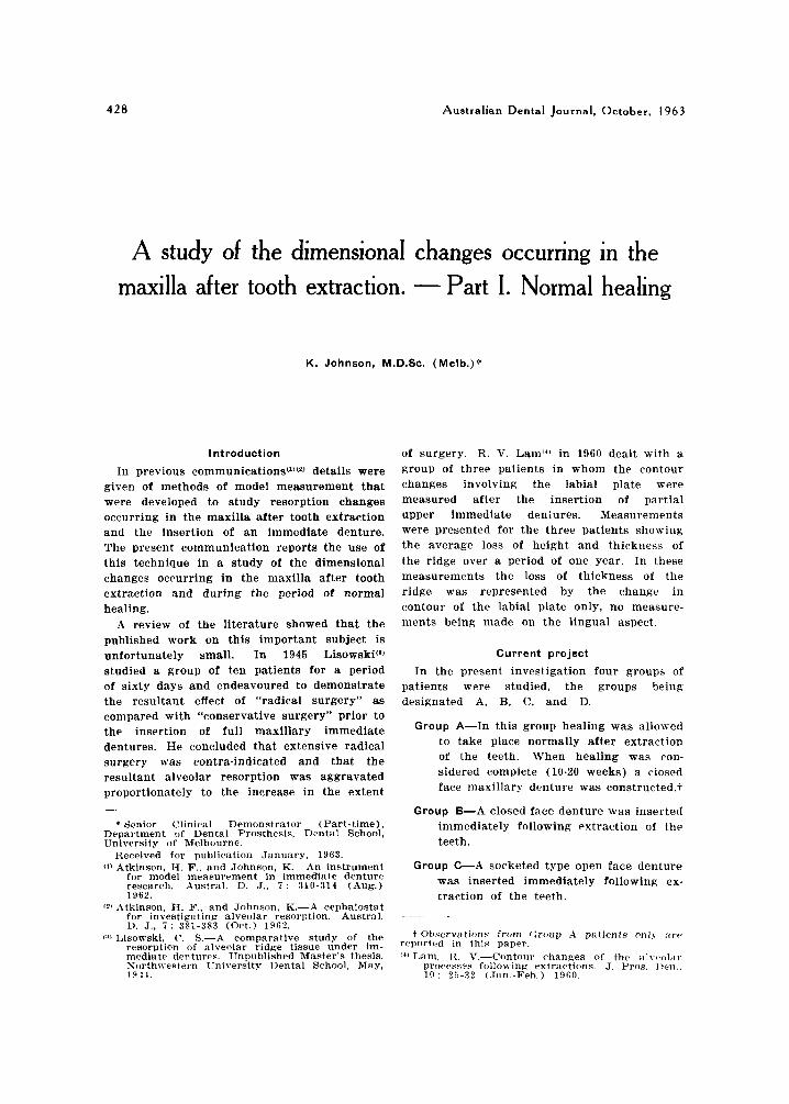
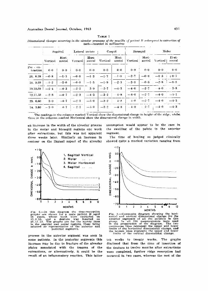
When the measurements from Table 1 are plotted against time, the gradient of the graph gives an indication of the rate of resorption, and furthermore, the amount of resorption can be ascertained after any given time interval (as shown in Fig. 2 ) .
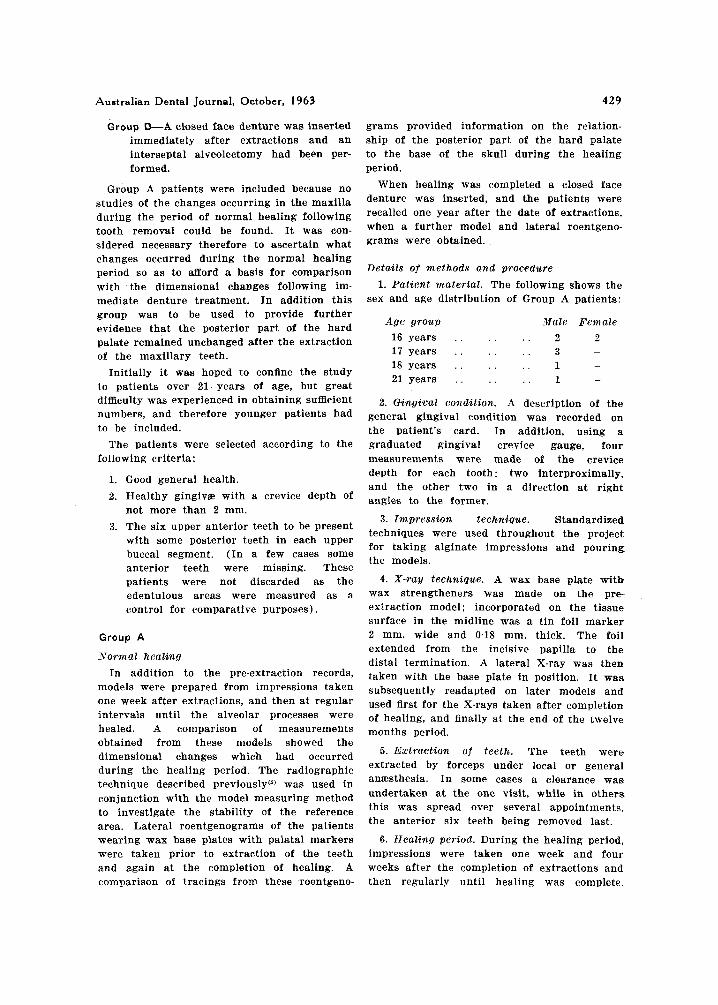
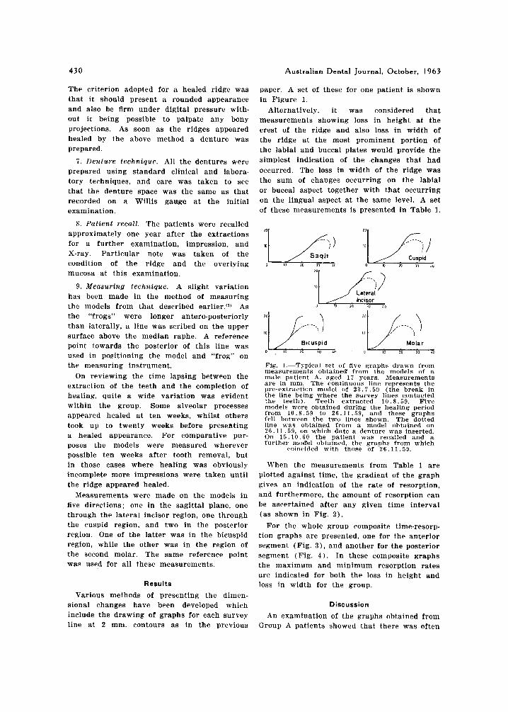
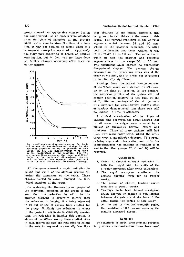
For the whole group composite time-resorp- tion graphs are presented, one for the anterior segment (Fig. 3 ) , and another for the posterior segment (Fig. 4 ) . In these composite graphs the maximum and minimum resorption rates are indicated for both the loss in height and loss in width for the group.
Discussion
An examination of the graphs obtained from Group A patients showed that there was often
Australian Dental Journal, October, I963 43 1
TABLE 1 Dimenstom1 changes occurring in the alveolar processes of the mcixtllcc nf pcctzent R s i tbxq i i e t i t to extlrrction of
teetLrecorded in millimelres - ~ _ ______
I I Sagittal Lateral incisor Cuspid 1 Bicuspd Molar
1 Hori- I . Vertical 1 zontal i I'ertical
10.10.59 I -2.4 i -4.3 -2 .2
14. 8.60 j -3.0 I -4.7 I -2.:! I I
_~ ~~ _~~ ~ - -- The readings in the columns markc
rhost. 111 the columns marked Horizont
a n increase in the width of the alveolar process in the molar and bicuspid regions one week after extractions, but this was not apparent three weeks later. Similarly an increase in contour on the lingual aspect of the alveolar
+ 1.0
1. Sagittal Vertical 2. Molar . . . .
4. Sagittal . . . . - 1.0 3. Molar Horizontal
1 ---- - - - - -_ _-- _-- - _ - - - -_ - -_ '.- z a 5 - 3 -
-6.01 . ' . . ' h *' 0 1 2 3 4 5 6 9 12
assumption would appear to be the case in the swelling of the palate in the anterior segment.
The time of healing as judged clinically showed quite a marked variation ranging from
- 3 0
J =l g -4 -
MONTHS
Fig. 2.-In this diagram the time-resorption graphs are shown for a male patient B aged 16 years, whose teeth were extracted on 13.8.59. and a denture was inserted on 26.11.59. The graphs are for the survey lines in the sagittal and molar regions, these being selected as representative of the anterior and
posterior segments.
process in the anterior segment was seen in some patients. In the posterior segments this increase may be due to fracture of the alveolar plates associated with the trauma of the extractions, or alternatively, it could be the result of an inflammatory reaction. This latter
MONTHS Fig. 3.-Coniposite diagram showing the hori- zontal and vertical dimensional change for the anterior segments of all the patients in this group. In all 236 measurements were used in the preparation of the diagram. The two continuous lines represent the upper and lower limits of the horizontal dimensional change, and the broken lines represent the upper and lower
limits of the vertical dimensional change.
ten weeks to twenty weeks. The graphs disclosed that from the time of insertion of the denture to twelve months after extractions were completed, further ridge resorption had occurred in two cases, whereas the rest of the
432 Australian Dental Journal, October, 1963
group showed no appreciable change during the same period. As no models were obtained from the time of insertion of the denture until twelve months after the time of extrac- tion, it was not possible to decide when this subsequent resorption occurred . Apparently the ridge may appear to be healed on clinical examination, but in fact may not have done so, further changes occurring after insertion of the denture.
+*'O c + 1 , o t A
- 8.0 -w- 0 1 2 3 6 5 6 9 12
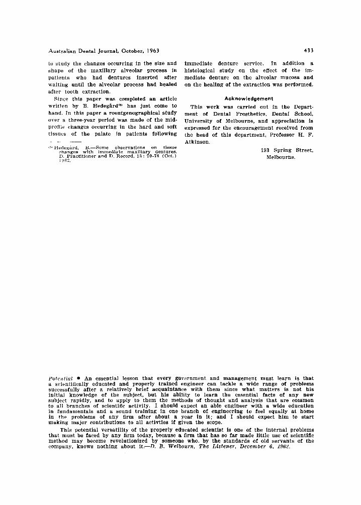
MONTHS Fig. 4.-Composite diagram showing the hori- zontal and vertical dimensional change for the posterior segments of all the patients in this group. In all, 140 measurements were used in the preparation of the diagram. The two continuous lines represent the upper and lower limits of the horizontal dimensional change, and the broken lines represent the upper and lower limits of the vertical dimensional change.
All the cases showed a rapid reduction in height and width of the alveolar process fol- lowing the extraction of the teeth. These changes varied in extent amongst the indi- vidual members of the group.
On reviewing the time-resorption graphs of the individual members of the group it was seen that the reduction in width in the anterior segment is generally greater than the reduction in height, this being observed in 2 1 out of the 25 survey lines studied for the group. Similarly the reduction in width in the posterior segments is generally greater than the reduction in height; this applied to eleven of the fifteen survey lines studied. Also in each individual case the reduction in height in the anterior segment is generally less than
that observed in the buccai segments, this being seen in two thirds of the cases in this group. The vertical reduction in the anterior segments varied between 2.5 and 5.0 mm., whilst in the posterior segments, including both the bicuspid and molar regions, it was in the range 3.0 to 7.0 mm. The reduction i n width in both the anterior and posterior segments was in the range 3.0 to 7.0 mm. The edentulous areas showed no appreciable dimensional change. The average change measured in the edentulous areas was of the order of 0.2 mm., and this was not considered to be clinically significant.
Tracings from the lateral roentgenograms of the whole group were studied. In all cases, up to the time of insertion of the denture, the posterior portion of the palate did not rhange position relative to the base of the skull. Similar tracings of the six patients who answered the recall twelve months after extractions demonstrated that there was still no change in this relationship.
A clinical examination of the ridges of patients who answered the recall showed that in all cases the ridges were covered by a mucosa of apparently normal texture and thickness. Three of these patients still had their own mandibular teeth, whilst the other three wore a mandibular denture. This group is being kept under observation, and in further communications the findings in relation to it and to the other groups (B, C, and D) will be reported.
Conclusions 1. Group A showed a rapid reduction in
both the height and the width of the alveolar processes after tooth extractions.
2. The rapid resorption continued for periods varying from ten to twenty weeks.
3. The period of clinical healing varied from ten to twenty weeks.
4. Tracings made from lateral roentgeno- grams showed no change in relationship between the palate and the base of the skull during the period of this study.
5. At the end of the twelve-month period the condition of the mucosa covering the maxilla appeared normal.
Summary The methods of model measurement reported
in previous communications have been used
Australian Dental Journal, October, I963
to study the changes occurring in the size and shape of the maxillary alveolar process in patients who had dentures inserted after waiting until the alveolar process had healed after tooth extraction.
Since this paper was completed a n article written by B. HedegBrd(6) has just come to hand. In this paper a roentgenographical study over a three-year period was made of the mid- profile changes occurring in the hard and soft tissues of the palate in patients following
G ) Hedegird, B.-Some obscrvations on tissue changes with immediate maxillary dentures. D. Practitioner and D. Record, 13 : 70-78 (Oct.) 1!162,
~ ~~ ~
433
immediate denture service. In addition a histological study on the effect of the im- mediate denture on the alveolar mucosa and on the healing of the extraction was performed.
Acknowledgement
This work was carried out in the Depart- ment of Dental Prosthetics, Dental School, University of Melbourne, and appreciation is expressed for the encouragement received from the head of this department, Professor H. F. Atkinson.
193 Spring Street, Melbourne.
I’ote,itial An essential lesson that every govarnment and management must learn is that a scientifically educated and properly trained engineer can tackle a wide range of problems successfully after a relatively brief acquaintance with them since what matters is not his initial knowledge of the subject, but his ability to learn the essential facts of any new subject rapidly, and to apply to them the methods of thought and analysis that are common to all branches of scientific activity. I should expect a n able engineer with a wide education in fundamentals and a sound training in one branch of engineering to feel equally at home in the problems of any firm after about a year in i t ; and I should expect him to s tar t making major contributions to all activties if given the scope.
This potential versatility of the properly educated scientist is one of the internal problems that must be faced by any firm today, because a firm that has so far made little use of scientiflc method may become revolutionized by someone who. by the standards of old servants of the company, knows nothing about it.-D. B. Welbourn, The Listener, December 6, 1962.