Embed Size (px)
Citation preview
ISI Impact Factor:3.461 Bas.J.Vet.Res.Vol.14,No.2,2015
265
A STUDY OF SOME PATHOLOGICAL LESIONS IN THE LUNG OF SHEEP AND DUHOK ABATTOIR
Mahdi, A.A. *, Al-Naqshabendy, A.A. ** , Haddel, B.T.***
* Department of Pathology and Microbiology, Faculty of Veterinary Medicine, University of Duhok-Iraq
** Department of Internal Medicine and Surgery, Faculty of Veterinary Medicine, University of Duhok-Iraq
*** Department of Pathology, College of Veterinary Medicine, University of Tikrit-Iraq (Received 4 January 2015 ,Accepted 3May 2015)
Keywords: sheep, Duhok abattoir, pathological lesions.
ABSTRACT
An abattoir study was conducted on 21854 sheep, and 3659 goats are slaughtered in
Duhok abattoir, from October 2013 to January 2014. The objective was to determine the
prevalence of disease conditions affecting the lungs. Routine meat inspection procedures
were done to detect the presence of the pathological lesions. A total of 21854 (13%) and
3659 (4.3%) lungs of sheep and goat, respectively, were examined and the main
condemned diseases in this study were recorded as namely pneumonia, and hydatidosis.
From this study the pathological features was concluded that 4 main forms of different
types of pneumonia and hydatid cyst which recorded according to macroscopical and
histological features.
INTRODUCTION
Respiratory diseases are common in all species of domestic animals, and they are
appear due to the interaction of many of infectious agents like (bacteria, mycoplasma,
viruses, fungi, parasite, host defense and environmental factors which are causes high
mortality rate and economic losses associated with respiratory diseases of sheep and
goats (1, 2, 4).
The main respiratory disease is occur due to inflammation of the lung tissues called
pneumonia which is widespread among sheep and goat all over the world and it is
considered to be one of the most important causes of losses in the small ruminant
ISI Impact Factor:3.461 Bas.J.Vet.Res.Vol.14,No.2,2015
266
industry (9, 11). The other important respiratory disease caused mainly by parasitic
infections called Echinococcus granulosus which caused by the larval stages of these
(Echinococcosis/hydatidosis) (tapeworm) species have been recognized as the most
important helminthes and zoonotic diseases, with great economic and public health
significances is developed in many countries (12,13, 14, 15).
Sheep and goats are regarded as the most principal slaughtered animals for human
consumption in Duhok province and due to a differentiation of the geographic location,
nutrition and climate are determining factors on the type of microorganism causing
pneumonia. In addition, rearing systems, stress factors, climatic changes, unhygienic
conditions, sudden changes in feed and a low level of herd health status are stated as
predisposing factors to bacteria, parasites and viruses infection which are recorded the
one common cause of lung lesions and death of infected animals, therefore, the aims of
this work are to know some epidemiological factors and determine the common
pathological lesions of pulmonary tissues in sheep and goats at Duhok abattoir in
Kurdistan region of Iraq.
MATERIALS AND METHODS
Data collection
In this study, data collected from 25513 sheep and goats were slaughtered at Duhok
abattoir during four months of the study was extended from October 2013 to January
2014. All pulmonary tissues are inspected the presence of different pneumonic lesions
using case history , gross macroscopically inspection and recorded of the results. Twenty
pulmonary tissue samples were suspected to any pathgnomic lesions with 5 normal once
were subjected to histopathological technique at Duhok Research Center, Faculty of
Veterinary Medicine- University of Duhok.
Histopathological study
Pulmonary tissue samples about 1 cm3 in thickness were directly taken from different
lesions using a sterile scalpel and were fixed with 10% of neutral-buffered formalin for
histopathological examination. The samples were then dehydrated in graded ethanol,
clearance with xylen, embedded in paraffin as blocks and sectioning from blocks were
cut at 4-5 μm in thickness using rotary microtome (Leica, Germany). Finally, the samples
ISI Impact Factor:3.461 Bas.J.Vet.Res.Vol.14,No.2,2015
267
were stained with Haematoxylin and Eosin stains (3) for examination using ordinary
light microscope to taken the photography (Lecia, Germany)
RESULTS On the basis of gross macroscopic inspection survey, the inflammatory lesions were
found 856 (3.8 %) samples in sheep and 115 (3.1) samples in goats. In addition, hydatid
cysts were appeared in 1998 (3.9 %) and 43 (1 %) samples in sheep and goats
respectively (Table: 1).
Table 1: The number of slaughtered animals in abattoir with lung lesions Species Examined animals Pulmonary inflammatory
lesions Infected with hydatid cyst
Sheep 21854 856 (3.9%) 1998 (9.1%) Goats 3659 115 (3.9%) 43 (1.1%)
Total 25513 971 (3.9%) 2041(7.9%)
Pulmonary lesions and hydatid cyst were appeared in most of the different seasons
with varying degrees, but at November showed the presence of the pulmonary lesions as
high rates 5.5 %. Hydatid cysts gave highest rate 11 % at December during the present
study. Generally, the results of prevalence of the respiratory diseases showed in sheep are
more common than goats between October to January . The results of the study
according to months and species of animals were seen in Table (2).
ISI Impact Factor:3.461 Bas.J.Vet.Res.Vol.14,No.2,2015
268
Table 2: The number of slaughtered animals in abattoir with lung lesions by months
Animal species
Months of study Examined animals
Pulmonary inflammatory
lesions
Infected with hydatid cyst
Sheep October 7451 200 (2.6%) 556 (7.4%)
November 4539 216 (4.7%) 368 (8.1%)
December 4972 208 (4.1%) 548 (11%)
January 4892 232 (4.7%) 526 (10%)
Total 21854 856 (3.9%) 1998 (9.1%)
Goats October 1224 34 (2.7%) 13 (1%)
November 806 45 (5.5%) 12 (1.4%)
December 878 23 (2.6%) 14 (1.5%) January 751 13 (1.7%) 4 (0.5%)
Total 3659 115 (3.1%) 43 (1.1%)
Pathological observation of lung lesions of sheep and goat
In the current study, a total of 20 samples of pulmonary tissues where found 18
samples affected with pathological lesions. According to macroscopic appearance
(texture, exudation, distribution and sites of lesions) and microscopic findings of lesions,
the samples were classified to different types of pneumonia, hydatid cyst and nodular
reaction. The pathological observation of the lung lesions included:
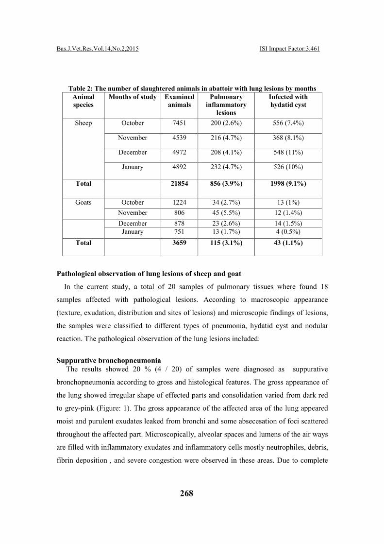
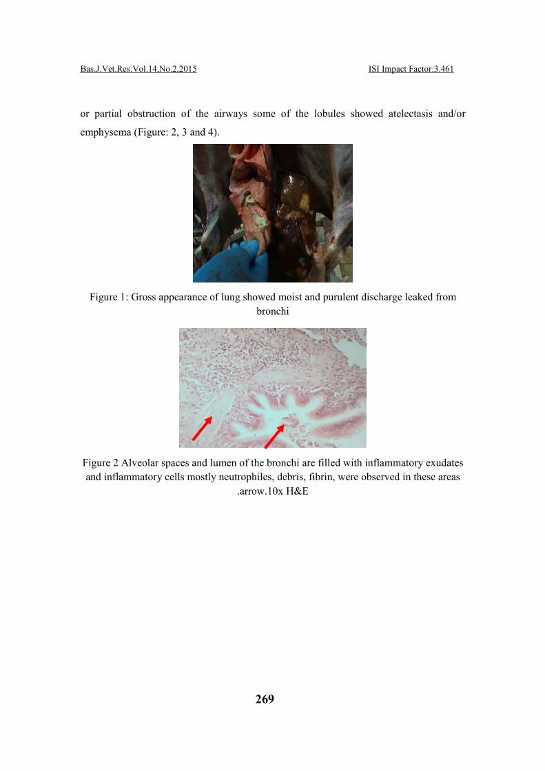
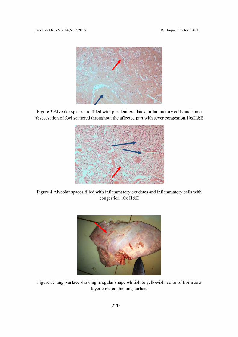
Suppurative bronchopneumonia The results showed 20 % (4 / 20) of samples were diagnosed as suppurative
bronchopneumonia according to gross and histological features. The gross appearance of
the lung showed irregular shape of effected parts and consolidation varied from dark red
to grey-pink (Figure: 1). The gross appearance of the affected area of the lung appeared
moist and purulent exudates leaked from bronchi and some absecesation of foci scattered
throughout the affected part. Microscopically, alveolar spaces and lumens of the air ways
are filled with inflammatory exudates and inflammatory cells mostly neutrophiles, debris,
fibrin deposition , and severe congestion were observed in these areas. Due to complete
ISI Impact Factor:3.461 Bas.J.Vet.Res.Vol.14,No.2,2015
269
or partial obstruction of the airways some of the lobules showed atelectasis and/or
emphysema (Figure: 2, 3 and 4).
Figure 1: Gross appearance of lung showed moist and purulent discharge leaked from
bronchi
Figure 2 Alveolar spaces and lumen of the bronchi are filled with inflammatory exudates
and inflammatory cells mostly neutrophiles, debris, fibrin, were observed in these areas
.arrow.10x H&E
ISI Impact Factor:3.461 Bas.J.Vet.Res.Vol.14,No.2,2015
270
Figure 3 Alveolar spaces are filled with purulent exudates, inflammatory cells and some
absecesation of foci scattered throughout the affected part with sever congestion.10xH&E
Figure 4 Alveolar spaces filled with inflammatory exudates and inflammatory cells with
congestion 10x H&E
Figure 5: lung surface showing irregular shape whitish to yellowish color of fibrin as a
layer covered the lung surface
ISI Impact Factor:3.461 Bas.J.Vet.Res.Vol.14,No.2,2015
271
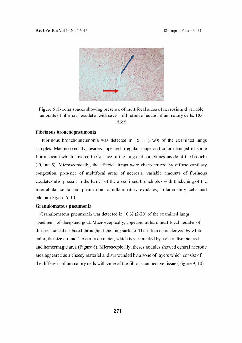
Figure 6 alveolar spaces showing presence of multifocal areas of necrosis and variable
amounts of fibrinous exudates with sever infiltration of acute inflammatory cells. 10x
H&E
Fibrinous bronchopneumonia
Fibrinous bronchopneumonia was detected in 15 % (3/20) of the examined lungs
samples. Macroscopically, lesions appeared irregular shape and color changed of some
fibrin sheath which covered the surface of the lung and sometimes inside of the bronchi
(Figure 5). Microscopically, the affected lungs were characterized by diffuse capillary
congestion, presence of multifocal areas of necrosis, variable amounts of fibrinous
exudates also present in the lumen of the alveoli and bronchioles with thickening of the
interlobular septa and pleura due to inflammatory exudates, inflammatory cells and
edema. (Figure 6, 10)
Granulomatous pneumonia
Granulomatous pneumonia was detected in 10 % (2/20) of the examined lungs
specimens of sheep and goat. Macroscopically, appeared as hard multifocal nodules of
different size distributed throughout the lung surface. These foci characterized by white
color, the size around 1-6 cm in diameter, which is surrounded by a clear discrete, red
and hemorrhagic area (Figure 8). Microscopically, theses nodules showed central necrotic
area appeared as a cheesy material and surrounded by a zone of layers which consist of
the different inflammatory cells with zone of the fibrous connective tissue (Figure 9, 10)

ISI Impact Factor:3.461 Bas.J.Vet.Res.Vol.14,No.2,2015
272
Figure 7 Microscopical changes of lung due to fibrinous-bronchpnumonia showing
thickening of the interlobular septa and pleura due to inflammatory exudates,
inflammatory cells and edema 10x H&E
Figure 8: lung showing hard multifocal nodules with different size distributed throughout
the lung surface
Figure 9 central caseous necrotic area appears as Gessy and surrounded by a zone of
layers consist of different inflammatory cells and fibrous connective tissue .10x H&E
ISI Impact Factor:3.461 Bas.J.Vet.Res.Vol.14,No.2,2015
273
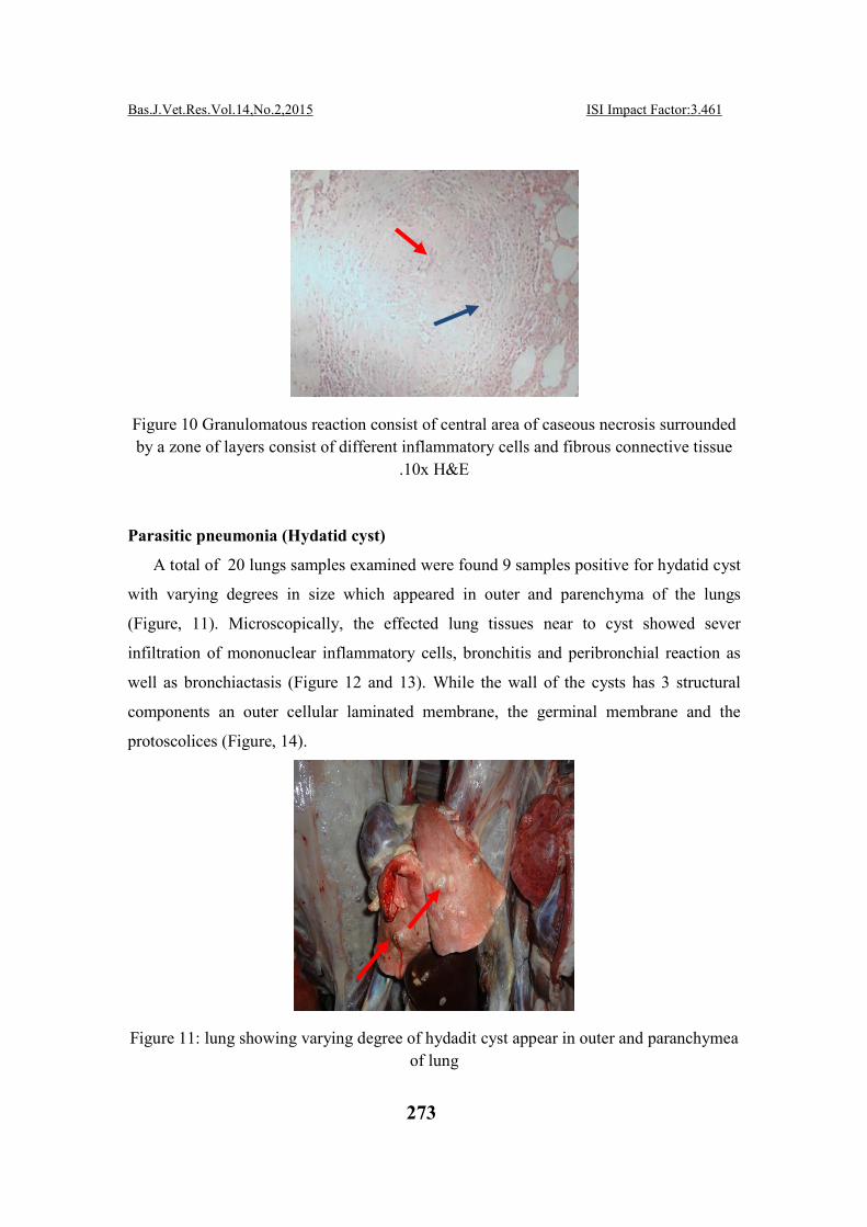
Figure 10 Granulomatous reaction consist of central area of caseous necrosis surrounded
by a zone of layers consist of different inflammatory cells and fibrous connective tissue
.10x H&E
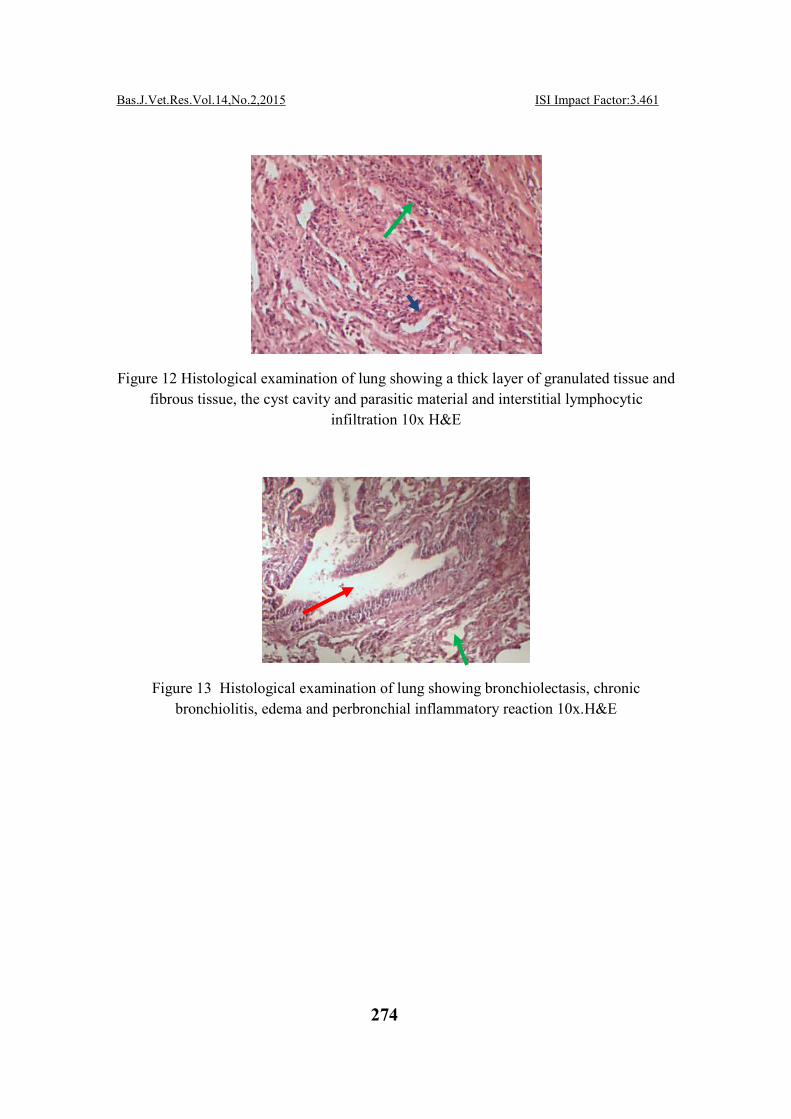
Parasitic pneumonia (Hydatid cyst)
A total of 20 lungs samples examined were found 9 samples positive for hydatid cyst
with varying degrees in size which appeared in outer and parenchyma of the lungs
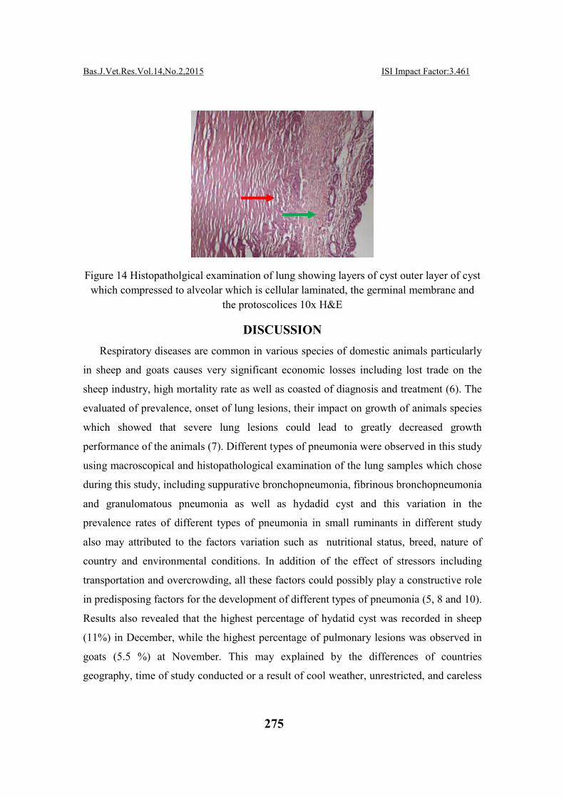
(Figure, 11). Microscopically, the effected lung tissues near to cyst showed sever
infiltration of mononuclear inflammatory cells, bronchitis and peribronchial reaction as
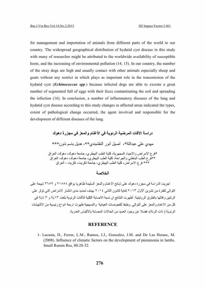
well as bronchiactasis (Figure 12 and 13). While the wall of the cysts has 3 structural
components an outer cellular laminated membrane, the germinal membrane and the
protoscolices (Figure, 14).
Figure 11: lung showing varying degree of hydadit cyst appear in outer and paranchymea
of lung
ISI Impact Factor:3.461 Bas.J.Vet.Res.Vol.14,No.2,2015
274
Figure 12 Histological examination of lung showing a thick layer of granulated tissue and
fibrous tissue, the cyst cavity and parasitic material and interstitial lymphocytic
infiltration 10x H&E
Figure 13 Histological examination of lung showing bronchiolectasis, chronic
bronchiolitis, edema and perbronchial inflammatory reaction 10x.H&E
ISI Impact Factor:3.461 Bas.J.Vet.Res.Vol.14,No.2,2015
275
Figure 14 Histopatholgical examination of lung showing layers of cyst outer layer of cyst
which compressed to alveolar which is cellular laminated, the germinal membrane and
the protoscolices 10x H&E
DISCUSSION
Respiratory diseases are common in various species of domestic animals particularly
in sheep and goats causes very significant economic losses including lost trade on the
sheep industry, high mortality rate as well as coasted of diagnosis and treatment (6). The
evaluated of prevalence, onset of lung lesions, their impact on growth of animals species
which showed that severe lung lesions could lead to greatly decreased growth
performance of the animals (7). Different types of pneumonia were observed in this study
using macroscopical and histopathological examination of the lung samples which chose
during this study, including suppurative bronchopneumonia, fibrinous bronchopneumonia
and granulomatous pneumonia as well as hydadid cyst and this variation in the
prevalence rates of different types of pneumonia in small ruminants in different study
also may attributed to the factors variation such as nutritional status, breed, nature of
country and environmental conditions. In addition of the effect of stressors including
transportation and overcrowding, all these factors could possibly play a constructive role
in predisposing factors for the development of different types of pneumonia (5, 8 and 10).
Results also revealed that the highest percentage of hydatid cyst was recorded in sheep
(11%) in December, while the highest percentage of pulmonary lesions was observed in
goats (5.5 %) at November. This may explained by the differences of countries
geography, time of study conducted or a result of cool weather, unrestricted, and careless
ISI Impact Factor:3.461 Bas.J.Vet.Res.Vol.14,No.2,2015
276
for management and importation of animals from different parts of the world to our
country. The widespread geographical distribution of hydatid cyst disease in this study
with many of researcher might be attributed to the worldwide availability of susceptible
hosts, and the increasing of environmental pollution (14, 15). In our country, the number
of the stray dogs are high and usually contact with other animals especially sheep and
goats without any restrict in which plays as important role in the transmission of the
hydatid cyst (Echinococcus spp.) because infected dogs are able to excrete a great
number of segmented full of eggs with their feces contaminating the soil and spreading
the infection (16). In conclusion, a number of inflammatory diseases of the lung and
hydatid cyst disease according to this study changes in affected areas indicated the types,
extent of pathological change occurred, the agent involved and responsible for the
development of different diseases of the lung.
دراسة اآلفات المرضیة الرئویة في األغنام والمعز في مجزرة دھوك
***، ھدیل باسم ذنون**، أصیل أنور ألنقشبندي*مھدي علي عبداللة
العراق ،الطب البیطري، جامعة دھوك، دھوك كلیةفرع االمراض واالحیاء المجھریة، * العراق ،وك، دھوكالطب البیطري، جامعة دھ كلیةفرع الطب الباطني والجراحة، **
العراق، فرع االمراض، كلیة الطب البیطري، جامعة تكریت، تكریت ***
ألخالصة
ذبیحة على ٣٦٥٩و ٢١٨٥٤اھریا بواقع مجزرة دھوك على ذبائح االغنام والمعز السلیمة ظاجریت الدراسة في
تحدید مدى انتشار االمراض التي تؤثر على بھدف ٢٠١٤لغایة كانون الثاني ٢٠١٣التوالي للفترة من تشرین أألول
في % ٤.٣و % ١٣أظھرت النتائج ان نسبة االصابة الكلیة لألفات الرئویة بلغت . ئتین وافاتھا بالطرق الروتینیةرال
والنسیجیة ظھرت اربعة انواع رئیسیة من االلتھابات وطبقا للفحوصات العیانیة . التوالي على عزكل من االغنام والم
.الت المصابة باألكیاس العدریةا، فضال عن وجود العدید من الح)ذات الرئة( ویة الرئ
REFERENCE
1- Lacasta, D., Ferrer, L.M., Ramos, J.J., Gonzalez, J.M. and De Las Herasc, M.
(2008). Influence of climatic factors on the development of pneumonia in lambs.
Small Rumin Res, 80:28-32.
ISI Impact Factor:3.461 Bas.J.Vet.Res.Vol.14,No.2,2015
277
2- Roy, J.H.B. (1990). Respiratory infections in the calf, management of health. Ed.
by Roy, T.H.B. Butterworths,
London. Pp. 132-153.
3- Bancroft, D.J., Cook, C.H.; Striling, R.W. and Turner, D.R. (1996). Manual of
histological technique and their diagnostic applications. Churchill Livingstone,
NewYork
4- Andrawis, A.H. (2001). Bacteriological studies on respiratory affection in sheep
and goats. PhD. Thesis, Fac.Vet. Med., Cairo Univ. (BeniSwif Branch).
5- Mohammed, Y., Hailu, M. and Mersha, C. (1012). Histopathological and
bacteriological examination of pneumonic lungs of small ruminants slaughtered
at Gondar, Ethiopia. American-Eurasian J. Sci. Res. 7 (6): 226-231.
6- Daniel, J.A., Held, J.E., Brake, D.G., Wulf, D.M. and Epperson, W. (2006).
Evaluation of the prevalence and onset of lung lesions and their impact on
growth of lambs. Am. J. Vet. Res. 67 (5): 890-894
7- Shahrzad, A., Farzad, K. and Ahmad, O. (2013). Pneumonia in slaughtered sheep
in south-western Iran: pathological characteristics and aerobic bacterial
aetiology. Veterinaria Italiana, 49 (1):109-118
8- Ezzi,
A., Moradi, B., and Jabbari, A. (2007). Survey on pneumonic pasteurellosis
in slaughtered sheep and goats at the Ziaran abattoir. Archives of Razi Institute,
62(4): 235-239
9- Hala, F., Fadel, N. and El-Shorbagy, M. (2009). Bacteriological and pathological
studies on the causes of mortalities among sheep in Sharkia-Governorate farms.
Egypt. J. Comp. Path. & Clinic. Path. 22(1): 130 – 146.
10- Ertan, O. (2006). The Pathologic and Bacteriologic Comparison of Pneumonia in
Lambs. Turk. J. Vet. Anim. Sci.30 : 593-599.
11- Mohamed, R. and Abdelsalam, E. (2008). A Review of pneumonic pasteurellosis
(respiratory mannheimiosis) with emphasis on pathogenesis, virulence
mechanism and predisposing factors. Bulgarian J. Vet. Med. 11(3): 139−160
12- Ansari-Lari, M. (2005). A retrospective survey of hydatidosis in livestock in
Shiraz, Iran, based on abattoir data during 1999-2004. Vet. Parasitol. 133: 119-
123.
13- Al-Khamesi, M. and Al- Hadithi, I. (2012). Study the prevalence of hydatid cyst
in cattle and sheep. Al-Anbar J. Vet. Sci. 4 (1): 22-25.
14- Baswaid, S. (2007). Prevalence of hydatid cyst in salghtered sheep and goat in haderamout (Yemen). Ass. Univ. Bull. Environ. Res. 10 (2): 12-18.
15- Daniel, G., Gizat, A. and Getachew, T (2012). Occurrence and fertility rates of
hydatid cysts in sheep and goats slaughtered at Modjo Luna Export Slaughter
House, Ethiopia. Ethiop. Vet. J., 16 (1): 83-91.
16- Benito, A. and Carmena, D. (2006). Double antibody sandwich ELISA for the
detection of E. granulosus coproantigens in dogs. Acta Trop. 95: 9-15.


![Isolation, molecular identification, and pathological lesions of ...2020/12/13 · Saprolegnia. Saprolegnia, Aphanomyces, and Achlya are important pathogens for aquaculture [7,8]](https://img.pdfslide.us/doc/110x75/614884192918e2056c22bd75/isolation-molecular-identification-and-pathological-lesions-of-20201213.jpg)