Embed Size (px)
Citation preview
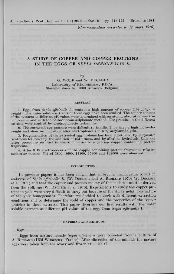
A STUDY OF COPPER AND COPPER PROTEINS IN THE EGGS OF SEPIA OFFICINALIS L.
byG. WOLF and W . DECLEIR
Laboratory of Biochemistry, RUCA,Slachthuislaan 68, 2000 Antwerp (Belgium)
ABSTRACT
1. Eggs from Sepia officinalis L. contain a high amount of copper (109 fxg/g dry weight ). The water soluble extracts of these eggs have been studied. The copper content of the extracts at different pH-values were determined with an atomic absorption spectrophotometer and with the bathocuproin sulphonate method. The proteins in the different extracts were studied by electrophoretic techniques.
2. The extracted egg proteins were difficult to handle. They have a high molecular weight and show no migration after electrophoresis in 6 % acrylamide gels.
3. Fragmentation of the extracted egg proteins has been effectuated by enzymatic treatment followed by the addition of 8M uruem, and by alkaline hydrolysis. Only the latter procedure resulted in electrophoretically migrating copper containing protein fragments.
4. After SDS electrophoresis of the copper containing protein fragments, relative molecular masses (Mr) of 3800, 8600, 17800, 35300 and 132000 were observed.
INTRODUCTION
In previous papers it has been shown that embryonic hemocyanin occurs in embryos of Sepia officinalis L. (W. D e c l e i r and A. R ic h a r d 1970; W. D e c le i r et al. 1971) and that the copper and protein moiety of this molecule must be derived from the yolk sac (W. D e c l e i r et al. 1970). Experiments to study the copper proteins in yolk were very difficult to carry out because of the sticky gelatinous nature of the yolk homogenates. Therefore we decided to work with different extraction conditions and to determine the yield of copper and the properties of the copper proteins in these extracts. This paper describes our first results with the water soluble extracts at different pH-values of the eggs from Sepia officinalis L.
m a t e r ia l a n d m eth od s
— EggsEggs from mature female Sepia officinalis were collected from a culture of
A. R ic h a r d (IBM-Wimereux, France). After dissection of the animals the mature eggs were taken from the ovary and frozen at — 20° C.
— Protein extractsAfter thawing the eggs were washed several times with a physiological salt
solution to remove copper containing contaminating agents such as hemocyanin from the maternal blood.
Protein extracts were made by homogénéisation of the eggs with an Ultra turrax in 50 mM buffer solutions with pH-values ranging between 2.0 and 9.0 (10 volumes/g wet weight). After centrifugation at 17000 rev/min during 20 min at 4° C (Sorval), the supernatant (I) was dialyzed during 22 hours againts 1 mM of the same buffer. The dialysate was submitted to centrigugation (20 min at 17000 rev/min) and the supernatant (II) was ready for use.
— Copper determinations1° Copper determinations with the bathocuproine sulphonate method.Digestion of 1 ml of the protein samples was carried out in pyrex tubes after
addition of 0.3 ml of perchloric acid (70 %), and heating at 240° C during several hours until evaporation was nearly complete. The copper was determined by measuring the absorbance of a cuprous bathocuproine sulphonate complex obtained after the addition of reagents as described by W. D e c l e i r et al. (1970). The yellow- orange colour of the copper complex was measured at 478 nm (Zeiss PM 6). The pH was in the range of 4.8; an intern copper standard was used.2° Copper determination with the atomic absorption spectrophotometer (AAS)
The samples were digested with HNO3 (Merck-Suprapur) at 110° C. The white residue was suspended in 0.1 M HNO3. The atomic absorption spectrometer was a Perkin Elmer 703 ; HGA-500 which included a graphyte tube atomizer. The furnace parameters were as follows : drying at 120° C for 20 sec, ashing at 900° C for 20 sec, atomization at 2650° C for 20 sec, tube cleaning at 2700° C for 3 sec. The furnace was flushed with argon (150 ml/min). The injected sample volume was 20 jzl. Calibration graphs were prepared with stock solutions from Merck « Titrisol ».
— Enzymatic treatmentThe reaction mixture contained 0.5 ml of the protein extract (supernatant II),
0.4 ml of 10 mM Tris-HCl buffer pH 7.3 and 0.1 ml of enzyme solution (I mg/ml). The mixture was incubated at 37° C during 24 hours. In some cases 8 M ureum was added 6 hours before termination of the reaction.
— Alkaline hydrolysisThe reaction mixture contained 1 ml of protein extract and 1 ml of a KOH
solution with a concentration ranging between 50 mM to 2.0 M. The mixture was heated during 1 to 60 min in a boiling water bath. Subsequently KOH was removed by dialysis against 10 mM Tris-HCl buffer pH 7.2.
— Polyacrylamide gel electrophoresis (PAGE)PAGE was carried out on 6 % acrylamide gels (T. W a e h n e ld t and E. S h o o t e r
1973). Protein staining of the gels was performed with 0.2 % amidoblack (in 10 % acetic acid). A colour test for the localization of copper in polyacrylamide gels based upon the quenching of fluorescence of bathocuproine sulphonate by Cu1+ was used (W . B r t ty n in c k x et al. 1978). The presence of copperproteins is indicated by dark bands on a field of blue fluorescence under U.V. light (366 nm).
— SDS electrophoresisElectrophoresis in the presence of SDS was performed as described by K. W e b e r
and M. O sb o rn (1969). The M r of the resulting protein fragments w'as calculated using the method of D. N e v i l l e (1971).
resu lts
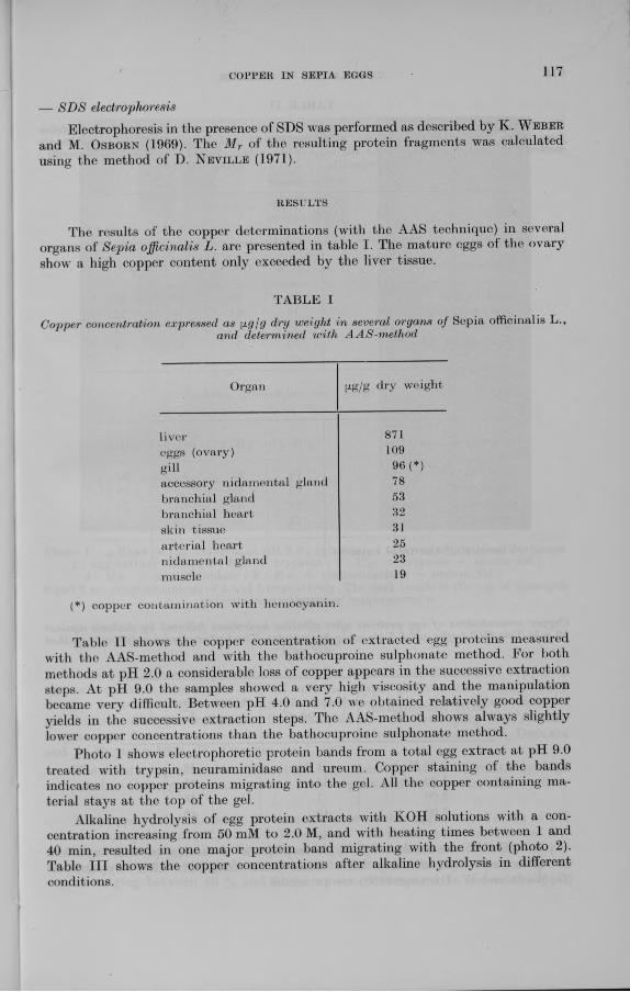
The results of the copper determinations (with the AAS technique) in several organs of Sepia officinalis L. are presented in table I. The mature eggs of the ovary show a high copper content only exceeded by the liver tissue.
TABLE I
Copper concentration expressed as ^g/g dry weight in several organs of Sepia officinalis L.,and determined with AAS-method
Organ (xg/g dry weight
liver 871eggs (ovary) 109gill 96 (*)accessory nidamental gland 78branchial gland 53branchial heart 32skin tissue 31arterial heart 25nidamental gland 23muscle 19
(*) copper contamination with hemocyanin.
Table II shows the copper concentration of extracted egg proteins measured with the AAS-method and with the bathocuproine sulphonate method. For both methods at pH 2.0 a considerable loss of copper appears in the successive extraction steps. At pH 9.0 the samples showed a very high viscosity and the manipulation became very difficult. Between pH 4.0 and 7.0 we obtained relatively good copper yields in the successive extraction steps. The AAS-method shows always slightly lower copper concentrations than the bathocuproine sulphonate method.
Photo 1 shows electrophoretic protein bands from a total egg extract at pH 9.0 treated with trypsin, neuraminidase and ureum. Copper staining of the bands indicates no copper proteins migrating into the gel. All the copper containing material stays at the top of the gel.
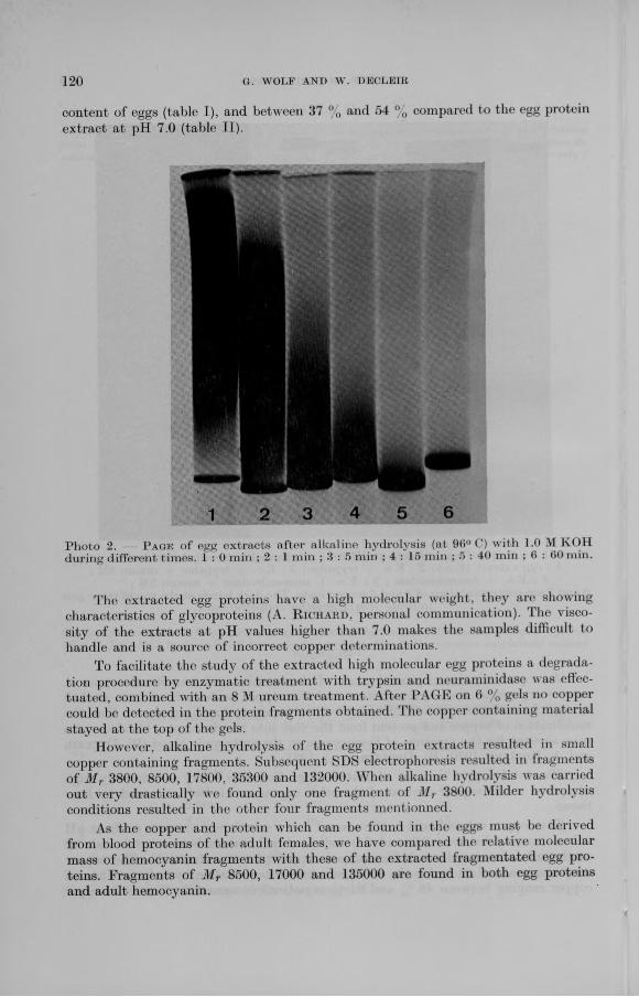
Alkaline hydrolysis of egg protein extracts with KOH solutions with a concentration increasing from 50 mM to 2.0 M, and with heating times between 1 and 40 min, resulted in one major protein band migrating with the front (photo 2). Table III shows the copper concentrations after alkaline hydrolysis in different conditions.
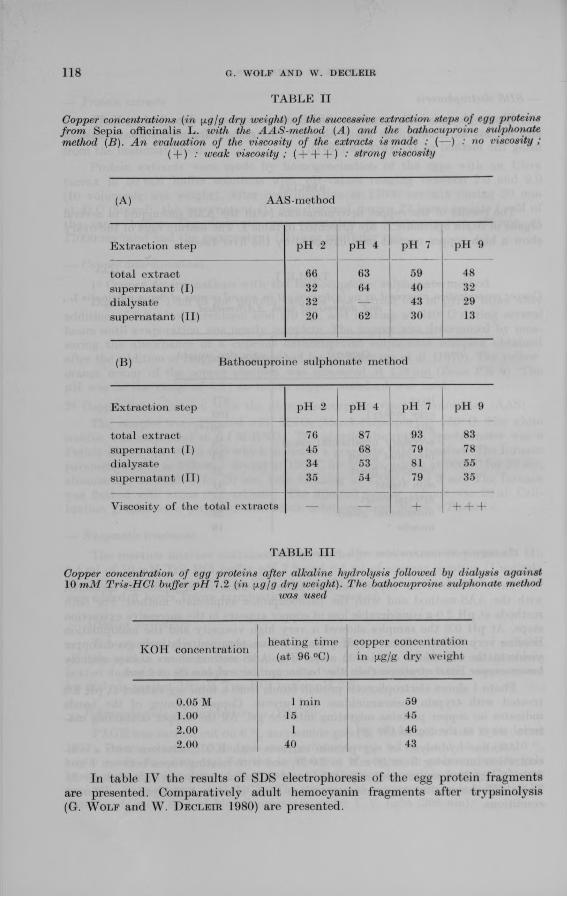
Copper concentrations (in [ig/g dry weight) of the successive extraction steps of egg proteins from Sepia officinalis L. with the AAS-method (A) and the bathocuproine sulphonate method (B). An evaluation of the viscosity of the extracts is made : (— ) : no viscosity ;
( + ) : weak viscosity ; ( + + + ) •' strong viscosity
(A) AAS-method
Extraction step pH 2 pH 4 pH 7 pH 9
total extract 66 63 59 48supernatant (I) 32 64 40 32dialysate 32 — 43 29supernatant (II) 20 62 30 13
(B) Bathocuproine sulphonate method
Extraction step pH 2 pH 4 pH 7 pH 9
total extract 76 87 93 83supernatant (I) 45 68 79 78dialysate 34 53 81 55supernatant (II) 35 54 79 35
Viscosity of the total extracts — — + -I— 1— (-
TABLE III
Copper concentration of egg proteins after alkaline hydrolysis followed by dialysis against10 mM Tris-HCl buffer pH 7.2 (in \i.g\g dry weight). The bathocuproine sulphonate method
was used
KOH concentrationheating time
(at 96 °C)copper concentration in [xg/g dry weight
0.05 M 1 min 591.00 15 452.00 1 462.00 40 43
In table IV the results of SDS electrophoresis of the egg protein fragments are presented. Comparatively adult hemocyanin fragments after trypsinolysis (G. W o l f and W . D e c le ir 1980) are presented.
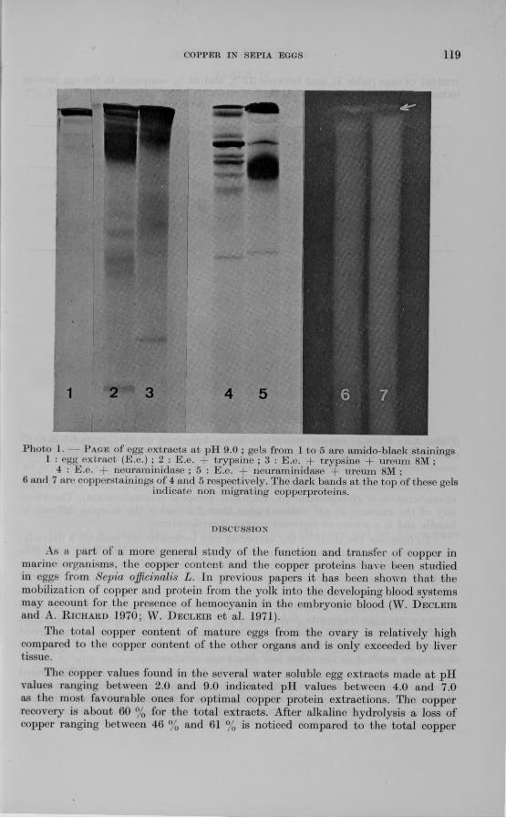
Photo 1. - P a g e of egg extracts at pH 9.0 ; gels from 1 to 5 are amido-black stainings1 : egg extract (E.e.) ; 2 : E.e. + trypsine ; 3 : E.e. + trypsine + ureum 8M ;
4 : E.e. -f neuraminidase ; 5 : E.e. + neuraminidase ureum 8M ;6 and 7 are copperstainings of 4 and 5 respectively. The dark bands at the top of these gels
indicate non migrating copperproteins.
DISCUSSION
As a part of a more general study of the function and transfer of copper in marine organisms, the copper content and the copper proteins have been studied in eggs from Sepia officinalis L. In previous papers it has been shown that the mobilization of copper and protein from the yolk into the developing blood systems may account for the presence of hemocyanin in the embryonic blood (W. D e c l e i r and A. R ic h a r d 1970; W. D e c l e i r et al. 1971).
The total copper content of mature eggs from the ovary is relatively high compared to the copper content of the other organs and is only exceeded by liver tissue.
The copper values found in the several water soluble egg extracts made at pH values ranging between 2.0 and 9.0 indicated pH values between 4.0 and 7.0 as the most favourable ones for optimal copper protein extractions. The copper recovery is about 60 % for the total extracts. After alkaline hydrolysis a loss of copper ranging between 46 % and 61 % is noticed compared to the total copper
content of eggs (table I), and between 37 % and 54 % compared to the egg protein extract at pH 7.0 (table II).
Photo 2. P age of egg extracts after alkaline hydrolysis (at 96° (') with 1.0 M KOH during different times. 1 : 0 mill ; 2 : 1 min ; 3 : 5 min ; 4 : 15 min ; 5 : 40 min ; 6 : 60 min.
The extracted egg proteins have a high molecular weight, they are showing characteristics of glycoproteins (A. R ic h a r d , personal communication). The viscosity of the extracts at pH values higher than 7.0 makes the samples difficult to handle and is a source of incorrect copper determinations.
To facilitate the study of the extracted high molecular egg proteins a degradation procedure by enzymatic treatment with trypsin and neuraminidase was effectuated, combined with an 8 M ureum treatment. After PAGE on t> % gels no copper could be detected in the protein fragments obtained. The copper containing material stayed at the top of the gels.
However, alkaline hydrolysis of the egg protein extracts resulted in small copper containing fragments. Subsequent SDS electrophoresis resulted in fragments of M r 3800, 8500, 17800, 35300 and 132000. When alkaline hydrolysis was carried out very drastically we found only one fragment of M r 3800. Milder hydrolysis conditions resulted in the other four fragments mentionned.
As the copper and protein which can be found in the eggs must be derived from blood proteins of the adult females, we have compared the relative molecular mass of hemocyanin fragments with these of the extracted fragmentated egg proteins. Fragments of M r 8500, 17000 and 135000 are found in both egg proteins and adult hemocyanin.
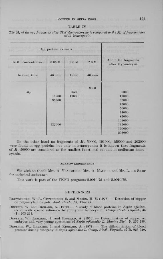
TABLE IV
The Mr of the egg fragments after SDS electrophoresis is compared to the Mr of fragmentatedadult hemocyanin
Egg protein extracts
KOH concentration 0.05 M 2.0 M 2.0 M Adult He fragments after trypsinolysis
heating time 40 min 1 min 40 min
3800M r 8500 8300
17800 17800 1700035300 32000
42000500007400083000
101000132000 135000
150000203000
On the other hand no fragments of M r 50000, 101000, 150000 and 203000 were found in egg proteins but only in hemocyanin; it is known that fragments of M r 50000 are considered as the smallest functional subunit in molluscan hemocyanin.
a c k n o w l e d g e m e n t s
We wish to thank Mrs. A. V la e m in c k , Mrs. A. M a u ru s and Mr. L. d e Sm et for technical assistance.
This w’ork is part of the FKFO programs 2.9010.75 and 2.0010.78.
REFERENCES
B r u y n i n c k x , W . J., G u t t e r i d g e , S. and M a s o n , H. S. (1978) — Detection of copper on polyacrylamide gels. Anal. Bioch., 89, 174-177.
D e c l e i r , W . and R i c h a r d , A. (1970) — A study of blood proteins in Sepia officinalis L. with special reference to embryonic hemocyanin. Comp. Bioch. Physiol., 34 (1), 203-221.
D e c l e i r , W ., L e m a i r e , J. and R i c h a r d , A. (1970) — Determination of copper on embryos and very young specimens of Sepia officinalis L. Marine Biol., 5, 256-258.
D e c l e i r , W ., L e m a i r e , J. and R i c h a r d , A. (1971) — The differentiation of blood proteins during ontogeny in Sepia officinalis L. Comp. Bioch. Physiol., 40 B , 923-930.
N e v i l l e , D . (1971) — Molecular weight determination of protein dodecylsulphate complexes by gel electrophoresis in a discontinuous buffer system. J . Biol. Chem. 246, 6328-6334.
W a e h n e l d t , T. and S h o o t e r , E. (1973) — A comparison of the protein composition of the brains of rodents. Brain Research, 57, 361-371.
W e b e r , K . and O s b o r n , M. (1969) — The reability of molecular weight determinations by sodium dodecylsulphate polyacrylamide gel electrophoresis. J. Biol. Chem., 244. 4406-4412.
W o l f , G. and D e c l e i r , W . (1980) (a) — Partial purification of hemocyanin fragments from Sepia officinalis L., Arch. Inter. Bioch. Physiol., 88. 45.
W o l f , G. and D e c l e i r , W . (1980) (b) A study of embryonic copper proteins and hemocyanin in Sepia officinalis L., Biol. Jb. Dodonaea, 47, 130-136.


![SEPIA OFFICINALIS - Homepage | Vlaams … of S. oficinalis from stage 7 of Naef onwards (Naef, 1928). but they did not notice any difference between embryonic and adult hemocyanin.]](https://img.pdfslide.us/doc/110x75/5ba0731309d3f242318ce6a8/sepia-officinalis-homepage-vlaams-of-s-oficinalis-from-stage-7-of-naef-onwards.jpg)