Embed Size (px)
Citation preview
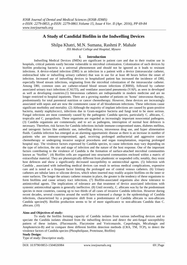
IOSR Journal of Dental and Medical Sciences (IOSR-JDMS)
e-ISSN: 2279-0853, p-ISSN: 2279-0861.Volume 15, Issue 4 Ver. II (Apr. 2016), PP 69-84
www.iosrjournals.org
DOI: 10.9790/0853-1504026984 www.iosrjournals.org 69 | Page
A Study of Candidal Biofilm in the Indwelling Devices
Shilpa Khatri, M.N. Sumana, Rashmi P. Mahale JSS Medical College and Hospital, Mysore
I. Introduction Indwelling Medical Devices (IMDs) are significant in patient care and due to their routine use in
hospitals, critical patients easily become vulnerable to microbial colonization. Colonization of such devices by
biofilm producing bacteria is a universal phenomenon and should not be ignored as it leads to resistant
infections. A device-related infection (DRI) is an infection in a patient with a device (intravascular catheter,
endotracheal tube or indwelling urinary catheter) that was in use for at least 48 hours before the onset of
infection. Increased use of indwelling devices in hospitalized patient has increased the incidence of DRI,
especially blood stream infections, originating from the microbial colonization of the intravascular catheter.
Among DRI, common ones are catheter-related blood stream infections (CRBSI), followed by catheter
associated urinary tract infections (CAUTI), and ventilator associated pneumonia (VAP), as seen in developed
as well as developing countries.(1) Intravenous catheters are indispensable in modern medicine and are no
longer restricted to hospital inpatients. There are a growing number of patients on ‗home‘ intravenous therapy,
predominantly for total parenteral nutrition or cancer chemotherapy. However, these devices are increasingly
associated with sepsis and are now the commonest cause of all bloodstream infections. These infections cause
significant morbidity and mortality. (2) Although the majority of implant infections are caused by gram-positive
bacteria, mainly staphylococci, infections due to Gram-negative bacteria and fungi tend to be more serious.
Fungal infections are most commonly caused by the pathogenic Candida species, particularly C. albicans, C.
tropicalis and C. parapsilosis. These organisms are regarded as increasingly important nosocomial pathogens.
(3) Candida organisms are commensals; and to act as pathogens, interruption of normal host defences is
necessary. Therefore, risk factors for Candida infections include immune-compromised states, diabetes mellitus,
and iatrogenic factors like antibiotic use, indwelling devices, intravenous drug use, and hyper alimentation
fluids. Candida infection has emerged as an alarming opportunistic disease as there is an increase in number of
patients who are immune-compromised, aged, receiving prolonged antibacterial and aggressive cancer
chemotherapy or undergoing invasive surgical procedures and organ transplantation, requiring prolonged
hospital stay. The virulence factors expressed by Candida species, to cause infections may vary depending on
the type of infection, the site and stage of infection and the nature of the host response. One of the important
factors contributing to the virulence of Candida is the formation of surface-attached microbial communities
known as ―biofilm‖. (4) Biofilm cells are organised into structured communities enclosed within a matrix of
extracellular material. They are phenotypically different from planktonic or suspended cells; notably, they resist
host defences and show a significantly decreased susceptibility to antimicrobial agents. (5) Infection with
Candida , associated with indwelling medical devices can result in serious medical complications, expensive
care and is noted as a frequent factor limiting the prolonged use of central venous catheters. (6) Urinary
catheters are tubular latex or silicone devices, which when inserted may readily acquire biofilms on the inner or
outer surfaces. The longer the urinary catheter remains in place, the greater is the tendency of these organisms to
form biofilms and cause urinary tract infections. (7) Biofilm-associated organisms also show tolerance to
antimicrobial agents. The implications of tolerance are that treatment of device associated infections with
systemic antimicrobial agents is generally ineffective. (8) Until recently, C. albicans was by far the predominant
species in most countries, causing up to two thirds of all cases of invasive Candida infection. However during
recent decades, several countries around the world have witnessed a change in the epidemiology of Candida
infections, characterized by a progressive shift from a predominance of Candida albicans to non-albicans
Candida species(9). Biofilm production seems to be of more significance to non-albicans Candida than C.
albicans. (10)
Aims and Objectives of study:
To study the biofilm forming capacity of Candida isolates from various indwelling devices and to
speciate the Candida isolates obtained from the indwelling devices and detect the anti-fungal susceptibility
pattern of these isolates. (Fluconazole, Flucytosine and Voriconazole, Caspofungin, Micafungin and
Amphotericin-B) and to compare three different biofilm detection methods (CRA, TM, TCP), to detect the
virulence factors of Candida species (Phospholipase, Proteinase, Biofilm)
Study Design:
Type of study: Descriptive study.
A Study of Candidal Biofilm in the Indwelling Devices
DOI: 10.9790/0853-1504026984 www.iosrjournals.org 70 | Page
Sample size estimation: from reported cases of Candida infections of 5%.
Taking p=5, and applying the formula n=4pq/d2, where in ‗n‘ is sample size,
‗p‘=40,q=100-p, d is absolute allowable error= 5%.
By this formula our sample size to be taken is approximately 80.
Inclusion Criteria: Candida isolated from patients admitted in JSS hospital undergoing management with
indwelling devices.
Exclusion criteria: Organisms other than Candida isolated from these indwelling devices.
Statistical methods applied
Both descriptive and inferential statistics were employed for data analysis.
Descriptive statistics
The Descriptive statistics procedure displays univariate summary statistics for several variables in a
single table and calculates standardized values. Variables can be ordered by the size of their means
alphabetically, or by the order in which the researcher selects the variables. In the present study following
descriptive statistics have been employed
a. Frequencies
b. Percentages
Inferential statistics
Crosstabs (Cramer’s V)
The Crosstabs procedure forms two-way and multi-way tables and provides a variety of tests and
measures of association for two-way tables. The structure of the table and whether categories are ordered
determine what test or measure to use. Cramer‘s V as a measure of association between rows and columns was
employed.
II. Material & Methods Patients admitted to JSS Hospital, with indwelling catheters, were included in the study over a period
of two years from March 2013 to March 2015, after getting the ethical clearance from the Ethical Committee. A
detailed history of these patients was taken specially with regard to the following - History of fever, Diabetes,
Chronic infection, Smoking, Drug intake and Underlying conditions like immune compromised state, presence
of indwelling devices. Samples were collected from IV catheter, Endotracheal aspirate, Urine. Samples were
processed for Microscopy and Culture. Catheter and Endotracheal aspirates were used for microscopy. For
Culture 5% sheep blood agar, Mac Conkey's agar, Sabouraud's dextrose agar with Chloramphenicol was used.
All the samples were processed as per standard guidelines and both quantitative and semiquantitative analyses
of samples were done. Intravenous catheter and Endotracheal aspirate was processed for semi quantitative
culture and Urine from urinary catheter was processed for Quantitative culture. The organism was grown on
Sabouraud's dextrose agar (SDA) at 37ºC for 24 hours.the organisms was inoculated in test tubes containing 2%
carbohydrate solutions of dextrose, maltose, sucrose, and lactose. Andrade's indicator 0.005%, was used in all
these carbohydrate solutions. Inverted durham's tubes are used for detection of gas. These tubes were incubated
at 37ºC for 3-7 days. Growth of yeast around individual discs indicates assimilation of that compound. When the
carbohydrate is not utilized, growth around disc is absent. Species differentiation was done on the basis of
combined pattern of Carbohydrate fermentation test and Carbohydrate assimilation test according to a standard
chart. Virulence testing of the following was also done as per the standard guidelines
1. Production of germ tube
2. Phospholipase estimation
3. Proteinase estimation
4. Adherence assay
Production of germ Tube - Strains of C.albicans produce germ tubes from their yeast cells when placed in a
liquid enviornment and incubated at 37ºC for 1-2 hours. It is also known as Reynolds Braude phenomenon.
Protinase test Protein substance (Bovine Serum Albumin) markedly influences the production of extracellular
proteases by Candida species .Candida species when grown in a medium containing Bovine Serum Albumin
causes hydrolysis of the substrate and hence proteolysis beyond the hinge of colonies which is visible as a clear
halo. Adherence assay
Candida species when inoculated into SDB (Subourauds dextrose broth ) medium that contains high glucose
(8%) and protein( 1%); adheres to the polystyrene tubes and plates and form biofilms.
Tissue Culture Method
A Study of Candidal Biofilm in the Indwelling Devices
DOI: 10.9790/0853-1504026984 www.iosrjournals.org 71 | Page
Isolates from freshly sub-cultured plates were inoculated in Subarouds dextrose broth (SDB) with 8%
w/v glucose and incubated for 24 hours at 37˚C in stationary conditions and then diluted to 1:100 with fresh
SDB. Individual wells of sterile polystyrene 96 well flat bottom microtitre plates were filled with 200μl aliquots
of diluted culture. Un-inoculated SDB served as a control to check sterility and nonspecific binding of media.
Control strains were also inoculated in triplicate. The microtitre plate was incubated for 48 hours at 37˚C. After
incubation contents of each well was removed by tapping the plates. After washing the wells for four times with
200μl of phosphate buffer saline (PBS pH 7.2), the floating planktonic bacteria were removed. The biofilms thus
formed in plates were fixed using 2% w/v sodium acetate for 10 minutes and tainted with 0.1% w/v crystal
violet for 10minutes. After washing thoroughly with de- ionized water to remove any excess stain, the plates
were dried. Micro-ELISA auto-reader at the wavelength of 540 nm was used to measure the Optical Density
(OD) of the stained adherent micro-organisms.(12,13,14) The OD540 value of sterile medium, fixative and dye
were averaged and subtracted from all test values. The mean OD540 value from a control well was deducted
from all test OD540 values. These OD540 values were considered as an index of bacteria adhering to surface
and forming biofilms. Experiments were performed in triplicate.
Congo Red Agar Method (CRA): Freeman et al (15) had described an alternative method of screening
biofilm formation by Candida isolates. In the present study the Congo red agar (CRA) was optimized to get
strong black pigmentation at 48hrs incubation and then for 2-4 days room temperature. Black coloured colonies
with dry crystalline consistency interpreted as positive biofilm producing strains. Red coloured colonies-
interpreted as negative for biofilm production.
The TCP method was considered the gold-standard for this study and compared with data from TM and
CRA methods.( Parameters like sensitivity, specificity, negative predictive value, positive predictive value were
calculated.(16) For flucytosine, isolates showing MIC‘s ≤4μg/ml were considered as susceptible, 8- 16μg/ml as
intermediate and ≥32μg/ml as resistant. For amphotericin B, isolates showing a MIC of ≤1.0μg/ml were taken as
susceptible and those with MIC>1μg/ml were considered as resistant.10, 11 MIC interpretative criteria were
referred to those described in the CLSI document M 27-S3 for amphotericin B, 5-flucytosine
III. Results: The results are in the tabular form as under
The samples collected from patients belonged to the wide range of age groups of 18 and above. It was
found that maximum number of samples which yielded Candida, belonged to age group 31-45 years (41.3%),
followed by 18-30 years (23.8%), and 46-60 years (22.5%) and more than 60 years (12.5%)
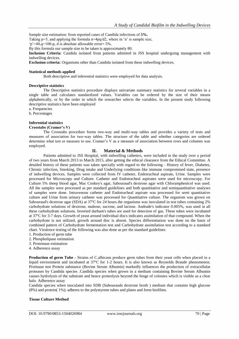
TABLE 1: Age Wise Distribution of the Samples Collected (N=80)
TABLE 3: Age and sex
distribution:
Graph 1:
AGE (YEARS) Frequency(f) SEX Total
% M F
18-30 F 13 6 19
% 27.1% 18.8% 23.8%
31-45 F 17 16 33
% 35.4% 50.0% 41.3%
46-60 f 11 7 18
% 22.9% 21.9% 22.5%
>60 F 7 3 10
% 14.6% 9.4% 12.5%
Total F 48 32 80
% 100.0% 100.0% 100.0%
AGE GROUPS (YEARS) Frequency (f) %
18-30 19 23.8
31-45 33 41.3
46-60 18 22.5
>60 10 12.5
Total 80 100.0
A Study of Candidal Biofilm in the Indwelling Devices
DOI: 10.9790/0853-1504026984 www.iosrjournals.org 72 | Page
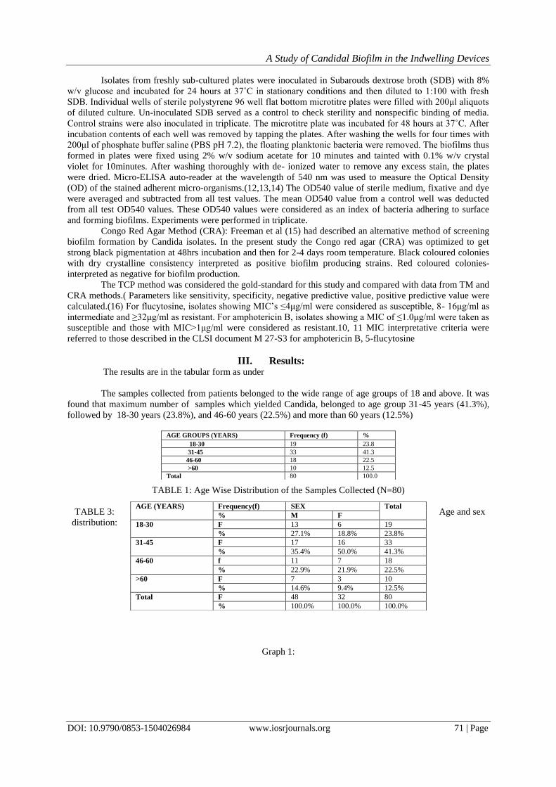
The male female sex ratio in our patients is 3:2. There is male predominance in all the age groups.
Table 4 : Duration Of Stay In Hospital
TABLE 4 shows that most of the patients in our study were admitted for an average 1-2 weeks (47.5%),
followed by <1week (46.3%), and only 5 patients were admitted in the hospital for more than 2 weeks (6.3%).
Table 5:Co-Morbid Conditions In The Study Group: Number %
SURGICAL 49 61.3
MEDICAL 25 31.3
TRAUMA 6 7.5
TOTAL 80 100.0
Table 5,shows that among the 80 patients in our study, 49 patients were surgical cases, 25 patients had
medical co-morbid conditions, and 6were trauma cases.
Table 6:Associated Risk Factors In The Study Group: RISK FACTORS NUMBER %
INDWELLING DEVICE 80 100
HTN 39 48.8
SMOKING 33 41.3
DM 36 45
Table 6 shows that all the patients in our study were associated with some indwelling devices, 48.8%
were hypertensives, 41.3% were smokers, and 45% were diabetics.
Table 7: Haematological Parameters In The Study Group:: CRITERIA FREQUENCY PERCENTAGE(%0
HB <10gm/dl 50 62.5
TLC >11000 / cumm 54 67.5
LYMPHOCYTES >40% 4 5.0
NEUTROPHILS >70% 41 51.3
Table7, shows that in our study 62.5% patients were anaemic, 67.5% patients had TLC count more than
>11,000, 5% patients had lymphocytosis,and 51.3% had neutrohils >70%
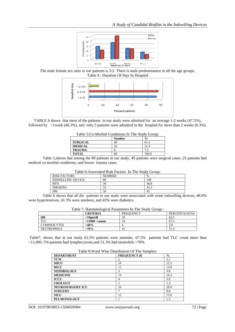
Table 8:Ward Wise Distribution Of The Samples: DEPARTMENT FREQUENCY (f) %
TCW 2 2.5
MICU 10 12.5
RICU 11 13.8
NEPHROLOGY 3 3.8
MEDICINE 13 16.3
ICCU 4 5.0
UROLOGY 1 1.3
NEUROSURGERY ICU 16 20.0
SURGERY 7 8.8
SICU 12 15.0
PULMONOLOGY 1 1.3
A Study of Candidal Biofilm in the Indwelling Devices
DOI: 10.9790/0853-1504026984 www.iosrjournals.org 73 | Page
Graph 3:
Table 8, and Graph 3, shows that maximum number of samples were received from Neurosurgery ICU (17),
and minimum from Pulmonology ward (1).
Table 9 shows that total of 850 indwelling devices, samples received during the course of study. Out of
these, 500 were from urine from Catheterised patients, 200 ET secretion, 150 from intravascular catheter tips.
Table 9: Samples Total Number Culture Positive Culture Negative Candida Positives (Out Of All
Culture Positives)
Urine From Catheterised
Patients
500 400 32 Number %
68 17.0%
Et Secretions 200 170 20 10 5.88%
I.V. Catheter Tips 150 68 80 2 2.94
Total 850 638 132 80 25.82
Table 10, shows that ,68 urine samples from catheterised patients (85%), 10 Endotracheal tubes secretions
(12.5%), and 2 , I.V catheter tips (2.5%) were Candida culture positive
Table 10: Samples Collected From The Study Group: SAMPLE NUMBER %
URINE 68 85.0
ET SECRETION 10 12.5
I.V.CATHETER TIP 2 2.5
Total 80 100.0
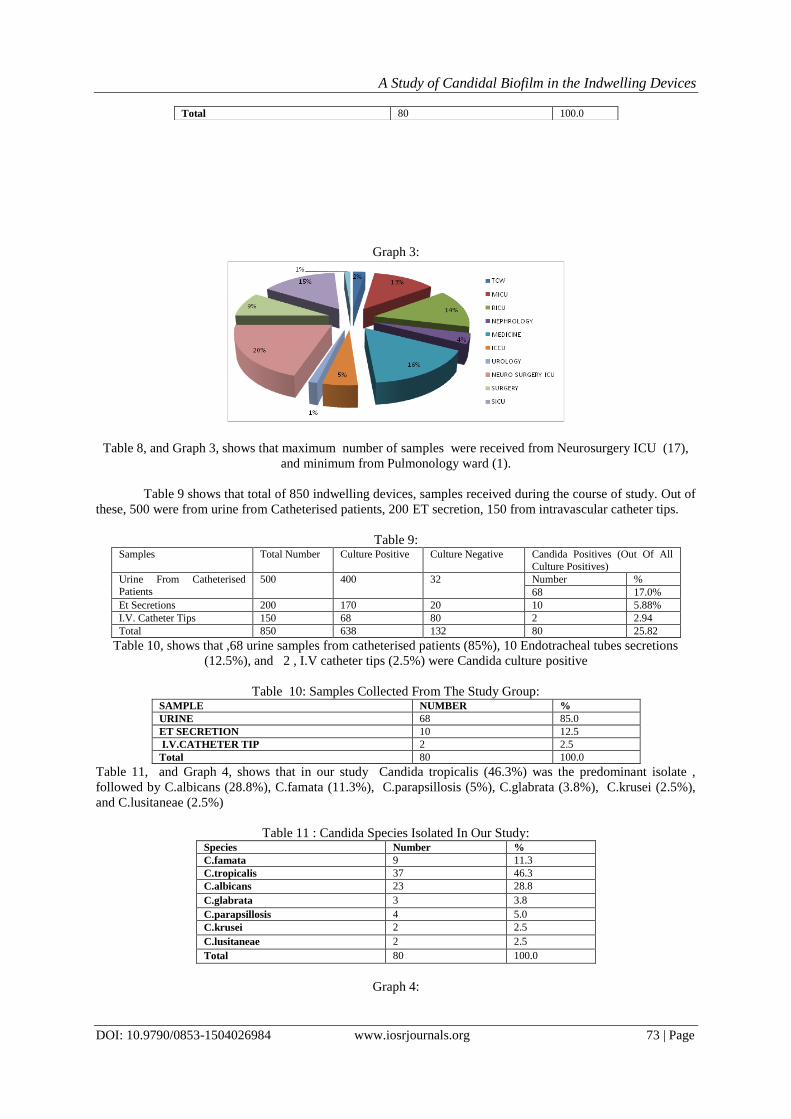
Table 11, and Graph 4, shows that in our study Candida tropicalis (46.3%) was the predominant isolate ,
followed by C.albicans (28.8%), C.famata (11.3%), C.parapsillosis (5%), C.glabrata (3.8%), C.krusei (2.5%),
and C.lusitaneae (2.5%)
Table 11 : Candida Species Isolated In Our Study: Species Number %
C.famata 9 11.3
C.tropicalis 37 46.3
C.albicans 23 28.8
C.glabrata 3 3.8
C.parapsillosis 4 5.0
C.krusei 2 2.5
C.lusitaneae 2 2.5
Total 80 100.0
Graph 4:
Total 80 100.0
A Study of Candidal Biofilm in the Indwelling Devices
DOI: 10.9790/0853-1504026984 www.iosrjournals.org 74 | Page
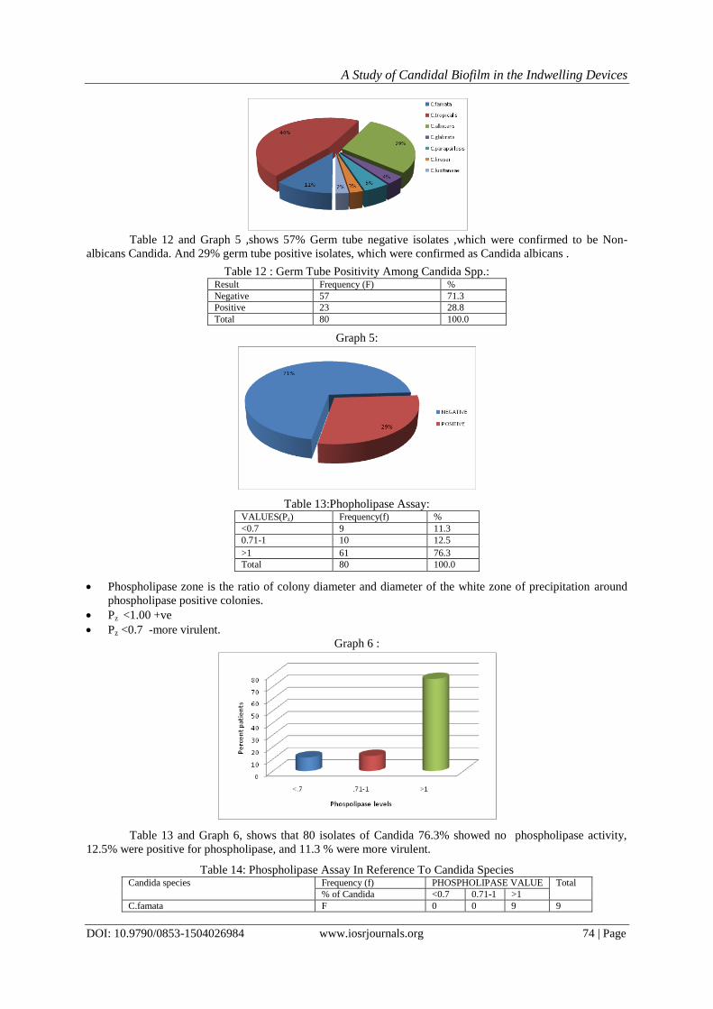
Table 12 and Graph 5 ,shows 57% Germ tube negative isolates ,which were confirmed to be Non-
albicans Candida. And 29% germ tube positive isolates, which were confirmed as Candida albicans .
Table 12 : Germ Tube Positivity Among Candida Spp.: Result Frequency (F) %
Negative 57 71.3
Positive 23 28.8
Total 80 100.0
Graph 5:
Table 13:Phopholipase Assay: VALUES(Pz) Frequency(f) %
<0.7 9 11.3
0.71-1 10 12.5
>1 61 76.3
Total 80 100.0
Phospholipase zone is the ratio of colony diameter and diameter of the white zone of precipitation around
phospholipase positive colonies.
Pz <1.00 +ve
Pz <0.7 -more virulent.
Graph 6 :
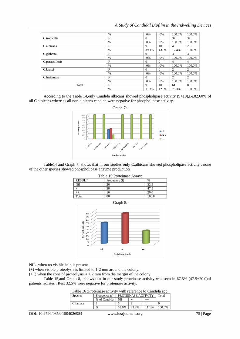
Table 13 and Graph 6, shows that 80 isolates of Candida 76.3% showed no phospholipase activity,
12.5% were positive for phospholipase, and 11.3 % were more virulent.
Table 14: Phospholipase Assay In Reference To Candida Species Candida species Frequency (f) PHOSPHOLIPASE VALUE Total
% of Candida <0.7 0.71-1 >1
C.famata F 0 0 9 9
A Study of Candidal Biofilm in the Indwelling Devices
DOI: 10.9790/0853-1504026984 www.iosrjournals.org 75 | Page
% .0% .0% 100.0% 100.0%
C.tropicalis F 0 0 37 37
% .0% .0% 100.0% 100.0%
C.albicans F 9 10 4 23
% 39.1% 43.5% 17.4% 100.0%
C.glabrata F 0 0 3 3
% .0% .0% 100.0% 100.0%
C.parapsillosis F 0 0 4 4
% .0% .0% 100.0% 100.0%
C.krusei F 0 0 2 2
% .0% .0% 100.0% 100.0%
C.lusitaneae F 0 0 2 2
% .0% .0% 100.0% 100.0%
Total F 9 10 61 80
% 11.3% 12.5% 76.3% 100.0%
According to the Table 14,only Candida albicans showed phospholipase activity (9+10),i.e.82.60% of
all C.albicans.where as all non-albicans candida were negative for phospholipase activity.
Graph 7:.
Table14 and Graph 7, shows that in our studies only C.albicans showed phospholipase activity , none
of the other species showed phospholipase enzyme production
Table 15:Proteinase Assay: RESULT Frequency (f) %
Nil 26 32.5
+ 38 47.5
++ 16 20.0
Total 80 100.0
Graph 8:
NIL- when no visible halo is present
(+) when visible proteolysis is limited to 1-2 mm around the colony.
(++) when the zone of proteolysis is > 2 mm from the margin of the colony
Table 15,and Graph 8, shows that in our study proteinase activity was seen in 67.5% (47.5+20.0)of
patients isolates . Rest 32.5% were negative for proteinase activity.
Table 16 :Proteinase activity with reference to Candida spp. Species Frequency (f) PROTEINASE ACTIVITY Total
% of Candida Nil + ++
C.famata f 5 3 1 9
% 55.6% 33.3% 11.1% 100.0%
A Study of Candidal Biofilm in the Indwelling Devices
DOI: 10.9790/0853-1504026984 www.iosrjournals.org 76 | Page
C.tropicalis f 11 22 4 37
% 29.7% 59.5% 10.8% 100.0%
C.albicans f 8 6 9 23
% 34.8% 26.1% 39.1% 100.0%
C.glabrata f 2 1 0 3
% 66.7% 33.3% .0% 100.0%
C.parapsillosis f 0 3 1 4
% .0% 75.0% 25.0% 100.0%
C.krusei f 0 1 1 2
% .0% 50.0% 50.0% 100.0%
C.lusitaneae f 0 2 0 2
% .0% 100.0% .0% 100.0%
Total f 26 38 16 80
% 32.5% 47.5% 20.0% 100.0%
Graph 9:
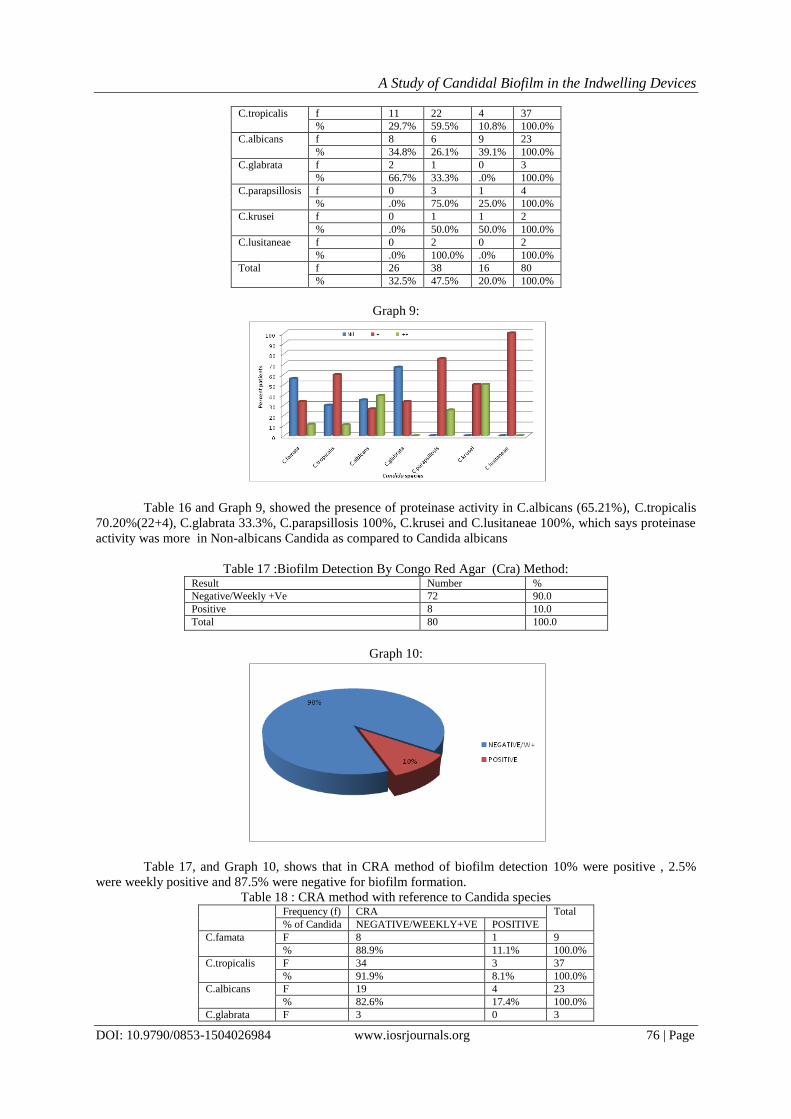
Table 16 and Graph 9, showed the presence of proteinase activity in C.albicans (65.21%), C.tropicalis
70.20%(22+4), C.glabrata 33.3%, C.parapsillosis 100%, C.krusei and C.lusitaneae 100%, which says proteinase
activity was more in Non-albicans Candida as compared to Candida albicans
Table 17 :Biofilm Detection By Congo Red Agar (Cra) Method: Result Number %
Negative/Weekly +Ve 72 90.0
Positive 8 10.0
Total 80 100.0
Graph 10:
Table 17, and Graph 10, shows that in CRA method of biofilm detection 10% were positive , 2.5%
were weekly positive and 87.5% were negative for biofilm formation.
Table 18 : CRA method with reference to Candida species Frequency (f) CRA Total
% of Candida NEGATIVE/WEEKLY+VE POSITIVE
C.famata F 8 1 9
% 88.9% 11.1% 100.0%
C.tropicalis F 34 3 37
% 91.9% 8.1% 100.0%
C.albicans F 19 4 23
% 82.6% 17.4% 100.0%
C.glabrata F 3 0 3
A Study of Candidal Biofilm in the Indwelling Devices
DOI: 10.9790/0853-1504026984 www.iosrjournals.org 77 | Page
% 100.0% .0% 100.0%
C.parapsillosis F 4 0 4
% 100.0% .0% 100.0%
C.krusei F 2 0 2
% 100.0% .0% 100.0%
C.lusitaneae F 2 0 2
% 100.0% .0% 100.0%
Total F 72 8 80
% 90.0% 10.0% 100.0%
Graph 11:
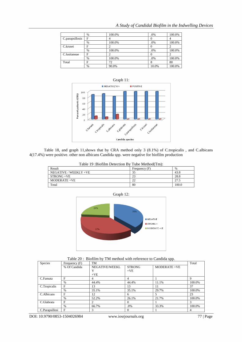
Table 18, and graph 11,shows that by CRA method only 3 (8.1%) of C.tropicalis , and C.albicans
4(17.4%) were positive. other non albicans Candida spp. were negative for biofilm production
Table 19 :Biofilm Detection By Tube Method(Tm): Result Frequency (F) %
NEGATIVE / WEEKLY +VE 35 43.8
STRONG +VE 23 28.8
MODERATE +VE 22 27.5
Total 80 100.0
Graph 12:
Table 20 : Biofilm by TM method with reference to Candida spp. Species Frequency (F) TM Total
% Of Candida NEGATIVE/WEEKL
Y
+VE
STRONG
+VE
MODERATE +VE
C.Famata F 4 4 1 9
% 44.4% 44.4% 11.1% 100.0%
C.Tropicalis F 13 13 11 37
% 35.1% 35.1% 29.7% 100.0%
C.Albicans F 12 6 5 23
% 52.2% 26.1% 21.7% 100.0%
C.Glabrata F 2 0 1 3
% 66.7% .0% 33.3% 100.0%
C.Parapsillosi F 3 0 1 4
A Study of Candidal Biofilm in the Indwelling Devices
DOI: 10.9790/0853-1504026984 www.iosrjournals.org 78 | Page
s % 75.0% .0% 25.0% 100.0%
C.Krusei F 0 0 2 2
% .0% .0% 100.0% 100.0%
C.Lusitaneae F 1 0 1 2
% 50.0% .0% 50.0% 100.0%
Total F 35 23 22 80
% 43.8% 28.8% 27.5% 100.0%
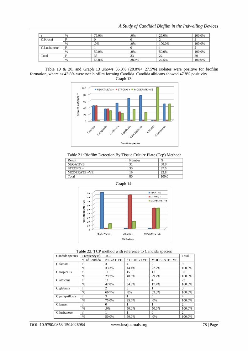
Table 19 & 20, and Graph 13 ,shows 56.3% (28.8%+ 27.5%) isolates were positive for biofilm
formation, where as 43.8% were non biofilm forming Candida. Candida albicans showed 47.8% positivity.
Graph 13:
Table 21 :Biofilm Detection By Tissue Culture Plate (Tcp) Method:
Graph 14:
Table 22: TCP method with reference to Candida species Candida species Frequency (f) TCP Total
% of Candida NEGATIVE STRONG +VE MODERATE +VE
C.famata f 3 4 2 9
% 33.3% 44.4% 22.2% 100.0%
C.tropicalis f 11 15 11 37
% 29.7% 40.5% 29.7% 100.0%
C.albicans f 11 8 4 23
% 47.8% 34.8% 17.4% 100.0%
C.glabrata f 2 0 1 3
% 66.7% .0% 33.3% 100.0%
C.parapsillosis f 3 1 0 4
% 75.0% 25.0% .0% 100.0%
C.krusei f 0 1 1 2
% .0% 50.0% 50.0% 100.0%
C.lusitaneae f 1 1 0 2
% 50.0% 50.0% .0% 100.0%
Result Number %
NEGATIVE 31 38.8
STRONG + 30 37.5
MODERATE +VE 19 23.8
Total 80 100.0
A Study of Candidal Biofilm in the Indwelling Devices
DOI: 10.9790/0853-1504026984 www.iosrjournals.org 79 | Page
TOTAL f 31 30 19 80
% 38.8% 37.5% 23.8% 100.0%
Graph 14:
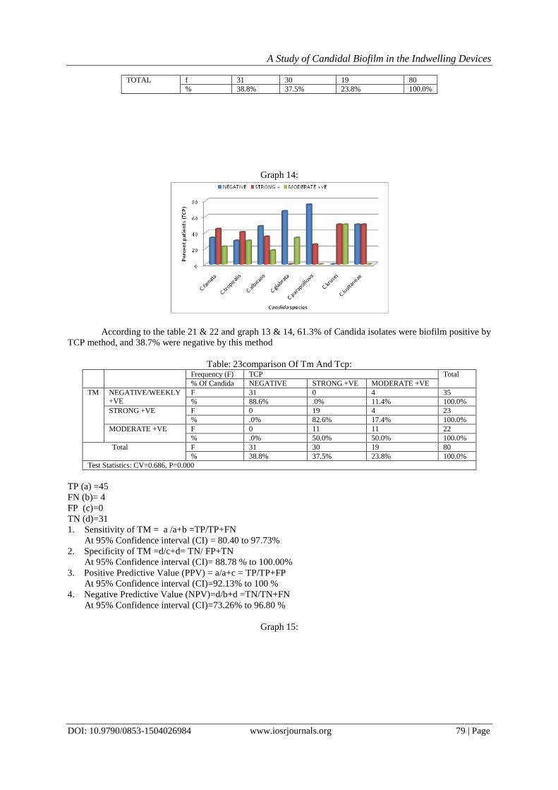
According to the table 21 & 22 and graph 13 & 14, 61.3% of Candida isolates were biofilm positive by
TCP method, and 38.7% were negative by this method
Table: 23comparison Of Tm And Tcp:
Frequency (F) TCP Total
% Of Candida NEGATIVE STRONG +VE MODERATE +VE
TM NEGATIVE/WEEKLY +VE
F 31 0 4 35
% 88.6% .0% 11.4% 100.0%
STRONG +VE F 0 19 4 23
% .0% 82.6% 17.4% 100.0%
MODERATE +VE F 0 11 11 22
% .0% 50.0% 50.0% 100.0%
Total F 31 30 19 80
% 38.8% 37.5% 23.8% 100.0%
Test Statistics: CV=0.686, P=0.000
TP (a) =45
FN (b)= 4
FP (c)=0
TN (d)=31
1. Sensitivity of TM = a /a+b =TP/TP+FN
At 95% Confidence interval (CI) = 80.40 to 97.73%
2. Specificity of TM =d/c+d= TN/ FP+TN
At 95% Confidence interval (CI)= 88.78 % to 100.00%
3. Positive Predictive Value (PPV) = a/a+c = TP/TP+FP
At 95% Confidence interval (CI)=92.13% to 100 %
4. Negative Predictive Value (NPV)=d/b+d =TN/TN+FN
At 95% Confidence interval (CI)=73.26% to 96.80 %
Graph 15:
A Study of Candidal Biofilm in the Indwelling Devices
DOI: 10.9790/0853-1504026984 www.iosrjournals.org 80 | Page
The above Table 23 and graph 15, shows P value of 0.000 between TM and TCP methods, which
shows a high significance and states that TM is highly sensitive and equally good method as TCP.
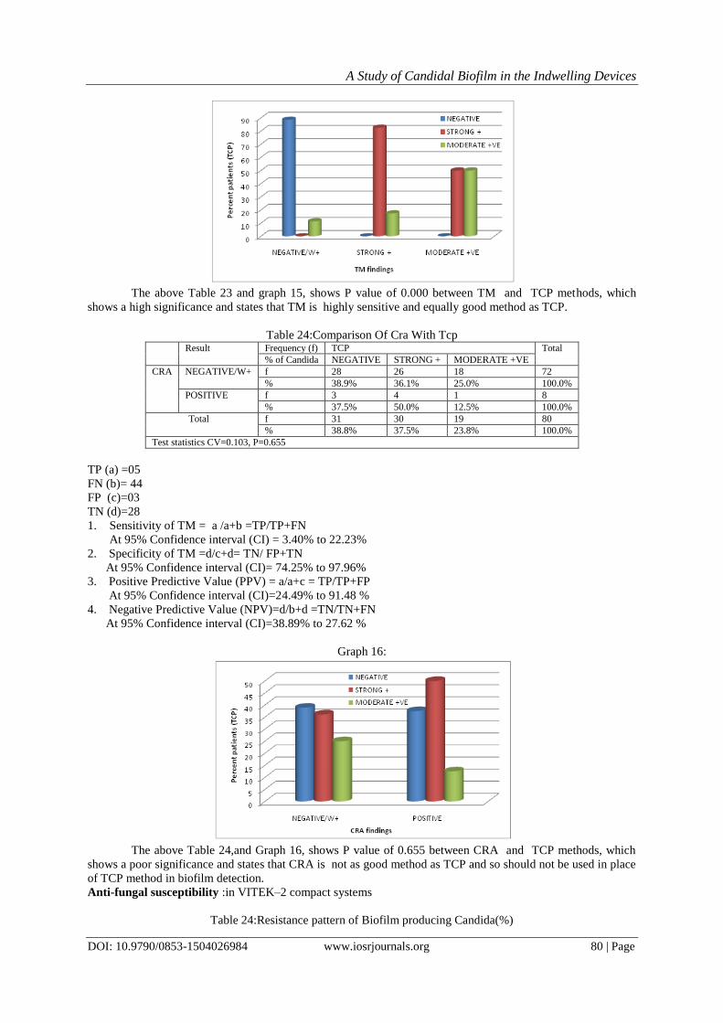
Table 24:Comparison Of Cra With Tcp
Result Frequency (f) TCP Total
% of Candida NEGATIVE STRONG + MODERATE +VE
CRA NEGATIVE/W+ f 28 26 18 72
% 38.9% 36.1% 25.0% 100.0%
POSITIVE f 3 4 1 8
% 37.5% 50.0% 12.5% 100.0%
Total f 31 30 19 80
% 38.8% 37.5% 23.8% 100.0%
Test statistics CV=0.103, P=0.655
TP (a) =05
FN (b)= 44
FP (c)=03
TN (d)=28
1. Sensitivity of TM = a /a+b =TP/TP+FN
At 95% Confidence interval (CI) = 3.40% to 22.23%
2. Specificity of TM =d/c+d= TN/ FP+TN
At 95% Confidence interval (CI)= 74.25% to 97.96%
3. Positive Predictive Value (PPV) = a/a+c = TP/TP+FP
At 95% Confidence interval (CI)=24.49% to 91.48 %
4. Negative Predictive Value (NPV)=d/b+d =TN/TN+FN
At 95% Confidence interval (CI)=38.89% to 27.62 %
Graph 16:
The above Table 24,and Graph 16, shows P value of 0.655 between CRA and TCP methods, which
shows a poor significance and states that CRA is not as good method as TCP and so should not be used in place
of TCP method in biofilm detection.
Anti-fungal susceptibility :in VITEK–2 compact systems
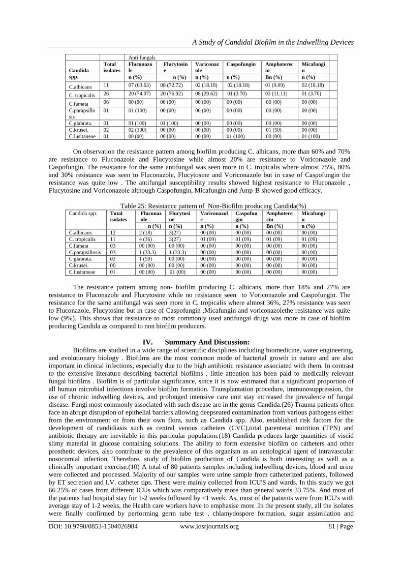
Table 24:Resistance pattern of Biofilm producing Candida(%)
A Study of Candidal Biofilm in the Indwelling Devices
DOI: 10.9790/0853-1504026984 www.iosrjournals.org 81 | Page
Anti fungals
Candida
spp.
Total
isolates
Fluconazo
le
Flucytosin
e
Variconaz
ole
Caspofungin Amphoterec
in
Micafungi
n
n (%) n (%) n (%) n (%) Bn (%) n (%)
C.albicans 11 07 (63.63) 08 (72.72) 02 (18.18) 02 (18.18) 01 (9.09) 02 (18.18)
C. tropicalis 26 20 (74.07) 20 (76.92) 08 (29.62) 01 (3.70) 03 (11.11) 01 (3.70)
C.famata 06 00 (00) 00 (00) 00 (00) 00 (00) 00 (00) 00 (00)
C.parapsillo
sis
01 01 (100) 00 (00) 00 (00) 00 (00) 00 (00) 00 (00)
C.glabrata. 01 01 (100) 01 (100) 00 (00) 00 (00) 00 (00) 00 (00)
C.krusei. 02 02 (100) 00 (00) 00 (00) 00 (00) 01 (50) 00 (00)
C.lusitaneae 01 00 (00) 00 (00) 00 (00) 01 (100) 00 (00) 01 (100)
On observation the resistance pattern among biofilm producing C. albicans, more than 60% and 70%
are resistance to Fluconazole and Flucytosine while almost 20% are resistance to Voriconazole and
Caspofungin. The resistance for the same antifungal was seen more in C. tropicalis where almost 75%, 80%
and 30% resistance was seen to Fluconazole, Flucytosine and Voriconazole but in case of Caspofungin the
resistance was quite low . The antifungal susceptibility results showed highest resistance to Fluconazole ,
Flucytosine and Voriconazole although Caspofungin, Micafungin and Amp-B showed good efficacy.
Table 25: Resistance pattern of Non-Biofilm producing Candida(%) Candida spp. Total
isolates Fluconaz
ole Flucytosi
ne Variconazol
e Caspofun
gin Amphotere
cin Micafungi
n
n (%) n (%) n (%) n (%) Bn (%) n (%)
C.albicans 12 2 (18) 3(27) 00 (00) 00 (00) 00 (00) 00 (00)
C. tropicalis 11 4 (36) 3(27) 01 (09) 01 (09) 01 (09) 01 (09)
C.famata 03 00 (00) 00 (00) 00 (00) 00 (00) 00 (00) 00 (00)
C.parapsillosis 03 1 (33.3) 1 (33.3) 00 (00) 00 (00) 00 (00) 00 (00)
C.glabrata. 02 1 (50) 00 (00) 00 (00) 00 (00) 00 (00) 00 (00)
C.krusei. 00 00 (00) 00 (00) 00 (00) 00 (00) 00 (00) 00 (00)
C.lusitaneae 01 00 (00) 01 (00) 00 (00) 00 (00) 00 (00) 00 (00)
The resistance pattern among non- biofilm producing C. albicans, more than 18% and 27% are
resistance to Fluconazole and Flucytosine while no resistance seen to Voriconazole and Caspofungin. The
resistance for the same antifungal was seen more in C. tropicalis where almost 36%, 27% resistance was seen
to Fluconazole, Flucytosine but in case of Caspofungin ,Micafungin and voriconazolethe resistance was quite
low (9%). This shows that resistance to most commonly used antifungal drugs was more in case of biofilm
producing Candida as compared to non biofilm producers.
IV. Summary And Discussion: Biofilms are studied in a wide range of scientific disciplines including biomedicine, water engineering,
and evolutionary biology . Biofilms are the most common mode of bacterial growth in nature and are also
important in clinical infections, especially due to the high antibiotic resistance associated with them. In contrast
to the extensive literature describing bacterial biofilms , little attention has been paid to medically relevant
fungal biofilms . Biofilm is of particular significance, since it is now estimated that a significant proportion of
all human microbial infections involve biofilm formation. Transplantation procedure, immunosuppression, the
use of chronic indwelling devices, and prolonged intensive care unit stay increased the prevalence of fungal
disease. Fungi most commonly associated with such disease are in the genus Candida.(26) Trauma patients often
face an abrupt disruption of epithelial barriers allowing deepseated contamination from various pathogens either
from the environment or from their own flora, such as Candida spp. Also, established risk factors for the
development of candidiasis such as central venous catheters (CVC),total parenteral nutrition (TPN) and
antibiotic therapy are inevitable in this particular population.(18) Candida produces large quantities of viscid
slimy material in glucose containing solutions. The ability to form extensive biofilm on catheters and other
prosthetic devices, also contribute to the prevalence of this organism as an aetiological agent of intravascular
nosocomial infection. Therefore, study of biofilm production of Candida is both interesting as well as a
clinically important exercise.(10) A total of 80 patients samples including indwelling devices, blood and urine
were collected and processed. Majority of our samples were urine sample from catheterized patients, followed
by ET secretion and I.V. catheter tips. These were mainly collected from ICU'S and wards. In this study we got
66.25% of cases from different ICUs which was comparatively more than general wards 33.75%. And most of
the patients had hospital stay for 1-2 weeks followed by <1 week. As, most of the patients were from ICU's with
average stay of 1-2 weeks, the Health care workers have to emphasise more .In the present study, all the isolates
were finally confirmed by performing germ tube test , chlamydospore formation, sugar assimilation and
A Study of Candidal Biofilm in the Indwelling Devices
DOI: 10.9790/0853-1504026984 www.iosrjournals.org 82 | Page
fermentation reactions. Sugar assimilation and fermentation tests are therefore considered to be important in
identifying Candida species especially non-albicans Candida, but it is a tedious procedure.
Germ tubes ,which mark the onset of hyphal growth, and are induced by contact with serum ,are particularly
involved in the pathogenesis of Candida infection. The formation of germ tube is accompanied by an increased
adherence to epithelial cells . In our study, germ tube formation was observed in 28.8% of Candida strains ,
which were identified as C.albicans, whereasremaining strains (71.3%) failed to produce germ tube(NAC).
A study by Alhussaini M S et al (2013), observed germ tube production in 27/50 (54%) of Candida
strains, which were identified as C.albicans and remaining strains failed to produce germ tube which were
identified as non albicans Candida(NAC).(19) The study performed by Mohammadi P et al. (2012) also
concluded that Glucose, Galactose, Maltose &Trehalose were used by all the yeasts as far as the identification of
Candida species. are concerned which was close to our study.(20) Kumar A et al. (2014) also observed from
their study that most species of
Candida assimilated glucose, galactose, maltose, trehalose and sucrose,the finding which was also at
par with our study.(21) Among 80 isolates from all the indwelling devices and urine Candida tropicalis (46.3%),
followed by C.albicans (28.8%), C.famata (11.3%), C.parapsillosis (5%), C.glabrata (3.8%), C.krusei (2.5%),
and C.lusitaneae (2.5%) . that is, Non -albicans candida were 71.2%, where as C.albicans were only 28.8 %. In
our study Candida tropicalis were 46.3%, and C.albicans were 28.8%. In our study male population were more
35.4%and they were more in the age group 31-45 years. The major risk factors were indwelling devices(urinary
catheter),followed by HTN, DM, and smoking. A study by J.K. Oberoi et al(2012), New Delhi, states that there
is a shift from Candida albicans to non-albicans Candida species causing fungaemia.(9) Nadeem SG et al. (2010,
Pakistan) in their study showed isolation of C.albicans to be maximum 201 (41.2 %) followed by C.tropicalis
140 (28.7%), C.parapsilosis 32 (6.5%), C.krusei 30 (6.1 %), C. glabrata 21 (4.3 %) and C. guilliermondii 21 (4.3
%) which was contradictory to our finding.(22) On the contrary, Kobayashi CC et al (2004, Brazil) in their study
showed Candida albicans was isolated in 35.6% and C.tropicalis (22%) was the second most frequent species
isolated .Most patients were women (57.8%) with a mean age of 48.7 years. The principal risk factors that were
observed in patients with candiduria included antibiotics therapy (100%), urinary catheterization (84.4%),
surgical procedure (66.7%), female sex and extended hospitalization.(23). In our study we found biofilm
forming Candida albicans 52.2%, which was quite low as compared to non- albicans Candida, specially
C.tropicalis, which gave 70.27% biofilm positives. Similarly, In a study by Vinitha Mohandas et al (2011),a
total of 81 (73%) out of 111 Candida species isolates obtainedfrom the clinical isolates produced biofilm. Only
51% (25 of 49) of C.albicans isolates produced biofilm, which was significantly lower than the percentage of all
non-albicans Candida species isolates producing slime.(24). Similarly, In a study by Saroj Golia et al(2012),Out
of 108 Candida species tested 71(65.74%) were found to be biofilm producers. Biofilm production was found to
occur most frequently among non-albicansCandida44 (61.97%) than Candida albicans 27(38.03%).(4) Saurabh
Muni et al also found that The biofilm positivity was found more with Non albicans Candida species (78.9%) as
compared to Candida albicans (54.8%) .(10) Jong Hee Shin et al in their study also says that Only 8% (11 of
146) of C. albicans isolates produced biofilms, which was significantly lower than the percentage of all non-
Candida albicans Candida species isolates producing biofilms.(25) On the contrary, D. M. Kuhn in his study
mentions that C.albicans produces quantitatively larger and qualitatively more complex biofilms than other
species, in particular to C.parapsillosis.(26) In our study with CRA method of biofilm detection, the sensitivity
and specificity of CRA method was evaluated by using TCP method as a gold standard .We observed Sensitivity
80.40 to 97.73%, Specificity 88.78 % to 100.00%, PPV 92.13% to 100 %, and NPV was 73.26% to 96.80 %( At
95% Confidence interval). Similar to our study, a study by Ilknur Dag et al (2010, Turkey) found that by the
congo red method, classification of existing biofilm in Candida species was problematic. It is known that,
Congo red has interaction with various polysaccharides, however it shows high affinity to chitin and glucan
(Roncero and Duran, 1985). Congo red not only binds to the carbohydrates of extracellular matrix (ECM)
generated by the Candida but also to chitin and glucan present in the cell wall; therefore, we conclude that the
interaction of the Congo Red with extracellular matrix and the cell wall composition could limit its use in the
evaluation of fungal biofilm formation. So we cannot recommend CRA test as a general screening for biofilm
formation of Candida species.(27)Whereas , a study by Naveen Saxena et al.(2014, Kota) for Biofilm production
of Candida isolates by Congo Red Agar Method (CRA). They subjected 120 Candida isolates for biofilm
production & detected 38.33% as biofilm positive and 61.66% as biofilm negative. C.albicans were found to be
the most common species 32 (80%). The sensitivity and specificity of CRA method was evaluated by using
microtiter plate method as a gold standard. Out of total biofilm positive Candida, 21.73% were strong biofilm
producers and 78.27% were weak biofilm producers. They gave the opinion that CRA method is a quantitative
and reliable method for the detection of biofilm forming microorganisms and this method can be recommended
as a general screening method for detection of biofilm producing Candida in laboratories.(81) If we compare
and correlate TCP method then we found that our study is correlated with Vinitha et al (2011)(79) in which a
total of 81(73%) out of 111 Candida species isolates obtained from the clinical isolates produced biofilm. We
A Study of Candidal Biofilm in the Indwelling Devices
DOI: 10.9790/0853-1504026984 www.iosrjournals.org 83 | Page
have got 49(61.25%) out of 80 Candida species isolates obtained from the clinical isolates produced biofilm
,with high resistance pattern among biofilm producers. In our study by TM method, we have got 45(56.25%)
biofilm positives which includes both moderate and strongly positives, and 35 (43.75%) negative for biofilm.
TM when compared to TCP method we found Sensitivity and specificity, were 91.8%and 100%, respectively
and the positive predictive value (PPV) was 100% and the negative predictive value (NPV) was 100%. So we
can say that The TM correlates well with the TCP test for strongly biofilm producing isolates but it was difficult
to discriminate between weak and biofilm negative isolates due to the variability in observed results by
different observersSimilarly , in a study by Ilknur Dag etal (2010, Turkey)Sensitivity and specificity of the
TM as compared to standard TCP method, were 68 and 98%, respectively and the positive predictive value
(PPV) was 97% and the negative predictive value (NPV) was 83%.(27) According to an Indian study by
M.Bhatt et al (2015) there was no major discrepancies observed between visual and spectrophotometric reading,
for Candida species. This related well with our study as well where TM showed high sensitivity and specificity
when compared to TCP method.(28). In a paper on Staphylococcus aureus by M Gogoi et al The TCP, TM and
CRA detected 61.7%, 41.7% and 18.2% of biofilm producers, respectively which co relates with our study.(29)
According to our results by TCP method , we conclude that the TCP method is a reliable and practical method
for determining the biofilm formation of clinical Candida isolates. However, because the polystyrene tubes or
plates may not reflect exactly the ability to form a biofilm in vivo, clinical decisions must be given carefully.
Proteinase and phospholipase secretion has been implicated as potential virulence factors for some Candida
species responsible for catheter related candidemia in intensive care unit (ICU) patients with indwelling
devices.(24). According to our study only Candida albicans showed phospholipase activity (9+10),i.e.82.60% of
all C.albicans.where as all non-albicans Candida were negative for phospholipase activity.But on the other hand
proteinase activity was shown by( 9+6).15 C.albicans (65.21%), C.tropicalis 70.20%(22+4), C.glabrata 33.3%,
C.parapsillosis 100%, C.krusei and C.lusitaneae 100%, which says proteinase activity was more in Non-albicans
Candida. A study by Amit Kumar et al(2011,Himachal Pradesh) ,Protease activity was determined during the
study and two isolates i.e. Candida albicans and Candida tropicalis were strongly positive for protease activity,
whereas others species were mild positive. Phospholipase activity was reported in all the isolates.(83The
phospholipase and proteinase activities of C. albicans isolates were found to be higher than those of non-
albicans Candida isolates as shown by Fatma Mutlu Sariguzel et al (2015,Turkey).(30) Another study by
Concetta et al(2012,Italy), also showed phospholipase activity in all strains of C.albicans and 48% of them
exhibited protease activity whereas in non albicans Candida, none of them showed phospholipase activity and
only one strain of C.parapsilosis was found to be positive for protease activity.(31) Our result also indicate that
even though all the isolated strains were pathogenic, not all strains of Candida produced proteinase and
phospholipase as virulent factors. The virulence of Candida species is attributed not to a single factor but to a
combination of several factors, like proteinase, phospholipase , biofilm production etc.(32) According to our
study, the biofilm forming Candida spp. are more resistant to antifungals , as compared to non-albicans Candida.
Especially more resistance is seen to fluconazole, flucytosine and voriconazole . Although Caspofungin,
Micafungin and Amp-B showed good efficacy to biofilm and non-biofilm forming Candida. A study by Mary
Ann et al (2004,USA) also states , biofilm-associated infections are difficult to treat, which emphasizes the need
to develop antimicrobial drugs that show activity against biofilm-associated organisms and specifically target
biofilm-associated infections. The novel classes of agents, namely the lipid formulation of amphotericins and
the echinocandins, have been shown to have unique activities against the resistant Candida biofilms.(33)
V. Conclusion
The study demonstrates that biofilm formation and consequent infections of medical indwelling devices
is a serious problem in hospitals in this era. Despite the large number of antimicrobial agents available, it is
extremely difficult to eradicate microorganisms from biofilms as a high degree of resistance is demonstrated by
most of organisms isolated. Baring a few studies abroad, there are virtually no reports to date of studies with
Candida biofilm detection by three methods (CRA,TM and TCP) in our country. Several studies of the
epidemiology of Candida infections have been carried out but definitive relationships between strains, types,
and properties such as
Pathogen city , commensalism and infectivity have not been established. Hence , the purpose was to
study the spectrum and association of Candida with biofilm formation and to establish the role of virulence
factors in Candida in the causation and persistence of the disease. Our data indicates that the TCP is an accurate
and reproducible method for screening and can serve as a reliable quantitative tool for determining biofilm
formation by clinical isolates of Candida species along with susceptibility testing to reduce resistance pattern.
Accurate species identification is important for the treatment of Candida infections, as most of the non-albicans
Candida are inherently resistant to anti-fungal agents especially to azoles. Hence rapid identification and
speciation of Candida species is essential in guiding appropriate anti-fungal therapy. However, no single
phenotypic test is highly effective in identifying Candida species. Therefore, a combination of tests is sometimes
A Study of Candidal Biofilm in the Indwelling Devices
DOI: 10.9790/0853-1504026984 www.iosrjournals.org 84 | Page
necessary for the identification. Virulence factors in Candida species may indicate the invasiveness of the
infections. But further studies need to be done in this field to establish their role in infections.
References [1]. Monil Singhai, Abida Malik, Mohd. Shahid,1 Mohd. Ashraf Malik, Rajeev Goyal. A Study on Device-Related Infections with
Special Reference to Biofilm Production and Antibiotic Resistance.J Glob Infect Dis. 2012 Oct-Dec; 4(4): 193–198.
[2]. Robert Horvath and Peter Collignon Controlling intravascular catheter infections . Infectious Diseases and Microbiology Unit, The Canberra Hospital, Canberra, Australian Prescriber Vol. 26 No. 2 2003.
[3]. Stephen P. Hawser and l. Julia Douglas Biofilm Formation by Candida Species on the Surface of Catheter Materials In Vitro,
Department of Microbiology, University of Glasgow, Glasgow G12 8QQ, United Kingdom. infection and immunity, Mar. 1994, p. 915-921.
[4]. SarojGolia, Vivek Hittinahalli, Sangeetha K. T, Vasudha C. L. study of biofilm formation as a virulence marker in candida species isolated from various clinical specimens. Journal of evaluation of medical and dental sciences, volume 1/ issue 6/ december 2012
[5]. Berit Adam, George s. Baillie and l. Julia Douglas. Mixed species biofilms of Candida albicans and Staphylococcus epidermidis.
Division of Infection and Immunity, Institute of Biomedical and Life Sciences, University of Glasgow, Glasgow, G12 8QQ, UK . J. Med. Microbiol. — Vol. 51 (2002), 344–349.
[6]. Christophe Cocuaud, Marie-He´le`ne Rodier, Gyslaine Daniault and Christine Imbert.Anti-metabolic activity of caspofungin against
Candida albicans and Candida parapsilosis biofilms. Journal of Antimicrobial Chemotherapy (2005) 56, 507–512. [7]. Rodney M. Donlan.Biofilms and Device-Associated Infections .Centers for Disease Control and Prevention Atlanta, Georgia, USA.
[8]. R. M. Donlan. Biofilms on Central Venous Catheters: Is Eradication Possible? Division of Healthcare Quality Promotion ,Centers
for Disease Control and Prevention. [9]. Jaswinder Kaur Oberoi, Chand Wattal, Neeraj Goel, Reena Raveendran, S. Datta & Kamaljeet Prasad, Department of Clinical
Microbiology & Immunology, Sir Ganga Ram Hospital, New Delhi, India. Non- albicans Candida species in blood stream
infections in a tertiary care hospital at New Delhi, India. Indian J Med Res 136, December 2012, pp 997-1003. [10]. Muni S, Menon S, Chande C, Gohil A, Chowdhary A, Joshi A. Candida biofilm. Bombay Hosp J. 2012;54(1).
[11]. Shilpa Khatri, Sumana M. N, Rashmi P. Mahale, Arnaw Kishore. ―Analysing Three Different Screening Methods for Biofilm
Formation in Clinical Isolates of Candida‖. Journal of Evolution of Medical and Dental Sciences 2015; Vol. 4, Issue 83, October 15; Page: 14515-14524, DOI: 10.14260/jemds/2015/2065
[12]. Christensen GD, Simpson WA Younger JA et al. Adherance of coagulase negative Staphylococci to plastic tissue cultures ;a
qualitative model for the adherence of Staphylococci to medical devices J ClinMicrobiol 1995;22:996 – 1006. [13]. T Mathure et al ijmmaug 19 2011, ip;27.7.49.6
[14]. Stepanović, S.,Vuković, D., Hola, V., Bonaventura, G.D., Djukić, S., Ćirković, I., Ruzicka, Quantification of biofilm in microtiter
plates: Overview of testing conditions and practical recommendations for assessment of biofilm production by Staphylococci..APMIS. 2007; 115(8): 891-899.
[15]. Freeman DJ, Falkiner FR, Keane CT. New method for detecting slimevproduction by coagulase negative Staphylococci. J
ClinPathol. 1989 Aug;v42(8):872-4. [16]. Hassan a, J Usmaan, Fattima Kaleem, Maria Omair, Muhammad Iqbal; Evaluation of different detection methods of biofilm
formation in clinical isolates. The Brazilian Journal of Infectious Diseases Volume 15, Issue 4, July– August 2011, Pages 305–311.
[17]. Chandra J, Kuhn DM, Mukherjee PK, Hoyer LL, McCormick T, Ghannoum MA. Biofilm formation by the fungal pathogen Candida albicans: development, architecture, and drug resistance. J Bacteriol. 2001 Sep;183(18):5385–94.
[18]. Manolakaki D, Velmahos G, Kourkoumpetis T, Chang Y, Alam HB, De Moya MM, et al. Candida infection and colonization
among trauma patients. Virulence. 2010 Oct;1(5):367–75. [19]. Mohammed S. Alhussaini, Noha F. El-Tahtawi, Ahmad M. Moharram, Phenotypic and molecular characterization of Candida
species in urine samples from renal failure patients, Science Journal of Clinical Medicine2013; 2(1) : 14- 25.
[20]. Mohammadi P, Shoaie N, Mohammadi SR. Isolation & Detection of Yeast Biofilms from Urine Catheters of Infectious Patients. Jundishpur Journal of Microbiology 2012; 5 (4): 533-536.
[21]. Kumar A, Sharma P.C, Kumar A, Negi V. A study on phenotypic traits of Candida species isolated from blood stream infections
and their in vitro susceptibility to fluconazole. Al Ameen Journal of Medical Science 2014; 7 (1): 83-91. [22]. Nadeem SG, Hakim ST, Kazmi SU. Use of Chromagar Candida for the presumptive identification of Candida species directly from
clinical specimens in resource-limited settings. Libyan Journal of Medicine 2010; 5 : 2144 - DOI: 10.3402/ijm.v5i0.2144
[23]. Kobayashi CC1, de Fernandes OF, Miranda KC, de Sousa ED, Silva Mdo R.Candiduria in hospital patients: a study prospective. Mycopathologia. 2004 Jul;158(1):49-52.
[24]. Mohandas V, Ballal M. Distribution of Candida Species in Different Clinical Samples and Their Virulence: Biofilm Formation,
Proteinase and Phospholipase Production: A Study on Hospitalized Patients in Southern India. J Glob Infect Dis. 2011;3(1):4–8. [25]. Shin JH, Kee SJ, Shin MG, Kim SH, Shin DH, Lee SK, et al. Biofilm production by isolates of Candida species recovered from
non-neutropenic patients: comparison of bloodstream isolates with isolates from other sources. J Clin Microbiol. 2002
Apr;40(4):1244–8.
[26]. Kuhn DM, Chandra J, Mukherjee PK, Ghannoum MA. Comparison of biofilms formed by Candida albicans and Candida
parapsilosis on bioprosthetic surfaces. Infect Immun. 2002 Feb;70(2):878–88.
[27]. Ilknur Dag, NuriKiraz and Yasemin OZ. Evaluation of different detection methods of biofilm formation in clinical Candida isolates. African Journal of Microbiology Research Vol. 4(24), pp. 2763-2768, 18 December, 2010.
[28]. M Bhatt, N Chayani, P Das, AMahapatra, D Mohapatra, BP Paty, D Sahoo, G Sarangi.Biofilm as a virulence marker in Candida
species in Nosocomial blood stream infection and its correlation with antifungal resistance. Indian Journal of Medical Microbiology, Vol. 33, No. 5, , 2015, pp. 112-114.
[29]. Gogoi M, Sharma A, Hazarika NK. Biofilm formation by bacterial isolates from patients on indwelling medical devices.Indian J Med Microbiol. 2015 Apr-Jun; 33(2):319-20. Doi: 10.4103/0255-0857.154896.
[30]. Fatma Mutlu Sariguzel, Elife Berk, Ayse Nedret Koc, Hafize Sav, Gonca Demir .Investigation of the relationship between virulence
factors and genotype of Candida spp. isolated from blood cultures. J Infect Dev Ctries 2015; 9(8): 857-864 [31]. Concetta De Luca, Maria Guglielminetti, Antonella Ferrario, Maria Calabrò, Erminia Casari. Candidemia: species involved,
virulence factors and antimycotic susceptibility. New microbiologica, 35, 459-468, 2012.
[32]. Vinitha Mohan das and Mamatha Ballal. Proteinase and phospholipase activity as virulence factors in Candida species isolated from blood. Rev Iberoam Micol 2008; 25: 208-210
[33]. Mary Ann Jabra-Rizk, William A. Falkler, and Timothy F. Meiller. Fungal Biofilms and Drug Resistance. Emerging Infectious
Diseases www.cdc.gov/eid • Vol. 10, No. 1, January 2004.