Embed Size (px)
Citation preview
RESEARCH
A Stimulus Paradigm Inducing Long-term Desensitization of AMPA Receptors Evokes a Specific Increase in BDNF mRNA in Cerebellar Slices Michisuke Yuzaki, 1'3 Teiichi Furuichi, 2 Katsuhiko Mikoshiba, 2 and
Yasuo Kagawal ~Department of Biochemistry Jichi Medical School Minamikawachi-machi T0chigi 329-04, Japan 2Department of Neur0bi010gy Institute of Medical Science University of Tokyo Shir0kanedai, Minat0-ku
Tokyo 108, Japan
Abstract
Long- te rm desens i t i za t ion of AMPA receptors (LTDA) is a core m e c h a n i s m of long- t e rm depress ion , a m o d e l o f m o t o r l ea rn ing in the ce rebe l lum. In this s tudy we invest igated the express ion of n e u r o t r o p h i c factor genes after i n d u c t i o n of LTDA in cu l t u r ed cerebe l la r slices. LTDA was i n d u c e d by appl ica t ion of qu isqua la te and m o n i t o r e d as a p o p u l a t i o n r e s p o n s e wi th a wedge r eco rd ing t echn ique . The levels of mRNA were quan t i f i ed by reverse t ranscr ip t ion fo l lowed by po lymerase chain react ion. Quisqualate , at a d o s e and du ra t ion that rel iably i n d u c e d LTDA, el ici ted a s ignif icant and specific increase in BDNF mRNA with a p e a k at four h o u r s after the appl icat ion. By cell f ract ionat ion, the major source of BDNF mRNA increase was f o u n d to be in g ranu le cells. In addi t ion, a small b u t s ignif icant increase of t ranscr ipts wi th specific exon usage was obse rved in a Purk in je cell fraction. These resul ts indicate that BDNF m a y be c o i n d u c e d wi th LTDA and sugges t that the s low and sus ta ined
3Present address: Roche Institute of Molecular Biology, Nutley, New Jersey 07110.
increase of BDNF mRNA m i g h t play a ro le in later phases of synaptic plasticity in the ce rebe l lum.
Introduction
Maintenance of long-term memory is reported to require protein and mRNA syntheses, whereas induction of long-term memory, in some models of synaptic plasticity (Montarolo et al. 1986; Tey- ler and DiScenna 1987), does not. Several genes have been identified that are coinduced in long- term sensitization, a model of Aplysia learning (Kennedy et al. 1992; Mayford et al. 1992), and in long-term potentiation (LTP), a model of memory in the cerebral cortex and hippocampus of mam- mals (Wisden et al. 1990; Patterson et al. 1992). This suggests that neural activity induced by learn- ing generally initiates a cascade of gene expres- sion, but it is not clear whether these genes are necessary and sufficient for the long-lasting synap- tic modification. To answer this question clearly, ablation or suppression of specific gene expres- sion will be of great help, but for that purpose candidate genes coinduced in long-lasting mem- ory models must be identified first. Little is known, however, about gene expression in long-term de- pression (LTD), a model of motor learning in the cerebellum.
LEARNING & MEMORY 1:230-242 © 1994 by Cold Spring Harbor Laboratory Press ISSN1072-0502/94 $5.00
& 230
L E A R N I N G M E M O R Y
Cold Spring Harbor Laboratory Press on May 30, 2018 - Published by learnmem.cshlp.orgDownloaded from

ACTIVITY-DEPENDENT CHANGE IN CEREBELLAR BDNF mRNA
LTD is a persistent depression of transmission efficacy at synapses between parallel fibers and Purkinje cells induced by conjunctive stimulation of parallel and climbing fibers (Ito 1989). Electro- physiological studies have shown that persistently reduced sensitivity of the 0t-amino-3-hydroxy-5- methyl-4-isoxazolone propionate (AMPA) type of glutamate receptor in Purkinje cells is the basic mechanism of LTD (Ito 1989; Linden and Connor 1991; H~mart et al. 1994), which we refer to as long-term desensitization of AMPA receptor (LTDA). One of the main reasons why little is yet known at the gene level about cerebellar LTD has been the difficulty in inducing, and recording LTD in large numbers of cells. LTD is difficult to detect with mass field potentials and thus induction of LTD by electrical stimulation was initially re- stricted to limited populations of cells (Ito 1989). Recently, however, recording from a population of Purkinje cells has become possible. Using a grease gap method in cerebellar slices t r immed to a wedge shape (wedge recording), Ito and Karachot (1989, 1990, 1992)have shown LTDA, which lasts up to 13 hr, in a population of Purkinje cells. It has also been found that conjoint application of the metabotropic glutamate receptor (mGluR) agonist ( _+ )- 1 -aminocyclopentane-trans- 1,3-dicar- boxylic acid (trans-ACPD) and AMPA, or applica- tion of quisqualate alone, which activates both mGluR and AMPA receptors, can induce LTDA (Ito and Karachot 1989; Linden et al. 1991). The model of LTDA induced by pharmacological stim- ulus and monitored by wedge recording has a great advantage in the stability of recording and the number of neurons sampled, which are essen- tial for the molecular biological study.
As a first step toward understanding the regu- lation of gene expression in cerebellar LTD, we adopted the quisqualate-induced LTDA model in cerebellar slices with monitoring by wedge re- cording. In this study we focused on the changes in the expressions of neurotrophic factors because they are necessary for the normal development and maintenance of neural functions (Ot ten et al. 1980; Diamond et al. 1987; Kalman et al. 1990; Nawa et al. 1993). Their expression in forebrain, mostly in the hippocampus, has also been shown to be increased by a variety of stimuli (Gall and Isackson 1989; Zafra et al. 1990; Ernfors et al. 1991; Isackson et al. 1991; Lu et al. 1991; Lindvall et al. 1992), including LTP (Patterson et al. 1992; Castr~n, et al. 1993). Quantification of neurotro- phin mRNAs in the cerebellum demonstrated that
brain-derived neurotrophic factor (BDNF) mRNA was specifically induced by LTDA-evoking stimu- lation.
M a t e r i a l s a n d M e t h o d s
SLICE CULTURE
Cerebellar slices were cultured as described by Stoppini et al. (1991 ), with some modification. Cerebella were dissected from 14 - to 15-day-old Wistar rats and placed on agar immersed in ice- cooled basal salt solution (BSS) containing 124 mM NaCI, 3.5 mM KCI, 1.25 mM NaH2PO 4, 2.0 mM MgSO4, 2.5 mM CaC12, 10 mM glucose, and 22.0 mM NaHCO 3 equilibrated with 95% O2 + 5% CO2 gas. The vermis was cut sagitally at 400 ~m thickness with a rotating blade (Rotorslicer, Dosaka-EM, Kyoto, Japan). The slices were then transferred to porous transparent membranes (Cell culture insert, pore size 0.45 ~m, Becton Dickinson, Bedford, MA), placed in six-well cham- bers with 1.5 ml of culture medium, and main- tained in a humidified atmosphere of 5% CO2 in air at 37°C. The culture medium was a mixture of 50% of Dulbecco's modified Eagle medium, 25% of Hanks' balanced salt solution, and 25% of horse serum, supplemented with HEPES (final concen- tration 16.7 mM, buffered to pH 7.3 with NaOH), glucose (4.5 g/l), L-glutamine (0.39 g/liter), NaHCO 3 (18.9 mM), s treptomycin (0.1 g/liter), and penicillin (100 U/ml) (all from GIBCO BRL, New York).
ELECTROPHYSIOLOGY
After cultivation for 2-3 days in vitro, a slice was tr immed to a wedge shape containing a few cortical folia of lobule VI with connected white matter and transferred to a recording chamber (Fig. 1A, below). The population responses of Purkinje cells in a slice to various agonists were recorded by a wedge recording technique (Garth- waite et al. 1986; Ito and Karachot 1989). Briefly, a wedge-like slice was placed in a two-compart- ment bath and sealed with grease in such a way that its white matter traversed a narrow slit in the septum. The direct current (d.c.) potential be- tween the two compartments was monitored con- tinuously with Ag/AgCI electrodes embedded in 4% agar in saline and a high-input impedance am-
& 231
L E A R N I N G M E M O R Y
Cold Spring Harbor Laboratory Press on May 30, 2018 - Published by learnmem.cshlp.orgDownloaded from
Yuzaki et al.
plifier (AN-601G, Nihon-koden, Tokyo, Japan), and displayed on a chart recorder. The d.c. poten- tial reflects the depolarization of the population of the Purkinje cells, because the only axons passing through the grease gap that give rise to the poten- tial difference between the chambers are those of Purkinje cells. The sealing resistance was 50-120 k~. The chamber was perfused with BSS contain- ing 0.5 IXM tetrodotoxin (TTX) (Sankyo, Tokyo, Japan) equilibrated with 95% 02 + 5% CO z at a rate of 1.5 ml/min at 35-36°C. Test drugs were added to the perfusate.
RNA ISOLATION AND QUANTITATIVE RT PCR
For RNA analysis of many slices, from which accompanying electrophysiological recordings were not made, whole slices were stimulated as a batch in the same condition, as in wedge record- ing. Slices on culture membranes were transferred to the chambers ("resting chamber") containing 0.5 gtM TTX in culture medium and stabilized for 2 hr. They were then rapidly transferred to cham- bers containing test drugs and TTX in medium, stimulated for a defined period, rinsed in another chamber, and returned to the resting chamber. To- tal cellular RNA was extracted from slices as de- scribed by Chomczynski and Sacchi (1987). In some experiments total RNA was recovered from a wedge-like slice after electrophysiological study. The yields of total RNA from whole slices and wedge-like slices were - 1 0 p~g and 1 Ixg, respec- tively. Reverse transcription was performed to synthesize cDNA from 0.5-p~g samples of total RNA using 5 U/pA of Moloney murine leukemia virus (MMLV)--reverse transcriptase (RT) (Superscript II, GIBCO-BRL, NY) at 42°C for 1 hr in 20 gtl of reaction buffer containing 50 mM Tris-HC1 (pH 8.3), 75 mM KCI, 3 mM MgCI2, 10 mM DTT, 0.5 mM dNTP, and 25 ~g/ml of oligo(dT)~ 6.
A volume of 1 ixl of the cDNA solution (cor- responding to 25 ng of total RNA) was amplified by polymerase chain reaction (PCR) with 0.02 U/Ixl of Taq polymerase (AmpliTaq, Perkin-Elmer Cetus, Norwalk, CT) in 50 pA of reaction buffer containing 10 mM Tris-HCl (pH 8.3), 50 mM KCI, 1.5 mM MgCI2, 0.001% gelatin, 0.2 mM dNTP, 0.21 tZM of each primer, and 1.11 kBq/txl (--0.01 IXM) [0t-32P]dCTP. The amplified cDNA was resolved by 4% polyacrylamide gel electrophoresis (PAGE), and the dried gel was exposed to an imaging plate and analyzed with a laser-scanning imaging system (BAS2000, Fuji film, Tokyo, Japan). The imaging
plate has a very wide and linear dynamic range of about four digits (i.e., 104), in addition to its high sensitivity to radioactivity, and is thus best suited for quantitative measurement of the mRNA level.
To correct for variation in recovery of RNA, in efficiency of reverse transcription, and in effi- ciency of amplification, we adopted the method of coamplification of endogenous mRNA (Chelly et al. 1988; Noonan et al. 1990). Glyceraldehyde phosphate dehydrogenase (GAPDH) cDNA, which is expressed ubiquitously and constitutively, was thus amplified with a second set of primers in the same PCR tube and used as an internal control. The radioactivity of the cDNA of interest was nor- malized to that of GAPDH cDNA in each sample. To assure the linearity of our RT-PCR assay, we performed preliminary experiments for each cDNA to be analyzed, by sampling 5-1xl aliquots at every third cycle of the reaction. We found, for example, that the linear range for GAPDH was be- tween 21 and 30 cycles, and for BDNF, between 31 and 40 cycles. Thus PCR was usually carried out first with a set of primers for BDNF for seven cycles, and then a set of primers for GAPDH was added to the PCR tube for a further 27 cycles. One normalized value means that the level of BDNF mRNA is about 2 -7 of that of GAPDH mRNA in this case.
One cycle of PCR consisted of denaturation at 94°C for 30 sec, primer annealing at 60°C for 45 sec, and primer extension at 72°C for 45 sec. The sequences of primer sets were as follows (from 5' to 3'): (NGF) sense TCTCCTTCAACAGGACTCA- CAG, antisense ACACACGCAGGCTGTATCTATC; (CNTF) sense GAGAACCTCCAGGCTTACCGT, antisense TGGCTCTCAAGTGCTGAGATTC; (FGF) sense ATCCCAAGCGGCTCTACTGCA, antisense AACAGTATGGCCTTCTGTCCAG; (GAPDH) sense CTGGAGAAACCTGCCAAGTATG, antisense CAC- CCTGTI'GCTGTAGCCATAT; [BDNF, exon 5 (com- mon)] sense GCACGTGATCGAAGAGCTGCT, an- tisense GTCTATCCTTATGAACCGCCAG; (BDNF, exon 1) sense TAAGACACTGAGTCTCCAGGAC; (BDNF, exon 2 ) sense AGTGTTTATCTCCAGGA- TCTAGC; (BDNF, exon 3) sense GAGCAGCTGC- CTTGATGTTTAC; (BDNF, exon 4) sense AGCG- TGACAACAATGTGACTCC; (BDNF, exon 1-4) antisense GCCTTCATGCAACCGAAGTATG.
VIABILITY ASSAY
Lactate dehydrogenase (LDH) released from damaged cells was determined from aliquots of
& 232
L E A R N / N G M E M O R Y
Cold Spring Harbor Laboratory Press on May 30, 2018 - Published by learnmem.cshlp.orgDownloaded from
ACTIVITY-DEPENDENT CHANGE IN CEREBELLAR BDNF mRNA
culture medium. The remaining cellular LDH was determined by lysing cells with a 0.2% Triton X-IO0 in PBS for 10 min. LDH activity in 50-txl samples of medium or cell lysates was measured using an LDH assay kit (Sigma, St. Louis, MO) and procedures specified by the supplier. The amount of LDH activity in the medium was normalized to total activity.
ISOLATION OF PURKINJE CELLS AND GRANULE CELLS
Purldnje and granule cells were purified by reported methods (Sellinger et al. 1974; Mikoshiba et al. 1979), with minor modifications for small- scale samples. Briefly, 45 cultured slices, which were either stimulated or unstimulated (control), were cut into -0 .5-mm squares on ice and sus- pended in 7.5% polyvinyl pyrrolidone containing 1% bovine serum albumin (BSA) and 3.77 m i CaCI 2. The suspension was pushed gently through a series of nylon meshes with pore sizes of 750, 300, 108, and 70 txm. The sieved suspension was layered over a three-step gradient of 2.3, 1.75, and 1.0 M sucrose containing 1% BSA and centrifuged at 41,O00g for 30 min at 4°C in a Beckman SW55Ti rotor. The fine band at the 2.30--1.75 i interface, which corresponds to cerebellar neurons, was col- lected, filtered through a nylon mesh of 59 Ixm pore size, layered on a three-step gradient of 2.3, 1.9, and 1.75 i sucrose containing 1% BSA, and centrifuged at l lO,O00g for 2 hr at 4°C in an SW55Ti rotor. Purkinje cells were recovered from the 1.9-1.75 M interface and granule cells from the 2.3-1.9 i interface.
The purity of preparations was determined by immunocytochemical examination of inositol trisphosphate receptor (IP3R) protein, a specific marker of Purkinje cells. Isolated cells were plated on coverslips coated with poly-L-lysine (Sigma, St. Louis, MO), treated with anti-IP3R monoclonal an- tibody (18A10), and stained by the avidin-biotin complex method with diaminobenzidine as a chromogen, as described previously (Yuzaki and Mikoshiba 1992).
DRUGS
AMPA, quisqualate, trans-ACPD, and L-aspar- tate (all from Tocris Neuramin, Bristol, UK) were dissolved in water as 1000× stock solutions (the pH was adjusted when necessary) and frozen at - 40°C until use.
STATISTICAL ANALYSIS
Data are expressed as means -+ standard error of the mean (S.E.M.). Statistical significance was an- alyzed by Student's t-test.
Results
STIMULUS CONDITIONS FOR INDUCTION OF LTDA IN CULTURED SLICES
Our initial experiments showed that the pro- cedures for preparing acute slices sometimes sig- nificantly increased the expressions of immediate early genes (IEGs) such as c-fos, cjun, and zif/ 268, which could conceivably interfere with the expression of late genes. Therefore, to investigate long-lasting changes in gene expression associated with cerebellar neuronal activity reproducibly, we used short-term cultured cerebellar slices instead of freshly isolated slices. The low levels of expres- sion of IEGs were restored and remained at low levels after 1-2 days in culture. The slices could be maintained for >2 weeks.
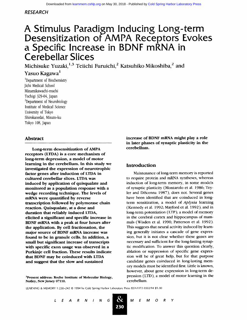
The responses of Purkinje cells in cultured slices were recorded by a wedge recording tech- nique (Garthwaite et al. 1986; Ito and Karachot 1989) (Fig. 1A). Application of 10 ~LM AMPA for 2 min induced responses of "--0.2-0.6 mV, which could be evoked repeatedly with little sign of de- sensitization (data not shown). Application of 100 bLi quisqualate for 5 min as a conditioning stimu- lus caused the desensitization of succeeding AMPA responses (Fig. 1B, upper trace) for at least 4 hr, whereas application of 10 ~LM AMPA for 5 min did not (Fig. 1B lower trace). The desensitization of AMPA responses was not caused by deterioration of neurons in the slices because the responses to 3 m i L-aspartate before and after the conditioning stimuli were similar (Fig. 1B). These data are sum- marized in Figure 1C. In addition, lactate dehydro- genase (LDH) leakage in the medium 2 hr after stimulation, expressed as a percentage of that be- fore stimulation, was 108---8% ( n = 3 ) , 102+-7% ( n = 3 ) , and 111+-6% ( n - - 3 ) after a 5-min appli- cation of 10 ~ i AMPA, 100 ~ i quisqualate, and medium only, respectively, indicating that the de- sensitization of AMPA response induced by quis- qualate was not attributable to damaged neurons. Thus, LTDA of Purkinje cells could be induced in cultured slices.
Next, we investigated the stimulation condi- tions for induction of LTDA in cultured slices in detail. As shown in Figure 3A (below), application
& 233
L E A R N / N G M E M O R Y
Cold Spring Harbor Laboratory Press on May 30, 2018 - Published by learnmem.cshlp.orgDownloaded from
Yuzaki et aL
A
B AMPA Asp Quis AMPA Asp
AMPA Asp AMPA AMPA Asp
_ j - - _
_l 0.16 mV 1 min
C ~ 100
[ 60 "-=I AMPA
20
O 0 ~" quisqualate AMPA
Conditioning stimulus
Figure 1: Wedge recording and induction of long-term desensitization of AMPA responses in cultured cerebellar slices. (A) Diagram of the perfusion chamber. A cultured slice cut into a wedge shape was placed in a two-com- partment bath. Gray matter containing Purkinje cell so- mata was perfused with test solution and separated from white matter in the adjacent compartment by grease. The potential difference between the two compartments was monitored with Ag/AgCI electrodes embedded in 4% agar in saline. (B) Representative wedge recordings. One hour after conditioning with 100 I, LM quisqualate (Quis) for 5 min, the response to 10 t£M AMPA remained de- sensitized to -35% of the response 10 rain before con- ditioning, whereas the response to 3 mM L-aspartate (Asp) was not changed significantly (upper trace). The AMPA and Asp responses were not changed by condi- tioning with 10 IJ, M AMPA for 5 min (lower trace). (C) Summary of results on conditioning stimulus. The de- grees of desensitization at 1 hr after conditioning are expressed as percentages of the control response before conditioning stimulus. Conditioning with quisqualate (100 I~M), but not AMPA (10 DM), for 5 min significantly desensitized subsequent AMPA responses (open col- umns). Responses to aspartate (3 mM) remained un- changed (shaded columns) with both conditioning stim- uli. The numbers in parentheses show the number of slices. Bars with asterisks indicate significant change: (*) P<O.O5; and (**) P<O.01, compared with the AMPA response after conditioning with AMPA (10 p,M) for 5 min.
& 234
L E A R N / N G
of 100 ~LM quisqualate for 3 or 4 min did not in- duce LTDA, whereas conditioning with quis- qualate for 5 min induced LTDA consistently. Con- ditioning with quisqualate for 6 min also induced LTDA consistently, but application of 10 ~M AMPA for 6 min as a conditioning stimulus also caused sizable LTDA. This suggests that prolonged appli- cation of AMPA can substitute for quisqualate, pos- sibly because a large influx of Ca 2 + can activate phospholipase C without activation of mGluR (Eberhard and Holz 1988). As shown in Figure 3B, quisqualate at 10 and at 30 ~LM did not induce LTDA reliably, whereas at 100 ~LM it consistently induced LTDA. Thus, the application duration and the dose of quisqualate for the induction of LTDA were in good agreement with those on freshly iso- lated cerebellar slices (Ito and Karachot 1989).
In the following study where electrophysio- logical recording was not routinely conducted, we thus applied 100 ~LM quisqualate for 5 min as a specific stimulus for induction of LTDA, and 10 ttM AMPA or medium alone for 5 min as controls. This condition reliably induced LTDA that could be fol- lowed at least up to 4 hr without signs of recovery.
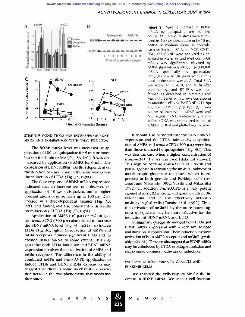
QUISQUALATE SPECIFICALLY INCREASED BDNF MRNA
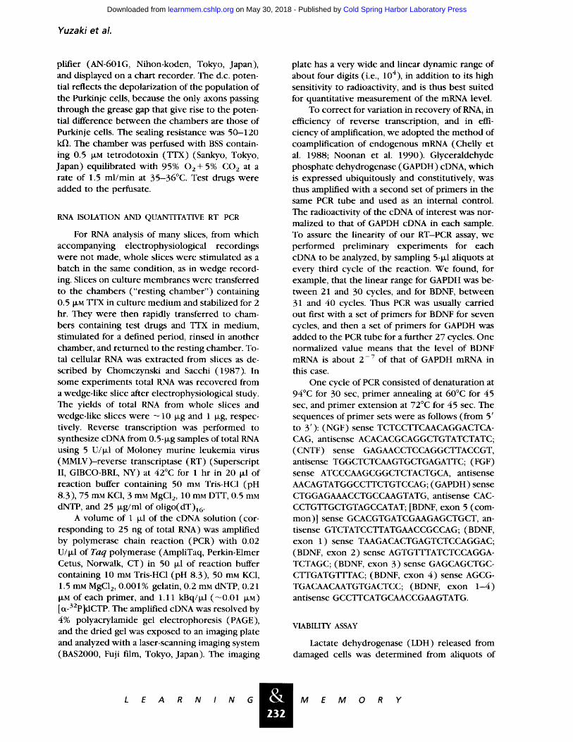
As shown in Figure 2A, quisqualate, applied at 100 ~LM for 5 min increased the expression of BDNF significantly. Stimulation with 10 ~LM AMPA did not increase the BDNF mRNA but it caused a significant and larger increase in nerve growth fac- tor (NGF) mRNA than quisqualate did. This sug- gests that quisqualate-induced NGF expression is mediated by AMPA receptor activation by quis- qualate. The other neurotrophic factors studied, ciliary neurotrophic factor (CNTF) and basic fi- broblast growth factor (FGF), did not show any increase on AMPA or quisqualate treatment. Thus the BDNF mRNA was increased specifically by the quisqualate stimulus.
Next, we analyzed the time course of expres- sion of BDNF mRNA. As shown in Figure 2, B and C, BDNF mRNA increased significantly 4 hr after application of quisqualate and remained at a high level even 16 hr after the stimulus. NGF mRNA also followed a similar time course of expression with a peak at 4 hr after stimulation (Fig. 2C, right), but the other neurotrophic factors tested remained relatively constant during the observed period (data not shown). The level of GAPDH mRNA expression, the internal control of mRNA quantification, remained almost constant (Fig. 2B).
M E M 0 R Y
Cold Spring Harbor Laboratory Press on May 30, 2018 - Published by learnmem.cshlp.orgDownloaded from
ACTIVITY-DEPENDENT CHANGE IN CEREBELLAR BDNF mRNA
A p<o.o5 p<0.01 B
i~ Quisqualate AMPA
~ n ~ P ~
0 1 4 8 16 1 4 8 16
Time after stimulus (hours)
Z . _ _ u NGF CNTF F G F 8 D N F
C 2.0
.ca "7. e~ 1.6
O .,..~
1.2
~q
~'~ 0.8
Z 0.4
BDNF 2.0
squalate 1.2 1.6
0.8
6 " 3 " lb " 1~ " 0.4
NGF
A
alate
Control 0 5 10 15
Time after stimulus (hours)
Figure 2: Specific increase in BDNF mRNA by quisqualate and its time course. (A) Cerebellar slices were stimu- lated by 100 t.LM quisqualate or by 10 IJ.M AMPA or medium alone as controls, each for 5 min. mRNAs for NGF, CNTF, FGF, and BDNF were analyzed as de- scribed in Materials and Methods. NGF mRNA was significantly elevated by AMPA stimulation (P<0.05), and BDNF mRNA specifically by quisqualate (P<0.01) (n=3). (B) Slices were stimu- lated in the same way as A. Total RNA was extracted 1, 4, 8, and 16 hr after conditioning, and RT-PCR was per- formed as described in Materials and Methods. Bands with arrows correspond to amplified cDNAs for BDNF (511 bp) and for GAPDH (228 bp). (C) Time course of increase in BDNF (/eft) and NGF (right) mRNA. Radioactivity of am- plified cDNA was normalized to that of GAPDH cDNA and plotted against time.
STIMULUS CONDITIONS FOR INCREASES OF BDNF MRNA AND COMPARISON WITH THAT FOR LTDA
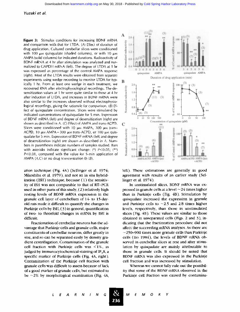
The BDNF mRNA level was increased by ap- pl icat ion of 1 O0 I~i quisqualate for 5 min or more, but not for 4 min or less (Fig. 3A, left). It was also increased by appl icat ion of AMPA for 6 min. The express ion of BDNF mRNA was thus dependen t on the durat ion of s t imulat ion in the same way as was the induc t ion of LTDA (Fig. 3A, right).
The dose response of BDNF mRNA express ion indicated that an increase was not observed on applicat ion of 10 I~M quisqualate, but at h igher concent ra t ions of quisqualate up to 100 I~M, it in- creased in a dose-dependent manner (Fig. 3B, left). This f inding was also consis tent wi th results on induc t ion of LTDA (Fig. 3B, right).
Applicat ion of AMPA (10 I~i) or mGluR ago- nist trans-ACPD (300 I~i) alone failed to increase the BDNF mRNA level (Fig. 3C, left) or to induce LTDA (Fig. 3C, right). Coactivation of AMPA and mGlu receptors induced significant LTDA and in- creased BDNF mRNA to some extent. This sug- gests that both LTDA induct ion and BDNF mRNA express ion involves the coact ivat ion of AMPA and mGlu receptors. The difference in the ability of c o m b i n e d AMPA and trans-ACPD applicat ion to induce LTDA and BDNF mRNA express ion may suggest that there is some mechanis t ic dissocia- t ion b e t w e e n the two phenomena , that needs fur- ther study.
It should also be noted that the BDNF mRNA express ion and the LTDA induced by coapplica- t ion of AMPA and trans-ACPD (300 p~M)were less than those induced by quisqualate (Fig. 3C). This was also the case w h e n a h igher concen t ra t ion of trans-ACPD (1 mM) was used (data not shown). This may be because trans-ACPD is a weak and partial agonist in activating the mGluR1 subtype of metabot ropic glutamate receptors, w h i c h is ex- pressed in both granule and Purkinje cells (Ar- amori and Nakanishi 1992; Yuzaki and Mikoshiba 1992). In addition, trans-ACPD is a very po ten t agonist of mGluR2 in Golgi and granule cells in the cerebel lum, and it also effectively activates mGluR3 in glial cells (Tanabe et al. 1992). Thus, the activation of mGluR1 by the more po ten t ag- onist quisqualate may be more effective for the induct ion of BDNF mRNA and LTDA.
In summary, quisqualate induced both LTDA and BDNF mRNA expression with a very similar dose and duration of application. Their induct ion involves activation of both AMPA receptor and mGluR (prob- ably mGluR1). These results suggest that BDNF mRNA may be coinduced by LTDA-evoking stimulation and shares some c o m m o n pathways of induction.
INCREASE IN BDNF MRNA IN GRANULE AND PURKINJE CELLS
We analyzed the cells responsible for the in- crease in BDNF mRNA. We used a cell fraction-
& 235
L E A R N I N G M E M O R Y
Cold Spring Harbor Laboratory Press on May 30, 2018 - Published by learnmem.cshlp.orgDownloaded from
Yuzak i e t al.
Figure 3: Stimulus conditions for increasing BDNF mRNA and comparison with that for LTDA. (A) Effect of duration of drug application. Cultured cerebellar slices were conditioned with 100 IJ, M quisqualate (shaded columns), or with 10 DM AMPA (solid columns) for indicated durations. Radioactivity of BDNF mRNA at 4 hr after stimulation was analyzed and nor- malized to GAPDH mRNA (/eft). The degree of LTDA at 1 hr was expressed as percentage of the control AMPA response (right). Most of the LTDA results were obtained from separate experiments using wedge recording to monitor LTDA for typ- ically 1 hr. From at least one wedge in each treatment, we recovered RNA after electrophysiological recordings. The de- sensitization values at 1 hr were quite similar to those at 4 hr after induction of LTDA, and increases in BDNF mRNA were also similar to the increases observed without electrophysio- logical recordings, giving the rationale for comparison. (B) Ef- fect of quisqualate concentration. Slices were stimulated by indicated concentrations of quisqualate for 5 min. Expression of BDNF mRNA (/eft) and degree of desensitization (right) are shown as described in A. (C) Effect of AMPA and trans-ACPD. Slices were conditioned with 10 I,Jt, M AMPA, 300 ~M trans- ACPD, 10 ]J,M AMPA+300 ILLM trans-ACPD, or 100 t,.LM quis- qualate for 5 min. Expression of BDNF mRNA (/eft) and degree of desensitization (right) are shown as described in A. Num- bers in parentheses indicate numbers of samples studied. Bars with asterisks indicate significant change: (*) P<0.05; (**) P<0.01, compared with the value for 5-min application of AMPA (A,C) or no drug (concentration 0) (B).
i3i ¿llt~, 13)
A ** 2.0 ** ~ I00
o
O.8
B 2.0
..g ,~ 1.6
1.2
~ 0.8
Z 0.4
0 3 4 5 6 3 4 5 6 ~ 3 4 5 6 5 6 quisqualate AMPA quisqualate AMPA
Duration of drug application (min)
ll* ~ 100'
< e~
(3) ~ 80
) (3) w
~ 60 ; 10 3'0 100 3'00
~. i ~ ~ 3) r (7)
0 10 3'0 1~10 Concentration of quisqualate (gM)
C ilt 2.0- (11~
°
.~MPAACPD ,~MPAQuis ~ A~PAACPD .~MPA~i~ +ACPD +Ai~I)
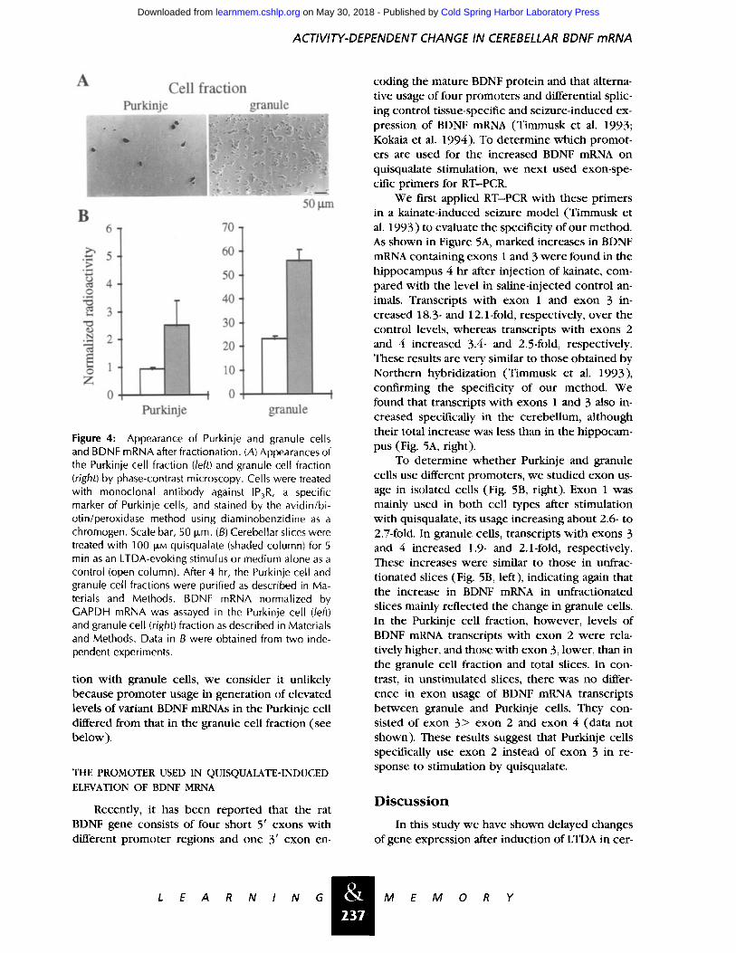
ation technique (Fig. 4A) (Sellinger et al. 1974; Mikoshiba et al. 1979), and not an in situ hybrid- ization (ISH) technique, because (1) the sensitiv- ity of ISH was not comparable to that of RT-PCR used in other parts of this study, (2) relatively high resting levels of BDNF mRNA expression in the granule cell layer of cerebellum of 14- to 15-day- old rats made it difficult to quantify the changes in Purkinje cells by ISH, (3) in general, quantification of two- to threefold changes in mRNA by ISH is difficult.
Fractionation of cerebellar neurons has the ad- vantage that Purkinje cells and granule cells, major constituents of cerebellar neurons, differ greatly in size, and so can be separated easily by density gra- dient centrifugation. Contamination of the granule cell fraction with Purkinje cells was <1%, as judged by immunocytochemical staining of IP3R, a specific marker of Purkinje cells (Fig. 4A, right). Contamination of the Purkinje cell fraction with granule cells was difficult to assess because of lack of a good marker of granule cells, but estimated to be - 2 % by morphological examination (Fig. 4A,
left). These estimations are generally in good agreement with results of an earlier study (Sel- linger et al. 1974).
In unstimulated slices, BDNF mRNA was ex- pressed in granule cells at a level --24 times higher than in Purkinje cells (Fig. 4B). Stimulation by quisqualate increased the expression in granule and Purkinje cells to - 2 . 5 and 2.8 times higher levels, respectively, than those in unstimulated slices (Fig. 4B). These values are similar to those obtained in unseparated cells (Figs. 2 and 3), in- dicating that the fractionation procedure did not affect the succeeding mRNA analyses. As there are - -250-900 times more granule cells than Purkinje cells (Ito 1984), the levels of BDNF mRNA ob- served in cerebellar slices at rest and after stimu- lation by quisqualate are mainly attributable to those in granule cells. It should be noted that BDNF mRNA was also expressed in the Purkinje cell fraction and was increased by stimulation.
Whereas we cannot fully rule out the possibil- ity that some of the BDNF mRNA observed in the Purkinje cell fraction was caused by contamina-
& 2 3 6
L E A R N / N G M E M O R Y
Cold Spring Harbor Laboratory Press on May 30, 2018 - Published by learnmem.cshlp.orgDownloaded from
ACTIVITY-DEPENDENT CHANGE IN CEREBELLAR BDNF mRNA
A Cell fraction
Purkinje granule
B 50 vu'n
6 "t 70
5
4 0 • ,e-,i
~D
• ~ 2
o 2:
60
50
40
30
20
10
0 0 .¢......l........J~_~ Purldnje granule
Figure 4: Appearance of Purkinje and granule cells and BDNF mRNA after fractionation. (A) Appearances of the Purkinje cell fraction (left) and granule cell fraction (right) by phase-contrast microscopy. Cells were treated with monoclonal antibody against IP3R, a specific marker of Purkinje cells, and stained by the avidin/bi- otin/peroxidase method using diaminobenzidine as a chromogen. Scale bar, 50 Dm. (B) Cerebellar slices were treated with 100 I£M quisqualate (shaded column) for 5 min as an LTDA-evoking stimulus or medium alone as a control (open column). After 4 hr, the Purkinje cell and granule cell fractions were purified as described in Ma- terials and Methods. BDNF mRNA normalized by GAPDH mRNA was assayed in the Purkinje cell (left) and granule cell (right) fraction as described in Materials and Methods. Data in B were obtained from two inde- pendent experiments.
tion with granule cells, we consider it unlikely because promoter usage in generation of elevated levels of variant BDNF mRNAs in the Purkinje cell differed from that in the granule cell fraction (see below).
THE PROMOTER USED IN QUISQUALATE-INDUCED ELEVATION OF BDNF MRNA
Recently, it has been reported that the rat BDNF gene consists of four short 5' exons with different promoter regions and one 3' exon en-
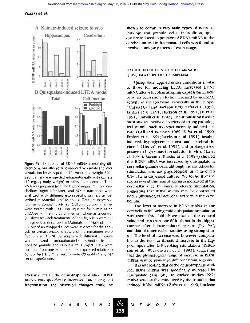
coding the mature BDNF protein and that alterna- tive usage of four promoters and differential splic- ing control tissue-specific and seizure-induced ex- pression of BDNF mRNA (Timmusk et al. 1993; Kokaia et al. 1994). To determine which promot- ers are used for the increased BDNF mRNA on quisqualate stimulation, we next used exon-spe- cific primers for RT-PCR.
We first applied RT-PCR with these primers in a kainate-induced seizure model (Timmusk et al. 1993) to evaluate the specificity of our method. As shown in Figure 5A, marked increases in BDNF mRNA containing exons 1 and 3 were found in the hippocampus 4 hr after injection of kainate, com- pared with the level in saline-injected control an- imals. Transcripts with exon 1 and exon 3 in- creased 18.3- and 12.1-fold, respectively, over the control levels, whereas transcripts with exons 2 and 4 increased 3.4- and 2.5-fold, respectively. These results are very similar to those obtained by Northern hybridization (Timmusk et al. 1993), confirming the specificity of our method. We found that transcripts with exons 1 and 3 also in- creased specifically in the cerebellum, although their total increase was less than in the hippocam- pus (Fig. 5A, right).
To determine whether Purkinje and granule cells use different promoters, we studied exon us- age in isolated cells (Fig. 5B, right). Exon 1 was mainly used in both cell types after stimulation with quisqualate, its usage increasing about 2.6- to 2.7-fold. In granule cells, transcripts with exons 3 and 4 increased 1.9- and 2.1-fold, respectively. These increases were similar to those in unfrac- tionated slices (Fig. 5B, left), indicating again that the increase in BDNF mRNA in unfractionated slices mainly reflected the change in granule cells. In the Purkinje cell fraction, however, levels of BDNF mRNA transcripts with exon 2 were rela- tively higher, and those with exon 3, lower, than in the granule cell fraction and total slices. In con- trast, in unstimulated slices, there was no differ- ence in exon usage of BDNF mRNA transcripts between granule and Purkinje cells. They con- sisted of exon 3> exon 2 and exon 4 (data not shown). These results suggest that Purkinje cells specifically use exon 2 instead of exon 3 in re- sponse to stimulation by quisqualate.
D i s c u s s i o n
In this study we have shown delayed changes of gene expression after induction of LTDA in cer-
L E A R N / N G M E M O R Y
Cold Spring Harbor Laboratory Press on May 30, 2018 - Published by learnmem.cshlp.orgDownloaded from
Yuzak i e t al.
A Kainate-induced seizure in vivo
Hippocampus Cerebellum
i
g
B Quisqualate-induced Total
g
LTDA model Cell fraction
I i .o 3.0[ I" granule[ 5 2.5 2.5
2.0 2.0
1.5
1.5 1.0
1.0
g g
Figure 5" Expression of BDNF mRNA containing dif- ferent 5' exons after seizure induced by kainate and after stimulation by quisqualate. (A) Adult rats (weight 210- 220 grams) were injected intraperitoneally with kainate (12 mg/kg body weight) or saline as a control. Total RNA was prepared from the hippocampus (left) and cer- ebellum (right) 4 hr later, and BDNF transcripts were analyzed with different exon-specific primers as de- scribed in Materials and Methods. Data are expressed relative to control levels. (B) Cultured cerebellar slices were treated with 100 ixMquisqualate for 5 min as an LTDA-evoking stimulus or medium alone as a control (45 slices for each treatment). After 4 hr, slices were cut into pieces as described in Materials and Methods, and ~1 out of 45 chopped slices were reserved for the anal- ysis of unfractionated slices, and the remainder were fractionated. BDNF transcripts with different 5' exons were analyzed in unfractionated slices (/eft) or in frac- tionated granule and Purkinje cells (right). Data were obtained from one experiment and expressed relative to control levels. Similar results were obtained in another set of experiments.
ebellar slices. Of the neurotrophins studied, BDNF mRNA was specifically increased, and using cell fractionation, the observed changes could be
shown to occur in two main types of neurons, Purkinje and granule cells. In addition, quis- qualate-induced expression of BDNF mRNA in the cerebellum and in fractionated cells was found to involve a unique pattern of exon usage.
SPECIFIC INDUCTION OF BDNF MRNA BY QUISQUALATE IN THE CEREBELLUM
Quisqualate, applied under conditions similar to those for inducing LTDA, increased BDNF mRNA after 4 hr. Neurotrophin expression in neu- rons has been shown to be increased by neuronal activity in the forebrain, especially in the hippo- campus (Gall and Isackson 1989; Zafra et al. 1990; Ernfors et al. 1991; Isackson et al. 1991; Lu et al. 1991; Lindvall et al. 1992). The stimulation used in most studies involved a variety of strong patholog- ical stimuli, such as experimentally induced sei- zure (Gall and Isackson 1989; Zafra et al. 1990; Ernfors et al. 1991; Isackson et al. 1991), insulin- induced hypoglycemic coma and cerebral is- chemia (Lindvall et al. 1992), and prolonged ex- posure to high potassium solution in vitro (Lu et al. 1991). Recently, Bessho et al. (1993) showed that BDNF mRNA was increased by quisqualate in cerebellar granule cells, although the condition for stimulation was not physiological, as it involved 0.5-4 hr in dispersed culture. We found that the expression of this neurotrophin is increased in the cerebellar slice by more moderate stimulation, suggesting that BDNF mRNA may be controlled under physiological neuronal activity in the cere- bellum.
The level of increase in BDNF mRNA in the cerebellum following mild quisqualate stimulation was about threefold above that of the control value and less than one-fifth of that in the hippo- campus after kainate-induced seizure (Fig. 5A), and that of other earlier studies using strong stim- uli. The level of increase was, however, compara- ble to the two- to threefold increase in the hip- pocampus after LTP-evoking stimulation (Patter- son et al. 1992; Castr6n et al. 1993), suggesting that the physiological range of increase in BDNF mRNA may be similar in different brain regions.
It is interesting that of the neurotrophins stud- ied, BDNF mRNA was specifically increased by quisqualate (Fig. 3B). In earlier studies, NGF mRNA was usually coinduced by the stimulus that induced BDNF mRNA (Zafra et al. 1990; Isackson
& 238
L E A R N I N G M E M O R Y
Cold Spring Harbor Laboratory Press on May 30, 2018 - Published by learnmem.cshlp.orgDownloaded from
ACTIVITY-DEPENDENT CHANGE IN CEREBELLAR BDNF mRNA
et al. 1991; Lindvall et al. 1992; Zafra et al. 1992). One explanation for the specific induction of BDNF mRNA that was observed in the cerebellum is that it results from the milder stimulus used, consistent with specific increases in BDNF mRNA in the visual cortex following illumination (Cas- tr~n et al. 1992) and in the hippocampal CA1 area following LTP (Patterson et al. 1992). If this were the case, then NGF mRNA would be expected to be coinduced with BDNF mRNA by a stimulus of higher intensity. However, AMPA, which caused more depolarization than quisqualate (Fig. 1B) and increased NGF mRNA, failed to increase BDNF mRNA (Fig. 2A). This suggests the alternative ex- planation that the specific increase of BDNF mRNA may mainly be attributable to the activation of mGluR by quisqualate.
EXON USAGE OF BDNF MRNA IN THE CEREBELLUM AND CELLULAR LOCALIZATION
Recently, alternative usage of four exons of the rat BDNF gene has been reported to control tissue-specific and stimulus-specific regulation of BDNF mRNA expression (Timmusk et al. 1993; Kokaia et al. 1994). However, these reports con- cern the forebrain, and not the cerebellum. We have showed here that kainate also increases the expression of BDNF mRNA in the cerebellum, with very similar usage of exons as in the hippo- campus (Fig. 5A).
Basal and activity-dependent expression of BDNF has been found mainly in granule cells. We consider that at least part of the BDNF mRNA ob- served in the Purkinje cell fraction reflected the low level of expression of BDNF mRNA in those cells, because the exon usage was different from that in the granule cell fraction. Studies using ISH have shown that BDNF mRNA was not localized to Purkinje cells, but to granule cells in the cerebel- lum (Hofer et al. 1990; Rocamora et al. 1993). The discrepancy between our results and those of ear- lier studies can be explained by the higher sensi- tivity of RT-PCR over ISH. It should also be noted that granule cells are packed more densely than Purkinje cells, as there are ~250-900 times more granule cells than Purkinje cells (Ito 1984), and thus grain density in granule cell layers is some- times exaggerated in ISH. Further studies using RT-PCR with single Purkinje cells will be needed to clarify the discrepancy.
FUNCTIONAL SIGNIFICANCE OF INCREASED BDNF MRNA AND RELATIONSHIP WITH LTD
BDNF was initially identified as a t rophic fac- tor for peripheral sensory neurons (Barde et al. 1982) and recently has been shown to promote the survival and/or differentiation of mesenceph- alic dopaminergic neurons and basal forebrain cholinergic neurons (Alderson et al. 1990; Hyman et al. 1991; Kniisel et al. 1991). BDNF has also been shown to regulate the expression of neu- ropeptides in GABA-ergic neurons (Nawa et al. 1993). During development of the cerebellum, ex- pression of BDNF mRNA is mainly localized in the internal granule cell layer and increases dynami- cally with a peak around postnatal day 20 (Ro- camora et al. 1993), at which time the synapse formation between parallel fibers of granule cells and Purkinje cells is prominent (Altman and Win- free 1977). In contrast, transcripts of the func- tional BDNF receptor gene, trkB, are expressed predominantly in Purkinje cells (Klein et al. 1990). Although the effect of BDNF on Purkinje cells remains to be determined, these observations suggest that BDNF expressed in granule cells may have a trophic action on Purkinje cells such as maintenance or regulation of synapses between granule cells and Purkinje cells. The BDNF mRNA observed in the Purkinje cell fraction also raises the possibility of autocrine actions of BDNF in these neurons (see also Kokaia et al. 1993; Mi- randa et al. 1993).
It is not clear whether BDNF mRNA elevation and LTDA are causally related or whether BDNF is involved in the process of LTDA at all. LTD and LTDA are attributable to the reduced AMPA recep- tor sensitivity in Purkinje cells (Ito and Karachot 1989; Linden et al. 1991). The desensitization of AMPA receptors in Purkinje cells, probably by phosphorylation, is a phenomenon observed within a few minutes of conditioning stimulus that persists for many hours. But information storage, based solely on post-translational modification of proteins such as phosphorylation, is generally con- sidered to deteriorate rapidly (Dudai 1989). Thus, although the time course of BDNF mRNA expres- sion is too slow to account for initial induction, it is possible that BDNF may have some role in later phases of synaptic plasticity, because if LTDA is a basic mechanism of motor learning, it should eventually be transformed into a permanent mem- ory. The efficacy of synaptic transmission can be affected by the morphological changes in the syn-
& 239
L E A R N / N G M E M O R Y
Cold Spring Harbor Laboratory Press on May 30, 2018 - Published by learnmem.cshlp.orgDownloaded from
Yuzaki et aL
aptic spine or the formation of new synapses, as has been suggested in the hippocampal LTP (Dudai 1989). For example, if the electrical resis- tance of the synaptic spine neck is decreased or the new synapses are formed on more distal den- drites, the voltage change at the soma induced by the synaptic input may be depressed. The number of synapses per Purkinje cell is reported to in- crease after motor learning but not after simple exercise (Black et al. 1990; Isaaacs et al. 1992). Consequently, BDNF mRNA expression in granule and Purkinje cells might play a role in such mor- phological manifestations. Further studies, for ex- ample, involving functional ablation of BDNF, will be useful in clarifying the role of this neurotrophin in cerebellar learning.
Acknowledgments We thank Dr. H. Miyagawa for technical advice in
preparing cerebellar slices, Drs. L. Karachot and M. Ito for generously showing us their experimental setup, and Drs. M. Kano, J.A. Connor, J.J. Petrozzino, B. Lu, and D. Forrest for critical reading of the manuscript. This work was supported by a research grant from the Human Frontier Science Program (to T.F.), a grant from the Brain Science Foundation (to M.Y.), and a grant for the Promotion of Sciences for Japanese Junior Scientists from the Japan Ministry of Education, Science and Culture (to M.Y.)
The publication costs of this article were defrayed in part by payment of page charges. This article must therefore be hereby marked "advertisement" in accordance with 18 USC section 1734 solely to indicate this fact.
References Alderson, R.F., A.L. Alterman, Y.A. Barde, and R.M. Lindsay. 1990. Brain-derived neurotrophic factor increases survival and differentiated functions of rat septal cholinergic neurons in culture. Neuron 5: 297-306.
Altman, J. and A.T. Winfree. 1977. Postnatal development of the cerebellar cortex in the rat. V. Spatial organization of Purkinje cell perikarya. J. Comp. Neurol. 171: 1-16.
Aramori, I. and S. Nakanishi. 1992. Signal transduction and pharmacological characteristics of a metabotropic glutamate receptor, mGluR1, in transfected CHO cells. Neuron 8" 757-765.
Barde, Y.A., D. Edgar, and H. Thoenen. 1982. Purification of a new neurotrophic factor from mammalian brain. EMBO J. 1: 549-553.
Bessho, Y., S. Nakanishi, and H. Nawa. 1993. Glutamate-receptor agonists enhance expression of BDNF mRNA in cultured cerebellar granule cells. Mol. Brain. Res. 18: 201-208.
Black, J.E., K.R. Isaacs, B.J., Anderson, A.A. AIcantara, and W.T. Greenough. 1990. Learning causes synaptogenesis, whereas motor activity causes angiogenesis, in cerebellar cortex of rats. Proc. Natl. Acad. Sci. 87: 5568-5572.
Castr~n, E., F. Zafra, H. Thoenen, and D. Lindholm. 1992. Light regulates expression of brain-derived neurotrophic factor mRNA in rat visual cortex. Proc. Natl. Acad. Sci. 89: 9444-9448.
Castr~n, E., M. Pitk~nen, J. Sirvi6, A. Parsadanian, D. Lindholm, H. Thoenen, and P.J. Riekkinen. 1993. The induction of LTP increases BDNF and NGF mRNA but decreases NT-3 mRNA in the dentate gyrus. NeuroReport 4: 895-898.
Chelly, J., J.C. Kaplan, P. Maire, S. Gautron, and A. Kahn. 1988. Transcription of the dystrophin gene in human muscle and non-muscle tissues. Nature 330: 858-860.
Chomczynski, P. and N. Sacchi. 1987. Single-step method of RNA isolation by acid-guanidinium thiocyanate-phenol- chloroform extraction. Anal. Biochem. 162: 156-159.
Diamond, J., M. Coughlin, L. Macintyre, M. Holmes, and B. Visheau. 1987. Evidence that endogenous nerve growth factor is responsible for the collateral sprouting, but not the regeneration, of nociceptive axons in adult rats. Proc. Natl. Acad. Sci. 84" 6596-6600.
Dudai, Y. 1989. The neurobiology of memory. Oxford University Press, New York.
Eberhard, D.A. and R.W. Holz. 1988. Intracellular Ca 2÷ activates phospholipase C. Trends Neurosci. 11" 51 7-520.
Ernfors, P., J. Bengzon, Z. Kokaia, H. Persson, and O. Lindvall. 1991. Increased levels of messenger RNAs for neurotrophic factors in the brain during kindling epileptogenesis. Neuron 7" 165-176.
Gall, C.M. and P.J. Isackson. 1989. Limbic seizures increase neuronal production of messenger RNA for nerve growth factor. Science 245." 758-761.
Garthwaite, J., G. Garthwaite, and F. Hojos. 1986. Amino acid neurotoxicity: Relationship to neuronal depolarization in rat cerebellar slices. Neuroscience 18" 449-460.
H~mart, N., H. Daniel, D. Jailard, and F. Cr~pel. 1994. Properties of glutamate receptors are modified during long-term depression in rat cerebellar Purkinje cells. Neurosci. Res. 19" 213-221.
Hofer, M., S.R. Pagliusi, A. Hohn, J. Leibrock, and Y.A. Barde. 1990. Regional distribution of brain-derived neurotrophic factor mRNA in the adult mouse brain. EtvlBO J. 9: 2459-2464.
Hyman, C., M. Hofer, Y.A. Barde, M. Juhasz, G.D. Yancopoulos, S.P. Squinto, and R.M. Lindsay. 1991. BDNF is a neurotrophic factor for dopaminergic neurons of the substantia nigra. Nature 350: 230-232.
& 2 4 0
L E A R N / N G M E M O R Y
Cold Spring Harbor Laboratory Press on May 30, 2018 - Published by learnmem.cshlp.orgDownloaded from

ACTIVITY-DEPENDENT CHANGE IN CEREBELLAR BDNF mRNA
Isaacs, K.R., B.J. Anderson, A.A. AIcantara, J.E. Black, and W.T. Greenough. 1992. Exercise and the brain: Angiogenesis in the adult rat cerebellum after vigorous physical activity and motor skill learning. J. Cereb. Blood Flow Metab. 12: 110-119.
Isackson, P.J., M.H. Huntsman, K.D. Murray, and C.M. Gall. 1991. BDNF mRNA expression is increased in adult rat forebrain after limbic seizures: Temporal patterns of induction distinct from NGF. Neuron 6" 937-948•
Ito, M. 1984. The cerebellum and neural control. Raven Press, New York.
• 1989. Long-term depression• Annu. Rev. Neurosci. 12" 85-102.
Ito, M. and L. Karachot. 1989. Long-term desensitization of quisqualate-specific glutamate receptors in Purkinje cells investigated with wedge recording from rat cerebellar slices. Neurosci. Res. 7:168-171.
• 1990. Receptor subtypes involved in, and time course of, the long-term desensitization of glutamate receptors in cerebellar Purkinje cells• Neurosci. Res. 8: 303-307.
• 1992. Protein kinases and phosphatase inhibitors mediating long-term desensitization of glutamate receptors in cerebellar Purkinje cells. Neurosci. Res. 14: 27-38•
Kalman, D., B. Wong, A.E. Horvai, M.J. Cline, and P.H. O'Lague. 1990. Nerve growth factor acts through cAMP-dependent protein kinase to increase the number of sodium channels in PC12 cells. Neuron 4: 355-366.
Kennedy, T.E., D. Kuhl, A. Barzilai, J.D. Sweat, and E.R. Kandel. 1992. Long-term sensitization in Aplysia leads to an increase in calreticulin, a major presynaptic calcium-binding protein. Neuron 9:1013-1024.
Klein, R., D. Martin-Zanca, M. Barbacid, and L.F. Parada. 1990. Expression of the tyrosine kinase receptor gene trkB is confined to the murine embryonic and adult nervous system. Development 109: 845-850.
Kn~isel, B., J.W. Winslow, A. Rosenthal, L.E. Burton, D.P. Seid, K. Nikolics, and F. Hefti. 1991. Promotion of central cholinergic and dopaminergic neuron differentiation by brain-derived neurotrophic factor but not neurotrophin-3. Proc. Natl. Acad. Sci. 88" 961-965.
Kokaia Z, J. Bengzon, M. Metsis, M. Kokaia, H. Persson, and O. Lindvall. 1993. Coexpression of neurotrophins and their receptors in neurons of the central nervous system• Proc. Natl. Acad. Sci. 90:6711-6715.
Kokaia, Z., M. Metsis, M. Kokaia, J. Bengzon, E. Elmer, M.L. Smith, T. Timmusk, B.K. Siesj6, H. Persson, and O. Lindval. 1994. Brain insults in rats induce increased expression of the BDNF gene through differential use of multiple promoters• Eur. J. Neurosci. 6: 587-596.
Linden, D.J. and J.A. Connor. 1991. Participation of postsynaptic PKC in cerebellar long-term depression in culture• Science 254:1656-1659.
Linden, D.J., M.H. Dickinson, M. Smeyne, and J.A. Connor. 1991. A long-term depression of AMPA currents in cultured cerebellar Purkinje neurons. Neuron 7" 81-89.
Lindvall, O., P. Ernfors, J. Bengzon, M.L. Smith, B.K. Siesj6, and H. Persson. 1992. Differential regulation of mRNAs for nerve growth factor, brain-derived neurotrophic factor, and neurotrophin 3 in the adult rat brain following cerebral ischemia and hypoglycemic coma. Proc. Natl. Acad. Sci. 89: 648-652•
Lu, B., M. Yokoyama, C. Dreyfus, and I.B. Black. 1991. Depolarizing stimuli regulate nerve growth factor gene expression in cultured hippocampal neurons. Proc. Natl. Acad. Sci. 88: 6289-6292•
Mayford, M., A. Barzilai, F. Keller, S. Schacher, and E.R. Kandel. 1992. Modulation of an NCAM-related adhesion molecule with long-term synaptic plasticity in Aplysia. Science 256: 638-644.
Mikoshiba, K., M. Huchet, and J.P. Changeux. 1979. Biochemical and immunological studies on the P4oo protein, a protein characteristic of the Purkinje cell from mouse and rat cerebellum• Dev. Neurosci. 2" 254-275•
Miranda R.C., F. Sohrabji, and C.D. Toran-Allerand. 1993. Neuronal colocalization of mRNAs for neurotrophins and their receptors in the developing central nervous system suggests a potential for autocrine interactions• Proc. Natl. Acad. Sci. 90: 6439-6443.
Montarolo, P.G., P. Goelet, V.F. Castellucci, J. Morgan, E.R. Kandel, and S. Schacher. 1986. A critical period for macromolecular synthesis in long-term heterosynaptic facilitation in Aplysia. Science 234: 1249-1254.
Nawa, H., Y. Bessho, J. Carnahan, S. Nakanishi, and K. Mizuno. 1993. Regulation of neuropeptide expression in cultured cerebral cortical neurons by brain-derived neurotrophic factor• J. Neurochem. 60" 772-775•
Noonan, K.E., C. Beck, T.A. Holzmayer, J.E. Chin, J.S. Wunder, I.L. Andrulis, A.F. Gazdar, C.L. Willman, B. Griffith, D.D. Von Hoff, and I.B. Roninson. 1990. Quantitative analysis of MDR1 (multidrug resistance) gene expression in human tumors by polymerase chain reaction. Proc. Natl. Acad. Sci. 87:7160-7164.
Otten, U., M. Goedert, N. Mayer, and F. Lembeck. 1980. Requirement of nerve growth factor for development of substance P-containing sensory neurons. Nature 287:158-159.
Patterson, S.L., L.M. Grover, P.A. Schwartzkroin, and M. Bothwell. 1992. Neurotrophin expression in rat hippocampal slices: a stimulus paradigm inducing LTP in CA1 evokes increases in BDNF and NT-3 mRNAs. Neuron 9" 1081-1088•
L E A R N I N G I M E M 0 R Y
Cold Spring Harbor Laboratory Press on May 30, 2018 - Published by learnmem.cshlp.orgDownloaded from
Yuzaki et al.
Rocamora, N., F.J. Garcfa-Ladona, J.M. Palacios, and G. Mengod. 1993. Differential expression of brain-derived neurotrophic factor, neurotrophin-3, and low-affinity nerve growth factor receptor during the postnatal development of the rat cerebellar system. Mol. Brain. Res. 17" 1-8.
Sellinger, O.Z., J. Legrand, J. Clos, and W.G. Ohlsson. 1974. Unequal patterns of development of succinate-dehydrogenase and acetylcholinesterase in Purkinje cell bodies and granule cells isolated in bulk from the cerebellar cortex of the immature rat. J. Neurochem. 23:1137-1144.
Stoppini, L., P.A. Buchs, and D. Muller. 1991. A simple method for organotypic cultures of nervous tissue. J. Neurosci. Methods 37:173-182.
Tanabe, Y., M. Masu, T. Ishii, R. Shigemoto, and S. Nakanishi. 1992. A family of metabotropic glutamate receptors. Neuron 8" 169-1 79.
Teyler, T.J. and P. DiScenna. 1987. Long-term potentiation. Annu. Rev. Neurosci. 10: 131-161.
Timmusk, T., K. Palm, M. Metsis, T. Reintam, V. Paalme, M. Saarma, and H. Persson. 1993. Multiple promoters direct tissue-specific expression of the rat BDNF gene. Neuron 10: 475-489.
Wisden, W., M.L. Errington, S. Williams, S.B. Dunnett, C. Waters, D. Hitchcock, G. Evan, T.V.P. Bliss, and S.P. Hunt. 1990. Differential expression of immediate early genes in the hippocampus and spinal cord. Neuron 4: 603-614.
Yuzaki, M. and K. Mikoshiba. 1992. Pharmacological and immunocytochemical characterization of metabotropic glutamate receptors in cultured Purkinje cells. J. Neurosci. 12" 4253-4263.
Zafra, F., B. Hengerer, J. Leibrock, H. Thoenen, and D. Lindholm. 1990. Activity dependent regulation of BDNF and NGF mRNAs in the rat hippocampus is mediated by non-NMDA glutamate receptors. EMBO J. 9" 3545-3550.
Zafra, F., D. Lindholm, E. Castr~n, J. Hartikka, and H. Thoenen. 1992. Regulation of brain-derived neurotrophic factor and nerve growth factor mRNA in primary cultures of hippocampal neurons and astrocytes. J. Neurosci. 12" 4793-4799.
Received August 15, 1994; accepted in revised form November 9, 1994.
& 242
L E A R N I N G M E M O R Y
Cold Spring Harbor Laboratory Press on May 30, 2018 - Published by learnmem.cshlp.orgDownloaded from
10.1101/lm.1.4.230Access the most recent version at doi: 1:1994, Learn. Mem.
M Yuzaki, T Furuichi, K Mikoshiba, et al. slices.receptors evokes a specific increase in BDNF mRNA in cerebellar A stimulus paradigm inducing long-term desensitization of AMPA
References
http://learnmem.cshlp.org/content/1/4/230.full.html#ref-list-1
This article cites 52 articles, 16 of which can be accessed free at:
License
ServiceEmail Alerting
click here.top right corner of the article or
Receive free email alerts when new articles cite this article - sign up in the box at the
Copyright © Cold Spring Harbor Laboratory Press
Cold Spring Harbor Laboratory Press on May 30, 2018 - Published by learnmem.cshlp.orgDownloaded from