Embed Size (px)
Citation preview

RSC Advances
PAPER
Ope
n A
cces
s A
rtic
le. P
ublis
hed
on 0
9 M
arch
202
1. D
ownl
oade
d on
3/1
4/20
22 1
1:53
:20
AM
. T
his
artic
le is
lice
nsed
und
er a
Cre
ativ
e C
omm
ons
Attr
ibut
ion-
Non
Com
mer
cial
3.0
Unp
orte
d L
icen
ce.
View Article OnlineView Journal | View Issue
A specific seleniu
aNational Engineering Research Center of JU
Forestry University, No. 15, Shangxiadian
350002, China. E-mail: liubin618@hotmailbCollege of Food Science, Fujian Agriculture
350002, ChinacInstitute of Emergency Medicine, Depart
Hospital, Fuzhou, Fujian 350001, China. E-
† Co-rst authors.
Cite this: RSC Adv., 2021, 11, 10272
Received 28th December 2020Accepted 26th February 2021
DOI: 10.1039/d0ra10886c
rsc.li/rsc-advances
10272 | RSC Adv., 2021, 11, 10272–10
m-chelating peptide isolated fromthe protein hydrolysate of Grifola frondosa
Yu Xiong,†a Zi-Hong Chen,†b Feng-Li Zhang,a Zhi-Ying Yu,a Bin Liu, *ab
Chong Zhang*c and Li-Na Zhao *a
Background: Grifola frondosa is a type of edible medicinal mushroom with abundant proteins. Selenium
(Se) is an essential micronutrient for human. Many animal experiments and clinical studies had indicated
that Se plays an important role in diverse physiologic actions. Most inorganic selenium compounds are
toxic, and the lowest lethal dose is relatively small. Peptide-Se chelate can probably be dietary
supplements in functional foods for humans with Se deficiency. Methods: In this study, a specific
tripeptide Arg-Leu-Ala (RLA) with strong Se-chelating capacity was purified from Grifola frondosa
through ultrafiltration, reversed-phase HPLC and gel filtration chromatography. The UV, SEM, XRD, 1H
NMR spectra are shown to provide more information about characterization of RLA-Se chelates. The
bioavailability of RLA-Se chelate in Caco-2 cell line was investigated by using human colon cancer
Caco-2 cells as model. iTRAQ comparative proteomics approach were used to identify the differentially
expressed proteins. Results: The Se binding capacity of RLA was 84.47 � 1.21 mg g�1. The results of UV,
X-ray diffraction (XRD), 1H NMR and SEM structure analysis showed that the binding of selenium in the
hydrolysate of Grifola frondosa protein was successful, and the amino and carboxyl groups of RLA were
involved in the coordination of Se, which was the main site of chelation. The results of absorption of
RLA-Se chelate in Caco-2 cells showed that RLA-Se chelate could be used as selenium supplement
source. Using iTRAQ comparative proteomics approach, 40 proteins found significant. RLA-Se treatment
had been demonstrated to present a higher accumulation of Se compared with control treatment and
show an effective absorption by Caco-2 with the result that E3 protein performed up regulation. RLA-Se
may play roles in cell cycle and apoptosis as an essential micronutrient. To sum up, our research results
show that Grifola polypeptide-Se chelate is a promising multifunctional organic selenium product, which
can be used as a new functional supplement for selenium deficiency.
1 Introduction
Selenium (Se) is an essential trace element necessary for thenormal development and maintenance of immune function inboth humans and animals.1 It has many physiological effects,such as anti-oxidant, anti-tumor, anti-bacterial, radioprotective,and immune-boosting properties.2–4 Se deciency is mainly dueto environmental pollution, poor living habits, and aging. Se isa trace element required by the human body and about onebillion people suffer from Se deciency worldwide.5 Se de-ciency and homeostasis are related to many diseases anddisorders, such as cardiovascular disease, infertility, gestational
NCAO Technology, Fujian Agriculture and
rd, Cangshan District, Fuzhou, Fujian
.com; [email protected]
and Forestry University, Fuzhou, Fujian
ment of Emergency, Fujian Provincial
mail: [email protected]
284
diabetes, and obstetric cholestasis.6–8 The development of arti-cial selenium supplements has become a hotspot in nutritionresearch. However, most inorganic Se compounds are toxic, andthe minimum lethal dose is relatively low.9,10 Therefore, organicSe compounds with high biological activity, low toxicity, and fewside effects are the desirable features of these supplements.Peptides with mineral-chelating abilities and other biologicallyactive functions have been isolated from the proteolysis prod-ucts of foods, such as proteins from chickpeas,11 scallops,12
shrimp,13 sugarcane yeast,14 and corn.15 These peptides arecapable of chelating minerals such as calcium,16–18 zinc,19 andiron and enhancing their bioavailability.20 Nevertheless, the Se-chelating peptides in various proteins remain mostlyunexplored.
Grifola frondosa (GF, Basidiomycetes, Aphyllophorales, Poly-poraceae) is an edible, medicinal mushroom, known for its highlevels of polysaccharides, steroids, phenols, and proteins. Theprotein content of Grifola frondosa is up to 35% per 100 g of thedry product, which is twice as much as that of shiitake mush-rooms.21 The biological components of Grifola frondosa have
© 2021 The Author(s). Published by the Royal Society of Chemistry

Paper RSC Advances
Ope
n A
cces
s A
rtic
le. P
ublis
hed
on 0
9 M
arch
202
1. D
ownl
oade
d on
3/1
4/20
22 1
1:53
:20
AM
. T
his
artic
le is
lice
nsed
und
er a
Cre
ativ
e C
omm
ons
Attr
ibut
ion-
Non
Com
mer
cial
3.0
Unp
orte
d L
icen
ce.
View Article Online
been shown to have antitumor,22 immunity-enhancing,23 anti-viral,24 and antioxidant activity.25,26 Several studies have shownthat the bioactive polypeptides obtained using proteolytichydrolysis have better solubility, can be easily digested andabsorbed, and have many unique physiological functions.27 Thewhey polypeptide–ferrous chelate28 and oyster–zinc chelate29
showed better ion solubility than their corresponding inorganicsalts in a simulated gastrointestinal digestion experiment.Using animal experiments, Chaud et al.30 proved that the caseinpeptide–ferrous chelate has an effect similar to that of ferroussulfate in the conversion rate of rat hemoglobin. By chelatingcovalent chemical bonds with metal ions, the cost of traceelements can be reduced, and the bioavailability of proteins andtrace elements can be improved. Most studies have focused onthe characterization of inorganic Se or the metabolic functionsrelated to Se. The purication of Se-binding peptides extractedfrom GF protein hydrolysate (GFPH) has not been extensivelyexplored. The rapid growth in various omics technologies overrecent years has provided avenues and opportunities forselenium-related bioinformatics research.31 Isobaric tags forrelative and absolute quantitation (iTRAQ) labeling technologyenables high-throughput screening of proteins, which isa useful approach in the interpretation of genomic informationthat facilitates the search for new therapeutic mechanisms.
In this study, a new chelating process was developed,wherein GFPH and Se were combined to form a new organicselenium supplement. The chelating mechanism was analyzedusing ultraviolet spectroscopy (UV), scanning electron micros-copy (SEM), X-ray diffraction (XRD), and 1H-nuclear magneticresonance (1H-NMR). At the cellular level, the absorptionproperties of the novel organic selenium supplements wereinvestigated, and the peptide fragments were identied usingmass spectrometry. Lastly, proteomics data showed that our Se-chelating peptide had specic physiological effects.
2 Material and methods2.1 Materials
GF was provided by Jingxiang Ecological Technology IndustryCo., Ltd. Alkaline protease and Sephadex G-25 were purchasedfrom Beijing Solarbio Technology Co., Ltd. All other chemicalsand solvents were of analytical grade.
2.2 Preparation of GFPH
The GF powder was sieved through a #40 mesh, dissolved indeionized water to yield a concentration of 3% (w/v), andhydrolyzed using alkaline protease. The dosage of the enzymewas 5.0% (w/w, dened as enzyme mass/substrate mass �100%) and the pH was adjusted to 10. The reaction was carriedout in a constant temperature oscillator at 50 �C for 1 h.32 Thesupernatant was centrifuged aer cooling and stored at 4 �Cuntil further analysis.
2.3 Purication of Se-binding peptides
2.3.1 Ultraltration. To isolate peptides with differentmolecular weights, the GFPH was ultraltered using 10 kDa and
© 2021 The Author(s). Published by the Royal Society of Chemistry
3 kDa Pellicon XL ultraltration diskmembranes (Millipore Co.,USA), and the ltrates were concentrated and lyophilized. TheSe-binding ability of each component was evaluated bymeasuring the selenium content. Components showing thestrongest chelating ability were collected for subsequent sepa-ration. Se levels were determined using 3,3-diaminobenzidinecolorimetry33 and the following equation:
Se�mg g�1
� ¼ pV
mN(1)
where p represents the standard mass concentration equivalentto Se determined using the standard curve (mg mL�1), V repre-sents the sample volume obtained using toluene extraction(mL),m represents the mass of sample (g), and N represents theratio of sample volume/total volume (%).
2.3.2 Gel ltration using Sephadex G-25 column. Thesample with the strongest Se-chelating ability obtained usingultraltration was weighed (100 mg), dissolved in 5 mL deion-ized water, ltered through a 0.2 mmmembrane, and applied toa pre-equilibrated Sephadex G-25 column (100 � 2.0 cm). Theperistaltic pump was set to a speed of 0.3 mLmin�1 and washedwith deionized water. The absorbance was measured using a UVspectrophotometer at 214 nm. Aer determining the chelatingability of selenium, the most active fractions were mixed andlyophilized.
2.3.3 Reversed-phase high-performance liquid chromatog-raphy (RP-HPLC) using a C18 column. In this study, a C18 semi-preparative RP-HPLC column (F 250 � 10 mm; PhenomenexInc., Torrance, CA, USA) was used. The lyophilized sample wasdissolved in deionized water to yield a concentration of 50 mgmL�1. The sample volume was 200 mL and the ow rate was setto 4.0 mL min�1. Deionized water containing 0.05% TFA(solvent A) and acetonitrile with 0.05% TFA (solvent B) was usedas the mobile phase. The detection wavelength was 214 nm.Aer determining the binding capacity of Se, the most activefractions were mixed, concentrated, and lyophilized.
2.3.4 Identication of peptides using liquidchromatography-electrospray ionization/mass spectrometry(LC-ESI/MS) and peptide synthesis. LC-ESI/MS (Delta Prep 4000,Waters Co., Milford, MA) was used to identify the molecularweight and amino acid sequence of the puried Se-chelatingpeptide in the range of 300–3000 m/z. The identied peptidewas synthesized using the solid-phase method by XinghaoMedicine Limited (Wuhan, China). The synthetic peptides werepuried using HPLC. A Gemini-NX 100A C18 column (F 250 �4.6 mm) was used and 10 mL of the sample was injected. Theow rate was set to 1.0 mL min�1 and the detection wavelengthwas 220 nm. The molecular weights were determined using MS.The samples were freeze-dried and stored at �20 �C.
2.4 Synthesis of GFPH-Se chelate
To the GFPH solution, 0.5 M sodium selenite solution wasadded in a volume ratio of sodium selenite : polypeptide 4 : 6.The pH was adjusted to 9.0 and the reaction was performed at50 �C for 1.5 h. Aer cooling, the sample was centrifuged at3970g for 10 min. To the resulting supernatant, ve volumes of95% ethanol were added and mixed well, and the chelate was
RSC Adv., 2021, 11, 10272–10284 | 10273

RSC Advances Paper
Ope
n A
cces
s A
rtic
le. P
ublis
hed
on 0
9 M
arch
202
1. D
ownl
oade
d on
3/1
4/20
22 1
1:53
:20
AM
. T
his
artic
le is
lice
nsed
und
er a
Cre
ativ
e C
omm
ons
Attr
ibut
ion-
Non
Com
mer
cial
3.0
Unp
orte
d L
icen
ce.
View Article Online
allowed to sediment for 12 h. Then, the sample was re-centrifuged at 5000 rpm for 10 min. The precipitated sedi-ment at the bottom was collected and washed several times withanhydrous ethanol. The Se content of the GFPH-Se chelate wasdetermined aer freeze-drying using eqn (1).
2.5 Characterization of the RLA-Se chelates
2.5.1 Ultraviolet (UV) spectroscopy. RLA, sodium seleniteand RLA-Se chelate samples were prepared at a concentration of0.2 mg mL�1, the pH was adjusted to 7.0, and their UVabsorption spectra were obtained at a wavelength of 200–800 nm using a Spectra Mr instrument (Persee, Beijing) aspreviously described.34 Each sample was analyzed in triplicate.
2.5.2 Scanning electron microscopy (SEM). Samples weresputter coated using gold and themorphologies of RLA and RLA-Sechelates were characterized using SEM (FEI, USA) under 1000� and5000� magnication using the high vacuum mode. Samples ofRLA and RLA-Se chelates were bonded to the sample table usinga conductive adhesive. Aer the gold-plated treatment, themorphologies of RLA and RLA-Se chelates were characterized usingSEM (FEI, USA) under 1000 and 5000 high vacuum conditions.
2.5.3 X-ray diffraction (XRD). The samples of RLA and RLA-Se chelates were analyzed using X-ray diffraction (Philips, Nether-lands) using a scanning speed of 4� min�1 ranging from 5–75�,with a stepwise increment of 1�, and an emission slit 0.3 mm.
2.5.4 1H-NMR spectroscopy. RLA and RLA-Se chelate(about 0.5 mg each) were dissolved in 500 mL Milli-Q water; 500mL tritium water was added and the sample was transferred toa 5 mm NMR (Bruker, Switzerland) test tube to obtain the 1H-NMR spectra at 600 MHz.
2.6 The effect of RLA-Se chelate on the cellular uptake of Se
2.6.1 Cell culture. The human colon adenocarcinoma cellline, Caco-2, was obtained from the Institute of Bioengineering,Fuzhou University, and passed for 20–40 generations. DMEMsupplemented with 20% fetal bovine serum and 1% double-antibody were used as cell culture media and the cells werecultured in an incubator at 37 �C in an atmosphere of 5%carbon dioxide. Cells were subcultured with 0.25% trypsin–EDTA at a ratio of 1 : 3.
2.6.2 Cytotoxicity test. The MTT assay with slight modi-cations35 was used for cytotoxicity determination. The MTTassay reects cell viability by detecting the activity of succinatedehydrogenase in the mitochondria of live cells. Caco-2 cellswere seeded in a 96-well-plate at a density of 1 � 104 cells percm�2 and incubated at 37 �C with 5% CO2 for 24 h, aer which,the medium was removed. DMEM (150 mL) containing 2, 4, 6, 8,and 10 mmol L�1 RLA-Se chelates and sodium selenite wereadded to each well of the treatment group. Aer incubation for24 h, the solution was aspirated and 20 mL PBS solution con-taining 5 mg mL�1 MTT was added. Aer 4 h of incubation,MTT was removed and 150 mL dimethyl sulfoxide (DMSO) wasadded to each well to dissolve the purple formazan crystals. TheOD of each well was measured using enzyme-linked immuno-sorbent assay (ELISA) at 560 nm. The results are expressed asa ratio to the control.
10274 | RSC Adv., 2021, 11, 10272–10284
2.6.3 Cellular uptake studies. Caco-2 cells were incubatedwith different concentrations of the RLA-Se chelate and sodiumselenite at 37 �C in a 5% CO2 environment for one hour. Next, thecells were washed with PBS three times. Then, the cells wereincubated with 1 mL of cell lysis buffer for 30 min on ice, trans-ferred into 1.5 mL Eppendorf tubes, centrifuged at 4 �C for 5 min,and the supernatant was collected. The total protein concentrationand Se content were determined using a published methoddescribed by Chen.36 The results are expressed in mg mg�1 protein.
2.7 Proteomics of Caco-2 cells detected using iTRAQ
2.7.1 Protein extraction, digestion, and iTRAQ labeling.The following protocol was used to extract the total protein from theCaco-2 cell samples (control) and RLA-Se chelate-injected cellsamples (treated). The cell samples were boiled in SDT buffer (8 MUrea, 100 mM Tris–HCl, 1 mM PMSF, 0.1 M DTT, pH 7.6) for 5 minand homogenized using FastPrep-24 (MP Biomedicals, 6M/S, 30 s,two cycles), treated using ultrasound (100 w, 30 s on, 30 s off, 10cycles) and centrifuged. Protein levels were quantied using theBradford method.37 Protein digestion was performed according tothe FASP procedure described by Wisniewski, Zougman et al.38 Theresulting peptidemixturewas then labeledwith 8-plex iTRAQ reagentfollowing the manufacturer's instructions (Applied Biosystems). Thesampleswere labeled as Treated (M,N, andO) andControl (P, Q, andR), and were subjected to multiple treatments and vacuum drying.
2.7.2 Two-dimensional LC-MS/MS analysis using Q exac-tive. The peptide mixture (1 mL) was added to the C18 reversed-phase column (Thermo Scientic Easy Column, 15 cm long, 75mm internal diameter, 3 mm resin) in buffer A (0.1% formic acidin water) and buffer B (0.1% formic acid in acetonitrile). Thelinear gradient was separated at a ow rate of 400 nL min�1.Mass spectrometry data uses the top 20 methods associatedwith the data to dynamically select the most abundantprecursor ions from the measurement scan (300–1500 m/z) ofHCD fragmentation. The determination of the target value wasbased on predictive Automatic Gain Control (pAGC). Theduration of the dynamic exclusion was 30 s. Survey scans wereacquired at a resolution of 70 000 at m/z 200, and resolution forHCD spectra was set to 17 500 at m/z 200. The normalizedcollision energy was 27 eV. The instrument was operated withthe peptide recognition mode enabled.
2.7.3 Bioinformatics analysis. The original data werecollected and processed using the international mainstreamproteomics analysis soware, Proteome Discoverer 2.1, anda human database downloaded from the UniProt website (9606sequences) was used. The differential proteins identied in thisstudy were screened using differential multiple >1.2 or #0.83and P-value #0.05. Using the bioinformatics analysis tool,David on-line analysis, GO (gene ontology) function annotation,location analysis, and function enrichment analysis werecarried out for the identied proteins.
2.8 Statistical analysis
All results are expressed as means� standard deviations (SD) ofthree experiments. Statistical analyses were performed usingStudent's t-test. A value of P < 0.05 was considered statistically
© 2021 The Author(s). Published by the Royal Society of Chemistry

Table 1 Se-Chelating ability of peptides with different molecularweights
Fraction Molecular sizeSe-Chelating ability(mg g�1)
F1 <3 kDa 55.61 � 4.16a
F2 3–10 kDa 21.99 � 1.93a
F3 >10 kDa 38.74 � 4.03a
GFPH Untreated 23.50 � 3.43a
a Signicant difference was evaluated by Duan’s t-test (p < 0.01), anddata with the same letter are not signicantly different (p > 005). Allthe samples were determined three times, and the results werereported as means � SD.
Table 2 Sequences of Se-chelating peptides identified from GFPH
No. MWa Sequence Fraction Abbreviation
1 219 Ser-Leu 13 SL2 233 Thr-Leu 17 TL3 231 Val-Leu 17 VL4 359 Arg-Leu-Ala 17 RLA5 263 Met-Leu 22 ML
a Molecular weight.
Paper RSC Advances
Ope
n A
cces
s A
rtic
le. P
ublis
hed
on 0
9 M
arch
202
1. D
ownl
oade
d on
3/1
4/20
22 1
1:53
:20
AM
. T
his
artic
le is
lice
nsed
und
er a
Cre
ativ
e C
omm
ons
Attr
ibut
ion-
Non
Com
mer
cial
3.0
Unp
orte
d L
icen
ce.
View Article Online
signicant, while a value of P < 0.01 suggested extremelystatistically signicant.
3 Results3.1 Purication of Se-chelating peptide
The Se-chelating peptides with a molecular weight lower than 3kDa were extracted from the enzymatic hydrolysate of the GFprotein using ultraltration. Three peptides with different molec-ular weights were identied aer ultraltration and their chelatingabilities are shown in Table 1. Sephadex G-25 gel ltration chro-matography was used to separate the component with the highestSe binding ability, which was named F1. Three components wereseparated, namely, F11, F12, and F13 (Fig. 1A). The chelatingcapacity of F12 was the highest (Fig. 1B). The eluent correspondingto the peak of F12 was collected and separated using semi-preparative C18 RP-HPLC. Fraction F12 was divided into 29different main fractions (Fig. 2A). The peptide was dissociatedusing a 0.05%TFA-acetonitrile system. The high-signal fractions (1,5, 13, 17, 22) were separated and lyophilized for LC-MS analysis.
3.2 Identication of peptides using LC-ESI/MS
For peptide identication, the high-signal fractions (1, 5, 13, 17,22) were subjected to LC-MSD Trap Mass Spectrometry. The ve
Fig. 1 Sephadex G-25 gel filtration chromatography from ultrafiltration
© 2021 The Author(s). Published by the Royal Society of Chemistry
peptides were sequence as Ser-Leu (SL), Thr-Leu (TL), Val-Leu(VL), Arg-Leu-Ala (RLA) and Met-Leu (ML), and its character-istic information is shown in Table 2. MS/MS spectra of RLAwith m/z is shown in Fig. 2B. These sequences matched wellwith those stored in the National Center for BiotechnologyInformation (NCBI) database. The results showed that the Se-chelating peptides in GF contained 2–3 residues. However, nopolypeptides were detected in fractions 1 and 5.
3.3 Se-Chelating ability of peptides
To ascertain whether the puried peptide had appreciable Se-binding ability, the Se-binding activities of the ve peptideswere determined (Fig. 2C). All peptides were found to bepotential Se-chelating peptides. The highest Se-chelating abilityof RLA was determined to be 84.47 � 1.21 mg g�1, which wassignicantly higher than that of TL (67.83 � 1.49 mg g�1) andML (67.96 � 3.35 mg g�1).
3.4 Structural characterization
3.4.1 UV spectroscopy. It can be seen from Fig. 3 that thecharacteristic absorption peak of the tripeptide, RLA, is near269 nm, which is caused by the chromogenic moieties such asthe carbonyl and carboxyl groups and the amide bond. Aerchelating with Se, the characteristic absorption peak shied to275 nm, resulting in the redshi phenomenon. It is speculatedthat during the process of chelation, n / p* energy level
and Se-chelating ability of the fractions from Sephadex G-25 column.
RSC Adv., 2021, 11, 10272–10284 | 10275

Fig. 2 Purification and identification of the Se-chelating peptide using semi-preparative C18 RP-HPLC and LC-ESI/MS. (A) Semi-preparative C18RP-HPLC of fraction F12. (B) The mass spectrum of the RLA. (C) Se-Chelating ability of synthetic peptides. (A)–(C) Significant difference wasevaluated by Duncan's multiple range test (p < 0.01), and data with the same letter are not significantly different (p > 0.05).
Fig. 3 UV spectrum of RLA, sodium selenite and RLA-Se chelates.
RSC Advances Paper
Ope
n A
cces
s A
rtic
le. P
ublis
hed
on 0
9 M
arch
202
1. D
ownl
oade
d on
3/1
4/20
22 1
1:53
:20
AM
. T
his
artic
le is
lice
nsed
und
er a
Cre
ativ
e C
omm
ons
Attr
ibut
ion-
Non
Com
mer
cial
3.0
Unp
orte
d L
icen
ce.
View Article Online
transition occurs in –NH2 or –OH owing to the convolution andfolding of the peptide chain, which increases the range of theconjugated system, thereby promoting the redshi. And there isno absorption peak in the range of 200–400 nm in sodiumselenite solution, which proves that the peptide reacts withselenium. At the same time, compared to the tripeptide RLA, theRLA-Se chelate also had a chromogenic effect at the maximumabsorption peak. When the molecular orbital of –CH3 on theside chain group folds with the p system, it has a certain
Fig. 4 The SEM images of RLA (left) and RLA-Se (right) chelates.
10276 | RSC Adv., 2021, 11, 10272–10284
chromogenic effect. A new characteristic absorption peak wasobserved with RLA-Se near 385 nm, which belongs to the R-bandabsorption peak, usually caused by the unsaturated group-containing lone electron pair in a p–p conjugate system suchas C]O. By comparing the UV spectra of RLA and RLA-Sechelates, it can be seen that the amino and carboxyl groups ofRLA are involved in the coordination of the Se ions and have anauxiliary effect on the side-chain –CH3 group, which is the mainsite of chelation.
3.4.2 SEM. Accurately weighed dried sample powder wasadhered to the sample table using conductive double-sidedadhesive. Aer blowing gently with ear ball, ion sputteringwas used to spray the gold coating, and the samples wereobserved and photographed using eld emission scanningelectron microscopy (Fig. 4). The particle size of RLA wasdetermined to be 5 mm and it showed a occulent structure.When the microstructure was observed at 10 000�, RLA showeda noticeable cluster bulge, which had a uniform and regulardistribution. The electron micrographs of RLA-Se chelatesreveal that the particle size of the chelate becomes smaller,about 2 mm, owing to the phenomenon of aggregation. Thechelate has a granular structure. Usingmicroscopy, it was foundthat some particles were attached to the granular clusterstructure, imparting it an irregular distribution.
3.4.3 XRD measurements. The diffraction pattern of thematerial was analyzed using X-ray diffraction, and the compo-sition and structure of the material were obtained. The X-raydiffraction patterns of RLA and RLA-Se chelates are shown in
© 2021 The Author(s). Published by the Royal Society of Chemistry

Fig. 5 XRD pattern of RLA and RLA-Se complexes.
Paper RSC Advances
Ope
n A
cces
s A
rtic
le. P
ublis
hed
on 0
9 M
arch
202
1. D
ownl
oade
d on
3/1
4/20
22 1
1:53
:20
AM
. T
his
artic
le is
lice
nsed
und
er a
Cre
ativ
e C
omm
ons
Attr
ibut
ion-
Non
Com
mer
cial
3.0
Unp
orte
d L
icen
ce.
View Article Online
Fig. 5. Compared to that of RLA, there were apparent differencesin the number, angle, and relative strength of the RLA-Sechelate, indicating the differences in the composition andstructure of the chelates. Aer chelating with selenium ions, thediffraction peaks of most RLA-Se chelates became wider and theparticles became smaller, which was consistent with theanalytical conclusions derived using SEM. The crystal structureof RLA changed upon the combination of carboxyl, amino, andselenium ions aer forming the coordination bond.
3.4.4 1H-NMR spectroscopy. Fig. 6 shows the 1H-NMRspectrum of the tripeptide, RLA, before and aer chelation. Itcan be seen that compared to that of RLA, the spectra of theRLA-Se chelates showed apparent changes. The chemical shisof the cleavage peak generated by H spin coupling at differentpositions of chemical shis have changed. It is speculated thataer chelating with selenium ions, there is a change in thestructure of the peptide chain, which affects the chemicalenvironment around H at different positions, resulting in an
Fig. 6 1H NMR spectrum of RLA (A) and RLA-Se (B) complexes.
© 2021 The Author(s). Published by the Royal Society of Chemistry
adjustment of the electron cloud density around the hydrogennucleus. When the electron cloud density increased, the reso-nance frequency and the chemical shi both decreased owingto the strong electron shielding effect. The proton signalcoupling in RLA was observed at 0.8–0.9 ppm, and the peak ofthe spectrum showed a split, which indicated that aerchelating with the selenium ion, the H of –CH3 on RLA wasreplaced by a selenium ion to form an ionic bond. These nd-ings were consistent with the results obtained using UV spec-troscopy. The peak at 4.2–4.3 ppm increased to 4.5 ppm andalmost disappeared. We speculated that –NH2 participated inthe chelation reaction, which led to a peak shi to the higheld. Similarly, proton coupling also occurred at the position of1.5–1.6 ppm. The peak split into multiple peaks, and the peaknear 1.33 ppm disappeared aer chelation. We also speculatedthat the coordination reaction between the –OH on the carboxylgroup and the selenium ion in the chelation process may havecaused a change in the spectrum. By comparing the 1H-NMRspectra of RLA and RLA-Se chelates, it was observed that theamino and carboxyl groups of RLA were involved in the coor-dination of the Se ions and constituted the main sites ofchelation. The –CH3 group of R was also involved in thechelation reaction.
3.5 Determination of Se bioavailability using humanintestinal Caco-2 cell line
An MTT assay was carried out to determine the cytotoxicity ofthe RLA-Se chelate and sodium selenite on Caco-2 cells. Bothcompounds inhibited the proliferation of Caco-2 cells in a dose-dependent manner, and Caco-2 cells were not inactivated at a Seconcentration of 8 mM (Fig. 7A). Uptake studies were performedusing Caco-2 cells pre-incubated with different concentrationsof RLA-Se chelate and cells incubated with sodium Se were usedas a control. The change in Se levels in cells was determinedusing ICP-MS. The Se levels in Caco-2 cells treated with sodiumselenite rst increased, and then decreased with the additionalsodium selenite. The effect of RLA-Se chelate on the absorptionof Se in Caco-2 cells increased with an increase in dose. When
RSC Adv., 2021, 11, 10272–10284 | 10277
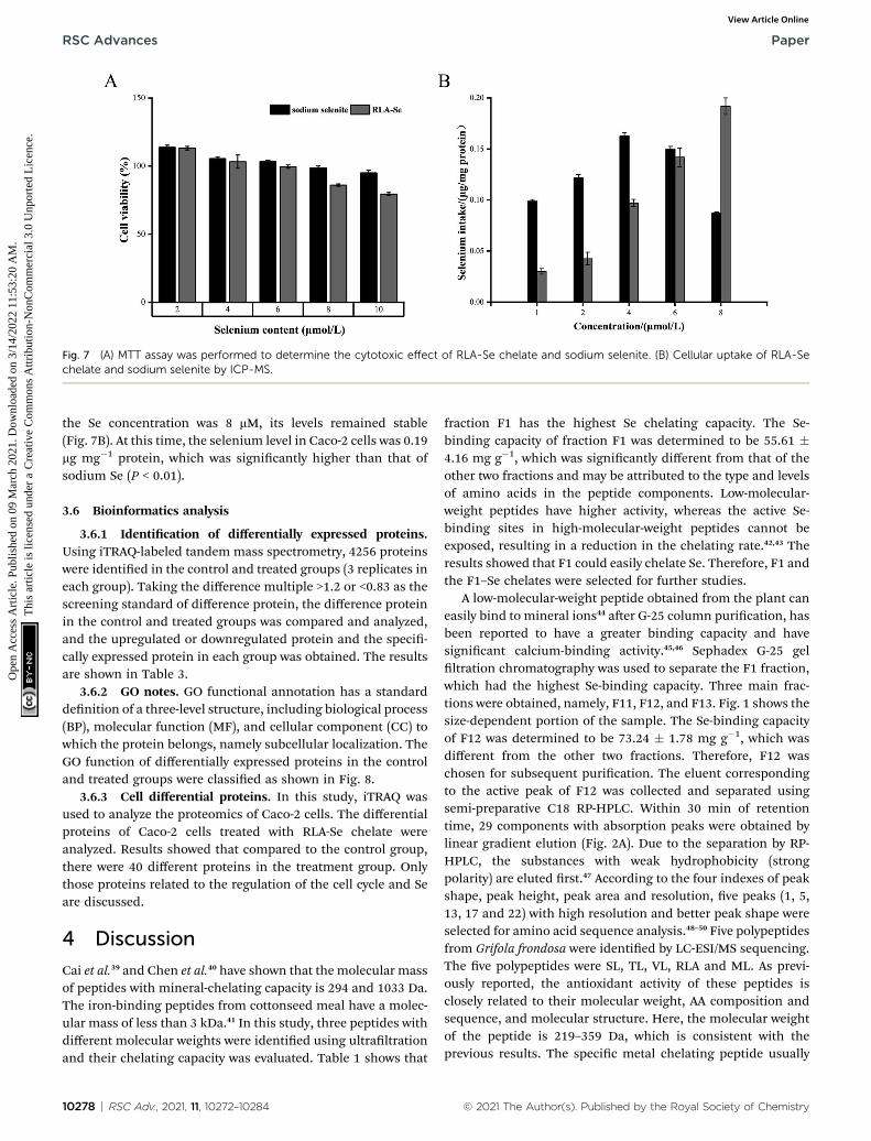
Fig. 7 (A) MTT assay was performed to determine the cytotoxic effect of RLA-Se chelate and sodium selenite. (B) Cellular uptake of RLA-Sechelate and sodium selenite by ICP-MS.
RSC Advances Paper
Ope
n A
cces
s A
rtic
le. P
ublis
hed
on 0
9 M
arch
202
1. D
ownl
oade
d on
3/1
4/20
22 1
1:53
:20
AM
. T
his
artic
le is
lice
nsed
und
er a
Cre
ativ
e C
omm
ons
Attr
ibut
ion-
Non
Com
mer
cial
3.0
Unp
orte
d L
icen
ce.
View Article Online
the Se concentration was 8 mM, its levels remained stable(Fig. 7B). At this time, the selenium level in Caco-2 cells was 0.19mg mg�1 protein, which was signicantly higher than that ofsodium Se (P < 0.01).
3.6 Bioinformatics analysis
3.6.1 Identication of differentially expressed proteins.Using iTRAQ-labeled tandem mass spectrometry, 4256 proteinswere identied in the control and treated groups (3 replicates ineach group). Taking the difference multiple >1.2 or <0.83 as thescreening standard of difference protein, the difference proteinin the control and treated groups was compared and analyzed,and the upregulated or downregulated protein and the speci-cally expressed protein in each group was obtained. The resultsare shown in Table 3.
3.6.2 GO notes. GO functional annotation has a standarddenition of a three-level structure, including biological process(BP), molecular function (MF), and cellular component (CC) towhich the protein belongs, namely subcellular localization. TheGO function of differentially expressed proteins in the controland treated groups were classied as shown in Fig. 8.
3.6.3 Cell differential proteins. In this study, iTRAQ wasused to analyze the proteomics of Caco-2 cells. The differentialproteins of Caco-2 cells treated with RLA-Se chelate wereanalyzed. Results showed that compared to the control group,there were 40 different proteins in the treatment group. Onlythose proteins related to the regulation of the cell cycle and Seare discussed.
4 Discussion
Cai et al.39 and Chen et al.40 have shown that the molecular massof peptides with mineral-chelating capacity is 294 and 1033 Da.The iron-binding peptides from cottonseed meal have a molec-ular mass of less than 3 kDa.41 In this study, three peptides withdifferent molecular weights were identied using ultraltrationand their chelating capacity was evaluated. Table 1 shows that
10278 | RSC Adv., 2021, 11, 10272–10284
fraction F1 has the highest Se chelating capacity. The Se-binding capacity of fraction F1 was determined to be 55.61 �4.16 mg g�1, which was signicantly different from that of theother two fractions and may be attributed to the type and levelsof amino acids in the peptide components. Low-molecular-weight peptides have higher activity, whereas the active Se-binding sites in high-molecular-weight peptides cannot beexposed, resulting in a reduction in the chelating rate.42,43 Theresults showed that F1 could easily chelate Se. Therefore, F1 andthe F1–Se chelates were selected for further studies.
A low-molecular-weight peptide obtained from the plant caneasily bind to mineral ions44 aer G-25 column purication, hasbeen reported to have a greater binding capacity and havesignicant calcium-binding activity.45,46 Sephadex G-25 gelltration chromatography was used to separate the F1 fraction,which had the highest Se-binding capacity. Three main frac-tions were obtained, namely, F11, F12, and F13. Fig. 1 shows thesize-dependent portion of the sample. The Se-binding capacityof F12 was determined to be 73.24 � 1.78 mg g�1, which wasdifferent from the other two fractions. Therefore, F12 waschosen for subsequent purication. The eluent correspondingto the active peak of F12 was collected and separated usingsemi-preparative C18 RP-HPLC. Within 30 min of retentiontime, 29 components with absorption peaks were obtained bylinear gradient elution (Fig. 2A). Due to the separation by RP-HPLC, the substances with weak hydrophobicity (strongpolarity) are eluted rst.47 According to the four indexes of peakshape, peak height, peak area and resolution, ve peaks (1, 5,13, 17 and 22) with high resolution and better peak shape wereselected for amino acid sequence analysis.48–50 Five polypeptidesfrom Grifola frondosa were identied by LC-ESI/MS sequencing.The ve polypeptides were SL, TL, VL, RLA and ML. As previ-ously reported, the antioxidant activity of these peptides isclosely related to their molecular weight, AA composition andsequence, and molecular structure. Here, the molecular weightof the peptide is 219–359 Da, which is consistent with theprevious results. The specic metal chelating peptide usually
© 2021 The Author(s). Published by the Royal Society of Chemistry

Table 3 List of differentially expressed proteins
Uniprot accession Name PeptidesTreated/controlfold p-Value
F1T0J3 Interphotoreceptor matrix proteoglycan 2 OS ¼ Homo sapiensGN ¼ IMPG2 PE ¼ 2 SV ¼ 1
1241 1.575937 0.002209
A0A024R2K4 Leucine rich repeat (in FLII) interacting protein 2, isoformCRA_b OS ¼ Homo sapiens GN ¼ LRRFIP2 PE ¼ 4 SV ¼ 1
742 1.545024 0.001082
Q9BRX9 WD repeat domain-containing protein 83 OS ¼ Homo sapiensGN ¼ WDR83 PE ¼ 1 SV ¼ 1
315 1.4814 0.020892
O00257 E3 SUMO-protein ligase CBX4 OS ¼ Homo sapiens GN ¼ CBX4PE ¼ 1 SV ¼ 3
560 1.478102 0.003381
Q13625 Apoptosis-stimulating of p53 protein 2 OS ¼ Homo sapiens GN¼ TP53BP2 PE ¼ 1 SV ¼ 2
1128 1.380386 0.035219
E7EQB3 tRNA-splicing endonuclease subunit Sen34 OS ¼ Homosapiens GN ¼ TSEN34 PE ¼ 1 SV ¼ 2
315 1.371411 0.034023
Q96BW5 Phosphotriesterase-related protein OS ¼ Homo sapiens GN ¼PTER PE ¼ 1 SV ¼ 1
349 1.334326 0.034962
A0A024R7N7 Interferon, gamma-inducible protein 30, isoform CRA_b OS¼Homo sapiens GN ¼ IFI30 PE ¼ 4 SV ¼ 1
250 1.32053 0.031198
Q5JPC1 Putative uncharacterized protein DKFZp667O1614 OS¼Homosapiens GN ¼ DKFZp667O1614 PE ¼ 2 SV ¼ 1
320 1.320405 0.002719
A0A087WXL6 Vacuolar protein sorting 11 (yeast), isoform CRA_a OS¼Homosapiens GN ¼ VPS11 PE ¼ 1 SV ¼ 1
941 1.268012 0.031961
O14965 Aurora kinase A OS¼ Homo sapiens GN¼ AURKA PE¼ 1 SV¼2
403 1.265934 0.041726
A8K2T7 Receptor protein-tyrosine kinase OS¼Homo sapiens PE¼ 2 SV¼ 1
1210 1.265171 0.019797
A0A024R438 ATG9 autophagy related 9 homolog A (S. cerevisiae), isoformCRA_a OS ¼ Homo sapiens GN ¼ ATG9A PE ¼ 4 SV ¼ 1
839 1.26087 0.035512
B2RDK3 Oxysterol-binding protein OS ¼ Homo sapiens PE ¼ 2 SV ¼ 1 480 1.256281 0.034395A7YA96 Gamma-glutamyl carboxylase OS ¼ Homo sapiens GN ¼ GGCX
PE ¼ 2 SV ¼ 1758 1.254265 0.040682
Q4VCS5 Angiomotin OS ¼ Homo sapiens GN ¼ AMOT PE ¼ 1 SV ¼ 1 1084 1.253906 0.00667Q5JTZ9 Alanine-tRNA ligase, mitochondrial OS ¼ Homo sapiens GN ¼
AARS2 PE ¼ 1 SV ¼ 1985 1.252551 0.0296
Q15642 Cdc42-interacting protein 4 OS ¼ Homo sapiens GN ¼ TRIP10PE ¼ 1 SV ¼ 3
601 1.244809 0.03779
A0A044PY82 Protein LOC113230 OS ¼ Homo sapiens GN ¼ LOC113230 PE¼ 1 SV ¼ 1
361 1.244755 0.042187
Q96CP7 Calfacilitin OS ¼ Homo sapiens GN ¼ TLCD1 PE ¼ 2 SV ¼ 1 247 1.236526 0.020604Q08AI8 Uncharacterized protein C2orf54 OS ¼ Homo sapiens GN ¼
C2orf54 PE ¼ 2 SV ¼ 2447 1.235443 0.008035
Q9P0R6 GSK3-beta interaction protein OS¼Homo sapiensGN¼ GSKIPPE ¼ 1 SV ¼ 2
139 1.234663 0.034995
A0A0A0MSV9 Tapasin OS ¼ Homo sapiens GN ¼ TAPBP PE ¼ 1 SV ¼ 1 504 1.230369 0.016178Q969U7 Proteasome assembly chaperone 2 OS ¼ Homo sapiens GN ¼
PSMG2 PE ¼ 1 SV ¼ 1264 1.225194 0.038636
V9GYM8 Rho guanine nucleotide exchange factor 2 OS ¼ Homo sapiensGN ¼ ARHGEF2 PE ¼ 1 SV ¼ 1
1031 1.21831 0.045723
Q9P2D8 Protein unc-79 homolog OS ¼ Homo sapiens GN ¼ UNC79 PE¼ 2 SV ¼ 4
2635 1.217964 0.01716
E5KBQ3 TRAF2 OS ¼ Homo sapiens GN ¼ TRAF2 PE ¼ 2 SV ¼ 1 501 1.214193 0.014041P82675 28S ribosomal protein S5, mitochondrial OS ¼ Homo sapiens
GN ¼ MRPS5 PE ¼ 1 SV ¼ 2430 1.211803 0.014183
A0A024R880 Cyclin-dependent kinase 9 (CDC2-related kinase), isoformCRA_a OS ¼ Homo sapiens GN ¼ CDK9 PE ¼ 3 SV ¼ 1
372 1.211151 0.027363
P29590 Protein PML OS ¼ Homo sapiens GN ¼ PML PE ¼ 1 SV ¼ 3 882 1.205636 0.006654Q8TB72 Pumilio homolog 2 OS¼ Homo sapiens GN¼ PUM2 PE ¼ 1 SV
¼ 21066 1.202853 0.032181
P62875 DNA-directed RNA polymerases I, II, and III subunit RPABC5OS ¼ Homo sapiens GN ¼ POLR2L PE ¼ 1 SV ¼ 1
67 1.200165 0.017111
A0A0U1RR79 Glutamine-tRNA ligase (fragment) OS ¼ Homo sapiens GN ¼QARS PE ¼ 1 SV ¼ 1
75 0.827804 0.036984
© 2021 The Author(s). Published by the Royal Society of Chemistry RSC Adv., 2021, 11, 10272–10284 | 10279
Paper RSC Advances
Ope
n A
cces
s A
rtic
le. P
ublis
hed
on 0
9 M
arch
202
1. D
ownl
oade
d on
3/1
4/20
22 1
1:53
:20
AM
. T
his
artic
le is
lice
nsed
und
er a
Cre
ativ
e C
omm
ons
Attr
ibut
ion-
Non
Com
mer
cial
3.0
Unp
orte
d L
icen
ce.
View Article Online

Table 3 (Contd. )
Uniprot accession Name PeptidesTreated/controlfold p-Value
H7BXP1 NF-kappa-B inhibitor-interacting Ras-like protein 2 OS ¼Homo sapiens GN ¼ NKIRAS2 PE ¼ 1 SV ¼ 1
229 0.817844 0.031648
B4DSI9 cDNA FLJ56483, highly similar to Eukaryotic translationinitiation factor 4 gamma 1 OS ¼ Homo sapiens PE ¼ 2 SV ¼ 1
1512 0.8104 0.016782
Q6ZSZ5 Rho guanine nucleotide exchange factor 18 OS ¼ Homosapiens GN ¼ ARHGEF18 PE ¼ 1 SV ¼ 3
1173 0.801527 0.03505
P53801 Pituitary tumor-transforming gene 1 protein-interactingprotein OS ¼ Homo sapiens GN ¼ PTTG1IP PE ¼ 1 SV ¼ 1
180 0.783099 0.039711
F1D8T1 Hepatocyte nuclear factor 4 4 alpha variant 2 OS ¼ Homosapiens GN ¼ NR2A1 PE ¼ 2 SV ¼ 1
474 0.774468 0.016323
Q8NE01 Metal transporter CNNM3 OS ¼ Homo sapiens GN ¼ CNNM3PE ¼ 1 SV ¼ 1
707 0.764848 0.014595
A0A024R2G1 Cysteine-rich with EGF-like domains 1, isoform CRA_b OS ¼Homo sapiens GN ¼ CRELD1 PE ¼ 4 SV ¼ 1
420 0.753902 0.020377
RSC Advances Paper
Ope
n A
cces
s A
rtic
le. P
ublis
hed
on 0
9 M
arch
202
1. D
ownl
oade
d on
3/1
4/20
22 1
1:53
:20
AM
. T
his
artic
le is
lice
nsed
und
er a
Cre
ativ
e C
omm
ons
Attr
ibut
ion-
Non
Com
mer
cial
3.0
Unp
orte
d L
icen
ce.
View Article Online
contains two or more AA residues, and the molecular weight isless than 1500 Da.51 In addition, hydrophobic and antioxidantAAs, such as Val (V), Leu (L) and Ile (I) can also scavenge freeradicals and contribute signicantly to antioxidant activitythrough their interaction with lipids or as strong proton/hydrogen donors.52,53 In this study, the ve peptides containedhydrophobic AA residues and antioxidant AA residues. Usually,high-molecular-weight peptides inhibit the absorption ofminerals in the intestine.54 Therefore, dipeptides and tripep-tides are considered suitable options. Leu might have likelyplayed a role in the Se-chelating activity of the ve peptides.Moreover, GF is a Leu-rich medicinal mushroom.55 In peptideno. 4, Arg56 and Ala57 are the main amino acids supporting themineral binding of the puried nonapeptide. In peptide no. 1,the phosphorylation of Ser introduces a negatively chargedphosphate group, which serves as a binding site for positivelycharged metal ions58 such as those of iron.59 Additionally, Thr,Val, and Met in peptides 2, 3, and 5 are the related amino acidsplaying a role in the mineral binding of the puried peptides.60
In general, according to the reported structure–activity rela-tionship of chelating peptides, the identied peptides seem tohave good chelating activity and antioxidant activity. However,further evaluation of synthetic peptides is needed to betterstudy their biological characteristics. The results showed thatRLA had the strongest Se-chelating ability with a chelatingcapacity of 84.47 mg g�1 (Fig. 2C). It was speculated that the Argand Ala residues in the tripeptide and the amino acid residuesbelonging to the hydrophobic group had more recognition sitesand could result in better binding to selenium. It has beendemonstrated that the oxygen atom on the carboxyl group of theamino acid residue can form a coordination bond through theelectron pair, thereby characterizing the structural characteris-tics of the puried peptide to create a favorable metal-chelatingenvironment. Therefore, we hypothesized that the carboxylategroup of Arg, as well as the imino (NH) and carbonyl (CO)groups in RLA might be involved in the formation of the coor-dination bond with Se.
10280 | RSC Adv., 2021, 11, 10272–10284
To provide additional information about the binding ofmetal ions to peptide organic ligands, the UV, SEM, XRD, and1H-NMR spectra are shown. The results from XRD and SEMshow an obvious change in the size and the structure of RLAaer chelating the selenium ions, which are consistent with theresults from previous studies.61,62 Combined with NMR data, wefound that the structure of peptide chain changes aerchelating selenium ions, thereby affecting the chemical envi-ronment of the H at different positions and changing the elec-tron density surrounding the hydrogen nucleus. In the range of0.8–0.9 ppm, proton signal-coupling appeared in RLA and thespectral peak showed splitting. These ndings indicated thataer chelating with selenium ions, the H of –CH3 in RLA wasreplaced by Se to form an ionic bond. The spatial orientation ofthe –CH3 group was also changed by rotation, and the internalhydrophobic group was exposed, which resulted in the redis-tribution of the molecular charge, giving rise to conformationchanges and resulting in the formation of a new type of chelate.Moreover, similar to that produced by RLA, the UV spectrum ofthe RLA-Se chelate also indicated a color enhancement at themaximum absorption peak. When the molecular orbital of–CH3 on the side chain group is folded with the p system, itproduces a particular color enhancement effect. In conclusion,the amino and carboxyl groups of RLA are involved in thecoordination of the Se ions with the participation of the –CH3
side chains, and constitute the primary chelating sites.The bioavailability of the RLA-Se chelate in the human colon
cancer Caco-2 cells was investigated. An MTT assay was used todetermine the effects of the RLA-selenium chelate and sodiumselenite in Caco-2 cells. The results showed that both the RLA-Sechelate and sodium selenite inhibited cellular proliferation ina dose-dependent manner, and that the Caco-2 cells were notinactivated at a Se concentration of 8 mM. Compared to sodiumselenite, RLA-Se chelates were more efficient in the absorptionof selenium in a dose-dependent manner. When the concen-tration of selenium was 4 mmol L�1, the absorption of sodiumselenite reached the peak, and when the concentration was 8
© 2021 The Author(s). Published by the Royal Society of Chemistry

Fig. 8 Gene ontology analysis. Biological processes (blue); cell components (purple); molecular functions (orange).
Paper RSC Advances
Ope
n A
cces
s A
rtic
le. P
ublis
hed
on 0
9 M
arch
202
1. D
ownl
oade
d on
3/1
4/20
22 1
1:53
:20
AM
. T
his
artic
le is
lice
nsed
und
er a
Cre
ativ
e C
omm
ons
Attr
ibut
ion-
Non
Com
mer
cial
3.0
Unp
orte
d L
icen
ce.
View Article Online
mmol L�1, the absorption of selenium in sodium selenitedecreased sharply, while the absorption of RLA-Se stillincreased. It shows that when the supplement of selenium is
© 2021 The Author(s). Published by the Royal Society of Chemistry
increased, the supplement of inorganic selenium cannot be wellabsorbed, while RLA-Se can. Numerous studies have shown thatorganic Se has a better Se-absorption capacity than inorganic
RSC Adv., 2021, 11, 10272–10284 | 10281

RSC Advances Paper
Ope
n A
cces
s A
rtic
le. P
ublis
hed
on 0
9 M
arch
202
1. D
ownl
oade
d on
3/1
4/20
22 1
1:53
:20
AM
. T
his
artic
le is
lice
nsed
und
er a
Cre
ativ
e C
omm
ons
Attr
ibut
ion-
Non
Com
mer
cial
3.0
Unp
orte
d L
icen
ce.
View Article Online
Se.63 It has been veried that RLA-Se has higher biologicalactivities and lower toxicity compared to sodium selenite.Therefore, the differences in metabolism of RLA-Se warrantfurther investigation.
In this study, 4256 proteins labeled with quantitative infor-mation using iTRAQ were identied. Compared to the healthycontrol group, there were 40 differentially expressed proteins inthe RLA-Se group, among which 22 were signicantly upregu-lated and 8 were signicantly downregulated. GO functionalannotation, including cell component (CC), biological process(BP), and molecular function (MF), was carried out for theidentied differential proteins, to determine the molecularfunctions and biological processes of proteins that weresignicantly enriched. The results revealed that for the biolog-ical process, differential proteins were mainly involved in theregulation of the apoptotic signaling pathway, regulation of themulti-organism process, and cell-cycle checkpoint amongothers. In cell composition, these proteins were mainly locatedin the endosomal membrane and transferase complex. In termsof molecular function, these differential proteins mainly involveSUMO binding, cyclin-dependent protein kinase activity, andNF-kappa B binding.
Se plays a key role in terminal differentiation, cell growth,and development by controlling the cell cycle. Because the“checkpoint” relationship between the occurrence of cancerand G1-S and G2-M transformation in the cell cycle is becomingincreasingly prominent and the cell cycle control mechanismhas conservative characteristics, the mechanism of Se incontrolling the cell cycle is also the embodiment of Se nutrition.It has been shown that Se reduces cell proliferation and growthby blocking the cell cycle and/or promoting apoptosis.64–66 Secompounds have shown varying chemopreventive effects.67
Studies indicate that the levels of TP53BP2 (Tumor protein p53binding protein 2),68 AURKA (Aurora kinase A),69 CDK9 (Cyclin-dependent kinase 9),70 and ARHGEF2 (RHO guanine nucleotideexchange factor 2)71 were upregulated in the treated groupcompared to those in the control group, which contributed tothe regulation of cell-cycle progression and cell growth via theirinteractions. These ndings indicate that the Se-chelatingpeptide obtained from GF had a positive effect on transcrip-tion regulation, cell apoptosis, and aging.
The requirement of Se also depends on its role at the cata-lytic site of multiple selenoproteins.72 Glutathione peroxidase(GSH-Px) is a Se-containing enzyme, which plays a crucial role inreproduction, anti-oxidation, muscle function, and tumorprevention. The reduction of hydroperoxide and hydrogenperoxide is catalyzed by GSH-Px through the reduced gluta-thione.73 E3 ubiquitin ligases participate in many physiologicalprocesses in cells by regulating the ubiquitination of regulatoryproteins. All E3 ligases can connect the target protein andspecic E2. Ubiquitin-conjugating enzymes (E2) intervene inthe ubiquitination and turnover of specic substrates of theubiquitin-dependent degradation pathway.74 A conserved coresequence comprising about 150 amino acids from the –NH2
terminus is present in E2 proteins, and the cysteine residueplays an essential role in the formation of the thiol ester bondwith the –COOH terminus of ubiquitin.75 In this study, the
10282 | RSC Adv., 2021, 11, 10272–10284
expression of E3 proteins in Caco-2 cells treated with Se washigher than that in the control groups. A higher expression ofE2 proteins indicated an increase in cysteine levels. Therefore,RLA-Se may accelerate cysteine increase in Caco-2 cells, therebypromoting the expression of E3 proteins.
5 Conclusions
To summarize, we puried a specic tripeptide, RLA, fromGFPH, which had strong Se-chelating ability. We also exploredthe mechanism of RLA in chelating Se ions by analyzing theprotein structure. The absorption proles of the RLA-Se chelatesand sodium selenite was studied using Caco-2 cells. The resultsfrom our study indicated that GF protein could be used asa promising source for the preparation of Se-binding peptides,and serve as a dietary supplement in selenium-decient indi-viduals. We also explored the effect of the RLA-Se chelates oncell growth using iTRAQ quantitative proteomics. The resultsshowed that supplementation with peptide-Se chelates couldresult in the positive regulation of multiple biological processesin cells and might play a specic anticancer role by regulatingcell apoptosis. Overall, the purpose of our study was to deter-mine the efficacy of Grifola frondosa-selenium chelates asa supplement in food additives and pharmaceutical products.
Conflicts of interest
There are no conicts to declare.
Acknowledgements
This work was supported by National Natural Science FonAgriundation of China (no. 31501432), Fujian Provincial School-enterprise Cooperative Major Project (no. 2017N5003), FujianAgriculture and Forestry University Leader Scholarship Program(no. xjq201608), Special Fund for Science and TechnologyInnovation of Fujiaculture and Forestry University(cxzx2019139G), Firestone Foundation of Fujian ProvincialHospital (2020HSJJ04), Regional development project of FujianProvincial Department of Science and Technology (2020N3002).
References
1 J. Avery and P. Hoffmann, Nutrients, 2018, 10, 1203.2 Y. Shu, M. Wu, S. Yang, Y. Wang and H. Li, Clin. Nutr., 2020,10, 3086–3691.
3 M. Kieliszek and S. Błazejak, Molecules, 2016, 21, 609–625.4 M. Brummer, S. Hayes, K. A. Dawson and L. M. Lawrence, J.Anim. Sci., 2013, 91, 2158–2168.
5 J. Hu, Q. Zhao, X. Cheng, C. Selomulya, C. Bai, X. Zhu, X. Liand H. Xiong, Food Chem., 2014, 146, 531–537.
6 H. D. Mistry, F. B. Pipkin, C. W. G. Redman and L. Poston,Am. J. Obstet. Gynecol., 2012, 206, 21–30.
7 L. Zhang, Y. Gao, H. Feng, N. Zou, K. Wang and D. Sun, J.Trace Elem. Med. Biol., 2019, 56, 21–30.
8 H. Ying and Y. Zhang, Biol. Trace Elem. Res., 2019, 192, 38–50.
© 2021 The Author(s). Published by the Royal Society of Chemistry

Paper RSC Advances
Ope
n A
cces
s A
rtic
le. P
ublis
hed
on 0
9 M
arch
202
1. D
ownl
oade
d on
3/1
4/20
22 1
1:53
:20
AM
. T
his
artic
le is
lice
nsed
und
er a
Cre
ativ
e C
omm
ons
Attr
ibut
ion-
Non
Com
mer
cial
3.0
Unp
orte
d L
icen
ce.
View Article Online
9 K. Mohsen, M. Chamani, H. Amanlou, A. Nikkhah andA. A. Sadeghi, Acta Sci., Anim. Sci., 2019, 41, 44392.
10 M. H. G. Berntssen, T. K. Sundal, P. A. Olsvik, H. Amlund,J. D. Rasinger, V. Sele, K. Hamre, M. Hillestad, L. Buttleand R. Ørnsrud, Aquat. Toxicol., 2017, 192, 116–126.
11 C. Torresfuentes, M. M. Contreras, I. Recio, M. Alaiz andJ. Vioque, Food Chem., 2015, 180, 194–202.
12 H. T. Wu, W. G. Jin, S. G. Sun, X. S. Li, X. H. Duan, Y. Li,Y. T. Yang, J. R. Han and B. W. Zhu, Eur. Food Res.Technol., 2016, 242, 713–722.
13 G. Huang, Z. Ren and J. Jiang, Food Bioprocess Technol., 2010,4, 1527–1532.
14 L. D. L. Hoz, A. N. Ponezi, R. F. Milani, V. S. N. D. Silva,A. S. D. Souza and M. T. Bertoldo-Pacheco, Food Chem.,2014, 142, 166–169.
15 D. Jin, X. Liu, X. Zheng, X. Wang and J. He, Food Chem., 2016,204, 427–436.
16 N. Sun, P. Cui, S. Lin, C. Yu, Y. Tang, Y. Wei, Y. Xiong andH. Wu, J. Sci. Food Agric., 2017, 97, 4604–4611.
17 B. Liu, Y. Zhuang and L. Sun, J. Food Sci., 2019, 85, 114–122.18 H. Hu, S. Wang, X. Zhu, Q. Li, Y. Fan, D. Cheng and B. Li,
Food Chem., 2017, 243, 389–395.19 N. Xie, J. Huang, B. Li, J. Cheng, Z. Wang, J. Yin and X. Yan,
Food Chem., 2014, 173, 210–217.20 M. E. Caetano-S, M. T. Bertoldo-P, A. F. Paes-L and
F. M. Netto, Food Res. Int., 2015, 71, 132–139.21 B. Boh and M. Berovic, Int. J. Med. Mushrooms, 2007, 9, 89–
108.22 E. N. Alonso, M. J. Ferronato, N. A. Gandini, M. E. Fermento,
D. J. Obiol, A. Lopez-R, J. Arevalo, M. E. Villegas,M. M. Facchinetti and A. C. Curino, Nutr. Cancer, 2017, 69,29–43.
23 N. Kodama, Y. Murata and H. Nanba, J. Med. Food, 2004, 7,141–145.
24 C. Q. Gu, J. W. Li, F. Chao, M. Jin, X. W.Wang and Z. Q. Shen,Antiviral Res., 2007, 75, 250–257.
25 W. Zhang, Y. Lu, Y. Zhang, Q. Ding, S. Hussain, Q. Wu,W. Pan and Y. Chen, Int. J. Food Sci. Technol., 2016, 51,1055–1061.
26 H. Lei, W. Wang, Q. Wang, S. Guo and L. Wu, Food Agric.Immunol., 2013, 24, 409–418.
27 W. Shen and T. Matsui, Int. J. Food Sci. Technol., 2019, 54,1942–1948.
28 I. B. O'Loughlin, P. M. Kelly, B. A. Murray, J. Richard andB. Andre, J. Agric. Food Chem., 2015, 63, 2708–2714.
29 Z. Zhang, F. Zhou, X. Liu and M. Zhao, Food Chem., 2018,258, 269–277.
30 M. V. Chaud, C. Izumi, Z. Nahaal, T. Shuhama,M. L. P. Bianchi and O. Freitaset, J. Agric. Food Chem.,2002, 50, 871–877.
31 M. J. Dunn and H. J. Kraus, Proteomics: Clin. Appl., 2016, 10,1–3.
32 Z. H. Chen, B. Liu and L. N. Zhao, Food Sci. Technol. Res.,2020, 26, 101–110.
33 P. W. Wesl and C. Cimerman, Anal. Chem., 2002, 36, 2013–2016.
© 2021 The Author(s). Published by the Royal Society of Chemistry
34 X. Y. Qin, J. T. Zhang, G. M. Li, M. Zhou, R. Z. Gu, J. Lu andW. Y. Liu, J. Funct. Foods, 2019, 64, 103619.
35 F. Hassan, G. A. El-Hiti, M. Abd-Allateef and E. Yousif, SaudiMed. J., 2017, 38, 359–365.
36 L. Chen, Z. Yu, Y. Lee, X. Wang, B. Zhao and Y. M. Jung,Analyst, 2012, 137, 5834–5838.
37 M. M. Bradford, Anal. Biochem., 1976, 72, 248–254.38 J. R. Wisniewski, A. Zougman, N. Nagaraj and M. Mann, Nat.
Methods, 2009, 6, 359–362.39 X. Cai, Q. Yang, J. Lin, N. Fu and S. Wang, Molecules, 2017,
22, 544.40 D. Chen, X. Mu, H. Huang, R. Nie, Z. Liu and M. Zeng, J.
Funct. Foods, 2014, 6, 575–584.41 N. H. Kim, S. H. Jung, J. Kim, S. H. Kim, H. J. Ahn and
K. B. Song, J. Korean Soc. Appl. Biol. Chem., 2014, 57, 91–95.42 X. Wang, A. Gao, Y. Chen, X. Zhang, S. Li and Y. Chen, Food
Chem., 2017, 229, 487–494.43 H. Tanzadehpanah, A. Asoodeh, M. Saidijam, J. Chamani
and H. Mahaki, J. Biomol. Struct. Dyn., 2018, 36, 3803–3818.44 L. Zhao, Q. Huang, S. Huang, J. Lin, S. Wang, Y. Huang,
J. Hong and P. Rao, J. Agric. Food Chem., 2014a, 62, 10274–10282.
45 L. Zhao, S. Huang, X. Cai, J. Hong and S. Wang, J. Funct.Foods, 2014b, 10, 46–53.
46 X. Y. Qin, J. T. Zhang, G. M. Li, M. Zhou, R. Z. Gu, J. Lu andW. Y. Liu, J. Funct. Foods, 2020, 64, 103619.
47 G. P. S. Jadaun, S. Dixit, V. Saklani, S. Mendiratta, R. Jain andS. Singh, Pharm. Methods, 2016, 8, 139–144.
48 S. B. Zhang, Z. Wang, S. Y. Xu and X. F. Gao, J. Am. Oil Chem.Soc., 2019, 86, 959–966.
49 P. Puchalska, M. L. Marina Alegre and M. C. Garcia Lopez,Crit. Rev. Food Sci. Nutr., 2015, 55, 521–551.
50 A. Sila and A. Bougatef, J. Funct. Foods, 2016, 21, 10–26.51 B. H. Sarmadi and A. Ismail, Peptides, 2010, 31, 1949–1956.52 A. G. P. Samaranayaka and C. Li, J. Funct. Foods, 2011, 3, 229–
254.53 G. C. Tenore, A. Ritieni, P. Campiglia, P. Stiuso, S. Di Maro,
E. Sommella, G. Pepe, E. D'Urso and E. Novellino, J. Funct.Foods, 2015, 15, 365–375.
54 C. Wang, B. Li and J. Ao, Food Chem., 2012, 134, 1231–1238.55 S. J. Huang, S. Y. Tsai, S. Y. Lin, C. H. Liang and J. L. Mau, Int.
J. Med. Mushrooms, 2011, 13, 265–272.56 C. Megıas, J. Pedroche, M. M. Yust, J. Giron-Calle, M. Alaiz,
F. Millan and J. Vioque, J. Agric. Food Chem., 2007, 55,3949–3954.
57 F. R. Liu, L. Wang, R. Wang and Z. X. Chen, J. Agric. FoodChem., 2013, 61, 7537–7544.
58 E. Miquel and R. Farre, Trends Food Sci. Technol., 2007, 18,139–143.
59 R. Palika, P. C. Mashurabad, M. K. Nair, G. B. Reddy andR. Pullakhandam, Food Res. Int., 2014, 67, 308–314.
60 G. R. Huang, Z. Y. Ren, J. X. Jiang and W. W. Chen, Adv. J.Food Sci. Technol., 2012, 4, 207–212.
61 B. Mannini, J. Habchi, S. Chia, F. S. Ruggeri, M. Perni,T. P. Knowles, C. M. Dobson and M. Vendruscolo, ACSChem. Neurosci., 2018, 9, 2959–2971.
RSC Adv., 2021, 11, 10272–10284 | 10283

RSC Advances Paper
Ope
n A
cces
s A
rtic
le. P
ublis
hed
on 0
9 M
arch
202
1. D
ownl
oade
d on
3/1
4/20
22 1
1:53
:20
AM
. T
his
artic
le is
lice
nsed
und
er a
Cre
ativ
e C
omm
ons
Attr
ibut
ion-
Non
Com
mer
cial
3.0
Unp
orte
d L
icen
ce.
View Article Online
62 Y. G. Jin, W. W. Fu and M. H. Ma, Afr. J. Biotechnol., 2011, 10,10204–10211.
63 S. Todd, D. Thomas and W. Hendriks, J. Anim. Physiol. Anim.Nutr., 2012, 96, 148–158.
64 S. Zhang, X. Peng, J. Fang, H. Cui, Z. Zuo and Z. Chen, Biol.Trace Elem. Res., 2014, 160, 32–40.
65 H. Zeng, J. Nutr., 2002, 132, 674–679.66 H. Zeng and J. H. Botnen, Biofactors, 2007, 31, 55–164.67 X. Cai, L. Zhao, S. Wang and P. Rao, Food Funct., 2015, 6,
816–823.68 Q. Song, J. Song, Q. Wang, Y. Ma, N. Sun, J. Ma, Q. Chen,
G. Xia, Y. Huo and L. Yang, Cancer Med., 2016, 5, 315–324.69 A. Jacobsen, L. J. Bosch, S. R. Martens-de, B. Carvalho,
A. H. Sillars-Hardebol, R. J. Dobson, E. De Rinaldis,G. A. Meijer, S. Abeln and J. Heringa, Sci. Rep., 2018, 8, 1–11.
10284 | RSC Adv., 2021, 11, 10272–10284
70 G. De Falco, L. M. Neri, M. De Falco, C. Bellan, Z. Yu, A. DeLuca, L. Leoncini and A. Giordano, Oncogene, 2002, 21, 7464–7470.
71 C. W. Cheon, D. H. Kim, Y. H. Cho and J. H. Kim, World J.Gastroenterol., 2009, 15, 310.
72 L. Fu, X. Yan, X. Ruan, J. Lin and Y. Wang, Nanoscale Res.Lett., 2014, 9, 589.
73 Y. Mehdi, J. L. Hornick, L. Istasse and I. Dufrasne,Molecules,2013, 18, 3292–3311.
74 M. T. Haldeman, G. Xia, E. M. Kasperek and C. M. Pickart,Biochemistry, 1997, 36, 10526–10537.
75 W. J. Cook, L. C. Jeffrey, M. L. Sullivan and R. D. Vierstra, J.Biol. Chem., 1992, 267, 15116–15121.
© 2021 The Author(s). Published by the Royal Society of Chemistry