Embed Size (px)
Citation preview

Supplementary Material (ESI) for Chemical Communications This journal is (c) The Royal Society of Chemistry 2011
A Small Fluorophore Reporter of Protein Conformation and
Redox State
Graham J. Pound, Alexandre A. Pletnev, Xiaomin Fang, and Ekaterina V. Pletneva*
Department of Chemistry, Dartmouth College, Hanover, NH 03755, USA
Supplementary Information
Experimental Methods
Synthesis of Atpt Iodoacetamide
Scheme S1 outlines a synthetic route for 1. Commercially available 3,5-dinitro-p-toluic acid
was converted into 2,6-dinitroterephthalic acid, which was fully esterified and reduced to afford
the corresponding diamine. The amino groups were then functionalized and the less hindered
methyl ester was selectively hydrolyzed with lithium hydroxide. Removal of the two
benzyloxycarbonyl protective groups furnished the target blue fluorescent 2,6-
bis(methylamino)terephthalic acid monomethyl ester 8. Reaction of 8 with 2-iodoacetyl chloride
yielded the Atpt iodoacetamide 1.
S1

Supplementary Material (ESI) for Chemical Communications This journal is (c) The Royal Society of Chemistry 2011
HOOC NO2
NO2
aHOOC NO2
NO2COOH
b
N
O
NHOOC
O
H
H N
O
N
OI
HOOC
OH
H3CO2C NO2
NO2CO2CH3
c, d
H3CO2C NHCbz
NHCbzCO2CH3 e
H3CO2C N
NCO2CH3
Cbz
Cbz
f
HOOC N
NCO2CH3
Cbz
Cbz
c
g
2 3 4
5 6 7
8 1
N
O
N
OS
HOOC
OH
NHAc
COOH
9
h
Reagents: (a) Na2Cr2O7, H2SO4; (b) (CH3)2SO4, K2CO3; (c) H2, cat. Pd/C; (d) CbzCl, Zn; (e) CH3I, NaH; (f) LiOH, H2O-MeOH; (g) 2-iodoacetyl chloride, NaHCO3; (h) N-acetyl-L-cysteine, Et3N.
Scheme S1. Synthesis of Atpt Iodoacetamide
S2

Supplementary Material (ESI) for Chemical Communications This journal is (c) The Royal Society of Chemistry 2011
Synthetic Procedures
2,6-Dinitroterephthalic acid (3) was prepared according to a literature procedure.1 A
stirred solution of 3,5-dinitro-p-toluic acid (2, 10 g, 44.2 mmol) in conc. H2SO4 (70 mL) was
cooled in an ice-water bath and sodium dichromate dihydrate (18.4 g, 61.7 mmol) was added in
portions keeping the internal temperature below 25 oC. The reaction mixture was allowed to stir
at rt overnight, poured onto ice (300 g) and the resulting mixture was extracted with EtOAc. The
organic phase was dried (Na2SO4) and concentrated to afford 10.55 g (93%) of an off-white
solid. M.p. 280 oC (decomp). 1H NMR (dmso-d6) δ 8.78 (s). 13C NMR (dmso-d6) δ 128.4, 130.0,
133.8, 146.6, 163.1, 163.6. IR (thin film) 1716, 1699, 1558, 1541, 1341 cm-1. HRMS (ESI, m/z)
calculated for [M+23]+ (C8H4N2O8Na) 278.9865, found 278.9866.
Dimethyl 2,6-dinitroterephthalate (4).1 Sodium hydrocarbonate (8.45 g, 100.58 mmol)
was added to a solution of 3 (10.3 g, 40.23 mmol) in acetone (200 mL) followed by dimethyl
sulfate (7.61 mL, 80.46 mmol). The resulting mixture was stirred at reflux overnight, allowed to
cool to rt and filtered. The filtrate was poured into 350 mL of water and the resulting white
precipitate was collected, washed with water and dried in air. Yield – 8.2 g (72%). An
analytically pure sample was obtained after column chromatography (5:1 hexanes-EtOAc). M.p.
147-148 oC. 1H NMR (CDCl3) δ 4.07 (two s, 6H), 9.07 (s, 2H). 13C NMR (CDCl3) δ 54.0, 54.6,
128.7, 130.4, 133.6, 147.1, 162.4, 162.5. IR (thin film) 3097, 2961, 1740, 1551, 1345, 1276 cm-1.
HRMS (ESI, m/z) calculated for [M+23]+ (C10H8N2O8Na) 307.0178, found 307.0183.
Dimethyl 2,6-bis(benzyloxycarbonylamino)terephthalate (5). Compound 4 (2 g, 7.04
mmol) was dissolved in EtOAc (50 mL) and hydrogenated using 10% Pd/C (0.3 g, 4 mol %)
under 1 atm of hydrogen for 4.5 h. Filtered through Celite and concentrated to afford a yellow
solid that was dissolved immediately in EtOAc (45 mL). This solution was added to a solution
S3
Supplementary Material (ESI) for Chemical Communications This journal is (c) The Royal Society of Chemistry 2011
of benzyl chloroformate (2.2 mL, 15.49 mmol) in EtOAc (20 mL) containing Zn dust (1.01 g,
15.49 mmol). The resulting suspension was stirred at rt for 2.5 h and filtered through a glass frit.
The precipitate (containing the poorly soluble product 5) was washed with EtOAc until no more
UV-active material went into solution. The combined filtrate was washed with water, dried
(Na2SO4) and concentrated to yield an amber oil. The oil was triturated with toluene to afford a
beige precipitate that was removed by filtration. The filtrate was concentrated to afford 2.88 g
(83%) of compound 5. M.p. 120-121 oC. 1H NMR (CDCl3) δ 3.92 (s, 3H), 3.97 (s, 3H), 5.23 (s,
4H), 7.35-7.44 (m, 10H), 8.57 (s, 2H), 9.20 (br s, 2H). 13C NMR (CDCl3) δ 52.7, 53.4, 67.4,
115.9, 128.5, 128.6, 128.7, 134.7, 136.0, 140.1, 153.3, 165.9, 167.1. IR (thin film) 3403, 3323,
3034, 2954, 1741, 1697, 1582, 1264, 1199 cm-1. HRMS (ESI, m/z) calculated for [M+23]+
(C26H24N2O8Na) 515.1430, found 515.1434.
Dimethyl N,N’-dimethyl-2,6-bis(benzyloxycarbonylamino)terephthalate (6) was
prepared according to a published procedure2. Sodium hydride (0.88 g of 60% suspension in oil,
22.07 mmol) was washed with hexanes and suspended in DMF (5 mL) at 0 oC under inert
atmosphere. A solution of 5 (2.68 g, 5.45 mmol) in DMF (20 mL) was then added dropwise and
the reaction mixture was stirred at 0 oC for 1 h. Methyl iodide (1.7 mL, 27.25 mmol) was then
added and stirring continued for 4 h at rt. The reaction was quenched by addition of ice and the
resulting mixture was extracted with EtOAc. The organic phase was washed with brine, dried
(Na2SO4) and concentrated. The crude product was purified by column chromatography (2:1
hexanes-EtOAc) to yield 1.6 g (56%) of compound 6 as a colorless oil. 1H NMR (CDCl3) δ 3.27
and 3.33 ( two br s, 6H), 3.57 (br s, 3H), 3.94 (s, 3H), 5.03 and 5.19 (two br s, 4H), 7.18-7.41 (br
m, 10H), 7.85 (br s, 2H). 13C NMR (CDCl3) δ 38.4, 52.7, 52.8, 67.7, 128.0, 128.4, 128.7, 133.5,
136.3, 142.3, 143.2, 155.0, 165.1 (two signals missing due to overlap). IR (thin film) 3033, 2952,
S4

Supplementary Material (ESI) for Chemical Communications This journal is (c) The Royal Society of Chemistry 2011
1716, 1570, 1434, 1339, 1252, 1154 cm-1. HRMS (ESI, m/z) calculated for [M+1]+
(C28H29N2O8) 521.1924, found 521.1931.
N,N’-dimethyl-2,6-bis(benzyloxycarbonylamino)terephthalic acid 1-methyl ester (7).
Lithium hydroxide monohydrate (0.2 g, 4.73 mmol) was added to a solution of 6 (1.54 g, 2.96
mmol) in 4:1 methanol-water mixture (25 mL). The resulting solution was stirred at rt for 4 h,
acidified with 2N HCl to pH 1, diluted with brine and extracted with EtOAc. The organic phase
was dried (Na2SO4) and concentrated to afford 1.54 g (100%) of compound 7 as a colorless oil.
1H NMR (CDCl3) δ 3.28 and 3.34 (two br s, 6H), 3.59 (br s, 3H), 5.05 and 5.21 (two br s, 4H),
7.20-7.42 (br m, 10H), 7.92 (br s, 2H), 8.90 (br s, 1H). 13C NMR (CDCl3) δ 38.4, 52.8, 67.9,
128.0, 128.3, 128.5, 128.7, 129.2, 132.9, 136.2, 136.4, 142.4, 143.1, 155.1, 155.8, 165.2, 169.0.
IR (thin film) 3065, 2952, 1717, 1433, 1342, 1265, 1157 cm-1. HRMS (ESI, m/z) calculated for
[M+23]+ (C27H26N2O8Na) 529.1587, found 529.1584.
2,6-Bis(methylamino)terephthalic acid 1-methyl ester (8). 10% Pd/C (0.2 g, 0.19
mmol) was added to a solution of 7 (1.5 g, 2.96 mmol) in methanol (30 mL) and the reaction
mixture was stirred under 1 atm of H2 for 3.5 h at rt, filtered through Celite and concentrated to
afford 0.67 g (95%) of compound 8 as a yellow solid. Decomp. above 175 oC without melting.
1H NMR (acetone-d6) δ 2.88 (s, 6H), 3.86 (s, 3H), 6.57 (s, 2H), 7.50 (br s, 1H). 13C NMR
(acetone-d6) δ 51.1, 97.7, 98.7, 136.0, 153.1, 167.9, 169.5 (one signal missing due to overlap). IR
(thin film) 3465, 3361, 1692, 1665, 1583, 1453, 1410, 1259, 1229, 1180, 1073 cm-1. HRMS
(ESI, m/z) calculated for [M+1]+ (C11H15N2O4) 239.1032, found 239.1033.
2-(2-Iodoacetyl)-2,6-bis(methylamino)terephthalic acid 1-methyl ester (Atpt-
iodoacetamide, 1) was prepared according to a literature procedure.3 Compound 8 (0.2 g, 0.84
mmol) was dissolved in a solution of sodium bicarbonate (0.54 g, 6.42 mmol) in water (20 mL).
S5
Supplementary Material (ESI) for Chemical Communications This journal is (c) The Royal Society of Chemistry 2011
The resulting solution was cooled to 0 oC and treated with 2-iodoacetyl chloride (79 μL, 0.88
mmol). The reaction mixture was stirred in the dark for 1 h at 0 oC and for 1 h more at rt,
acidified with 2N HCl to pH 2 and extracted with EtOAc. The organic phase was dried
(Na2SO4), concentrated and the residue purified by column chromatography (EtOAc) to afford
87 mg (24%) of compound 1 as a yellow solid. Decomp. above 140 oC without melting. 1H
NMR (CDCl3) δ 3.14 (s, 3H), 3.33 (s, 3H), 3.75-3.82 (m, 2H), 4.05 (s, 3H), 7.42 (s, 1H), 7.62 (s,
1H), 9.20 (br s, 1H). 13C NMR (CDCl3) δ -2.8, 30.4, 38.4, 53.3, 112.3, 113.5, 116.4, 134.6,
144.1, 152.4, 167.5, 168.6, 169.5. IR (thin film) 3373, 2921, 1717, 1698, 1685, 1616, 1572,
1419, 1243 cm-1. HRMS (ESI, m/z) calculated for [M+1]+ (C13H16N2O5I) 407.0104, found
407.0110. The solubility of 1 in water is 1.4±0.1 mmol/L, which is more than sufficient for high-
efficiency labeling of proteins.
N-Acetyl-S-Atpt-cysteine (9). A solution of triethylamine (15 μL, 108 μmol) and N-
acetyl-L-cysteine (6 mg, 37 μmol) in dichloromethane (0.5 mL) was added to a solution of
compound 1 (7 mg, 17 μmol) in dichloromethane (0.5 mL) at 0 oC. The resulting solution was
stirred at rt in the dark for 3 h, concentrated in vacuo, dissolved in water (3 mL), acidified to pH
2 with conc. HCl and extracted with EtOAc. The organic extracts were concentrated and the
product purified on the GE Healthcare Source 15RPC column HPLC (Buffer A: 0.1% TFA in
water, Buffer B: 10% Buffer A(v/v) and 90% acetonitrile; 0-100 % gradient of buffer B, eluted at
20% B) to afford 5 mg of the target compound 9 as a yellow solid. HRMS (ESI, m/z) calculated
for [M+1]+ (C18H24N3O8S) 442.1284, found 442.1285.
S6

Supplementary Material (ESI) for Chemical Communications This journal is (c) The Royal Society of Chemistry 2011
Site-Directed Mutagenesis, Protein Expression and Purification
Plasmids for iso-1 yeast cyt c bacterial expression have been prepared in our previous
work.4 Expression and purification of K72A/C102S (WT*) and E66C/K72A/C102S cyt c were
done as described.4
Mutations W253F and C85S/D121C/C138S/W253F were introduced in the pET3
vinculin D1 plasmid5 using the QuickChange method. DNA sequencing at Dartmouth Molecular
Biology Core confirmed the mutations. Vinculin plasmids were transformed into E. coli BL21
StarTM cells (Invitrogen) and the protein variants were expressed as described.5 Harvested cells
were broken by French Press and the solution was clarified from cell debris by centrifugation.
Protein purification followed a published protocol5 using GE Healthcare HisTrap and HiTrap Q
prepacked columns connected to an Akta FPLC system.
Natalie T. Burkhard and Dr. D. M. Indika Bandara have prepared some of the
E66C/K72A/C102S cyt c mutant for this work.
Atpt Labeling
The purified protein (300-500 μM) was pretreated with 5-10 mM DTT to break disulfide
protein adducts. DTT was removed and the buffer exchanged to a 100 mM NaPi buffer pH 7.4
using an FPLC desalting column. The protein was then diluted to a concentration of 50-100 μM
with a 100 mM NaPi buffer pH 7.4 buffer. A tenfold molar excess of Atpt-iodoacetamide 1 was
dissolved in 0.2-0.4 mL of dimethyl sulfoxide (DMSO) and added dropwise in the dark to the
stirring solution of the protein. The reaction proceeded for 5 hours, shielded from light. Upon
completion of the reaction, an excess of DTT was added to consume non-reacted labeling
reagent.
S7

Supplementary Material (ESI) for Chemical Communications This journal is (c) The Royal Society of Chemistry 2011
The buffer of the reaction mixture was exchanged by overnight dialysis to a 10 mM NaPi
at pH 7.0 (cyt c) and 10 mM Tris at pH 8.0 or 10 mM NaPi at pH 7.7 (vinculin). The same buffer
(Buffer A) was used to equilibrate an SP (cyt c) or Q (vinculin) column for purification of the
labeled product. Before applying onto a column, cyt c was reoxidized with an excess of
K3[Fe(CN)6]. The proteins were eluted with a shallow gradient from 0 M to 0.5 M NaCl in buffer
A, a procedure that resulted in separation of labeled and unlabeled proteins (Figure S2).
Formation of a monolabeled adduct was confirmed by ESI-MS done at the W.M. Keck
Foundation Biotechnology Resource Laboratory at Yale University School of Medicine.
Spectroscopic Measurements
All the experiments were done at 21±1 °C. Absorption and CD spectra were recorded
with an Agilent 8453 diode-array spectrophotometer and a Jasco J-715 spectropolarimeter,
respectively. Fluorescence spectra were recorded with a Horiba Jobin Yvon Fluorolog-3
spectrofluorimeter.
Fluorescence lifetimes were measured by time-correlated single photon counting
(TCSPC) using NanoLED-375L diode laser (λex=375 nm, <70 ps pulsewidth) as the excitation
source and a fast TBX-04 detector. The dye emission was observed at 460 nm. The
measurements were done under magic angle conditions. The fluorescence decay traces were
analyzed with the commercial DAS6 software from Horiba Jobin Yvon and previously described
routines in MATLAB.4, 6
Quantum Yield Calculations
The quantum yield Φ of Atpt-Cys was measured using quinine sulfate (Aldrich) in 0.1 M
H2SO4 (Φ=0.58)7 as the reference R with the following equation:8
2
2
RR
RR
nn
II
AA
Φ=Φ (1)
S8
Supplementary Material (ESI) for Chemical Communications This journal is (c) The Royal Society of Chemistry 2011
In eq 1, A is the absorbance at the excitation wavelength, I is the integrated emission, and n is the
solution refractive index. Corrected emission spectra were collected with 375 nm excitation.
Solution refractive indexes were determined with an AO Scientific Instrument ABBE Mark II
digital refractometer.
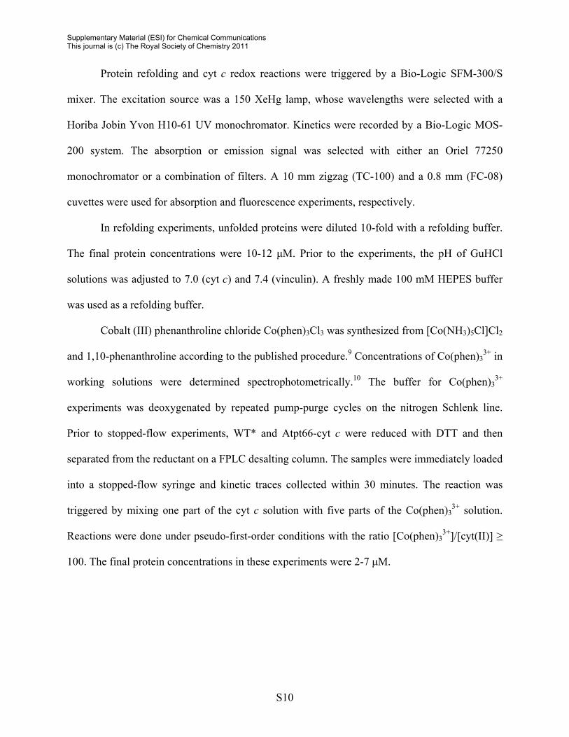
Critical Distance R0 Calculations
The critical distances, R0, for the Atpt-heme and Trp-Atpt donor-acceptor pairs were
calculated according to eq 2,8 where the value of the orientation parameter κ2 was taken as 2/3,
ΦD is the donor fluorescence quantum yield (0.138 for Trp and 0.28 for Atpt), n is the refractive
index of the solution, FD is the normalized fluorescence spectrum of the donor, and εA is the
molar absorbance spectrum of the acceptor:
( ) ( ) λλλελ⎟⎟⎠
⎞⎜⎜⎝
⎛ Φκ×= ∫− d107858 4
AD4D
256
0 Fn
.R (2)
The overlap integrals ( ) ( ) ( ) λλλελ=λ ∫ d4ADFJ (Figure S4) were calculated using the area
function in SigmaPlot 10.0 from Systat Software, Inc.
Unfolding Curves
The unfolding curves were obtained from CD and heme absorption measurements. To
create a series of GuHCl solutions with the same concentration of protein, aliquots of
concentrated protein were added to pH-adjusted GuHCl solutions via gas-tight Hamilton
syringes. The samples were incubated at room temperature for 15 minutes prior to
measurements. GuHCl concentrations were monitored for accuracy with refractive index
measurements. The protein concentrations were between 3 and 10 µM. Analyses of unfolding
curves were performed as previously described.4
Stopped-Flow Kinetics Measurements
S9
Supplementary Material (ESI) for Chemical Communications This journal is (c) The Royal Society of Chemistry 2011
Protein refolding and cyt c redox reactions were triggered by a Bio-Logic SFM-300/S
mixer. The excitation source was a 150 XeHg lamp, whose wavelengths were selected with a
Horiba Jobin Yvon H10-61 UV monochromator. Kinetics were recorded by a Bio-Logic MOS-
200 system. The absorption or emission signal was selected with either an Oriel 77250
monochromator or a combination of filters. A 10 mm zigzag (TC-100) and a 0.8 mm (FC-08)
cuvettes were used for absorption and fluorescence experiments, respectively.
In refolding experiments, unfolded proteins were diluted 10-fold with a refolding buffer.
The final protein concentrations were 10-12 μM. Prior to the experiments, the pH of GuHCl
solutions was adjusted to 7.0 (cyt c) and 7.4 (vinculin). A freshly made 100 mM HEPES buffer
was used as a refolding buffer.
Cobalt (III) phenanthroline chloride Co(phen)3Cl3 was synthesized from [Co(NH3)5Cl]Cl2
and 1,10-phenanthroline according to the published procedure.9 Concentrations of Co(phen)33+ in
working solutions were determined spectrophotometrically.10 The buffer for Co(phen)33+
experiments was deoxygenated by repeated pump-purge cycles on the nitrogen Schlenk line.
Prior to stopped-flow experiments, WT* and Atpt66-cyt c were reduced with DTT and then
separated from the reductant on a FPLC desalting column. The samples were immediately loaded
into a stopped-flow syringe and kinetic traces collected within 30 minutes. The reaction was
triggered by mixing one part of the cyt c solution with five parts of the Co(phen)33+ solution.
Reactions were done under pseudo-first-order conditions with the ratio [Co(phen)33+]/[cyt(II)] ≥
100. The final protein concentrations in these experiments were 2-7 μM.
S10

Supplementary Material (ESI) for Chemical Communications This journal is (c) The Royal Society of Chemistry 2011
NH
O
N
O
HOOC
O
Atpt
NO
N
NO2
NO
O
NBD
ON O
NH
O
DCIA
a
OHO O
HN
S
COOH
FITC
NN
O O
NH
O
Bimane
SO3
HNHN
O
Dns
OH2N
O3S
O
NH
ON
O
O4
Alexa350
Wavelength (nm)
400 450 500 550 600 650
Fluo
resc
ence
Inte
nsity
J(λ)
1/J(λ
) 2
1.0
1.1
1.2
1.3
1.4
1.5
DCIABimane
Alexa350Dns
NBD
FITCAtpt
b
c
FITC Atpt NBD DCIA Bimane Dns Alexa350
Figure S1. (a) Structures, (b) emission spectra,11-14 and (c) ratios of the overlap integrals of the fluorophore emission spectrum with the cyt c absorption spectra in the two protein redox states J(λ)red/J(λ)ox or J(λ)ox/J(λ)red for thiol adducts of Atpt and common commercial dyes. The Atpt-labeled protein is predicted to show the strongest heme redox response among the less bulky dyes.
S11

Supplementary Material (ESI) for Chemical Communications This journal is (c) The Royal Society of Chemistry 2011
Figure S2. FPLC chromatograms of cyt c (a) and vinculin D1 (b) after Atpt-labeling showing efficient separation of labeled and unlabeled proteins on ion exchange SP and Q resins, respectively. For cyt c, Buffer A was 10 mM NaPi at pH 7.0, Buffer B was 10 mM NaPi at pH 7.0 containing 0.5 M NaCl. For vinculin D1, Buffer A was 10 mM Tris at pH 8.0, Buffer B was 10 mM Tris at pH 8.0 containing 0.5 M NaCl.
S12

Supplementary Material (ESI) for Chemical Communications This journal is (c) The Royal Society of Chemistry 2011
Figure S3. (a) Circular dichroism (CD) spectra of Atpt66-cyt c and its parent (dashed line) yeast iso-1 K72A/C102S (WT*) variant in a 100 mM NaPi buffer at pH 7.0. The protein concentrations c are 10.0 μM, the pathlength l is 1.0 mm. (b) Heme absorption and CD signals of ferric Atpt66-cyt c as a function of GuHCl concentration at pH 7.0. (c) CD spectra of Atpt121-(blue) and Atpt85-(red) labeled vinculin D1 and their parent (dashed lines) variants in a 100 mM NaPi buffer at pH 7.4. The protein concentrations c are 5.0 μM, the pathlength l is 1.0 mm. (d) CD signals of Atpt-labeled and unlabeled vinculin D1 as a function of GuHCl concentration at pH 7.4. The curves suggest similar stepwise unfolding of the protein.
S13
Supplementary Material (ESI) for Chemical Communications This journal is (c) The Royal Society of Chemistry 2011
Figure S4. Overlap of Atpt-Cys absorption and emission spectra with (a) Trp emission and (b) cyt c heme absorption spectra, respectively. The calculated R0 values for Trp-Atpt, Atpt-heme(III), and Atpt-heme(II) donor (D)-acceptor (A) pairs are 22, 38, and 37 Å, respectively. The isotropic value of κ2=⅔ and Trp quantum yield Φ=0.13 were used in these calculations.
S14
Supplementary Material (ESI) for Chemical Communications This journal is (c) The Royal Society of Chemistry 2011
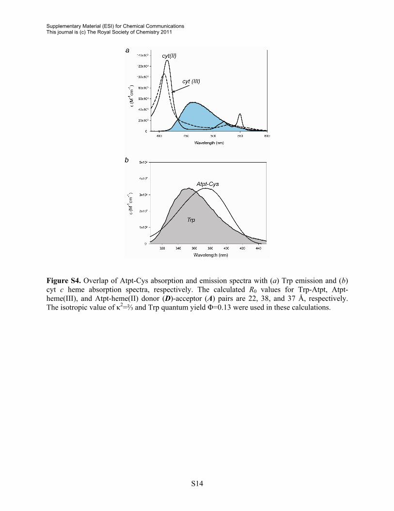
Figure S5. (a) Steady-state spectra and (b) fluorescence decays of Atpt-Cys in a 100 mM NaPi buffer at pH 7.4 in the absence (black) and presence (green) of 10 mM Trp. In both cases, the decays are best described by biexponential functions. The time constants are τ1=6.1±0.2 ns (23%) and τ2=8.6±0.1 ns (77% ) in the absence of Trp and τ1=2.7±0.1 ns (6%) and τ2=7.2±0.1 (94%) with 10 mM Trp.
S15
Supplementary Material (ESI) for Chemical Communications This journal is (c) The Royal Society of Chemistry 2011
S16
References
1. R. N. Warrener, R. A. Russell and S. M. Marcuccio, Aust. J. Chem., 1980, 33, 2777-
2779.
2. G. S. Reddy, H.-Y. Chen and I.-J. Chang, J. Chin. Chem. Soc., 2006, 53, 1303-1308.
3. G. Swoboda and W. Hasselbach, Hoppe-Seylers Z. Physiol. Chem. , 1973, 354, 1611-
1618.
4. E. V. Pletneva, H. B. Gray and J. R. Winkler, J. Mol. Biol., 2005, 345, 855-867.
5. T. Izard, G. Evans, R. A. Borgon, C. L. Rush, G. Bricogne and P. R. Bois, Nature, 2004,
427, 171-175.
6. E. V. Pletneva, H. B. Gray and J. R. Winkler, J. Am. Chem. Soc., 2005, 127, 15370-
15371.
7. J. W. Eastman, Photochem. Photobiol., 1967, 6, 55-72.
8. J. R. Lakowicz, Principles of Fluorescence Spectroscopy, Kluwer Academic/Plenum
Publishing, New York, 1999.
9. N. Maki, Bull. Chem. Soc. Jpn., 1969, 42, 2275-2281.
10. R. D. Farina and R. G. Wilkins, Inorg. Chem., 1968, 7, 514-518.
11. F. Fernandes, L. M. Loura, R. Koehorst, R. B. Spruijt, M. A. Hemminga, A. Fedorov and
M. Prieto, Biophys. J., 2004, 87, 344-352.
12. J. D. Pardee, P. A. Simpson, L. Stryer and J. A. Spudich, The Journal of Cell Biology,
1982, 94, 316-324.
13. E. Pletneva, V., H. B. Gray and J. R. Winkler, unpublished results.
14. C. E. Soltani, E. M. Hotze, A. E. Johnson and R. K. Tweten, J. Biol. Chem., 2007, 282,
15709-15716.