Embed Size (px)
Citation preview
International Journal of
Molecular Sciences
Technical Note
A Simple and Efficient Genetic ImmunizationProtocol for the Production of Highly SpecificPolyclonal and Monoclonal Antibodies againstthe Native Form of Mammalian Proteins
Julie Pelletier 1, Hervé Agonsanou 1,2, Fabiana Manica 1,2 , Elise G. Lavoie 1,2,Mabrouka Salem 1,2, Patrick Luyindula 1,2, Romuald Brice Babou Kammoe 1,2 andJean Sévigny 1,2,*
1 Centre de Recherche du CHU de Québec—Université Laval, Quebec City, QC G1V 4G2, Canada;[email protected] (J.P.); [email protected] (H.A.);[email protected] (F.M.); [email protected] (E.G.L.);[email protected] (M.S.); [email protected] (P.L.);[email protected] (R.B.B.K.)
2 Département de Microbiologie-Infectiologie et d’Immunologie, Faculté de Médecine, Université Laval,Québec City, QC G1V 0A6, Canada
* Correspondence: [email protected]
Received: 14 August 2020; Accepted: 21 September 2020; Published: 25 September 2020�����������������
Abstract: We have generated polyclonal and monoclonal antibodies by genetic immunization overthe last two decades. In this paper, we present our most successful methodology acquired over theseyears and present the animals in which we obtained the highest rates of success. The techniquepresented is convenient, easy, affordable, and generates antibodies against mammalian proteins intheir native form. This protocol requires neither expensive equipment, such as a gene gun, norsophisticated techniques such as the conjugation of gold microspheres, electroporation, or surgeryto inject in lymph nodes. The protocol presented uses simply the purified plasmid expressing theprotein of interest under a strong promoter, which is injected at intramuscular and intradermal sites.This technique was tested in five species. Guinea pigs were the animals of choice for the production ofpolyclonal antibodies. Monoclonal antibodies could be generated in mice by giving, as a last injection,a suspension of transfected cells. The antibodies detected their antigens in their native forms. Theywere highly specific with very low non-specific background levels, as assessed by immune-blots,immunocytochemistry, immunohistochemistry and flow cytometry. We present herein a detailed andsimple procedure to successfully raise specific antibodies against native proteins.
Keywords: immunization; antibody; protocol; guinea pig; cDNA
1. Introduction
Antibodies that detect native proteins with high specificity are essential research tools. To obtainthese precious immunoglobulins, different types of antigens can be used such as synthetic peptidesconjugated to a carrier. The antibodies generated against peptides often do not detect the proteinsof interest in their native forms. To circumvent this limitation, purified proteins can be utilized forimmunization. However, the techniques necessary to purify proteins are laborious and may denaturatethe proteins of interest during the purification steps, especially transmembrane proteins. Furthermore,the level of purity necessary to raise specific antibodies is high as some of the impurities are oftenimmunogenic. A genetic immunization approach represents an interesting alternative [1] but itgenerally generates sera with low titers.
Int. J. Mol. Sci. 2020, 21, 7074; doi:10.3390/ijms21197074 www.mdpi.com/journal/ijms

Int. J. Mol. Sci. 2020, 21, 7074 2 of 16
In genetic immunization, the protein of interest is expressed using a plasmid containing its geneunder the control of a strong enhancer-promoter such as the one from cytomegalovirus (CMV) fora high expression level. This construct is injected into the animal where it is taken up by cells andthe gene of interest is expressed. As a result, the animal reacts against this “non-self” antigen andproduces specific immunoglobulins. This technique has the advantage to produce an antigen 100%pure without any effort. When using the full coding sequence of a mammalian gene, the protein ofinterest undergoes normal post-translational modifications. Therefore, the antibodies produced aredirected against the protein in a normal mammalian form.
Genetic immunization has been used in different species such as rat [2], mouse [3], monkey [4],ferret [4] and rabbit [5]. Thus far, cDNA immunization in the guinea pig was mostly used inmodels of infectious challenge to verify the protective effect of cDNA vaccine. Most of these cDNAvaccine protocols in the guinea pig have been established using electroporation [6,7], which requiresfurther equipment.
In this study, we summarize our results obtained over the last two decades using cDNA injectionin different animal species and we propose an optimized, easy, and convenient protocol for thegeneration of polyclonal as well as monoclonal antibodies. During this work, we observed thatone species in particular produced polyclonal antibodies in a consistent and reproducible manner,namely the guinea pig. Monoclonal antibodies could also be obtained in mice with a similar cDNAimmunization procedure, to which we added a final injection constituted of a suspension of cellstransiently transfected with the protein of interest for a stronger and faster challenge.
2. Results
2.1. Polyclonal Antibodies
2.1.1. Analysis of the Antibodies Produced
Over the last two decades, we have tested different immunization conditions to raise antibodies.We have tested several conditions that allowed us to identify a protocol that is very convenient andreliable to successfully raise specific antibodies excellent for research purposes. This protocol canbe used by laboratories with minimal immunization experience. We will first present some of theimmunization procedures that led us to the protocol that we describe at the end of this manuscript.
The sera obtained were tested by western blot, immunocytochemistry and immunohistochemistryafter the third injection and compared to their respective pre-immune sera collected immediately beforethe first injection. We considered that an animal serum was positive when a specific signal was obtainedeither in western blot, immunohistochemistry or immunocytochemistry, and absent in the pre-immuneserum. The best antibodies were also tested by flow cytometry. It is noteworthy that most of theantibodies that we have generated by cDNA immunization detected the native protein with its normaldisulfide bridges. Therefore, the antibodies generally did not detect the proteins of interest in westernblots under reducing conditions, with either DTT or mercaptoethanol. Broadly speaking, when usingthis immunization technique, antisera that reacted positively in western blot under non-reducingconditions detected also efficiently the antigens by immunocytochemistry, immunohistochemistry andflow cytometry.
Most of the antibodies documented in this study can now be obtained commercially.The monoclonal and polyclonal antibodies to NTPDases, NPPs and CD73 can be obtained atectonucleotidases-ab.com. The antibodies to Robo4, Dectin-2 and RANK were licensed to Medimabsand a different monoclonal anti-human RANK was licensed to Millipore, where they can be obtained.
2.1.2. Administration Routes and Electroporation
Most of the immunization protocols were performed by intramuscular (IM) and intradermal(ID) injections, which is easy and convenient. Alternative routes of injection, some of which having
Int. J. Mol. Sci. 2020, 21, 7074 3 of 16
been reported to elicit a stronger and more rapid immunization [8], were also tested. Administrationin the subscapular area was tested in seven rabbits with different antigens and compared with IDand IM injections on 13 rabbits (see Table 1). Unfortunately, antibodies were produced only in oneof those groups with the plasmid expressing mouse NTPDase2. The serum of the animal injectedin the subscapular region did not give a better signal than those of the two rabbits injected at IDand IM sites. For the six other plasmids (angiomotin, Bmx/Etk, LCCP, mouse NTPDase8, RANK,RANKL), no antibody was produced either by the rabbits injected ID and IM or by those injected inthe subscapular region in addition to ID and IM injections.
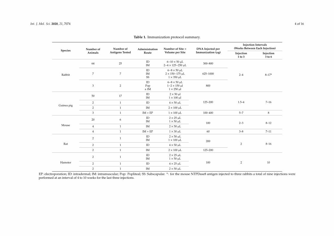
The injection of cDNA in lymph nodes has been reported to increase the antibody response withlower levels of cDNAs [8]. We tested the popliteal lymph node route in rabbits injected with plasmidscoding for mouse NTPDase3 and rat NTPDase6. Among the rabbits injected with the mouse NTPDase3expression plasmid, one was injected at IM and ID sites, and the other rabbit received the same amountof cDNA in ID sites as well as in both popliteal lymph nodes (see Table 1). In this assay, the serum of therabbit that received only IM and ID injection gave a better response by flow cytometry than the serumof the rabbit that received cDNA in the popliteal lymph nodes in addition to ID injections. Meanwhile,as the sera of both of these rabbits showed very high background in western blots (Figure 1D, rabbit “j”and “i”, respectively) it is difficult to conclude whether one of those bands actually corresponds tothe antigen. Similar results were obtained in another series with the plasmid encoding rat NTPDase6.In these experiments, one rabbit received only IM and ID injections and two rabbits received IM, IDand popliteal lymph node injections. The rabbits injected only at IM and ID sites gave a stronger signalby immunohistochemistry than the two rabbits injected in the popliteal lymph nodes (data not shown).
Although our limited study does not allow us to draw a conclusion on the effectiveness of thesetwo injections routes (subscapular and popliteal lymph nodes), we obtained better responses andreliability by ID and IM immunization in the animals tested. It is noteworthy that popliteal lymphnode injection requires a level of surgical technical skills that did not meet our goal of identifyingan easy and convenient procedure of immunization. Furthermore, the latter injection protocols werenot necessary to obtain a good antibody response. Therefore, as these injections routes were moredemanding technically and that they were not improving significantly antibody production with theplasmids tested, we decided to abandon the injections of the subscapular and popliteal lymph nodes.
Electroporation was also reported to increase antibody titer [9,10]. This technique was testedon three guinea pigs and four mice injected with plasmids expressing mouse NTPDase8 and humanNTPDase2, respectively. No specific antibodies were obtained using electroporation in guinea pigswhile positive antisera were obtained with the same plasmid in 10 out of 11 guinea pigs when injectedat ID and IM sites. In our hands, electroporation was very efficient in mice as a host where the sera ofall the treated mice showed a positive immune-blotting signal with a better signal versus backgroundratio than the sera of the two mice that received only ID and IM injections. In agreement with theliterature, electroporation induced a more rapid antibody production than that observed in responseto ID or IM injections alone [9]. For the mice that received DNA-electroporation, one mouse gave apositive signal in western blot after the second injection, two mice after the third injection and thefourth mouse after the fourth injection. In comparison with the two mice that received the sameplasmid at ID and IM sites, one mouse gave a positive signal after the third injection and the othermouse gave a weak positive signal after the fourth injection. Among the animals that gave a positiveresponse with ID and IM immunizations, a positive antiserum was observed after the third injectionin 10% (1 out of 10) in mice, 40% (12 out of 30) in guinea pigs and 62% (16 out of 26) in rabbits. Themajority of the successful animals responded following four sets of ID and IM injections (60% (six outof 10) in mice, 91% (30 out of 33) in guinea pigs and 100% (28 out of 28) in rabbits).
Since ID and IM injections do not require special surgical skills or specialized apparatus such asan electrical device, and as they have been shown to be very efficient routes of injection thus far, weselected those easy and convenient administration routes for our next assays.
Int. J. Mol. Sci. 2020, 21, 7074 4 of 16
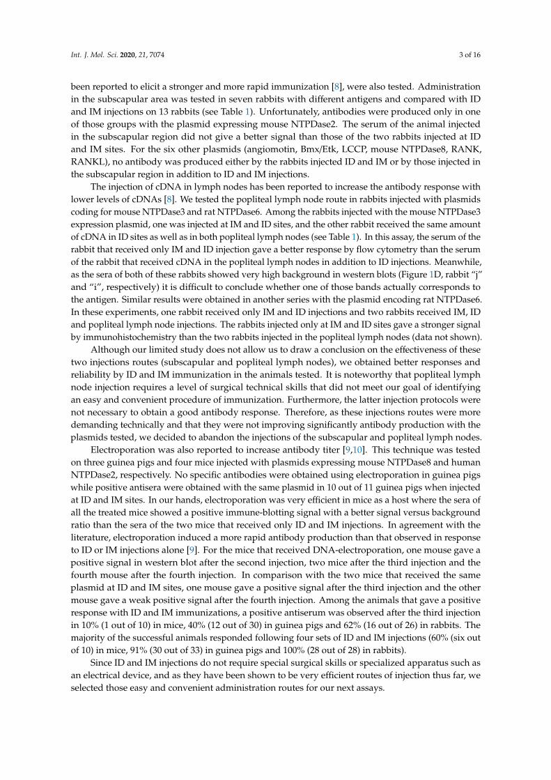
Table 1. Immunization protocol summary.
Species Number ofAnimals
Number ofAntigens Tested
AdministrationRoute
Number of Site ×Volume per Site
DNA Injected perImmunization (µg)
Injection Intervals(Weeks Between Each Injection)
Injection1 to 3
Injection3 to 6
Rabbit
64 25 IDIM
6−10 × 50 µL2−4 × 125−250 µL 300–800
2–4 8–17a7 7IDIMSS
6−8 × 50 µL2 × 150−175 µL
1 × 350 µL625–1000
3 2ID
Pop± IM
6−8 × 50 µL1−2 × 150 µl
1 × 250 µl800
Guinea pig
50 17 IDIM
2 × 50 µl1 × 100 µl
125–200 1.5–4 7–162 1 ID 4 × 50 µL
2 1 IM 2 × 100 µL
3 1 IM + EP 1 × 100 µL 100–400 5–7 8
Mouse
20 6 IDIM
2 × 25 µL1 × 50 µL 100 2–3 8–12
4 1 IM 2 × 50 µL
4 1 IM + EP 1 × 30 µL 60 3–8 7–11
Rat
2 1 IDIM
2 × 50 µL1 × 100 µL 200
2 8–162 1 ID 4 × 50 µL
2 1 IM 2 × 100 µL 125–200
Hamster
2 1 IDIM
2 × 25 µL1 × 50 µL
100 2 102 1 ID 4 × 25 µL
2 1 IM 2 × 50 µL
EP: electroporation; ID: intradermal; IM: intramuscular; Pop: Popliteal; SS: Subscapular. a: for the mouse NTPDase8 antigen injected to three rabbits a total of nine injections wereperformed at an interval of 4 to 10 weeks for the last three injections.
Int. J. Mol. Sci. 2020, 21, 7074 5 of 16
Figure 1. Immuno-blotting-based comparative analysis of rabbit and guinea pig antisera. Lysates forone large well from COS-7 cells or HEK 293T cells transfected with plasmids encoding humanecto-5′-nucleotidase (A), rat ecto-5′-nucleotidase (B), human NTPDase1 (C), mouse NTPDase3(D) or mouse NTPDase8 (E) were subjected to electrophoresis under non-reducing conditions,electrotransferred to an Immobilon-P membrane and probed with rabbits “a” to “p” (left panels)or guinea pig “1” to “17” (right panels) antisera. The sera presented are the pre-immune (Pi) negativecontrols and the immune sera collected after the third (I3), the fourth (I4), the fifth (I5) or the sixth (I6)injection. Specific bands are denoted with an arrow. Multimeric (M) and truncated (T) protein formsare indicated with an arrow head. The antibodies shown were diluted 1:500 except for the rabbit “g” inpanel C and rabbits “k,” “l” and “m” in panel D that were diluted 1:1000.

Int. J. Mol. Sci. 2020, 21, 7074 6 of 16
2.1.3. Immunization of Different Species
Most antibodies raised in this work were produced in rabbits. We injected 25 plasmids to 74rabbits. The cDNA immunization in rabbits led to specific antibodies in 35 out of 74 rabbits (47%).Specific antibodies were also efficiently produced in mice, the animal of choice to generate monoclonalantibodies, with a similar ID and IM immunization protocol as that used in rabbits. Among the 28mice injected with five different plasmids, 16 mice (57%) reacted positively. To compare this cDNAimmunization procedure in different species we have also immunized three rabbits, six hamsters, sixrats and 11 guinea pigs with the same antigen, a plasmid encoding mouse NTPDase8. No specificantibodies were obtained in three rabbits (Figure 1E) or in six hamsters. A positive signal was obtainedin two out of six rats, but only by immuno-cytochemistry (Table 2). In contrast, 10 out of 11 guinea pigsresponded to this plasmid. This prompted us to test this species further with the same immunizationtechnique. A majority of the 54 guinea pigs injected with 17 different plasmids produced a positiveand specific serum with low background (41 positives out of 54 or 76%) (Table 2).
Table 2. Polyclonal antibodies raised.
Species Number of Plasmids Tested Number of Animals Immunized Responding Animals (number, %)
Rabbit 25 74 35, 47%Guinea pig 17 54 41, 76%
Mouse 5 28 16, 57%Rat 1 6 2, 33% *
Hamster 1 6 0
Compilation of animal antisera that were considered positive when a specific signal was obtained in either westernblot, immunohistochemistry or immunocytochemistry, and absent in the pre-immune serum. * A positive signalcould be detected by immunocytochemistry but not by western blot.
As rabbits and guinea pigs are two species of interest for the production of polyclonal antibodiesand as both of these species generated specific antibodies with our protocol, we carried out a systematiccomparison between them by injecting the same six plasmids into these two species. For three of thoseplasmids (those expressing mouse NTPDase1, human ecto-5′-nucleotidase and rat ecto-5′-nucleotidase),antisera with similar signal intensities versus background were obtained in both species. The data arepresented for human ecto-5′-nucleotidase and rat ecto-5′-nucleotidase in Figure 1A,B, respectively.On the other hand, for the three other plasmids (human NTPDase1, mouse NTPDase3 and mouseNTPDase8), rabbits failed to produce specific antibodies, while guinea pigs produced, again, highlyspecific antibodies with low background, as seen by immune blot (Figure 1 and data not shown).Among the 10 rabbits immunized with the latter three plasmids, five rabbits gave a weak positivesignal with high background in western blot for the plasmids encoding human NTPDase1 (two/tworabbits), mouse NTPDase3 (three/five rabbits), mouse NTPDase8 (zero/three rabbits). In contrast,among the 17 guinea pigs injected with those three cDNAs, 11 guinea pigs gave a strong signal withoutbackground and five other guinea pigs showed a moderate, but clean, signal in western blot: humanNTPDase1 (three/three guinea pigs), mouse NTPDase3 (three/three guinea pigs), mouse NTPDase8(10/11 guinea pigs). Most of the antisera produced are presented in Figure 1. In general, guinea pigsgenerated antisera with higher titer and lower background than rabbits.
All antisera were also tested by immunocytochemistry and immunohistochemistry, and thepositive ones by flow cytometry. Nearly all of the antisera that gave a positive signal by westernblot in non-reducing conditions also detected the protein of interest by immunocytochemistry,immunohistochemistry and flow cytometry, and vice versa. The antiserum mN3-3c against mouseNTPDase3 (rabbit “12” in Figure 1D) is presented as an example in Figure 2 and the specificity for theantisera mN1-1c and rN3-1L are presented in Figure 3.

Int. J. Mol. Sci. 2020, 21, 7074 7 of 16
Figure 2. Specificity of the guinea pig anti-mouse NTPDase3 antibody mN3-3c. (A) Strong signals inimmunocytochemistry of transfected COS-7 cells with a plasmid encoding mouse NTPDase3 (mN3)are only detected with antiserum mN3-3c (rabbit “12” in Figure 1D). No signals are detected with thepre-immune serum or with the anti-serum on un-transfected cells (COS-7). (B) Immuno- histochemistryof serial sections from a mouse pancreas. The antiserum displays a positive reaction on the cells of theLangerhans islets. (C) Flow cytometry of transfected HEK 293T cells with a plasmid encoding mouseNTPDase3 shows a rightward shift (right panel) when compared to its pre-immune control (left panel).Nuclei were stained in blue with hematoxylin (A,B).
Int. J. Mol. Sci. 2020, 21, 7074 8 of 16
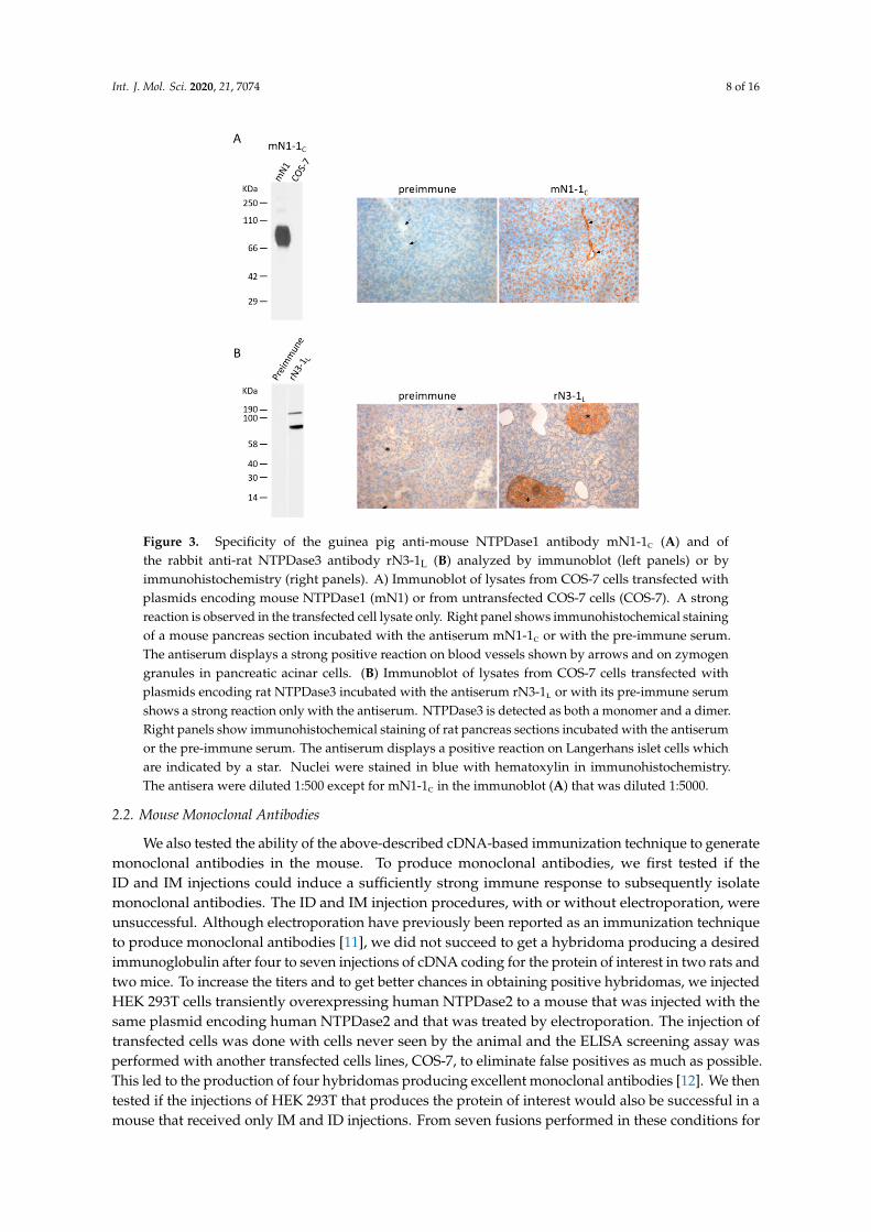
Figure 3. Specificity of the guinea pig anti-mouse NTPDase1 antibody mN1-1c (A) and ofthe rabbit anti-rat NTPDase3 antibody rN3-1L (B) analyzed by immunoblot (left panels) or byimmunohistochemistry (right panels). A) Immunoblot of lysates from COS-7 cells transfected withplasmids encoding mouse NTPDase1 (mN1) or from untransfected COS-7 cells (COS-7). A strongreaction is observed in the transfected cell lysate only. Right panel shows immunohistochemical stainingof a mouse pancreas section incubated with the antiserum mN1-1c or with the pre-immune serum.The antiserum displays a strong positive reaction on blood vessels shown by arrows and on zymogengranules in pancreatic acinar cells. (B) Immunoblot of lysates from COS-7 cells transfected withplasmids encoding rat NTPDase3 incubated with the antiserum rN3-1l or with its pre-immune serumshows a strong reaction only with the antiserum. NTPDase3 is detected as both a monomer and a dimer.Right panels show immunohistochemical staining of rat pancreas sections incubated with the antiserumor the pre-immune serum. The antiserum displays a positive reaction on Langerhans islet cells whichare indicated by a star. Nuclei were stained in blue with hematoxylin in immunohistochemistry.The antisera were diluted 1:500 except for mN1-1c in the immunoblot (A) that was diluted 1:5000.
2.2. Mouse Monoclonal Antibodies
We also tested the ability of the above-described cDNA-based immunization technique to generatemonoclonal antibodies in the mouse. To produce monoclonal antibodies, we first tested if theID and IM injections could induce a sufficiently strong immune response to subsequently isolatemonoclonal antibodies. The ID and IM injection procedures, with or without electroporation, wereunsuccessful. Although electroporation have previously been reported as an immunization techniqueto produce monoclonal antibodies [11], we did not succeed to get a hybridoma producing a desiredimmunoglobulin after four to seven injections of cDNA coding for the protein of interest in two rats andtwo mice. To increase the titers and to get better chances in obtaining positive hybridomas, we injectedHEK 293T cells transiently overexpressing human NTPDase2 to a mouse that was injected with thesame plasmid encoding human NTPDase2 and that was treated by electroporation. The injection oftransfected cells was done with cells never seen by the animal and the ELISA screening assay wasperformed with another transfected cells lines, COS-7, to eliminate false positives as much as possible.This led to the production of four hybridomas producing excellent monoclonal antibodies [12]. We thentested if the injections of HEK 293T that produces the protein of interest would also be successful in amouse that received only IM and ID injections. From seven fusions performed in these conditions for
Int. J. Mol. Sci. 2020, 21, 7074 9 of 16
human NTPDase1, human NTPDase2, human NTPDase3, human NTPDase8 and human Rank, threefusion procedures allowed us to obtain hybridomas that produced monoclonal antibodies againsthuman NTPDase3 [13,14], human NTPDase8 [15] and human RANK. These data suggest that ID andIM injections, without electroporation, can be sufficient to prime the animals, and that a final injectionwith the recombinant protein expressed by transfected cells induces a rapid and high production ofactivated B cells suitable for the isolation of specific hybridomas.
2.3. Protocol Proposed
The above data led us to propose the protocol in Table 3 that we now routinely use for rabbits,guinea pigs and mice. The plasmid containing the protein of interest is injected at ID sites and atIM sites at an interval of 2 to 3 weeks for the first three injections and then at 8 to 10 weeks intervalsfor the two subsequent injections. For guinea pigs and rabbits, we suggest testing a serum sampleafter the third and fourth injection to identify the good responders. If a strong immunoreaction isobserved after the third injection, rabbits and guinea pigs should be exsanguinated after a fourthand final immunization. If guinea pigs and rabbits do not respond after the fourth injection, theyshould be sacrificed as they will unlikely respond with more injections. On the other hand, five cDNAimmunization procedures should be done in mice before selecting the best one for the final injectionbefore fusion with myeloma cells.
Table 3. Proposed protocol to raise polyclonal and monoclonal antibodies by cDNA immunization.
Species RouteNumber of Sites× Volume per
Site
DNAConcentration
(mg/mL)
DNA Injectedper
Immunization(µg)
Injection Intervals(Weeks between Each
Injection)
BloodCollection(Days afterInjection)
SpleenCollection (Daysafter TransfectedCell Injection *)
Number ofAnimals
perAntigen
Injection1 to 3
Injection3 to 5 #
Rabbit IDIM
6–10 × 50 µL2–4 × 125–250 µL 0.5–0.8 500–800 & 2–3 8 ¶ 13–14 N/A 3–5
Guineapig
IDIM
2 × 50 µL1 × 100 µL 1 200 2–3 8 ¶ 12–13 N/A 2–3
Mouse IDIM
2 × 25 µL1 × 50 µL 1 100 2–3 7¶ 12–13 3 5–10
The final injection before the fusion with SP2/0 cells should be done with 10 to 18 million HEK 293T transfectedcells using a high efficiency transfection system. Other related cell lines can be used for transfection. # If a strongimmunoreaction is observed after the third injection, rabbits and guinea pigs should be exsanguinated after a fourthand final immunization. & A lower amount of plasmids (650 ± 50 µg) is suggested for the first four injections, and ahigher amount (800 µg) for the last injection. ¶ As the animals remain primed for several weeks to months, theintervals between the last injections can be longer.
As reliability to get antibodies is better in the guinea pig species, we suggest a lower number ofguinea pigs (two or three) than rabbits (three to five) or mice (five to 10). Indeed, we obtained similarexcellent antisera in all responding guinea pigs tested, which was not the case for the two other species.As mice exhibited a highly variable response from animal to animal (57% of the mice respondedpositively) while not requiring large amounts of plasmids, and having low housing costs, it would bewise to use two cages of mice (eight–10 mice) in order to get a better probability in obtaining a highresponder in the first four or five injections to be selected for the final injection with transfected cellsand for fusion with myeloma cells. The final injection can be done with 10 to 18 million HEK 293T cellstransfected with the same plasmid used for the ID and IM injections. We recommend collecting thespleen three days later to perform the fusion with SP2/0 myelomas cells.
3. Discussion
We produced in the last two decades several antibodies using cDNA immunization techniques.We tested different strategies that finally led us to propose the protocol presented in Table 3 that we nowuse routinely in guinea pigs. When larger amount of serum or when antibodies from different speciesare necessary, or in the rare case where guinea pigs do not respond, we also use rabbits. Obviously,
Int. J. Mol. Sci. 2020, 21, 7074 10 of 16
when a monoclonal antibody is necessary, we do it in mice with a similar protocol, also presented inTable 3, with the difference that we perform a final injection with the recombinant proteins to elicit afaster and stronger response using an expression cell system that has never been seen by the immunizedanimals. Indeed, the injection of HEK 293T cells expressing the protein of interest three days beforethe fusion procedure was more successful to obtain clones expressing the monoclonal antibodies ofinterest than injecting only DNA for all immunization steps. Other groups have also come to the sameconclusion [16,17]. Obviously, this last injection would be inappropriate when generating polyclonalantibodies as the background would be expected to increase, which is not an issue when producingmonoclonal antibodies as the desired B cells are cloned. Along the same line, it may be advantageousto transfect a mouse cell line for the final injection to reduce the number of false positive clones, butthis might trigger a weaker immunological response and cause lower chances in getting hybridomas.This could be a subject for future improvement.
Guinea pigs are often the only species that we now immunize as they are high responders to thisprotocol and the response is generally similar from one animal to the other, which was not the casewith mice and rabbits. There is therefore no need to inject five or six guinea pigs with our technique,as is often suggested in immunization protocols for most animals. Another advantage of using guineapigs over rabbits is the much lower amount of DNA necessary (three–four times less) to trigger anantibody response. Depending of the animal facilities, housing costs for guinea pigs are also generallyless expensive than rabbits.
Antibody response has been reported to be increased when injecting the cDNA in lymph nodes,or when using electroporation, but as plasmids can be easily produced in large amount, and with anextremely high purity, it is much easier to produce more plasmids to inject at ID and IM sites ratherthan to inject in the lymph nodes to save some plasmids. This is especially important when the surgeryskills are not at hand. The same situation applies when comparing the technique that we present inTable 3 with other techniques using expensive technologies such as the injection of gold or tungstenconjugated particles with a gene gun. As the technique presented in Table 3 is efficient and easy toperform, there is no need to buy expensive equipment or to use unnecessarily sophisticated techniques.
cDNA immunization technique, as presented here, elicited an antibody response that reactedagainst the antigen in its native form. This is of interest when using plasmids encodingmammalian protein antigens to immunize guinea pigs, rabbits or mice that will perform mammalianpost-translational modifications, although they can differ slightly between mammalian species.As post-translational modifications are different in non-mammalian species, the protocol proposed heremight not be as efficient to raise antibodies against antigens in their native forms when they originatefrom non-mammalian sources, such as for antigens from prokaryotes or plants. This will depend onthe epitope(s) selected by the host animal. Another issue, especially when using non mammalianantigens, is to make sure that the expression level of the gene is adequate in the host animal. This canbe corrected or ameliorated by codon optimization of the gene of interest. In agreement with this idea,an increased antibody response was reported by a few groups after optimizing the codon sequencesof some HIV genes [18–21]. Post-translational modifications and codon optimization could thereforerepresent a subject for future amelioration of cDNA immunization.
Another point to consider when using this technique is the protein location after its expressionfrom the plasmid. Indeed, as we did in this paper, cDNA immunization has mostly been usedto produce antibodies against transmembrane proteins. Several groups have also reported somesuccess in generating antibodies against intracellular and secreted proteins by gene gun [22,23] orelectroporation [24–26]. Efficiency to produce antibodies against an intracellular protein by injectingcDNA at ID or IM sites differs from group to group. Few studies have compared the same intracellularprotein with or without a signal peptide to mediate protein secretion. In one of these studies, a plasmidwith or without such a signal sequence was injected IM and a similar antibody response was reportedfor both plasmids [27]. Other studies reported a better efficiency when a signal sequence was added toa gene coding for an intracellular antigen [28,29]. In the results presented here, the majority of the
Int. J. Mol. Sci. 2020, 21, 7074 11 of 16
plasmids encoded a transmembrane protein which led to specific antibody in 80% (41 out of 51) inguinea pigs, 60% (33 out of 55) in rabbits and 57% (16 out of 28) in mice. In our study, we did notsucceed to raise antibodies in rabbits (zero out of 11) injected with a plasmid coding for an intracellularprotein. The immune responses to an antigen encoded by plasmids coding for secreted proteins wereslightly stronger. Although we did not succeed in guinea pigs (zero out of three) a few rabbits (twoout of eight) produced antibodies against a secreted protein. Another alternative to get antibodies tosoluble proteins is to modify the protein by adding a transmembrane domain to direct the protein tothe external surface of the plasma membrane. We tested this idea with one plasmid, which successfullyproduced antibodies in one out of five mice (unpublished data in collaboration with Dr J. Fietto,Universidade federal de Viçosa, Brazil). This is another avenue of study to make cDNA immunizationmore efficient for more protein types.
As there is no immunization technique that works all the time, when should we stop to immunizean animal with our protocol presented on Table 3? We observed that when there was no positivesignal after the fourth injection in guinea pigs and in rabbits, there was limited chance to obtain a goodantiserum by doing additional injections. Indeed, the few times that a fifth injection was positive forthe guinea pigs, the titer was too low to be useful for a research purpose. Therefore, we now sacrificeall guinea pigs and rabbits that do not give a good response in the first four immunizations. On theother hand, injecting a good responder too many times might not be beneficial, and on the contrary,the antibody response might decrease and the background signals may increase. Accordingly, when arabbit or a guinea pig responds well at the third injection, it should be exsanguinated after the fourthinjection. Our observations were different in mice as we observed some mice that did not react at thefourth injection but gave an interesting response after the fifth injection. But as this was seen onlyin a few mice (four out of 10) with the same plasmid, we cannot draw any definitive conclusions atthe moment concerning this point. Nevertheless, it might be safer to try a fifth DNA injection beforesacrificing mice. Indeed, according to our results, mice often required five cDNA injections to get astrong positive signal.
4. Materials and Methods
4.1. Materials
Hypoxanthine-aminopterin-thymidine solution, hypoxanthine-thymidine solution, poly-ethyleneglycol and bovine serum albumin were purchased from Sigma-Aldrich (Oakville, ON, Canada).Dulbecco’s modified Eagle’s medium and Lipofectamine were obtained from Life Technologies(Burlington, ON, Canada). Fetal bovine serum (FBS), phosphate-buffered saline (PBS),antibiotic/antimycotic and HCell-100 hybridomas media were purchased from Wisent (St-Bruno,QC, Canada). Secondary antibodies conjugated to horseradish peroxidase (HRP) were obtained from:goat anti-mouse IgG (H + L) from Jackson ImmunoResearch Laboratories Inc. (West Grove, PA, USA),goat anti-guinea pig from Santa Cruz Biotechnology (Dallas, TX, USA), donkey anti-rabbit from GEHealthcare Life Sciences (Baie d’Urfe, QC, Canada). The secondary goat anti-guinea pig antibodyconjugated to biotin was from Jackson ImmunoResearch Laboratories Inc. (West Grove, PA, USA).Enhanced K-Blue® Substrate was from Neogen Corporation (Lansing, MI, USA).
4.2. Animals
Female New Zealand rabbits (4 months), Hartley guinea pigs (6–7 weeks), BALB/c mice (4–6 weeks),Sprague-Dawley rats (7 weeks), LVG Golden Syrian hamsters (7 weeks), were obtained from CharlesRiver Laboratories (St-Constant, QC, Canada). For two antigen series, rabbits (four) were 9–10 weeks old.All procedures were approved by the Canadian Council on Animal Care and the Université LavalAnimal Welfare Committee (protocol number: 2001-120; 2004-179; 2007-124; 2010-102; 2013-108;2017-109).
Int. J. Mol. Sci. 2020, 21, 7074 12 of 16
4.3. Immunization
Complementary DNA (cDNA) was prepared using Endofree plasmid kits from Qiagen (Toronto,ON, Canada) and diluted in PBS 0.8× at a concentration range of 0.5–1 mg/mL. Intradermal (ID) orintramuscular (IM) injections were performed in the dorsal skin or in a thigh, respectively. Two groupsof rabbits received the cDNA in the popliteal lymph node(s). This injection route was performed onceduring the injection schedule and the rabbits received ID and IM injections for the other injections.In one of these groups, rabbits received DNA coding for mouse NTPDase3 in popliteal lymph nodes ofboth legs at the second injection in addition to ID injections. In the second group, rabbits receivedDNA coding for rat NTPDase6 in a popliteal lymph node of one leg at the first injection, in addition toID and IM injection. Specific information related to the number of sites, amount injected and injectionschedule is reported in Table 1. Each animal received at least five series of immunization to assureadequate priming. In some cases, animal received up to nine series of immunization.
4.4. Electroporation
Mice and guinea pigs received DNA followed by an electroporation as previously described [9,12].Briefly, an IM injection of DNA diluted in Hank’s balanced salt solution or in PBS, for the mouse orthe guinea pig, respectively, was made in the tibialis anterior. After application of an electrode creamon the skin, electroporation was carried out at the site of DNA injection with two electrode platesconnected to an electroporator according to the following parameters: seven pulses of 100 V.cm−1 anda duration of 20 ms.
4.5. Blood Collection
Blood was collected before the first injection and 11 to 15 days after the second injection whenan electroporation was performed, or after each injection after the third one. Blood was collected inspray-coated silica tubes with or without gel separator or in regular microcentrifuge tubes. Tubes werelet in the upright position for 1–2 h at room temperature (RT) to allow clot formation, and were thencentrifuged at 1500× g for 10–30 min depending on the blood volume. A final concentration of 10%glycerol was added to the serum which was then aliquoted and stored at −80 ◦C until needed.
4.6. Monoclonal Antibodies Raised in Mice
After four to six DNA injections with or without electroporation, as described in Table 1, some micereceived a final injection of DNA, and others an injection of human embryonic kidney (HEK 293T)transfected cells. These cells were transfected with Lipofectamine as previously described [30] withthe same plasmid used for the previous immunization procedures. Then, 2 days after transfection, thecells were washed twice with PBS and the cells were detached by a 2–5 min incubation at 37 ◦C ina citric saline solution (135 mM potassium chloride, 15 mM sodium citrate). Cells were centrifuged,resuspended in PBS and counted. The mouse received a final intraperitoneal injection of 10 to 18 millionof transfected HEK 293T. The fusion procedure was performed either 6–7 days, or 3 days, after thisfinal injection of DNA, or the cell suspension, respectively. Blood and spleen cells were collectedafter this final injection and fusion with SP2/0 cells was done as described [13]. The supernatant wasscreened by ELISA and the positive hybridomas were cloned by limiting dilution. In a few assays,a hybridomas optimized medium, Hcell-100, was used for the clone isolation procedures. The producedimmunoglobulins were purified on Protein A Sepharose CL-4B column as described [13].
4.7. ELISA
ELISA was performed as previously described [15]. In brief, protein extract (500 ng per well) fromuntransfected African green monkey kidney (COS-7) cells or transiently transfected with the plasmidused for immunization diluted in PBS was distributed in a 96 well ELISA plate and incubated overnightat 4 ◦C. The wells were then washed with PBS-Tween 0.05% (PBS-T) and incubated for 1 h at 37 ◦C in

Int. J. Mol. Sci. 2020, 21, 7074 13 of 16
a blocking solution (0.5% bovine serum albumin diluted in PBS-T). After washing, the supernatantfrom each hybridoma was added to a well and incubated for 2 h at RT, followed by four washing steps.Then, a goat anti-mouse IgG (H + L)-HRP (1:2500) diluted in the blocking solution was incubatedfor 2 h at RT, followed by four washing steps. The Enhanced K-Blue® Substrate was then added for15 min and the reaction was stopped by the addition of an equal volume of 2 N sulphuric acid. Theabsorbance at 450 nm was then recorded.
4.8. Isotyping
Mouse Immunoglobulin Isotyping ELISA Kit (BD Bioscience, Mississauga, ON, Canada) was usedto determine the isotypes produced by the hybridomas, according to the manufacturer’s instructionand as previously described [15]. In brief, monoclonal rat anti-mouse IgG1, IgG2a, IgG2b, IgG3, IgAand IgM were coated O/N in 96-well plates. A blocking treatment was performed after washingsteps, and then each monoclonal antibody was transferred to the wells. After four washing steps,rat anti-mouse Igs conjugated to HRP was added to each well, and revealed with a substrate providedin the kit. The plate was then read at 450 nm.
4.9. Western Blot
COS-7 cells and HEK 293T cells were cultured and transiently transfected with the indicatedcDNA construct as described previously [30]. For western blot assays, lysates from transfected COS-7cells or HEK 293T cells were resuspended in NuPAGE LDS sample buffer, separated on NuPAGE4–12% Bis-Tris gels under non-reduced conditions, and transferred to an Immobilon-P membrane(Millipore, Bedford, MA, USA) by electroblotting according to the manufacturer’s recommendation andas previously described [15]. Membranes were then blocked with 2.5% non-fat milk in PBS containing0.15% Tween20 (pH 7.4) O/N at 4◦C and subsequently probed with the primary antibodies using theMini-Protean II multiscreen apparatus (Bio-Rad Laboratories Ltd., Mississauga, ON, Canada) in which20 antibodies can be tested on one gel. Appropriate secondary HRP-conjugated antibodies were used,and the membranes developed with the western LightningTM Plus-ECL system (PerkinElmer Life andAnalytical Sciences, Waltham, MA, USA).
4.10. Immunocytochemistry and Immunohistochemistry
Immunocytochemistry and immunohistochemistry were performed as previously described [15].Tissues were frozen in Tissue-Tek®O.C.T.TM Compound (Sakura Finetek, Torrance, CA, USA). COS-7cells or tissue sections (6 µm thick) were fixed with cold acetone and 10% phosphate-buffered formalin(Fisher Scientific, Ottawa, ON, Canada) (19:1) and blocked in a PBS solution containing 7% normalgoat serum for 30 min. COS-7 cells and tissue sections were incubated with the indicated primaryantibody at 4 ◦C or pre-immune serum as a negative control. COS-7 cells and tissue sections werethen treated with 0.15% H2O2 in PBS for 10 min to inactivate endogenous peroxidase, and with anavidin/biotin solution (Avidin/Biotin Blocking kit; Vector Laboratories, Burlington, ON, Canada) toprevent non-specific staining due to endogenous biotin. This step was followed by incubation with anappropriate biotin-conjugated secondary antibody at a dilution of 1:1000. The avidin–biotinylatedHRP complex (VectaStain Elite ABC kit; Vector Laboratories) was added to optimize the reaction.Peroxidase activity was revealed with DAB as the substrate. Nuclei were counterstained with aqueoushematoxylin (Biomeda, Foster City, CA, USA) in accordance with the manufacturer’s instructions.
4.11. Flow Cytometry
Flow cytometry was performed as previously described [15]. Briefly, HEK 293T cells transfectedwith the plasmid expressing the antigen were detached from the plates with a citric saline solution(135 mM potassium chloride, 15 mM sodium citrate). Cells were washed with an ice-cold PBS solutioncontaining 1% FBS and 0.1% NaN3 (fluorescence-activated cell sorting (FACS) buffer) followed byincubation with the immune serum or the pre-immune serum in FACS buffer for 1 h. After washes
Int. J. Mol. Sci. 2020, 21, 7074 14 of 16
with FACS buffer solution, the cells were incubated with the appropriate FITC-conjugated secondaryantibody (Jackson ImmunoResearch Laboratories Inc., West Grove, PA, USA) for 30 min on ice, washedwith FACS buffer, and analyzed by flow cytometry (BD LSR II, BD Biosciences, San Jose, CA USA).
5. Conclusions
In this paper we propose a detailed protocol for cDNA immunization in guinea pigs and rabbitsfor the production of polyclonal antibodies and in mice for the generation of monoclonal antibodies.The procedure does not require any sophisticated technical expertise nor any specialized equipment.This protocol works especially well in guinea pigs which produce antisera with high specificity andextremely low background in a consistent manner, allowing the use of fewer animals per antigento reduce effective costs, especially when comparing with larger animals. To generate monoclonalantibodies in mice, we propose to perform the last injection with a suspension of cells expressing therecombinant protein at a high level.
Author Contributions: Conceptualization, J.S.; Methodology, J.P., E.G.L. and J.S.; Validation, J.P., H.A., F.M.,E.G.L., M.S., P.L., R.B.B.K. and J.S.; Formal analysis, J.P.; Investigation, J.S.; Resources, J.S.; Data Curation, J.P.,E.G.L. and J.S.; Writing—Original Draft Preparation, J.P.; Writing—Review and Editing, J.S.; Visualization, J.P.;Supervision, J.S.; Project Administration, J.S.; Funding Acquisition, J.S. All authors have read and agreed to thepublished version of the manuscript.
Funding: This work was supported by grants to J. Sévigny from the Natural Sciences and Engineering ResearchCouncil of Canada (NSERC; RGPIN-2016-05867). J.S. was also a recipient of a “Chercheur National” Scholarshipfrom the Fonds de Recherche du Québec-Santé (FRQS). H. Agonsanou was a recipient of a scholarship from theMinistère de la Santé publique du Bénin, F. Manica of a scholarship from The Canadian Bureau for InternationalEducation, E.G. Lavoie and M. Salem of a scholarship from the FRQS, P. Luyindula of a scholarship from theCanadian Francophonie Scholarship Program and R.B. Babou Kammoe of a studentship from Université Laval.
Acknowledgments: The authors thank the following research teams with whom some of the antibodies presentedhere were generated and characterized: Paul H. Naccache, Maria J. Fernandes, Patrice E. Poubelle, Sylvain G.Bourgoin (Québec, Canada), Simon C. Robson (Boston, USA) and Herbert Zimmermann (Frankfurt am Main,Germany). We also thank Aileen F. Knowles (San Diego, USA; human NTPDase2), Terence L. Kirley (Cincinnati,USA; human NTPDase3 and human NTPDase6), James W. Goding (Victoria, Australia; human NPP1), KimihikoSano (Kobe, Japan; human NPP2 and human NPP3) and Mathieu Bollen (Leuven, Belgium; mouse NPP1) forproviding the indicated expression plasmids used in this study. We are also grateful to Paul H. Naccache forediting this manuscript.
Conflicts of Interest: The authors declare no conflict of interest.
Abbreviations
cDNA Complementary DNACOS-7 African green monkey kidney cellsDAB 3, 3′ diaminobenzidineELISA Enzyme-linked immunosorbent assayFBS Fetal bovine serumFRQS Fonds de Recherche du Québec – SantéHEK 293T Human embryonic kidney 293T cellsHRP Horseradish peroxidaseID IntradermalIM IntramuscularPBS Phosphate-buffered salinePBS-T PBS-TweenPi Pre-immune serumRT Room temperatureNSERC Natural Sciences and Engineering Research Council of Canada
References
1. Tang, D.C.; DeVit, M.; Johnston, S.A. Genetic immunization is a simple method for eliciting an immuneresponse. Nature 1992, 356, 152–154. [CrossRef] [PubMed]
Int. J. Mol. Sci. 2020, 21, 7074 15 of 16
2. Aoyama, T.; Kamata, K.; Yamanaka, N.; Takeuchi, Y.; Higashihara, M.; Kato, S. Characteristics ofpolyclonal anti-human nephrin antibodies induced by genetic immunization using nephrin cDNA.Nephrol. Dial. Transplant. 2006, 21, 1073–1081. [CrossRef] [PubMed]
3. Morel, P.A.; Falkner, D.; Plowey, J.; Larregina, A.T.; Falo, L.D. DNA immunisation: Altering the cellularlocalisation of expressed protein and the immunisation route allows manipulation of the immune response.Vaccine 2004, 22, 447–456. [CrossRef] [PubMed]
4. Donnelly, J.J.; Friedman, A.; Martinez, D.; Montgomery, D.L.; Shiver, J.W.; Motzel, S.L.; Ulmer, J.B.; Liu, M.A.Preclinical efficacy of a prototype DNA vaccine: Enhanced protection against antigenic drift in influenzavirus. Nat. Med. 1995, 1, 583–587. [CrossRef]
5. Diestre, C.; Martínez-Lorenzo, M.; Bosque, A.; Naval, J.; Larrad, L.; Anel, A. Generation of rabbit antibodiesagainst death ligands by cDNA immunization. J. Immunol. Methods 2006, 317, 12–20. [CrossRef]
6. Schultheis, K.; Schaefer, H.; Yung, B.S.; Oh, J.; Muthumani, K.; Humeau, L.; Broderick, K.E.; Smith, T.R.Characterization of guinea pig T cell responses elicited after EP-assisted delivery of DNA vaccines to theskin. Vaccine 2016, 35, 61–70. [CrossRef]
7. Cashman, K.A.; Wilkinson, E.R.; Wollen-Roberts, S.E.; Shamblin, J.D.; Zelko, J.M.; Bearss, J.J.; Zeng, X.;Broderick, K.E.; Schmaljohn, C.S. DNA vaccines elicit durable protective immunity against individual orsimultaneous infections with Lassa and Ebola viruses in guinea pigs. Hum. Vaccines Immunother. 2017, 13,3010–3019. [CrossRef]
8. Maloy, K.J.; Erdmann, I.; Basch, V.; Sierro, S.; Kramps, T.A.; Zinkernagel, R.M.; Oehen, S.; Kündig, T.M.Intralymphatic immunization enhances DNA vaccination. Proc. Natl. Acad. Sci. USA 2001, 98, 3299–3303.[CrossRef]
9. Widera, G.; Austin, M.; Rabussay, D.; Goldbeck, C.; Barnett, S.W.; Chen, M.; Leung, L.; Otten, G.R.;Thudium, K.; Selby, M.J.; et al. Increased DNA vaccine delivery and immunogenicity by electroporationin vivo. J. Immunol. 2000, 164, 4635–4640. [CrossRef]
10. Sardesai, N.Y.; Weiner, D.B. Electroporation delivery of DNA vaccines: Prospects for success.Curr. Opin. Immunol. 2011, 23, 421–429. [CrossRef]
11. Yang, L.; Cheong, N.; Wang, D.Y.; Lee, B.W.; Kuo, I.C.; Huang, C.H.; Chua, K.Y. Generation of monoclonalantibodies against Blot 3 using DNA immunization with in vivo electroporation. Clin. Exp. Allergy 2003, 33,663–668. [CrossRef] [PubMed]
12. Pelletier, J.; Agonsanou, H.; Delvalle, N.; Fausther, M.; Salem, M.; Gulbransen, B.; Sévigny, J. Generationand characterization of polyclonal and monoclonal antibodies to human NTPDase2 including a blockingantibody. Purinergic Signal. 2017, 13, 293–304. [CrossRef]
13. Munkonda, M.N.; Pelletier, J.; Ivanenkov, V.V.; Fausther, M.; Tremblay, A.; Kunzli, B.; Kirley, T.L.; Sévigny, J.Characterization of a monoclonal antibody as the first specific inhibitor of human NTP diphosphohydrolase-3:Partial characterization of the inhibitory epitope and potential applications. FEBS J. 2009, 276, 479–496.[CrossRef] [PubMed]
14. Saunders, D.C.; Brissova, M.; Phillips, N.; Shrestha, S.; Walker, J.T.; Aramandla, R.; Poffenberger, G.;Flaherty, D.K.; Weller, K.P.; Pelletier, J.; et al. Ectonucleoside triphosphate diphosphohydrolase-3 antibodytargets adult human pancreatic beta cells for in vitro and in vivo analysis. Cell Metab. 2018, 29, 745–754.[CrossRef] [PubMed]
15. Pelletier, J.; Salem, M.; Lecka, J.; Fausther, M.; Bigonnesse, F.; Sévigny, J. Generation and characterization ofspecific antibodies to the murine and human ectonucleotidase NTPDase8. Front. Pharmacol. 2017, 8, 115.[CrossRef]
16. Nagata, S.; Salvatore, G.; Pastan, I. DNA immunization followed by a single boost with cells: A protein-freeimmunization protocol for production of monoclonal antibodies against the native form of membraneproteins. J. Immunol. Methods 2003, 280, 59–72. [CrossRef]
17. Chu, T.T.; Halverson, G.R.; Yazdanbakhsh, K.; Øyen, R.; Reid, M. A DNA-based immunization protocol toproduce monoclonal antibodies to blood group antigens. Br. J. Haematol. 2001, 113, 32–36. [CrossRef]
18. André, S.; Seed, B.; Eberle, J.; Schraut, W.; Bültmann, A.; Haas, J. Increased immune response elicited byDNA vaccination with a synthetic gp120 sequence with optimized codon usage. J. Virol. 1998, 72, 1497–1503.[CrossRef]
Int. J. Mol. Sci. 2020, 21, 7074 16 of 16
19. Deml, L.; Bojak, A.; Steck, S.; Graf, M.; Wild, J.; Schirmbeck, R.; Wolf, H.; Wagner, R. Multiple effects of codonusage optimization on expression and immunogenicity of DNA candidate vaccines encoding the humanimmunodeficiency virus type 1 gag protein. J. Virol. 2001, 75, 10991–11001. [CrossRef]
20. Megede, J.Z.; Chen, M.-C.; Doe, B.; Schaefer, M.; Greer, C.E.; Selby, M.; Otten, G.R.; Barnett, S.W. Increasedexpression and immunogenicity of sequence-modified human immunodeficiency virus type 1 gag gene.J. Virol. 2000, 74, 2628–2635. [CrossRef]
21. Wang, S.; Farfan-Arribas, D.J.; Shen, S.; Chou, T.H.W.; Hirsch, A.; He, F.; Lu, S. Relative contributions ofcodon usage, promoter efficiency and leader sequence to the antigen expression and immunogenicity ofHIV-1 Env DNA vaccine. Vaccine 2006, 24, 4531–4540. [CrossRef] [PubMed]
22. García, J.F.; García, J.F.; Maestre, L.; Lucas, E.; Sánchez-Verde, L.; Romero-Chala, S.; Piris, M.Á.; Roncador, G.Genetic immunization: A new monoclonal antibody for the detection of BCL-6 protein in paraffin sections.J. Histochem. Cytochem. 2006, 54, 31–38. [CrossRef] [PubMed]
23. Maestre, L.; Fontán, L.; Martinez-Climent, J.A.; Garcia, J.F.; Cigudosa, J.C.; Roncador, G. Generation of anew monoclonal antibody against MALT1 by genetic immunization. Hybridoma 2007, 26, 86–91. [CrossRef][PubMed]
24. Leinonen, J.; Niemelä, P.; Lövgren, J.; Bocchi, L.; Pettersson, K.; Nevanlinna, H.; Stenman, U.-H.Characterization of monoclonal antibodies against prostate specific antigen produced by geneticimmunization. J. Immunol. Methods 2004, 289, 157–167. [CrossRef] [PubMed]
25. Chen, Y.; Zhang, T.; Li, T.; Han, W.; Zhang, Y.; Ma, D. Preparation and characterization of a monoclonalantibody against CKLF1 using DNA immunization with in vivo electroporation. Hybridoma 2005, 24, 305–308.[CrossRef]
26. Daftarian, P.; Chowdhury, R.; Ames, P.; Wei, C.; King, A.D.; Vaccari, J.P.D.R.; Dillon, L.; Price, J.; Leung, H.;Ashlock, B.; et al. In vivo electroporation and non-protein based screening assays to identify antibodiesagainst native protein conformations. Hybridoma 2011, 30, 409–418. [CrossRef]
27. Haddad, D.; Liljeqvist, S.; Stahl, S.; Andersson, I.; Perlmann, P.; Berzins, K.; Ahlborg, N. Comparativestudy of DNA-based immunization vectors: Effect of secretion signals on the antibody responses in mice.FEMS Immunol. Med. Microbiol. 1997, 18, 193–202.
28. Svanholm, C.; Bandholtz, L.; Lobell, A.; Wigzell, H. Enhancement of antibody responses by DNAimmunization using expression vectors mediating efficient antigen secretion. J. Immunol. Methods 1999, 228,121–130. [CrossRef]
29. Inchauspé, G.; Vitvitski, L.; Major, M.E.; Jung, G.; Spengler, U.; Maisonnas, M.; Trepo, C. Plasmid DNAexpressing a secreted or a nonsecreted form of hepatitis C virus nucleocapsid: Comparative studies ofantibody and T-helper responses following genetic immunization. DNA Cell Biol. 1997, 16, 185–195.[CrossRef]
30. Kukulski, F.; Lévesque, S.A.; Lavoie, É.G.; Lecka, J.; Bigonnesse, F.; Knowles, A.F.; Robson, S.C.; Kirley, T.L.;Sévigny, J. Comparative hydrolysis of P2 receptor agonists by NTPDases 1, 2, 3 and 8. Purinergic Signal. 2005,1, 193–204. [CrossRef]
© 2020 by the authors. Licensee MDPI, Basel, Switzerland. This article is an open accessarticle distributed under the terms and conditions of the Creative Commons Attribution(CC BY) license (http://creativecommons.org/licenses/by/4.0/).


![Immunization Program Strategic Plan 2013 – 2017 · 1 2013-2017 Immunization Program [Immunization Program Strategic Plan 2013 – 2017] Maintaining and Improving Immunization Rates](https://img.pdfslide.us/doc/110x75/5e18e16c0228f448f3787c8f/immunization-program-strategic-plan-2013-a-2017-1-2013-2017-immunization-program.jpg)