Embed Size (px)
Citation preview
1442 Research Article
IntroductionIn mammals, adult neurogenesis was demonstrated to be activein two main brain regions, namely the subventricular zone(SVZ) and the dentate gyrus of the hippocampus, where neuralstem cells (NSCs) were isolated (Alvarez-Buylla and Garcia-Verdugo, 2002; Galli et al., 2003). NSCs are characterized byplasticity and multipotency so that they may proliferate and/orundergo differentiation into functional progeny according tothe environmental conditions (Bottai et al., 2003). Thesefeatures make them ideal candidate targets for the cure ofneurodegenerative diseases, although the molecularmechanisms that determine a NSC to proliferate or todifferentiate towards a neuronal or astroglial fate are onlypartially elucidated (Qian et al., 2000; Roegiers and Jan, 2004).Post-transcriptional regulation, exerted by RNA-bindingproteins (RBPs) as an alternative mechanism to the activity oftranscription factors in modulating gene expression, hasrecently been shown to have a role in the development of thecentral nervous system (CNS) and, in particular, in the controlof the NSC division mode (Okano et al., 2002). Several neural-specific RBPs have been described so far to affect splicing,transport, translation and stability of target mRNAs, and can
cause severe neurological disorders when altered (Perrone-Bizzozero and Bolognani, 2002).
The Musashi-1 (Msi1) RBP was first reported to be requiredfor the proper development of the neural sensory organ inDrosophila (Nakamura et al., 1994), whereas in mammals it iscommonly considered a specific marker for stem/progenitorcells of neural origin (Kaneko et al., 2000; Maslov et al., 2004).Msi1 acts as a translational suppressor by binding to the 3�-untranslated region (3�UTR) of specific mRNA targets. In thisway the proliferation state of NSCs is maintained by inhibitingthe translation of the membrane protein Numb, involved in theNotch/Delta signaling cascade (Imai et al., 2001), and of thecyclin-dependent kinase inhibitor p21WAF-1 (Battelli et al.,2006).
The neuronal-specific ELAV (nELAV) RBPs, which areassociated with the pathological condition of paraneoplasticencephalomyelitis (Szabo et al., 1991), are necessary andsufficient to induce neuronal differentiation in mammaliancells (Akamatsu et al., 1999; Kasashima et al., 1999), as is theirelav ortholog in Drosophila (Robinow and White, 1991). Thethree mammalian nELAV family members, HuB, HuC andHuD, are commonly referred to as early and specific markers
Post-transcriptional regulation exerted by neural-specificRNA-binding proteins plays a pivotal role in thedevelopment and maintenance of the nervous system.Neural ELAV proteins are key inducers of neuronaldifferentiation through the stabilization and/ortranslational enhancement of target transcripts bearing theAU-rich elements (AREs), whereas Musashi-1 maintainsthe stem cell proliferation state by acting as a translationalrepressor. Since the gene encoding Musashi-1 (Msi1)contains a conserved ARE in its 3�� untranslated region, wefocused on the possibility of a mechanistic relationshipbetween ELAV proteins and Musashi-1 in cell fatecommitment. Colocalization of neural ELAV proteins withMusashi-1 clearly shows that ELAV proteins are expressedat early stages of neural commitment, whereas interactionstudies demonstrate that neural ELAV proteins exert anARE-dependent binding activity on the Msi1 mRNA. This
binding activity has functional effects, since the ELAVprotein family member HuD is able to stabilize the Msi1ARE-containing mRNA in a sequence-dependent way in adeadenylation/degradation assay. Furthermore activationof the neural ELAV proteins by phorbol esters in humanSH-SY5Y cells is associated with an increase of Musashi-1protein content in the cytoskeleton. We propose that ELAVRNA-binding proteins exert an important post-transcriptional control on Musashi-1 expression in thetransition from proliferation to neural differentiation ofstem/progenitor cells.
Supplementary material available online athttp://jcs.biologists.org/cgi/content/full/119/7/1442/DC1
Key words: ELAV, Musashi-1, Neurogenesis, RNA-binding protein,Stem cell
Summary
A role for the ELAV RNA-binding proteins in neuralstem cells: stabilization of Msi1 mRNAAntonia Ratti1,*, Claudia Fallini1, Lidia Cova1, Roberto Fantozzi1, Cinzia Calzarossa1, Eleonora Zennaro1,Alessia Pascale2, Alessandro Quattrone3 and Vincenzo Silani11Department of Neuroscience, ‘Dino Ferrari’ Centre, University of Milan–IRCCS Istituto Auxologico Italiano, Via Zucchi 18, 20095 Cusano Milanino,Italy2Department of Experimental and Applied Pharmacology, University of Pavia, Via Taramelli 14, 27100 Pavia, Italy3Laboratory of Metabolomics and Systems Biology, Magnetic Resonance Center and FiorGen Foundation, University of Florence, Via Sacconi 6,50019 Sesto Fiorentino, Italy*Author for correspondence (e-mail: [email protected])
Accepted 21 December 2005Journal of Cell Science 119, 1442-1452 Published by The Company of Biologists 2006doi:10.1242/jcs.02852
Jour
nal o
f Cel
l Sci
ence
1443Regulation of Msi1 mRNA by the nELAV proteins
of post-mitotic neurons during CNS development (Barami etal., 1995; Okano and Darnell, 1997; Wakamatsu and Weston,1997), whereas the fourth member, HuR, is ubiquitouslyexpressed. The ELAV RNA-binding activity promotes thestabilization and/or translation of an array of transcriptscontaining the AU-rich consensus element (ARE) in their3�UTR (Antic and Keene, 1997). ARE sequences representwell-documented cis-acting regulatory motifs usuallyidentified in mRNAs of genes with a high turnover rate (Shawand Kamen, 1986), and are recognized by several ARE-bindingproteins which exert a different and often opposite role onmRNA fate (Bevilacqua et al., 2003). nELAV target genes areendowed with a wide variety of biological functions (Gao etal., 1994), from cell growth regulation (Levine et al., 1993) tobrain maturation and maintenance (Antic et al., 1999; Aranda-Abreu et al., 1999; Chung et al., 1997).
The expression of Msi1 and nELAV RBPs has been initiallydescribed to be spatiotemporally sequential during thedevelopment of murine CNS (Sakakibara et al., 1996),although recent data suggest that nELAV proteins might havean important role as early as the neuronal-specific commitmentof NSCs (Akamatsu et al., 2005).
In the present work we show that nELAV proteins areexpressed and colocalize with Msi1 in the adult mouse SVZand in cultured NSCs/progenitors, where they show a specificand ARE-dependent binding activity for the Msi1 transcript. Inparticular, we find that nELAV RBPs exert a stabilizing activityon Msi1 mRNA decreasing its turnover rate in vitro andpromoting its translation in vivo. Such findings suggest amechanistic correlation between nELAV and Msi1 RBPs incontrolling the proliferation/differentiation activities of neuralstem/progenitor cells.
ResultsThe mammalian Msi1 3�UTR contains a conserved AU-rich element which is bound by multiple RBPs includingthe nELAV proteinsExamination of the mouse Msi1 gene revealed the presence ofa putative ARE consensus sequence in its 3�UTR, cataloguedas belonging to group III in the ARE-mRNA database (ARED)(Bakheet et al., 2001). Since AREs are cis-acting regulatorysites, they are usually well conserved throughout phylogenesis(Asson-Batres et al., 1994), and indeed a comparative analysisof Msi1 3�UTRs in ortholog genes revealed that the humansequence showed a 75% identity with its murine counterpart(Fig. 1A). The EST-expanded rat Msi1 3�UTR displayed thesame degree of conservation (74%), and alignment of the threesequences gave almost full identity of a region of 24 bp,including the putative functional ARE. On the other hand,Danio rerio, Xenopus laevis, Caenorhabditis elegans andDrosophila melanogaster Msi1 orthologs neither contained theARE signature nor showed any significant homology along theentire 3�UTR region. Also Msi2 gene, the ubiquitouslyexpressed member of the Musashi family (Sakakibara et al.,2001), didn’t show any ARE consensus sequence in its 3�UTR.Therefore, we hypothesized that the ARE sequence present inthe Msi1 gene could have a role in influencing its mRNAstability and in the post-transcriptional regulation of itsexpression only in mammals. Prediction of the mouse Msi13�UTR secondary structure by the Sfold RNA-foldingalgorithm (Ding et al., 2004) clearly showed that the ARE
sequence is exposed in a single-stranded loop, which is likelyto be accessible to ARE-binding proteins (Fig. 1B).
In order to investigate the putative RNA-binding activitiesassociated with the Msi1 transcript, we performedRNA/protein UV crosslinking experiments in the presence ofradiolabeled Msi1 3�UTR riboprobe and protein extracts fromboth mouse brain and cultured NSCs. After UV irradiation, theresulting mRNA-protein (mRNP) complexes were separatedby SDS-PAGE and the electrophoretic pattern was comparedwith that obtained for the growth-associated protein 43(Gap43), a well-known mRNA target of the nELAV RBPs. Apartially overlapping profile was evident with common bandsof approximately 95, 65 and 42 kDa (Fig. 1C), the latter beingpreviously shown to correspond to the RNA-binding activityof HuD (Chung et al., 1997). We observed two additionalmRNP complexes specific to the Msi1 3�UTR, with apparentmolecular weights of 37 and 32 kDa, the lower band beingmore abundant in NSCs and the upper one in whole brainlysate.
These preliminary findings prompted us to investigate thepossible role of the nELAV RBPs as trans-acting factors in thebinding and regulation of the Msi1 transcript. To this purposeafter UV crosslinking assays, the resulting mRNP complexeswere immunoprecipitated with the pan anti-nELAV antibody(16A11), which is known to recognize the three neuronal RBPsHuB, HuC and HuD, but not HuR (Marusich et al., 1994) (Fig.1D). A 42 kDa complex was immunoprecipitated in thepresence of the Msi1 3�UTR riboprobe, confirming theexistence of an nELAV RNA-binding activity for the Msi1transcript both in mature brain and in NSCs.
As several RBPs are reported to bind to their own mRNA(Chu et al., 1991; Schaeffer et al., 2001), including nELAVproteins (Abe et al., 1996; Samson, 1998), we alsoimmunoprecipitated the products of the UV crosslinkingreaction using the anti-Msi1 antibody, but the Msi1 transcriptwas not recovered (Fig. 1E).
nELAV proteins are expressed in neural stem/progenitorcells in vitroOur preliminary data suggest the existence of a functionalcorrelation between the Msi1 gene, associated with themaintenance of NSC proliferation, and the nELAV RBPs,usually considered early markers of newly generated neurons.NSCs isolated from developing or mature brain can be grownin vitro as floating cell aggregates denominated neurospheres,which are characterized by an indefinite proliferationpotentiality and by the multipotency to differentiate into theprincipal neural phenotypes (Morshead and van der Kooy,2004). We therefore looked for nELAV protein expression inneurospheres obtained from adult mouse brains.Immunolabeling images with the anti-Msi1 antibody showedcolocalization with the nELAV antigens throughout the sphere(Fig. 2A-C), suggesting a stem identity of nELAV-positivecells. To further characterize the expression of these RBPs,from tridimensional neurospheres we obtained a monolayer ofstem/progenitor cells after dissociation and exposure toadhesive substrate for 1 hour. This limited period of timeallows the preservation of the typical NSC heterogeneousmorphologies and it is not sufficient to commit cells towards aspecific fate (Bez et al., 2003; Suslov et al., 2002). As inneurosphere cultures, double fluorescent signals for nELAV
Jour
nal o
f Cel
l Sci
ence

1444
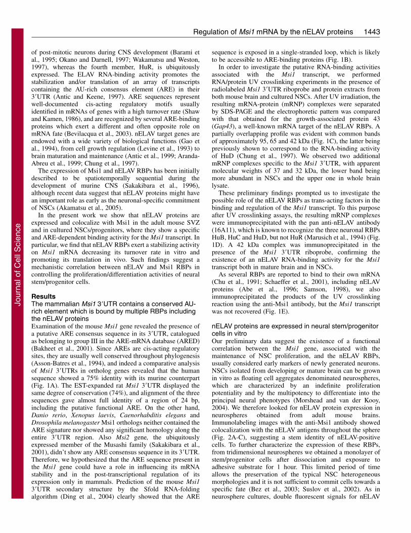
and Msi1 were still present although their cellular distributionappeared slightly different, Msi1 signals being prevalent in thenuclei and nELAV RBP signals more diffuse in the cytoplasm(Fig. 2D-F). Coexpression of the intermediate filament nestin,widely used as a neural stem/progenitor cell marker (Wiese etal., 2004), with both nELAV and Msi1 demonstrated the stillundifferentiated and proliferating state of our neurosphere-derived cell culture (data not shown). The specificity of theanti-Msi1 antibody used was previously verified in committedprogenitor cells with neuronal morphology, which resultedpositive for the differentiation marker �-tubulinIII and negativefor Msi1, and in immature astrocytes, positive for both GFAPand Msi1 (Sakakibara et al., 1996) (data not shown).
Furthermore, the uncommitted mitotic state of theneurosphere-derived cells was attested by the positive labelingfor the proliferation-associated antigen Ki67, a proteinproduced in all active phases of the cell cycle, but absent in G0(Kee et al., 2002). nELAV RBPs and Ki67 were co-expressed
Journal of Cell Science 119 (7)
in actively proliferating cells with a distinct cellulardistribution, nuclear for Ki67 and mainly cytoplasmic fornELAV proteins (Fig. 2G-I). nELAV protein staining was alsopresent in non-proliferating Ki67-negative cells.
nELAV RBPs are expressed in the neurogenic SVZ invivoSince our immunostaining data clearly indicate that nELAVproteins are not limited to early post-mitotic and matureneurons, we wanted to investigate nELAV expression in vivoin the SVZ region, one of the germinal areas whereneurogenesis occurs in the adult. Brain sections from adult ratswere immunolabeled with anti-nELAV and anti-Ki67antibodies (Fig. 3A,B): nELAV proteins were clearly expressedin the SVZ and overlapped the Ki67 fluorescent signals with acomplementary intracellular distribution pattern (Fig. 3C,D).As neuronal-specific markers, nELAV proteins were alsostrongly expressed in the cerebral areas surrounding the lateral
Fig. 1. Computational analysis of the Msi1 3�UTR sequence and identification of the associated RBP activities. (A) Alignment of themammalian Msi1 3�UTR sequences is shown: Homo sapiens (Hs, NM_002442) at the top, Rattus norvegicus (Rn, CD568097) in the middleand Mus musculus (Mm, NM_008629) at the bottom. Conserved nucleotides are highlighted in grey, whereas the ARE is indicated by the box.(B) The most stable RNA secondary structure of the mouse Msi1 3�UTR as predicted by the Sfold software (www.bioinfo.rpi.edu). Themagnification shows the loop exposing the ARE sequence, indicated by the arrow. (C) UV crosslinking assay of Msi1 and Gap43 3�UTRriboprobes and lysates from whole mouse brain and NSCs. The 95 kDa complex is detected only when brain extracts were used. The 37 and 32kDa RNA-binding activities specific for Msi1 sequence are indicated by asterisks. (D) Immunoprecipitation of Msi1 and Gap43 riboprobes withthe anti-nELAV antibody after UV irradiation on brain and NSC extracts. A 42 kDa complex (arrowhead) was precipitated in all samples.(E) Immunoprecipitation of the NSC UV crosslinked mRNPs with the anti-nELAV and the anti-Msi1 antibodies, respectively.
Jour
nal o
f Cel
l Sci
ence
1445Regulation of Msi1 mRNA by the nELAV proteins
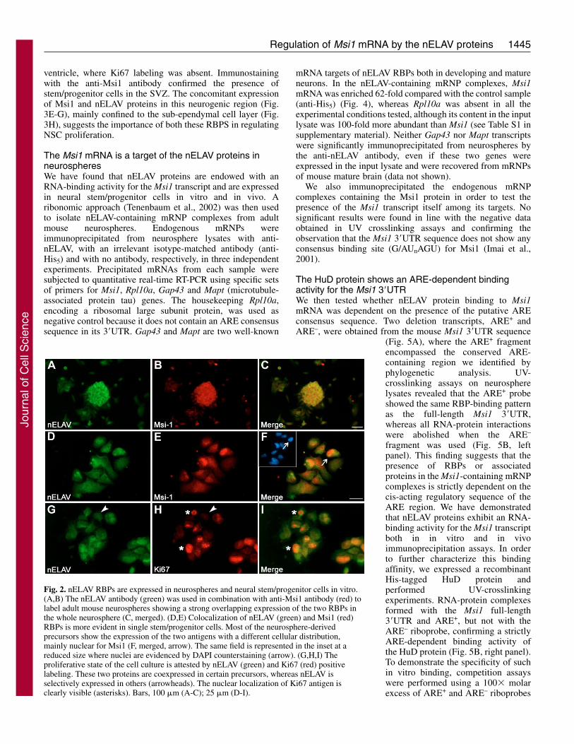
ventricle, where Ki67 labeling was absent. Immunostainingwith the anti-Msi1 antibody confirmed the presence ofstem/progenitor cells in the SVZ. The concomitant expressionof Msi1 and nELAV proteins in this neurogenic region (Fig.3E-G), mainly confined to the sub-ependymal cell layer (Fig.3H), suggests the importance of both these RBPS in regulatingNSC proliferation.
The Msi1 mRNA is a target of the nELAV proteins inneurospheresWe have found that nELAV proteins are endowed with anRNA-binding activity for the Msi1 transcript and are expressedin neural stem/progenitor cells in vitro and in vivo. Aribonomic approach (Tenenbaum et al., 2002) was then usedto isolate nELAV-containing mRNP complexes from adultmouse neurospheres. Endogenous mRNPs wereimmunoprecipitated from neurosphere lysates with anti-nELAV, with an irrelevant isotype-matched antibody (anti-His5) and with no antibody, respectively, in three independentexperiments. Precipitated mRNAs from each sample weresubjected to quantitative real-time RT-PCR using specific setsof primers for Msi1, Rpl10a, Gap43 and Mapt (microtubule-associated protein tau) genes. The housekeeping Rpl10a,encoding a ribosomal large subunit protein, was used asnegative control because it does not contain an ARE consensussequence in its 3�UTR. Gap43 and Mapt are two well-known
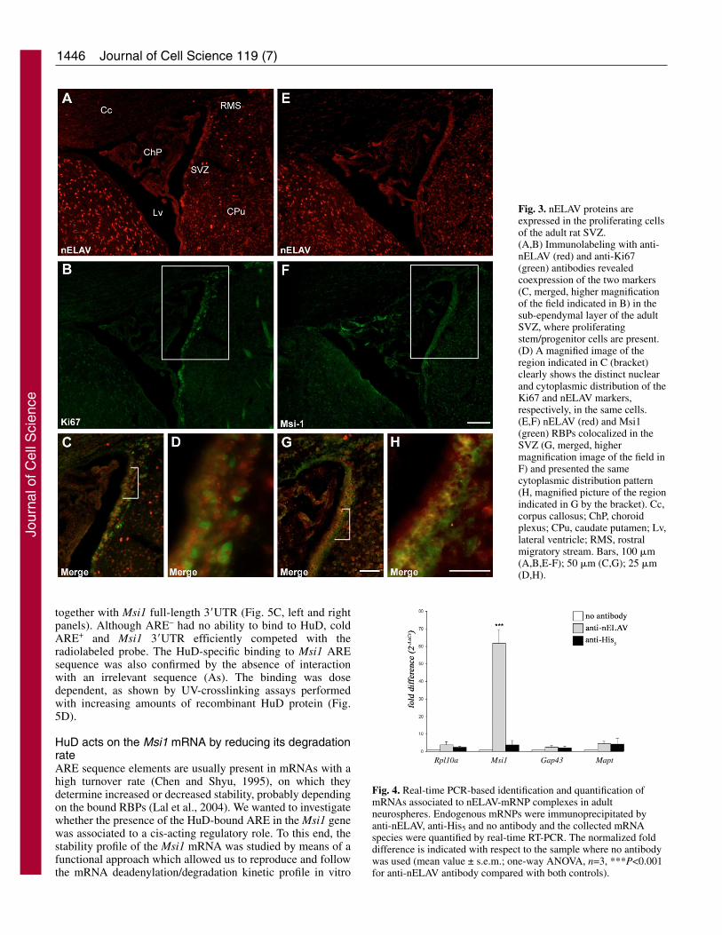
mRNA targets of nELAV RBPs both in developing and matureneurons. In the nELAV-containing mRNP complexes, Msi1mRNA was enriched 62-fold compared with the control sample(anti-His5) (Fig. 4), whereas Rpl10a was absent in all theexperimental conditions tested, although its content in the inputlysate was 100-fold more abundant than Msi1 (see Table S1 insupplementary material). Neither Gap43 nor Mapt transcriptswere significantly immunoprecipitated from neurospheres bythe anti-nELAV antibody, even if these two genes wereexpressed in the input lysate and were recovered from mRNPsof mouse mature brain (data not shown).
We also immunoprecipitated the endogenous mRNPcomplexes containing the Msi1 protein in order to test thepresence of the Msi1 transcript itself among its targets. Nosignificant results were found in line with the negative dataobtained in UV crosslinking assays and confirming theobservation that the Msi1 3�UTR sequence does not show anyconsensus binding site (G/AUnAGU) for Msi1 (Imai et al.,2001).
The HuD protein shows an ARE-dependent bindingactivity for the Msi1 3�UTRWe then tested whether nELAV protein binding to Msi1mRNA was dependent on the presence of the putative AREconsensus sequence. Two deletion transcripts, ARE+ andARE–, were obtained from the mouse Msi1 3�UTR sequence
(Fig. 5A), where the ARE+ fragmentencompassed the conserved ARE-containing region we identified byphylogenetic analysis. UV-crosslinking assays on neurospherelysates revealed that the ARE+ probeshowed the same RBP-binding patternas the full-length Msi1 3�UTR,whereas all RNA-protein interactionswere abolished when the ARE–
fragment was used (Fig. 5B, leftpanel). This finding suggests that thepresence of RBPs or associatedproteins in the Msi1-containing mRNPcomplexes is strictly dependent on thecis-acting regulatory sequence of theARE region. We have demonstratedthat nELAV proteins exhibit an RNA-binding activity for the Msi1 transcriptboth in in vitro and in vivoimmunoprecipitation assays. In orderto further characterize this bindingaffinity, we expressed a recombinantHis-tagged HuD protein andperformed UV-crosslinkingexperiments. RNA-protein complexesformed with the Msi1 full-length3�UTR and ARE+, but not with theARE– riboprobe, confirming a strictlyARE-dependent binding activity ofthe HuD protein (Fig. 5B, right panel).To demonstrate the specificity of suchin vitro binding, competition assayswere performed using a 100� molarexcess of ARE+ and ARE– riboprobes
Fig. 2. nELAV RBPs are expressed in neurospheres and neural stem/progenitor cells in vitro.(A,B) The nELAV antibody (green) was used in combination with anti-Msi1 antibody (red) tolabel adult mouse neurospheres showing a strong overlapping expression of the two RBPs inthe whole neurosphere (C, merged). (D,E) Colocalization of nELAV (green) and Msi1 (red)RBPs is more evident in single stem/progenitor cells. Most of the neurosphere-derivedprecursors show the expression of the two antigens with a different cellular distribution,mainly nuclear for Msi1 (F, merged, arrow). The same field is represented in the inset at areduced size where nuclei are evidenced by DAPI counterstaining (arrow). (G,H,I) Theproliferative state of the cell culture is attested by nELAV (green) and Ki67 (red) positivelabeling. These two proteins are coexpressed in certain precursors, whereas nELAV isselectively expressed in others (arrowheads). The nuclear localization of Ki67 antigen isclearly visible (asterisks). Bars, 100 �m (A-C); 25 �m (D-I).
Jour
nal o
f Cel
l Sci
ence
1446
together with Msi1 full-length 3�UTR (Fig. 5C, left and rightpanels). Although ARE– had no ability to bind to HuD, coldARE+ and Msi1 3�UTR efficiently competed with theradiolabeled probe. The HuD-specific binding to Msi1 AREsequence was also confirmed by the absence of interactionwith an irrelevant sequence (As). The binding was dosedependent, as shown by UV-crosslinking assays performedwith increasing amounts of recombinant HuD protein (Fig.5D).
HuD acts on the Msi1 mRNA by reducing its degradationrateARE sequence elements are usually present in mRNAs with ahigh turnover rate (Chen and Shyu, 1995), on which theydetermine increased or decreased stability, probably dependingon the bound RBPs (Lal et al., 2004). We wanted to investigatewhether the presence of the HuD-bound ARE in the Msi1 genewas associated to a cis-acting regulatory role. To this end, thestability profile of the Msi1 mRNA was studied by means of afunctional approach which allowed us to reproduce and followthe mRNA deadenylation/degradation kinetic profile in vitro
Journal of Cell Science 119 (7)
Fig. 3. nELAV proteins areexpressed in the proliferating cellsof the adult rat SVZ.(A,B) Immunolabeling with anti-nELAV (red) and anti-Ki67(green) antibodies revealedcoexpression of the two markers(C, merged, higher magnificationof the field indicated in B) in thesub-ependymal layer of the adultSVZ, where proliferatingstem/progenitor cells are present.(D) A magnified image of theregion indicated in C (bracket)clearly shows the distinct nuclearand cytoplasmic distribution of theKi67 and nELAV markers,respectively, in the same cells.(E,F) nELAV (red) and Msi1(green) RBPs colocalized in theSVZ (G, merged, highermagnification image of the field inF) and presented the samecytoplasmic distribution pattern(H, magnified picture of the regionindicated in G by the bracket). Cc,corpus callosus; ChP, choroidplexus; CPu, caudate putamen; Lv,lateral ventricle; RMS, rostralmigratory stream. Bars, 100 �m(A,B,E-F); 50 �m (C,G); 25 �m(D,H).
Rpl10a Msi1 Gap43 Mapt
Fig. 4. Real-time PCR-based identification and quantification ofmRNAs associated to nELAV-mRNP complexes in adultneurospheres. Endogenous mRNPs were immunoprecipitated byanti-nELAV, anti-His5 and no antibody and the collected mRNAspecies were quantified by real-time RT-PCR. The normalized folddifference is indicated with respect to the sample where no antibodywas used (mean value ± s.e.m.; one-way ANOVA, n=3, ***P<0.001for anti-nELAV antibody compared with both controls).
Jour
nal o
f Cel
l Sci
ence
1447Regulation of Msi1 mRNA by the nELAV proteins
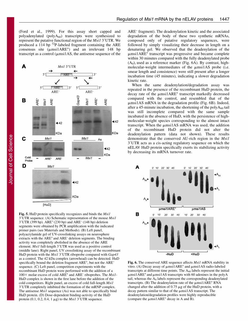
(Ford et al., 1999). For this assay short capped andpolyadenylated (polyA60) transcripts were synthesized torepresent the putative functional region of the Msi1 3�UTR. Weproduced a 114 bp 32P-labeled fragment containing the AREconsensus site (�msi1ARE+) and an irrelevant 148 bptranscript as a control (�msi1AS, the antisense sequence of the
ARE– fragment). The deadenylation kinetic and the associateddegradation of the body of these two synthetic mRNAs,composed only of putative regulatory sequences, werefollowed by simply visualizing their decrease in length on adenaturing gel. We observed that the deadenylation of the�msi1ARE+ transcript was progressive and became completewithin 30 minutes compared with the fully deadenylated probe(A0), used as a reference marker (Fig. 6A). By contrast, high-molecular-weight intermediates of the �msi1AS probe (i.e.smear length and consistence) were still present after a longerincubation time (45 minutes), indicating a slower degradationkinetic rate.
When the same deadenylation/degradation assay wasrepeated in the presence of the recombinant HuD protein, thedecay rate of the �msi1ARE+ transcript markedly decreasedcompared with the control, and resembled that of the�msi1AS mRNA in the degradation profile (Fig. 6B). Indeed,after a 45-minute incubation, the shortening of the polyA60 tailwas still incomplete compared with the same sampleincubated in the absence of HuD, with the persistence of high-molecular-weight species corresponding to the almost intacttranscript. When the �msi1AS mRNA was used, the additionof the recombinant HuD protein did not alter thedeadenylation pattern (data not shown). These resultsdemonstrate that the conserved AU-rich region in the Msi13�UTR acts as a cis-acting regulatory sequence on which thenELAV HuD protein specifically exerts its stabilizing activityby decreasing its mRNA turnover rate.
Fig. 5. HuD protein specifically recognizes and binds the Msi13�UTR sequence. (A) Schematic representation of the mouse Msi13�UTR (399 bp). ARE+ (230 bp) and ARE– (148 bp) deletionsegments were obtained by PCR amplification with the indicatedprimer pairs (see Materials and Methods). (B) Left panel,polyacrylamide gel of UV-crosslinking assays on neurosphereextracts with the ARE+ and ARE– deletion segments. The bindingactivity was completely abolished in the absence of the AREelement. Msi1 full-length 3�UTR was used as a positive control(middle lane). Right panel, UV crosslinking assay of the recombinantHuD protein with the Msi1 3�UTR riboprobe compared with Gap43as a control. The 42 kDa complex (arrowhead) can be detected. HuDspecifically bound the deletion fragment ARE+, but not the ARE–
sequence. (C) Left panel, competition experiments with therecombinant HuD protein were performed with the addition of a100� molar excess of cold ARE+ and ARE– riboprobes. The Msi1-HuD complex is shown in the first lane before the addition of thecold competitors. Right panel, an excess of cold full-length Msi13�UTR completely inhibited the formation of the mRNP complex.The antisense Msi1 sequence (As) was not able to specifically bindHuD protein. (D) Dose-dependent binding-activity of the HuDprotein (0.1, 0.2, 0.4, 1 �g) to the Msi1 3�UTR sequence.
Fig. 6. The conserved ARE sequence affects Msi1 mRNA stability invitro. (A) Decay assay of �msi1ARE+ and �msi1AS radio-labeledtranscripts at different time points. The A60 labels represent the initial�msi1ARE+ and �msi1AS transcripts with 60 adenines in the polyAtail, whereas the A0 labels represent the corresponding deadenylatedtranscripts. (B) The deadenylation rate of the �msi1ARE+ RNAchanged after the addition of 0.75 �g of the HuD protein, with adecay pattern similar to that of the �msi1AS transcript. Thedeadenylation/degradation profiles were highly reproducible(compare the �msi1ARE+ decay in A and B).
Jour
nal o
f Cel
l Sci
ence
1448
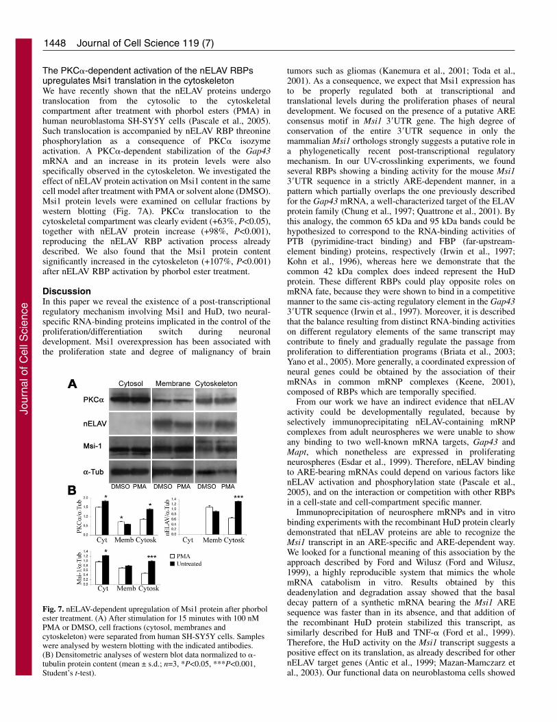
The PKC�-dependent activation of the nELAV RBPsupregulates Msi1 translation in the cytoskeletonWe have recently shown that the nELAV proteins undergotranslocation from the cytosolic to the cytoskeletalcompartment after treatment with phorbol esters (PMA) inhuman neuroblastoma SH-SY5Y cells (Pascale et al., 2005).Such translocation is accompanied by nELAV RBP threoninephosphorylation as a consequence of PKC� isozymeactivation. A PKC�-dependent stabilization of the Gap43mRNA and an increase in its protein levels were alsospecifically observed in the cytoskeleton. We investigated theeffect of nELAV protein activation on Msi1 content in the samecell model after treatment with PMA or solvent alone (DMSO).Msi1 protein levels were examined on cellular fractions bywestern blotting (Fig. 7A). PKC� translocation to thecytoskeletal compartment was clearly evident (+63%, P<0.05),together with nELAV protein increase (+98%, P<0.001),reproducing the nELAV RBP activation process alreadydescribed. We also found that the Msi1 protein contentsignificantly increased in the cytoskeleton (+107%, P<0.001)after nELAV RBP activation by phorbol ester treatment.
DiscussionIn this paper we reveal the existence of a post-transcriptionalregulatory mechanism involving Msi1 and HuD, two neural-specific RNA-binding proteins implicated in the control of theproliferation/differentiation switch during neuronaldevelopment. Msi1 overexpression has been associated withthe proliferation state and degree of malignancy of brain
Journal of Cell Science 119 (7)
tumors such as gliomas (Kanemura et al., 2001; Toda et al.,2001). As a consequence, we expect that Msi1 expression hasto be properly regulated both at transcriptional andtranslational levels during the proliferation phases of neuraldevelopment. We focused on the presence of a putative AREconsensus motif in Msi1 3�UTR gene. The high degree ofconservation of the entire 3�UTR sequence in only themammalian Msi1 orthologs strongly suggests a putative role ina phylogenetically recent post-transcriptional regulatorymechanism. In our UV-crosslinking experiments, we foundseveral RBPs showing a binding activity for the mouse Msi13�UTR sequence in a strictly ARE-dependent manner, in apattern which partially overlaps the one previously describedfor the Gap43 mRNA, a well-characterized target of the ELAVprotein family (Chung et al., 1997; Quattrone et al., 2001). Bythis analogy, the common 65 kDa and 95 kDa bands could behypothesized to correspond to the RNA-binding activities ofPTB (pyrimidine-tract binding) and FBP (far-upstream-element binding) proteins, respectively (Irwin et al., 1997;Kohn et al., 1996), whereas here we demonstrate that thecommon 42 kDa complex does indeed represent the HuDprotein. These different RBPs could play opposite roles onmRNA fate, because they were shown to bind in a competitivemanner to the same cis-acting regulatory element in the Gap433�UTR sequence (Irwin et al., 1997). Moreover, it is describedthat the balance resulting from distinct RNA-binding activitieson different regulatory elements of the same transcript maycontribute to finely and gradually regulate the passage fromproliferation to differentiation programs (Briata et al., 2003;Yano et al., 2005). More generally, a coordinated expression ofneural genes could be obtained by the association of theirmRNAs in common mRNP complexes (Keene, 2001),composed of RBPs which are temporally specified.
From our work we have an indirect evidence that nELAVactivity could be developmentally regulated, because byselectively immunoprecipitating nELAV-containing mRNPcomplexes from adult neurospheres we were unable to showany binding to two well-known mRNA targets, Gap43 andMapt, which nonetheless are expressed in proliferatingneurospheres (Esdar et al., 1999). Therefore, nELAV bindingto ARE-bearing mRNAs could depend on various factors likenELAV activation and phosphorylation state (Pascale et al.,2005), and on the interaction or competition with other RBPsin a cell-state and cell-compartment specific manner.
Immunoprecipitation of neurosphere mRNPs and in vitrobinding experiments with the recombinant HuD protein clearlydemonstrated that nELAV proteins are able to recognize theMsi1 transcript in an ARE-specific and ARE-dependent way.We looked for a functional meaning of this association by theapproach described by Ford and Wilusz (Ford and Wilusz,1999), a highly reproducible system that mimics the wholemRNA catabolism in vitro. Results obtained by thisdeadenylation and degradation assay showed that the basaldecay pattern of a synthetic mRNA bearing the Msi1 AREsequence was faster than in its absence, and that addition ofthe recombinant HuD protein stabilized this transcript, assimilarly described for HuB and TNF-� (Ford et al., 1999).Therefore, the HuD activity on the Msi1 transcript suggests apositive effect on its translation, as already described for othernELAV target genes (Antic et al., 1999; Mazan-Mamczarz etal., 2003). Our functional data on neuroblastoma cells showed
Fig. 7. nELAV-dependent upregulation of Msi1 protein after phorbolester treatment. (A) After stimulation for 15 minutes with 100 nMPMA or DMSO, cell fractions (cytosol, membranes andcytoskeleton) were separated from human SH-SY5Y cells. Sampleswere analysed by western blotting with the indicated antibodies.(B) Densitometric analyses of western blot data normalized to �-tubulin protein content (mean ± s.d.; n=3, *P<0.05, ***P<0.001,Student’s t-test).
Jour
nal o
f Cel
l Sci
ence
1449Regulation of Msi1 mRNA by the nELAV proteins
that phorbol-ester-induced stimulation of diacylglycerol-dependent PKC isozymes determines an increase of PKC� andnELAV protein content in the cytoskeleton. This effect,previously described to be associated with PKC� and nELAVprotein colocalization and with an increase in the threoninephosphorylation state of nELAV RBPs (Pascale et al., 2005),probably reflects an induction of nELAV activity at thepolysomal level (A.Q., unpublished results). We showed thatthese phenomena were also associated with an increase in theMsi1 protein content, which was evident especially in thecytoskeletal compartment, as already shown for the nELAVprotein target GAP-43.
Immunolabeling studies in cultured NSCs and in the SVZof adult rat brains clearly showed that the nELAV RBPs areexpressed and colocalize with the Msi1 protein in neuralstem/progenitor cells, thus extending their biological role inNSC fate determination. This observation is in agreement withthe recent description of the HuD-null mouse phenotype,where a perturbation of the proliferation and differentiationprocesses in neurospheres was observed (Akamatsu et al.,2005). An overall distinct and complementary expressionpattern for Msi1 and nELAV RBPs has been previouslyreported during rodent brain development (Kaneko et al., 2000;Sakakibara et al., 1996; Sakakibara and Okano, 1997), whichcorrelates with the observation of opposite effects of thyroidhormone on the transcription of the Msi1 and HuD genes(Cuadrado et al., 2002; Cuadrado et al., 2003). However,analyzing Msi1 and nELAV protein expression in neurogenicregions, they were described to colocalize in vivo at the borderof the mouse embryonic subventricular and intermediate zones(Sakakibara et al., 1996), and in differentiation-committedareas including the anterior horn of the spinal cord.Furthermore, their coexpression was also observed in vitro inMAP2-positive cells from embryonic neuroepithelial cultures(Kaneko et al., 2000).
Neurogenesis is a multi-step cell process that gradually leadsa self-renewing undifferentiated NSC to acquire a completelydifferentiated phenotype. During embryogenesis this isachieved through symmetric division of the initial NSC, whichincreases the body mass as a first step. Progenitors, slightlydifferentiated and committed cells, originate from NSCs bysequential asymmetric division and, although for a limitednumber of cell cycles, they maintain the proliferative capacity.Therefore the cell programs of proliferation and differentiation
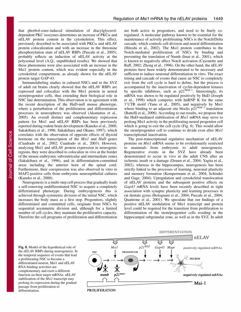
are both active in progenitors, and need to be finely co-regulated. A molecular pathway known to be essential for themaintenance of actively proliferating NSCs is the Notch/Deltacascade, which controls cell division and neural differentiation(Hitoshi et al., 2002). The Msi1 protein contributes to theNotch-mediated proliferation of NSCs by binding andpreventing the translation of Numb (Imai et al., 2001), whichis known to negatively affect Notch activation (Cayouette andRaff, 2002; Zhong et al., 1996). On the other hand, the nELAVproteins have been widely demonstrated to be necessary andsufficient to induce neuronal differentiation in vitro. The exacttiming and cascade of events that cause an NSC to completelyexit from the cell cycle is hard to define, but the process isaccompanied by the inactivation of cyclin-dependent kinasesby specific inhibitors, such as p21WAF-1. Interestingly, itsmRNA was shown to be regulated positively by HuD (Josephet al., 1998) which competes with hnRNP K for the same3�UTR motif (Yano et al., 2005), and negatively by Msi1whose binding to an adjacent site blocks p21WAF-1 translation(Battelli et al., 2006). According to our results, we propose thatthe HuD-mediated stabilization of Msi1 mRNA may serve toprolong Msi1 activity in the proliferating neural progenitor cellwhich is going to exit the cell cycle (Fig. 8). This would allowthe stem/progenitor cell to continue to divide even after Msi1transcriptional inactivation.
The post-transcriptional regulative mechanism of nELAVproteins on Msi1 mRNA seems to be evolutionarily restrictedto mammals from embryonic to adult neurogenesis.Regenerative events in the SVZ have already beendemonstrated to occur in vivo in the adult CNS after anischemic insult or a damage (Douen et al., 2004; Yagita et al.,2002), whereas in the hippocampus, neurogenesis has beenstrictly linked to the processes of learning, neuronal plasticityand memory formation (Kempermann et al., 2004; Schinderand Gage, 2004). Upregulation and cytoskeletal translocationof nELAV proteins and the subsequent positive effects onGap43 mRNA levels have been recently described in tightassociation with synaptic plasticity and learning processes inrat dentate gyrus (Bolognani et al., 2004; Pascale et al., 2004;Quattrone et al., 2001). We speculate that our findings of apositive nELAV modulation of Msi1 transcript and proteinlevel could be required for the transition from proliferation todifferentiation of the stem/progenitor cells residing in thehippocampal subgranular zone, as well as in the SVZ. In adult
Msi1 Gap43p21WAF MaptFig. 8. Model of the hypothetical role ofthe nELAV RBPs during neurogenesis. Inthe temporal sequence of events that leada proliferating NSC to become adifferentiated neuron, Msi1 and nELAVRNA-binding activities arecomplementary and exert a differentfunction on their target mRNAs. nELAVstabilization of the Msi1 transcript mayprolong its expression during the gradualpassage from proliferation todifferentiation.
Jour
nal o
f Cel
l Sci
ence
1450
neurogenic areas, nELAV activity could be assumed to betransiently induced by signals present only in restricted spatialand/or temporal conditions. We have recently demonstratedthat nELAV proteins are activated by a PKC�-dependentpathway in human neuroblastoma cells (Pascale et al., 2005),and it is known that PKC-dependent signal transduction is atthe basis of many aspects of CNS development (Metzger andKapfhammer, 2003). The combination of these observationswith the present results could open the possibility of apharmacological modulation of NSC dynamics.
In conclusion, our results suggest that in mammals nELAVproteins may have a key biological role in positively regulatingMsi1 gene expression along a molecular cascade leading aproliferating stem cell towards a multi-step neuraldifferentiation process. Since the intrinsic features of NSCsmake them attractive candidates for cell-based repair therapyof the CNS (Cova et al., 2004; Lakshmipathy and Verfaillie,2005), the nELAV/Msi1 pathway could also represent a newpharmacological target for enhancement of neurogenesis in thetreatment of neurodegenerative disorders.
Materials and MethodsCell cultureNeural stem/progenitor cell cultures were obtained from CD1 mouse brains aspreviously published (Gritti et al., 1999). After isolation of adult SVZ, tissue waspapain-digested and mechanically dissociated. Single cells were plated with adensity of 3.5�103 cells/cm2 in NS-A basal serum-free medium (Euroclone),supplemented with 20 ng/ml epidermal growth factor (EGF) and 10 ng/ml basicfibroblast growth factor (bFGF, PeproTech) and optimized for stem-cell growth. In7-10 days neurospheres were obtained, and every 5-7 days they were dissociated tosingle cells for 3-5 passages in order to amplify the neural stem compartment. Forimmunocytochemical assays, they were plated, as neurospheres or dissociatedstem/precursor cells, on an adhesive substrate (MatrigelTM, Becton Dickinson) for1 hour.
Human neuroblastoma SH-SY5Y cells were grown in MEM (Eagle’s minimalessential medium) with 10% fetal calf serum, penicillin/streptomycin, non-essentialamino acids and 1 mM sodium pyruvate (Invitrogen). Cells were exposed to 100nM phorbol 12-myristate-13-acetate (PMA, Sigma) or to the solvent alone (DMSO)for 15 minutes and then the incubation was stopped with ice-cold PBS.
Protein extracts, cell fractioning and western blotTotal mouse brain and NSC cultures were homogenized in lysis buffer (150 mMNaCl, 20 mM Tris-HCl, 1% Triton X-100, protease inhibitor cocktail; Roche) and400 U/ml RNase inhibitor (Promega). Samples were centrifuged at 12,000 g for 20minutes at 4°C and supernatant was collected. Proteins from different cell fractionswere obtained as previously published with slight modifications (Pascale et al.,1996). Briefly, SH-SY5Y cells were homogenized in buffer A (20 mM Tris-HCl pH7.4, 2 mM EDTA, 0.5 mM EGTA, 50 mM mercaptoethanol, 0.32 mM sucrose andprotease inhibitor cocktail) and centrifuged at 100,000 g for 1 hour. The supernatantcontaining the cytosolic fraction was collected. The pellet was resuspended in thesame buffer containing 1% Triton X-100, sonicated, and incubated for 45 minutesat 4°C, then centrifuged again at 100,000 g for 1 hour. The supernatant containingthe membrane fraction and the pellet with the cytoskeletal component werecollected. Proteins were resolved by SDS-PAGE, transferred to nitrocellulosemembranes and immunoblotted with Msi1 (1:200, Chemicon), PKC� (1:1000,Transduction Laboratories), �-tubulin (1:5000, Santa Cruz Biotechnology)antibodies, and Hu-positive serum (1:1000).
Plasmid constructsTotal RNA was extracted from whole mouse brain with TriZol® reagent (Invitrogen)and used to amplify Msi1 and Gap43 3�UTR sequences by RT-PCR with thefollowing primers: Msi1_fw TGAGGACCAGACTGAGCCAGCAAG andMsi1_rev GGGGCCTCAGTCTGCAGCAG; GAP-43_fw ATGCCTGAACTTT-AAGAAATGGCT and GAP-43_rev ATGAGGAAACAAAATGGTTTTTG. Theproducts were cloned into pGem®-T Easy Vector (Promega). ARE+ and ARE–
fragments were obtained by PCR amplification of the above Msi1-pGem® constructwith the following primers: Msi1_fw and ARE+_rev GTAGGGCAA-CTGGCTAATC; ARE–_fw GATTAGCCAGTTGCCCTAC and Msi1_rev. The�msi1ARE+ insert was subcloned from the Msi1-pGem® construct (primers:CTCATGTCTGGCTCCCCTACT and GTAGGGCAACTGGCTAATC) into thepCR®II-TOPO® (Invitrogen) vector and selected in order to have the T7 promoterat the 5� end and the HindIII restriction site at the 3� end of the cloning site for the
Journal of Cell Science 119 (7)
mRNA decay assay. The cDNA fragment encoding the open-reading frame for HuD(GenBank accession number D31953) was amplified by RT-PCR using thefollowing modified primers: HuD_fwTGATCTCATGAAGCCTCAGGTGTCAAATGGAC and HuD_rev CTG-CATCCCGGGGGATTTGTGGGCTTTGTTGGTT. The product was digested withRcaI/SmaI and directionally cloned into the expression vector pIVEX2.3d (Roche)with a His tag at the C-terminus. All the clones and their orientation were validatedby sequencing.
RNA labelingRadiolabeled riboprobes were obtained by transcribing 0.5 �g linearized constructDNA with 20 U T7 RNA polymerase (Roche), 20 �Ci [�-32P]UTP, 0.5 mM NTPs,20 U RNase inhibitor (Promega) for 30 minutes at 37°C. The reaction was stoppedat 65°C and template DNA was removed by DNaseI digestion (20 U, Roche). Theresulting 32P-labeled riboprobe was purified on ProbeQuant G-50 microcolumns(Amersham Biosciences).
In vitro TranslationRecombinant HuD protein was obtained in a cell-free system in which 0.5 �g HuD-pIVEX2.3d plasmid was incubated at 30°C for 6 hours in the reaction solution,containing E. coli lysate and amino acids according to the manufacturer’sinstructions (Roche). The fusion protein was purified on Ni-NTA spin kit columnsand visualized by western blotting using an Anti-His5 antibody (Qiagen).
UV crosslinking and immunoprecipitation300,000 cpm of 32P-labeled RNA transcripts were incubated with 40 �g of proteinextract or with 300 ng of recombinant HuD protein in 15 �l ligation buffer (1.3mM MgCl2, 19 mM HEPES-KOH pH 7.4, 1.5 mM ATP, 19 mM creatinephosphate) for 10 minutes at 30°C. After addition of 5 �g tRNA, samples wereirradiated with UV (Stratalinker®, Stratagene) for 5 minutes on ice and RNaseAtreated (25 U) for 30 minutes. Samples were run on a 12% SDS-PAGE, andanalyzed by autoradiography. For competition experiments, a 100� molar excessof cold riboprobe was added to the sample before UV irradiation.Immunoprecipitation was conducted on UV crosslinked samples by the additionof 4 �g of the selected antibody for 2 hours at 4°C. Samples were then incubatedwith 30 �l Protein A/G Sepharose beads (Amersham Biosciences) for 2 hours at4°C. Immunocomplexes were then collected by centrifugation at 14,000 g for 30seconds, washed several times in lysis buffer, run on a 12% SDS-PAGE andanalyzed by autoradiography.
Immunocytochemistry and immunohistochemistryCells were fixed with 4% paraformaldehyde in PBS (pH 7.4) for 20 minutes,blocked with 10% normal goat serum (NGS) and permeabilized with 0.3% TritonX-100 (Gritti et al., 1999). 20-�m-thick coronal brain sections from adult ratsintracardially perfused with 4% paraformaldehyde were mounted on glasses pre-coated with poly-L-lysine. Sections were boiled for 15 minutes in 50 mM Tris-HCl(pH 8.0), permeabilized with 0.5% Triton X-100 and blocked with 10% NGS.Samples were exposed to the selected antibodies overnight at 4°C (antibodies anddilutions used are specified in Table S2 in supplementary material). Slides weremounted with FluorsaveTM (Calbiochem) and acquired with a camera connected toa DMIRE2/HCS microscope (Leica Microsystems). For negative controls, theprimary antibody was replaced with NGS.
Isolation and immunoprecipitation of mRNP complexesAbout 5�106 NSCs were harvested for each condition, washed several times withcold PBS and resuspended in 1:1 (v/v) RNP buffer (100 mM KCl, 5 mM MgCl2,10 mM HEPES pH 7.4, 0.5% NP-40) (Tenenbaum et al., 2002). 50 �l protein A/GSepharose beads, pre-coated with 8 �g of the selected antibody, were added to 300�g NSC lysate in 1 ml NT2 buffer (50 mM Tris-HCl pH 7.4, 150 mM NaCl, 1 mMMgCl2, 0.05% NP-40), containing 400 U RNase inhibitor, 1 M DTT and 20 mMEDTA. 10% of the reaction mix was collected as the initial input and RNA wasextracted by TriZol® reagent (Invitrogen). After a 2-hour incubation, theimmunoprecipitated mRNPs were washed several times with cold NT2 buffer,incubated with 30 �g of proteinase K for 30 minutes and phenol-chloroformextracted. After DNaseI digestion, the isolated mRNAs were retro-transcribed usingSuperScriptII RT (Invitrogen), oligo dT and random primers.
Real-time quantitative PCRReal-time PCR was performed for 45 cycles with SYBRGreen PCR Master mix(Applied Biosystems) and processed on the ABI Prism 7900HT sequence detectionsystem. Oligonucleotide pairs for each gene were designed with Primer Express 2.0software (Applied Biosystems) on exon boundaries. Reactions were conducted intriplicate for each sample and a dissociation curve was produced at the end. For themRNP assays, the threshold cycle (Ct) values of immunoprecipitated samples werenormalized to the Ct value of the corresponding input (�Ct) to account fordifferences in initial mRNA quantities. ��Ct was calculated by subtracting the �Ct
value of the sample with no antibody to the �Ct of the sample with anti-nELAV oranti-His5 antibodies and fold differences were then expressed as 2-��Ct (Chakrabarti
Jour
nal o
f Cel
l Sci
ence
1451Regulation of Msi1 mRNA by the nELAV proteins
et al., 2002). Statistical analysis was conducted by one-way ANOVA withBonferroni t-test correction. For primer sequences and real-time PCR data seeTables S1 and S3 in supplementary material.
In vitro mRNA degradation/deadenylation assayThe mRNA decay experiments were conducted as described (Ford and Wilusz,1999) with some modifications. Whole-brain cytoplasmic extracts were used toreproduce a neural-like environment in vitro. The addition of the polyA60 tail to the�msi1ARE+ or �msi1AS constructs was carried out by the PCR-ligation procedure.Briefly, the double-stranded linker AGCTT(A)60TATTTACCTCGAGCACTC wasligated to the HindIII-digested plasmids, which were then amplified with the T7promoter primer and the complementary linker primer (GAGTGCTCGAG-GTAAATAT). Products were digested with SspI and in-vitro transcribed in thepresence of 7mGpppG (Roche) and [�-32P]UTP. Riboprobes were run on adenaturing 5% polyacrylamide gel (19:1) containing 8 M urea. After a shortautoradiographic exposure, bands were excized from the gel, eluted overnight inHSCB buffer (400 mM NaCl, 25 mM Tris-HCl pH 7.6, 0.1% SDS), phenol-chloroform extracted and ethanol precipitated. 200,000 cpm riboprobes wereincubated with 200 �g of mouse brain extract and 0.5 �g polyA (AmershamBiosciences) in 15 �l deadenylation buffer (2.6% polyvinyl alcohol, 1 mM ATP, 13mM creatine phosphate) at 30°C. At the indicated time intervals reactions werestopped with 400 �l HSCB buffer. RNA was extracted in phenol-chloroform,separated on an urea-denatured 5% polyacrylamide gel and visualized byautoradiography.
We thank C. Fenoglio, A. Grifa and M. G. Savino for their kindhelp. This work was supported by Italian Ministry of Health (Mal.Neurodegenerative, ex art.56 Anno 2003), Fondazione I.Monzino andFondazione FiorGen.
ReferencesAbe, R., Sakashita, E., Yamamoto, K. and Sakamoto, H. (1996). Two different RNA
binding activities for the AU-rich element and the poly(A) sequence of the mouseneuronal protein mHuC. Nucleic Acids Res. 24, 4895-4901.
Akamatsu, W., Okano, H. J., Osumi, N., Inoue, T., Nakamura, S., Sakakibara, S.,Miura, M., Matsuo, N., Darnell, R. B. and Okano, H. (1999). Mammalian ELAV-like neuronal RNA-binding proteins HuB and HuC promote neuronal development inboth the central and the peripheral nervous systems. Proc. Natl. Acad. Sci. USA 96,9885-9890.
Akamatsu, W., Fujihara, H., Mitsuhashi, T., Yano, M., Shibata, S., Hayakawa, Y.,Okano, H. J., Sakakibara, S., Takano, H., Takano, T. et al. (2005). The RNA-binding protein HuD regulates neuronal cell identity and maturation. Proc. Natl. Acad.Sci. USA 102, 4625-4630.
Alvarez-Buylla, A. and Garcia-Verdugo, J. M. (2002). Neurogenesis in adultsubventricular zone. J. Neurosci. 22, 629-634.
Antic, D. and Keene, J. D. (1997). Embryonic lethal abnormal visual RNA-bindingproteins involved in growth, differentiation, and posttranscriptional gene expression.Am. J. Hum. Genet. 61, 273-278.
Antic, D., Lu, N. and Keene, J. D. (1999). ELAV tumor antigen, Hel-N1, increasestranslation of neurofilament M mRNA and induces formation of neurites in humanteratocarcinoma cells. Genes Dev. 13, 449-461.
Aranda-Abreu, G. E., Behar, L., Chung, S., Furneaux, H. and Ginzburg, I. (1999).Embryonic lethal abnormal vision-like RNA-binding proteins regulate neuriteoutgrowth and tau expression in PC12 cells. J. Neurosci. 19, 6907-6917.
Asson-Batres, M. A., Spurgeon, S. L., Diaz, J., DeLoughery, T. G. and Bagby, G. C.,Jr (1994). Evolutionary conservation of the AU-rich 3� untranslated region ofmessenger RNA. Proc. Natl. Acad. Sci. USA 91, 1318-1322.
Bakheet, T., Frevel, M., Williams, B. R., Greer, W. and Khabar, K. S. (2001). ARED:human AU-rich element-containing mRNA database reveals an unexpectedly diversefunctional repertoire of encoded proteins. Nucleic Acids Res. 29, 246-254.
Barami, K., Iversen, K., Furneaux, H. and Goldman, S. A. (1995). Hu protein as anearly marker of neuronal phenotypic differentiation by subependymal zone cells of theadult songbird forebrain. J. Neurobiol. 28, 82-101.
Battelli, C., Nikopoulos, G. N., Mitchell, J. G. and Verdi, J. M. (2006). The RNA-binding protein Musashi-1 regulates neural development through the translationalrepression of p21(WAF-1). Mol. Cell Neurosci. 31, 85-96.
Bevilacqua, A., Ceriani, M. C., Capaccioli, S. and Nicolin, A. (2003). Post-transcriptional regulation of gene expression by degradation of messenger RNAs. J.Cell. Physiol. 195, 356-372.
Bez, A., Corsini, E., Curti, D., Biggiogera, M., Colombo, A., Nicosia, R. F., Pagano,S. F. and Parati, E. A. (2003). Neurosphere and neurosphere-forming cells:morphological and ultrastructural characterization. Brain Res. 993, 18-29.
Bolognani, F., Merhege, M. A., Twiss, J. and Perrone-Bizzozero, N. I. (2004).Dendritic localization of the RNA-binding protein HuD in hippocampal neurons:association with polysomes and upregulation during contextual learning. Neurosci.Lett. 371, 152-157.
Bottai, D., Fiocco, R., Gelain, F., Defilippis, L., Galli, R., Gritti, A. and Vescovi, L.A. (2003). Neural stem cells in the adult nervous system. J. Hematother. Stem Cell Res.12, 655-670.
Briata, P., Ilengo, C., Corte, G., Moroni, C., Rosenfeld, M. G., Chen, C. Y. and
Gherzi, R. (2003). The Wnt/beta-catenin–>Pitx2 pathway controls the turnover ofPitx2 and other unstable mRNAs. Mol. Cell 12, 1201-1211.
Cayouette, M. and Raff, M. (2002). Asymmetric segregation of Numb: a mechanism forneural specification from Drosophila to mammals. Nat. Neurosci. 5, 1265-1269.
Chakrabarti, S. K., James, J. C. and Mirmira, R. G. (2002). Quantitative assessmentof gene targeting in vitro and in vivo by the pancreatic transcription factor, Pdx1.Importance of chromatin structure in directing promoter binding. J. Biol. Chem. 277,13286-13293.
Chen, C. Y. and Shyu, A. B. (1995). AU-rich elements: characterization and importancein mRNA degradation. Trends Biochem. Sci. 20, 465-470.
Chu, E., Koeller, D. M., Casey, J. L., Drake, J. C., Chabner, B. A., Elwood, P. C.,Zinn, S. and Allegra, C. J. (1991). Autoregulation of human thymidylate synthasemessenger RNA translation by thymidylate synthase. Proc. Natl. Acad. Sci. USA 88,8977-8981.
Chung, S., Eckrich, M., Perrone-Bizzozero, N., Kohn, D. T. and Furneaux, H. (1997).The Elav-like proteins bind to a conserved regulatory element in the 3�-untranslatedregion of GAP-43 mRNA. J. Biol. Chem. 272, 6593-6598.
Cova, L., Ratti, A., Volta, M., Fogh, I., Cardin, V., Corbo, M. and Silani, V. (2004).Stem cell therapy for neurodegenerative diseases: the issue of transdifferentiation. StemCells Dev. 13, 121-131.
Cuadrado, A., Garcia-Fernandez, L. F., Imai, T., Okano, H. and Munoz, A. (2002).Regulation of tau RNA maturation by thyroid hormone is mediated by the neural RNA-binding protein musashi-1. Mol. Cell. Neurosci. 20, 198-210.
Cuadrado, A., Navarro-Yubero, C., Furneaux, H. and Munoz, A. (2003). NeuronalHuD gene encoding a mRNA stability regulator is transcriptionally repressed bythyroid hormone. J. Neurochem. 86, 763-773.
Ding, Y., Chan, C. Y. and Lawrence, C. E. (2004). Sfold web server for statisticalfolding and rational design of nucleic acids. Nucleic Acids Res. 32, W135-W141.
Douen, A. G., Dong, L., Vanance, S., Munger, R., Hogan, M. J., Thompson, C. S. andHakim, A. M. (2004). Regulation of nestin expression after cortical ablation in adultrat brain. Brain Res. 1008, 139-146.
Esdar, C., Oehrlein, S. A., Reinhardt, S., Maelicke, A. and Herget, T. (1999). Theprotein kinase C (PKC) substrate GAP-43 is already expressed in neural precursor cells,colocalizes with PKCeta and binds calmodulin. Eur. J. Neurosci. 11, 503-516.
Ford, L. P. and Wilusz, J. (1999). An in vitro system using HeLa cytoplasmic extractsthat reproduces regulated mRNA stability. Methods 17, 21-27.
Ford, L. P., Watson, J., Keene, J. D. and Wilusz, J. (1999). ELAV proteins stabilizedeadenylated intermediates in a novel in vitro mRNA deadenylation/degradationsystem. Genes Dev. 13, 188-201.
Galli, R., Gritti, A., Bonfanti, L. and Vescovi, A. L. (2003). Neural stem cells: anoverview. Circ. Res. 92, 598-608.
Gao, F. B., Carson, C. C., Levine, T. and Keene, J. D. (1994). Selection of a subset ofmRNAs from combinatorial 3� untranslated region libraries using neuronal RNA-binding protein Hel-N1. Proc. Natl. Acad. Sci. USA 91, 11207-11211.
Gritti, A., Frolichsthal-Schoeller, P., Galli, R., Parati, E. A., Cova, L., Pagano, S. F.,Bjornson, C. R. and Vescovi, A. L. (1999). Epidermal and fibroblast growth factorsbehave as mitogenic regulators for a single multipotent stem cell-like population fromthe subventricular region of the adult mouse forebrain. J. Neurosci. 19, 3287-3297.
Hitoshi, S., Alexson, T., Tropepe, V., Donoviel, D., Elia, A. J., Nye, J. S., Conlon, R.A., Mak, T. W., Bernstein, A. and van der Kooy, D. (2002). Notch pathway moleculesare essential for the maintenance, but not the generation, of mammalian neural stemcells. Genes Dev. 16, 846-858.
Imai, T., Tokunaga, A., Yoshida, T., Hashimoto, M., Mikoshiba, K., Weinmaster, G.,Nakafuku, M. and Okano, H. (2001). The neural RNA-binding protein Musashi1translationally regulates mammalian numb gene expression by interacting with itsmRNA. Mol. Cell. Biol. 21, 3888-3900.
Irwin, N., Baekelandt, V., Goritchenko, L. and Benowitz, L. I. (1997). Identificationof two proteins that bind to a pyrimidine-rich sequence in the 3�-untranslated regionof GAP-43 mRNA. Nucleic Acids Res. 25, 1281-1288.
Joseph, B., Orlian, M. and Furneaux, H. (1998). p21(waf1) mRNA contains aconserved element in its 3�-untranslated region that is bound by the Elav-like mRNA-stabilizing proteins. J. Biol. Chem. 273, 20511-20516.
Kaneko, Y., Sakakibara, S., Imai, T., Suzuki, A., Nakamura, Y., Sawamoto, K.,Ogawa, Y., Toyama, Y., Miyata, T. and Okano, H. (2000). Musashi1: anevolutionally conserved marker for CNS progenitor cells including neural stem cells.Dev. Neurosci. 22, 139-153.
Kanemura, Y., Mori, K., Sakakibara, S., Fujikawa, H., Hayashi, H., Nakano, A.,Matsumoto, T., Tamura, K., Imai, T., Ohnishi, T. et al. (2001). Musashi1, anevolutionarily conserved neural RNA-binding protein, is a versatile marker of humanglioma cells in determining their cellular origin, malignancy, and proliferative activity.Differentiation 68, 141-152.
Kasashima, K., Terashima, K., Yamamoto, K., Sakashita, E. and Sakamoto, H.(1999). Cytoplasmic localization is required for the mammalian ELAV-like proteinHuD to induce neuronal differentiation. Genes Cells 4, 667-683.
Kee, N., Sivalingam, S., Boonstra, R. and Wojtowicz, J. M. (2002). The utility of Ki-67 and BrdU as proliferative markers of adult neurogenesis. J. Neurosci. Methods 115,97-105.
Keene, J. D. (2001). Ribonucleoprotein infrastructure regulating the flow of geneticinformation between the genome and the proteome. Proc. Natl. Acad. Sci. USA 98,7018-7024.
Kempermann, G., Jessberger, S., Steiner, B. and Kronenberg, G. (2004). Milestonesof neuronal development in the adult hippocampus. Trends Neurosci. 27, 447-452.
Kohn, D. T., Tsai, K. C., Cansino, V. V., Neve, R. L. and Perrone-Bizzozero, N. I.
Jour
nal o
f Cel
l Sci
ence
(1996). Role of highly conserved pyrimidine-rich sequences in the 3� untranslatedregion of the GAP-43 mRNA in mRNA stability and RNA-protein interactions. BrainRes. Mol. Brain Res. 36, 240-250.
Lakshmipathy, U. and Verfaillie, C. (2005). Stem cell plasticity. Blood Rev. 19, 29-38.Lal, A., Mazan-Mamczarz, K., Kawai, T., Yang, X., Martindale, J. L. and Gorospe,
M. (2004). Concurrent versus individual binding of HuR and AUF1 to common labiletarget mRNAs. EMBO J. 23, 3092-3102.
Levine, T. D., Gao, F., King, P. H., Andrews, L. G. and Keene, J. D. (1993). Hel-N1:an autoimmune RNA-binding protein with specificity for 3� uridylate-rich untranslatedregions of growth factor mRNAs. Mol. Cell. Biol. 13, 3494-3504.
Marusich, M. F., Furneaux, H. M., Henion, P. D. and Weston, J. A. (1994). Huneuronal proteins are expressed in proliferating neurogenic cells. J. Neurobiol. 25, 143-155.
Maslov, A. Y., Barone, T. A., Plunkett, R. J. and Pruitt, S. C. (2004). Neural stem celldetection, characterization, and age-related changes in the subventricular zone of mice.J. Neurosci. 24, 1726-1733.
Mazan-Mamczarz, K., Galban, S., Lopez de Silanes, I., Martindale, J. L., Atasoy, U.,Keene, J. D. and Gorospe, M. (2003). RNA-binding protein HuR enhances p53translation in response to ultraviolet light irradiation. Proc. Natl. Acad. Sci. USA 100,8354-8359.
Metzger, F. and Kapfhammer, J. P. (2003). Protein kinase C: its role in activity-dependent Purkinje cell dendritic development and plasticity. Cerebellum 2, 206-214.
Morshead, C. M. and van der Kooy, D. (2004). Disguising adult neural stem cells. Curr.Opin. Neurobiol. 14, 125-131.
Nakamura, M., Okano, H., Blendy, J. A. and Montell, C. (1994). Musashi, a neuralRNA-binding protein required for Drosophila adult external sensory organdevelopment. Neuron 13, 67-81.
Okano, H. J. and Darnell, R. B. (1997). A hierarchy of Hu RNA binding proteins indeveloping and adult neurons. J. Neurosci. 17, 3024-3037.
Okano, H., Imai, T. and Okabe, M. (2002). Musashi: a translational regulator of cellfate. J. Cell Sci. 115, 1355-1359.
Pascale, A., Fortino, I., Govoni, S., Trabucchi, M., Wetsel, W. C. and Battaini, F.(1996). Functional impairment in protein kinase C by RACK1 (receptor for activatedC kinase 1) deficiency in aged rat brain cortex. J. Neurochem. 67, 2471-2477.
Pascale, A., Gusev, P. A., Amadio, M., Dottorini, T., Govoni, S., Alkon, D. L. andQuattrone, A. (2004). Increase of the RNA-binding protein HuD andposttranscriptional up-regulation of the GAP-43 gene during spatial memory. Proc.Natl. Acad. Sci. USA 101, 1217-1222.
Pascale, A., Amadio, M., Scapagnini, G., Lanni, C., Racchi, M., Provenzani, A.,Govoni, S., Alkon, D. L. and Quattrone, A. (2005). Neuronal ELAV proteins enhancemRNA stability by a PKCalpha-dependent pathway. Proc. Natl. Acad. Sci. USA 102,12065-12070.
Perrone-Bizzozero, N. and Bolognani, F. (2002). Role of HuD and other RNA-bindingproteins in neural development and plasticity. J. Neurosci. Res. 68, 121-126.
Qian, X., Shen, Q., Goderie, S. K., He, W., Capela, A., Davis, A. A. and Temple, S.(2000). Timing of CNS cell generation: a programmed sequence of neuron and glialcell production from isolated murine cortical stem cells. Neuron 28, 69-80.
Quattrone, A., Pascale, A., Nogues, X., Zhao, W., Gusev, P., Pacini, A. and Alkon,D. L. (2001). Posttranscriptional regulation of gene expression in learning by theneuronal ELAV-like mRNA-stabilizing proteins. Proc. Natl. Acad. Sci. USA 98,11668-11673.
Robinow, S. and White, K. (1991). Characterization and spatial distribution of the ELAVprotein during Drosophila melanogaster development. J. Neurobiol. 22, 443-461.
Roegiers, F. and Jan, Y. N. (2004). Asymmetric cell division. Curr. Opin. Cell Biol. 16,195-205.
Sakakibara, S. and Okano, H. (1997). Expression of neural RNA-binding proteins inthe postnatal CNS: implications of their roles in neuronal and glial cell development.J. Neurosci. 17, 8300-8312.
Sakakibara, S., Imai, T., Hamaguchi, K., Okabe, M., Aruga, J., Nakajima, K.,Yasutomi, D., Nagata, T., Kurihara, Y., Uesugi, S. et al. (1996). Mouse-Musashi-1,a neural RNA-binding protein highly enriched in the mammalian CNS stem cell. Dev.Biol. 176, 230-242.
Sakakibara, S., Nakamura, Y., Satoh, H. and Okano, H. (2001). Rna-binding proteinMusashi2: developmentally regulated expression in neural precursor cells andsubpopulations of neurons in mammalian CNS. J. Neurosci. 21, 8091-8107.
Samson, M. L. (1998). Evidence for 3� untranslated region-dependent autoregulation ofthe Drosophila gene encoding the neuronal nuclear RNA-binding protein ELAV.Genetics 150, 723-733.
Schaeffer, C., Bardoni, B., Mandel, J. L., Ehresmann, B., Ehresmann, C. and Moine,H. (2001). The fragile X mental retardation protein binds specifically to its mRNA viaa purine quartet motif. EMBO J. 20, 4803-4813.
Schinder, A. F. and Gage, F. H. (2004). A hypothesis about the role of adult neurogenesisin hippocampal function. Physiology (Bethesda) 19, 253-261.
Shaw, G. and Kamen, R. (1986). A conserved AU sequence from the 3� untranslatedregion of GM-CSF mRNA mediates selective mRNA degradation. Cell 46, 659-667.
Suslov, O. N., Kukekov, V. G., Ignatova, T. N. and Steindler, D. A. (2002). Neural stemcell heterogeneity demonstrated by molecular phenotyping of clonal neurospheres.Proc. Natl. Acad. Sci. USA 99, 14506-14511.
Szabo, A., Dalmau, J., Manley, G., Rosenfeld, M., Wong, E., Henson, J., Posner, J.B. and Furneaux, H. M. (1991). HuD, a paraneoplastic encephalomyelitis antigen,contains RNA-binding domains and is homologous to Elav and Sex-lethal. Cell 67,325-333.
Tenenbaum, S. A., Lager, P. J., Carson, C. C. and Keene, J. D. (2002). Ribonomics:identifying mRNA subsets in mRNP complexes using antibodies to RNA-bindingproteins and genomic arrays. Methods 26, 191-198.
Toda, M., Iizuka, Y., Yu, W., Imai, T., Ikeda, E., Yoshida, K., Kawase, T., Kawakami,Y., Okano, H. and Uyemura, K. (2001). Expression of the neural RNA-bindingprotein Musashi1 in human gliomas. Glia 34, 1-7.
Wakamatsu, Y. and Weston, J. A. (1997). Sequential expression and role of Hu RNA-binding proteins during neurogenesis. Development 124, 3449-3460.
Wiese, C., Rolletschek, A., Kania, G., Blyszczuk, P., Tarasov, K. V., Tarasova, Y.,Wersto, R. P., Boheler, K. R. and Wobus, A. M. (2004). Nestin expression–a propertyof multi-lineage progenitor cells? Cell. Mol. Life Sci. 61, 2510-2522.
Yagita, Y., Kitagawa, K., Sasaki, T., Miyata, T., Okano, H., Hori, M. and Matsumoto,M. (2002). Differential expression of Musashi1 and nestin in the adult rat hippocampusafter ischemia. J. Neurosci. Res. 69, 750-756.
Yano, M., Okano, H. J. and Okano, H. (2005). Involvement of Hu and heterogeneousnuclear ribonucleoprotein K in neuronal differentiation through p21 mRNA post-transcriptional regulation. J. Biol. Chem. 280, 12690-12699.
Zhong, W., Feder, J. N., Jiang, M. M., Jan, L. Y. and Jan, Y. N. (1996). Asymmetriclocalization of a mammalian numb homolog during mouse cortical neurogenesis.Neuron 17, 43-53.
Journal of Cell Science 119 (7)1452
Jour
nal o
f Cel
l Sci
ence


![PcG Proteins MSI1 and BMI1 Function Upstream of miR156 to ... · PcG Proteins MSI1 and BMI1 Function Upstream of miR156 to Regulate Aerial Tuber Formation in Potato1[OPEN] Amit Kumar,2](https://img.pdfslide.us/doc/110x75/5e55ed860cda997cf77628a1/pcg-proteins-msi1-and-bmi1-function-upstream-of-mir156-to-pcg-proteins-msi1.jpg)