Embed Size (px)
Citation preview

1
Supplemental Information for:
A Role for Streptococcal Collagen-Like Protein 1 (Scl-1) in M1T1 Group A
Streptococcus Resistance to Neutrophil Extracellular Traps
Simon Döhrmann1, Sabina Anik1, Joshua Olson1, Ericka L. Anderson1, Neelou
Etesami2, Hyewon No2, Joshua Snipper2, Victor Nizet1,3#, Cheryl Y.M. Okumura1,2#
Author affiliations
1. Department of Pediatrics, University of California, San Diego, La Jolla, CA, USA
2. Department of Biology, Occidental College, Los Angeles, CA, USA
3. Skaggs School of Pharmacy and Pharmaceutical Sciences, University of California,
San Diego, La Jolla, CA, USA
#Corresponding author ([email protected]; [email protected])
Running Title: Scl-1 Protects GAS From Neutrophil Extracellular Traps

2
Supplemental Methods
Immunofluorescence assays
Bacterial cultures were grown to log phase and seeded on coverslips in 24-well
plates for 30 min at 37°C. Bacteria were fixed with 4% paraformaldehyde and
incubated in 1mg/mL lysozyme in block buffer (10% heat-inactivated fetal equine
serum and 0.1% BSA in 20 mM HEPES, 140 mM NaCl, 5 mM CaCl2, 2.5 mM MgCl2,
pH 7.4). Bacteria were subsequently incubated with 0.005 mg/mL affinity purified
anti-Scl-1 or pre-immune rabbit serum (kind gifts of Magnus Rasmussen), followed by
incubation with goat anti-rabbit Alexa 488 antibody (Invitrogen). Bacteria were
imaged on an inverted Leica TCS SP5 confocal microscope using a 63x/1.40 oil
objective and 6x digital zoom at calibrated magnifications and recorded with LAS AF
software (Leica). Presence of bacteria in the field of view were verified by brightfield
images (data not shown). Linear adjustments to the entire image brightness were
applied uniformly for all images.
Hyaluronic acid capsule assay
Hyaluronic acid capsule from GAS was quantified as previously described (1). Briefly,
bacterial cultures were grown to log phase and resuspended in water. Capsule was
extracted with chloroform in Mini-BeadBeater-8 (Biospec Products). Following
centrifugation, the hyaluronic acid content in the aqueous phase was determined by
the hyaluronic acid test kit (Corgenix) according to the manufacturer's instructions.
The absorbance was measured with SpectraMax M3 plate reader at 450 nm using
SoftMax Pro software.
Fibrinogen-binding assay

3
Fibrinogen-binding of GAS was measured as previously described (2). Briefly,
human plasminogen-depleted fibrinogen (Calbiochem) was labeled with FITC using
the FluoReporter protein labeling kit (Invitrogen) and added at 100 µg/mL to GAS
strains grown to log phase. Samples were incubated for 30 min with rotating and
washed with PBS prior to flow cytometric analysis. Bacteria were gated by forward
and side scatter, and the fluorescence intensity was measured for a total of 50,000
bacteria. Unlabeled bacteria were used as a negative control. Flow cytometry data
were analyzed with FlowJo v. 9.4.10 (Tree Star, Inc.).
SpeB activity assays
Bacterial colonies were picked and grown in THB in a 96-well plate O/N at 37°C.
Plates were spun to pellet bacteria, and supernatant was transferred to a new plate
to an equivalent volume of activation buffer (0.1 M sodium acetate. 0.1 M acetic acid,
1 mM EDTA and 20 mM DTT, pH 5) and incubated for 30 min at 40°C. An equal
volume of 2% azocasein (Sigma) in activation buffer was added to supernatants and
incubated for and additional 1 h at 40°C. The absorbance was measured with
SpectraMax M3 plate reader at 460 nm using SoftMax Pro software.
Whole blood killing assays
Bacteria were diluted to a final inoculum of 104 CFU, added to 300 µL freshly drawn
blood, and rotated at 37°C. At 3 h post-infection, an aliquot of blood was removed,
blood cells were lysed with water, and bacteria were enumerated on THA plates.
Survival was calculated as the percentage of the initial inoculum, and due to large
donor-dependent variability, results were compared with the wild-type strain.
4
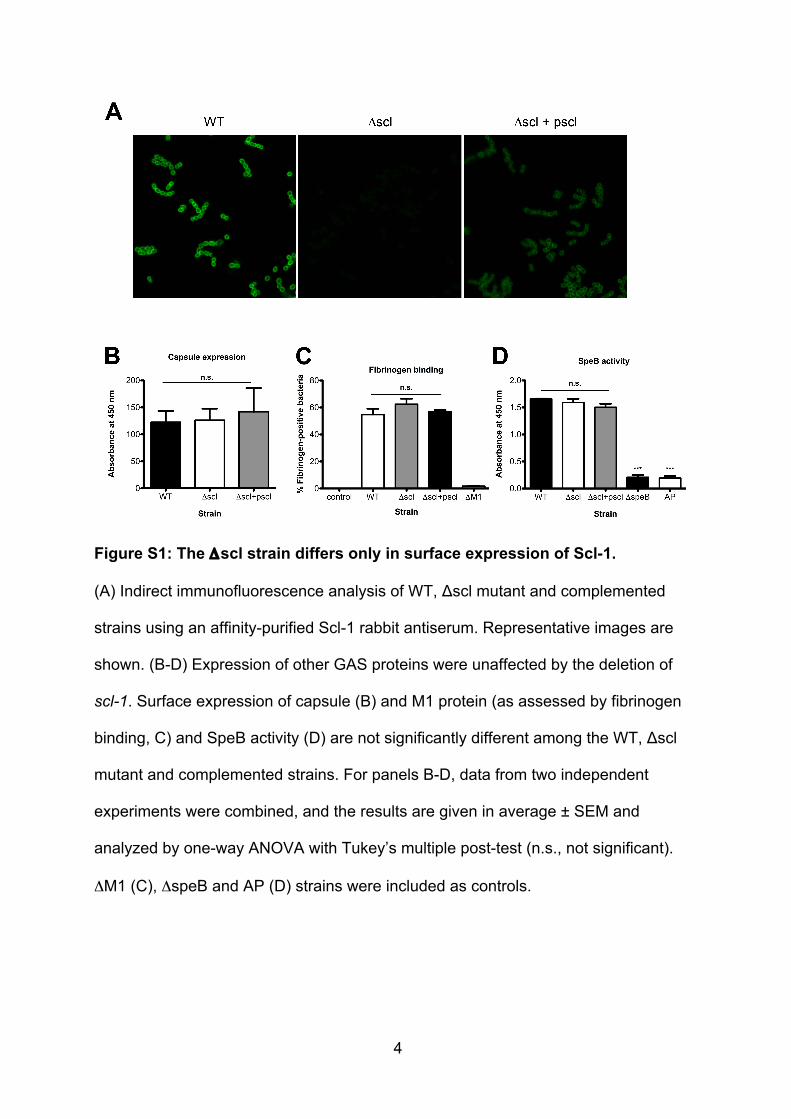
Figure S1: The Δscl strain differs only in surface expression of Scl-1.
(A) Indirect immunofluorescence analysis of WT, Δscl mutant and complemented
strains using an affinity-purified Scl-1 rabbit antiserum. Representative images are
shown. (B-D) Expression of other GAS proteins were unaffected by the deletion of
scl-1. Surface expression of capsule (B) and M1 protein (as assessed by fibrinogen
binding, C) and SpeB activity (D) are not significantly different among the WT, Δscl
mutant and complemented strains. For panels B-D, data from two independent
experiments were combined, and the results are given in average ± SEM and
analyzed by one-way ANOVA with Tukey’s multiple post-test (n.s., not significant).
ΔM1 (C), ΔspeB and AP (D) strains were included as controls.
5
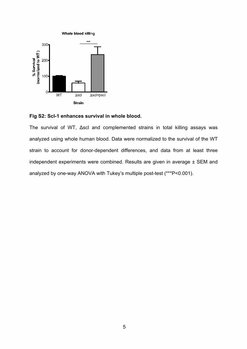
Fig S2: Scl-1 enhances survival in whole blood.
The survival of WT, Δscl and complemented strains in total killing assays was
analyzed using whole human blood. Data were normalized to the survival of the WT
strain to account for donor-dependent differences, and data from at least three
independent experiments were combined. Results are given in average ± SEM and
analyzed by one-way ANOVA with Tukey’s multiple post-test (***P<0.001).

6
Supplemental References 1. Hollands A, Pence MA, Timmer AM, Osvath SR, Turnbull L, Whitchurch
CB, Walker MJ, Nizet V. 2010. Genetic switch to hypervirulence reduces colonization phenotypes of the globally disseminated Group A Streptococcus M1T1 clone. J Infect Dis 202:11-19.
2. Anderson EL, Cole JN, Olson J, Ryba B, Ghosh P, Nizet V. 2014. The fibrinogen-binding M1 protein reduces pharyngeal cell adherence and colonization phenotypes of M1T1 Group A Streptococcus. J Biol Chem 289:3539-3546.