Embed Size (px)
Citation preview
ABSTRACT THz spectroscopy is a relatively new spectroscopic method in which the THz useful range of frequencies is from 300 cm-1 down to 3 cm-1, a spectral region unachievable by FTIR spectroscopy. Multiple resonances in the absorption spectra of organic molecules exist in the THz range around 0.1–10 THz (3 to 300 cm-1) where these resonances arise from electromagnetic wave interaction of the THz source with the low-frequency, and hence large- scale, vibrational modes within the organic macromolecules. These major vibrational modes correspond to the quaternary or overall structure of macromolecules. The concerted motion of protein structure can also be investigated by THz spectroscopy, which enables both energy and momentum transfer at the picosecond time scale. The short bursts of far-IR radiation in an ultra-fast time scale in THz spectroscopy al-low researchers to study the dynamic processes of various materials for a large variety of applications. This review paper discusses the probability of utilizing THz spectroscopy as a probe to determine the intermolecular structures
INTRODUCTION
Infrared (IR) spectroscopy is a classical chemical tool dating back to the early 20th century (Hagen & Rubens, 1902), whereas THz spectroscopy have only garnered serious attention about 20 years ago (Beard et al, 2002; Fattinger & Grischkowsky, 1989). The effort to study THz spectroscopy was triggered by the challenge to produce and detect ultra-short electric transients (changes in electrical voltage) as they were propagated along an electric transmission line (Beard et al, 2002). These electrical pulses create very small bursts of electromagnetic radiation in the far-IR frequency wavelength from 0.2 to 2.0 THz (6.66 to 66.6 cm-1) (Fattinger & Grischkowsky, 1989). THz spectroscopy, sometimes referred as T-ray spectroscopy (Markelz, 2008), allows direct interrogation of complex permittivity of samples spanning a broad bandwidth of 0.1–8.0 THz (3.33–266.4 cm-1) (Shen et al, 2003) without the need for cryogens and simultaneously allowing extremely flexible sample control and optics arrangements (Markelz, 2008).
THz rays provides an safer alternative to x-rays due to its non-ionizing radiative property for imaging purposes through cloth, skin, paper and many others imaging materials. The investigation of the THz fields has considerably grown with much emphasis concentrating on the THz spectroscopy of
biological materials. Several investigations that have linked THz spectroscopy to the biological or biochemistry fields have been conducted by various groups of researchers worldwide, including (i) the analysis of low-frequency molecular vibration of DNA and RNA strands (Fischer et al, 2005; Mickan et al, 2002), (ii) several types of proteins including lysozyme, myoglobin, bacteriorhodopsin and virus-like particles (Falconer et al, 2010; Markelz et al, 2002; Png et al, 2009; Zakaria et al, 2011; Zhang & Durbin, 2006), (iii) the interaction between electromagnetic radiation and biological materials (Doria et al, 2004; Pickwell et al, 2004; Smye et al, 2001), (iv) the detection of potentially harmful biological agents (Brown et al, 2004) and, (v) the application of THz imaging to detect skin cancer (Pickwell et al, 2004; Woodward et al, 2003).
THE TECHNICAL ASPECT OF THZ SPECTROSCOPY
The rapid growth of interest in the THz field is largely due to the emergence of the technology needed to access the THz region, including the development of THz source and detectors in recent years (Plusquellic et al, 2007). Vanexter and co-workers developed the THz photoconductive transmitter and detector based on silicon technology (Vanexter & Grischkowsky, 1990) spanning the frequency range of 0.1–2 THz (3.33–66.6 cm-1). In the last decade, photoconductive antennae technology was manipulated and engineered
of macromolecules as well as basic technical knowledge in THz spectroscopy.
A REVIEW: TERAHERTZ SPECTROSCOPY AS A VIABLE DYNAMIC TOOL FOR PROTEIN AND ORGANIC MOLECULES CHARACTERIZATION
Hidayatul Aini Zakaria*
(Keywords:Terahertz, spectroscopy, vibrational, proteins, organic molecules)
Malaysian Journal of Science 34 (1) : 93-102 (2015)
93
Universiti Malaysia Terengganu, 21030 Kuala Terengganu, Terengganu, Malaysia*Corresponding author: [email protected]

for the generation and detection of THz rays (Fattinger & Grischkowsky, 1989; Grischkowsky et al, 1990). Most THz sources relied on inorganic semiconductor materials—gallium arsenide (GaAs), zinc telluride (ZnTe), and indium arsenide (InAs). These semiconductors can be used to transform an ultra-short (around 100 fs) near infrared pulsed laser to a THz pulse by excitation of electron-hole pairs in a semiconductor structure (Davies et al, 2002). The higher photon energy of the near-infrared pulse over the semiconductor band gap (energy difference from the top of valence band to the bottom of the conduction band) will create electron hole-pairs close to the surface of the THz source. A THz pulse is generated by the photo-excited acceleration of electron-hole pairs of the semiconductor material when the near-IR laser pulse reaches the semiconductor surface (Davies et al, 2002).
The channel of field effect transistors may be utilized as resonators for plasma waves where the dimensions and gate length of of the plasma frequency of the resonator may reach the THz range (Knap & Dyakonov, 2013) For THz pulse detection, GaAs (Lu et al, 1998), silicon (Si), and gallium nitride (GaN) (Kachorovskii & Shur, 2008) are typically used as plasma (THz) wave detectors. The THz detection mechanism is as follows (Dyakonov & Shur, 1996): incoming THz radiation excites the plasma wave in the FET that is translated as a voltage drop across the FET structure. This voltage drop between the drain to the source of the FET is correlated with a resonance response from the incoming electromagnetic radiation. The electromagnetic radiation will then provide the spectra needed to study the intra-molecular characteristic being studied where the theoretical calculations will be discussed in the following paragraphs.
Figure 1. Schematics of a FET utilized as THz detector (upper image), and its corresponding equivalent circuit (lower image) (Knap & Dyakonov, 2013)
In general, THz data measurements are obtained in the time domain (raw data) by recording the temporal field strength of a short electromagnetic pulse that lasts only for a few picoseconds. The Fast Fourier Transform (FFT) is the common algorithm used to convert these time domain signals (i.e., an estimate of the impulse response of the sample) to the frequency domain (Fischer, 2005b; Yin et al, 2007). From this conversion, the frequency response
of the sample can be estimated. A THz pulse will be modified after propagating through a sample (dielectric material), because of the absorption and dispersion in the sample. By comparing this THz pulse with an appropriate reference pulse, an estimate can be obtained of the average absorption (absorption of EM waves by the sample) and the refraction (dispersion of light in the sample) from the sample measurements (Fischer 2005).
Malaysian Journal of Science 34 (1) : 93-102 (2015)
94
Absorptions of electromagnetic radiation in the THz region are frequently related to hydrogen- bonding interactions between separate molecules and variations in the crystal lattice (Fischer et al, 2007). THz data were acquired in the time domain by recording the temporal field strength of the short electromagnetic pulse. The following signal processing analysis is based on the THz studies of Bernd M Fischer (Fischer, 2005b).
The time domain signal, j (τ) *where τ represents the time delay+ is a convolution of the electric pulse of the THz pulse E (t) with the THz detector pulse D (t) as given:
Here, the waveform before propagation through the sample is represented by the time dependent term E0 (t) = E0 e
iwt.
A THz reference pulse propagating through vacuum is given by:
Consider now a THz pulse after propagating through a dielectric material of thickness d. A THz pulse will be modified after propagating through a sample (dielectric material), because of the absorption and dispersion in the sample. Thus, the frequency dependent field after the THz pulse propagation through a material is given by:
Where T is the Fresnel losses at the interface of the sample (Bea & Teich, 1974):
By dividing this THz sample pulse with an appropriate THz reference pulse, an expression for the frequency dependent ratio (complex refractive index) can be obtained:
The absorption of light by an optical medium can be quantified by a parameter called the absorption coefficient, α; the index of refraction, n, can be simplified as the ratio of t h e v e l ocity of light in free space to the velocity of light in a medium. The estimation of the average absorption coefficient (energy propagation through homogenous system) can be derived from the imaginary part of Equation [3.6], as shown in the equation below:
Absorption coefficient,
where A is the complex amplitude.
The index of refraction (change in direction of wave propagation) can be derived from the real part of Equation [3.6] as shown in Equation [3.8]:
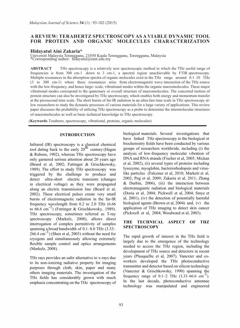
A schematic diagram of the THz spectroscopy system is illustrated in Figure 2. Measurements are typically conducted in a nitrogen-purged environment to reduce water vapour absorption by the THz radiation (Fischer, 2005a). An ultra-short (typically around 100 fs) laser pulse from the visible or near-IR laser (around 800 nm) is divided into 2 directions by a beam splitter (Picometrix, 2005). One laser pulse is directed to a THz emitter, which transforms the incident laser pulse into a THz pulse; the other pulse serves as a detector monitoring the temporal shape of the radiated THz waveform (Walther et al, 2002). A sample is deposited in the propagation path of the THz wave between the THz emitter and the detector. The sample is irradiated by the THz wave from the THz emitter, and the transmitted THz pulse is detected by the THz detector. The detected signal from the THz detector is then transformed by Fourier transform and the complex valued spectra of the THz wave is obtained (Fischer, 2005a). Far-IR optical properties (absorption coefficient and index of refraction) of the sample can be determined as a function of frequency from the resolved THz spectra (Ferguson & Zhang, 2002).
1
𝑇
𝑇
0 𝑗 𝑡 = 𝐸 𝑡 𝐷 (𝑡 − 𝜏) 𝑑𝑡 [1]
n= n+ik
The linear response of a material to a THz ray is determined by the dielectric properties of the material where the complete information of the dielectric function of that material is contained in its reflected THz pulse (Fischer, 2005b). Consider that an electromagnetic plane wave of frequency ω propagating through a sample in the z-direction with the dielectric constant of will produce a time dependent electric field as follows:
𝐸 𝑧, 𝑡 =E0 (t) exp 𝑖 (𝜔𝑡 + 𝑛𝜔
𝑐𝑧) = 𝐸
0
𝑛𝜔
𝑐𝑧 . 𝑒𝑥𝑝 − 𝑧
𝜅𝜔
𝑐 𝑖
𝑡 𝑒𝑥𝑝
[2]
0
𝑑
𝑐 𝜔 exp 𝑖𝜔= 𝐸 𝐸
𝑟𝑒𝑓 [3]
0 𝐸
𝑛(𝜔)𝜔
𝑐𝑑 . 𝑒𝑥𝑝 −
𝜅(𝜔)𝜔
𝑐𝑠𝑎𝑚(𝜔)= 𝐸 𝜔 exp 𝑇 𝑖 𝑑 [4]
T= 4𝑛(𝜔)
(𝑛 𝜔 +1)2 [5]
𝑖𝛷 𝜔 𝐸 𝑠𝑎𝑚
𝑟𝑒𝑓 𝜔 𝐸
= 𝐴𝑒
= 𝑑
2+ 𝑖𝑛(𝜔)2𝜋
𝑑
𝑐 [6]
4𝑛(𝜔)
(𝑛 𝜔 +1) −𝛼 𝜔
(𝑛 𝜔 +1)2
4𝑛(𝜔)
2
𝑑
2exp [6]
𝑛 𝜔 = 1 + Φ𝑐
𝑤𝑑 [8]
𝛼 𝜔 =− ln 𝐴 [7]
Malaysian Journal of Science 34 (1) : 93-102 (2015)
95

Figure 2. Schematic diagram of THz spectroscopy system adopted from Fischer (Fischer et al, 2007)
THz spectroscopy has several notable advantag-es over other forms of spectroscopic methods. Vi-brational modes in the THz region are more wide-spread and include motions of all of the atoms in the molecular structure, whereas vibrational modes in the mid-IR wavelength are typically located within specific sites in the molecular conformation (Plusquellic et al, 2007). THz radiation has a longer wavelength than the near infra-red radiation, thus it will provide sharper images with better spatial reso-lution because it is less affected by Rayleigh scatter-ing than near-infrared radiation (Beard et al, 2002). One of the many advantages of THz spectroscopy over far-IR spectroscopy is that far-IR spectroscopy uses blackbody radiation sources and phonon de-tectors, while THz spectroscopy incorporates bright sources and sensitive detectors that can provide a higher signal-to-noise ratio and a wider dynamic range than far-IR spectroscopy (Grischkowsky et al, 1990). Another advantage of THz spectrometry is
that it allows time-resolved far-IR spectroscopy to cover the spectral range of 0.1–20 THz (3–600 cm-1) in an ultra-fast time scale that provides sub-pico-second temporal resolution (Beard et al, 2002). Viv-id changes in nucleic acid and component protein properties were observed when they were inserted into chromosomes or functional membranes (Sau-er, 1995), which might be analysed using the pico-second time scale technology of THz spectroscopy.
There has been a notable increase in the interest in exploring the low-frequency collective vibration-al modes within large biomolecules because these modes might provide valuable information about the conformational state of the biomolecules (Brandt et al, 2008). These low-frequency collective structur-al vibrational modes consisting of concerted large-scale motions of biomolecules dictated by biomole-cule architecture were believed to bear crucial roles towards biomolecular function and conformational modification changes (Markelz, 2008). However,
Malaysian Journal of Science 34 (1) : 93-102 (2015)
96
spectroscopic studies in the low-frequency spectral region were often limited by technological con-straints, such as insufficient light source power and detector sensitivity (Fischer, 2005b), owing to which it was difficult to resolve both energy and momentum transfers in the picosecond time scale (He et al, 2011b). THz spectroscopy with its inherent ability to probe the intra-molecular struc-ture of materials in the low frequency spectral re-gion. 1–8.0 THz (3.33–266.4 cm-1 might fill in this gaping research gap. ANALYSIS OF ORGANIC MOLECULES BY THZ SPECTROSCOPY
Advancements in THz technology have enabled studies by THz spectroscopy for the characterisa-tion of very far-IR vibrational modes of chemical compounds (Ueno & Ajito, 2008). This intermo-lecular vibrational mode includes low-frequency vibrations around the hydrogen bonds (Walther et al, 2002), phonon modes that are IR-active inter-molecular modes in a crystalline sample (Siegrist et al, 2006), and low-frequency delocalised modes that consist of the samples’ entire vibrational mo-tions (Giraud et al, 2003). THz spectroscopy pro-vides the means to actively investigate the qua-ternary structure of organic molecules by the interaction of THz radiation with low-frequency vibrational modes of organic molecules. The di-electric functions of the organic molecules benzoic acid and its monosubstituted derivatives 2-, 3- and4-hydroxybenzoic acid in pressed pellet form were investigated at 0.5–4.5 THz (17–150 cm-1)using THz time domain spectroscopy by Walther and co-workers at the University of Freiburg, Ger-many. Despite similarities in the molecular struc-ture, the absorption spectra of these biomolecules had very little in common (Walther et al, 2002).
The high sensitivity of THz vibrational modes to small changes in the overall biomolecular structure in addition to the hydrogen bonding environment were evidenced again in a THz study of the enantio-meric crystalline structure of L-, D-, and DL-alanine (Yamaguchi et al, 2005) and the pure enantiomers and racemic compounds of tartaric acid (Fischer et al, 2007). Recently, the absorption spectra of organic crystals diglycine lithium nitrate, triglycine zinc chloride, and diglycine hydrobromide have shown clear resonant behavior in the permittivity
diagram with sharp absorption peaks at 32.11 cm-1 ,44.16 cm-1, and 51.72 cm-1 (Trzebiatowska- Gu-sowska et al, 2014).
Comparisons of hydrated and dehydrated tri-ala-nine samples caused calculative differences in its respective THz spectra, demonstrating that use of a hydrogen-bonded solvent has a huge effect on the intermolecular structure of organic molecules investigated in the THz spectral range (Siegrist et al, 2006). A study of the intermolecular charac-teristics of chlorobenzene in the THz region con-ducted by Fischer (2007) showed that at a higher temperature (295 K), the absorption spectrum of chlorobenzene was swamped by large water ab-sorption. In contrast, at a lower temperature (100 K), the constitution of chlorobenzene changed from liquid to crystalline; hence, a sharp absorp-tion peak could be observed in the THz spectrum of crystalline chlorobenzene samples (Fischer et al, 2007). The effects of water molecules in THz measurements were evidenced again by (Zhang & Durbin, 2006), who reported significant differ-ences in the THz absorption spectra of a myoglobin solution at various water concentrations, and (Liu & Zhang, 2006), with investigation of D-glucose monohydrate with THz- TDS. Liu and co-work-ers showed that the absorption peak of D-glucose monohydrate decreases at a higher sample dehydra-tion rate (Liu & Zhang, 2006) as shown in Figure 3.
Malaysian Journal of Science 34 (1) : 93-102 (2015)
97
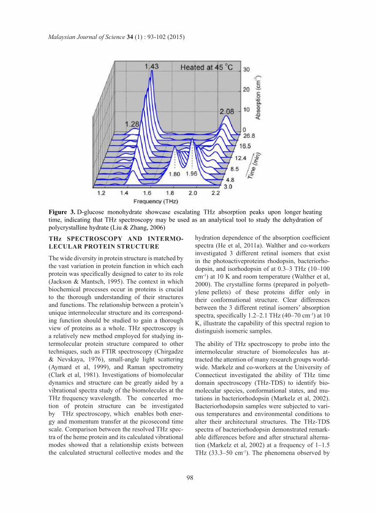
Figure 3. D-glucose monohydrate showcase escalating THz absorption peaks upon longer heating time, indicating that THz spectroscopy may be used as an analytical tool to study the dehydration of polycrystalline hydrate (Liu & Zhang, 2006)
THz SPECTROSCOPY AND INTERMO-LECULAR PROTEIN STRUCTURE
The wide diversity in protein structure is matched by the vast variation in protein function in which each protein was specifically designed to cater to its role (Jackson & Mantsch, 1995). The context in which biochemical processes occur in proteins is crucial to the thorough understanding of their structures and functions. The relationship between a protein’s unique intermolecular structure and its correspond-ing function should be studied to gain a thorough view of proteins as a whole. THz spectroscopy is a relatively new method employed for studying in-termolecular protein structure compared to other techniques, such as FTIR spectroscopy (Chirgadze & Nevskaya, 1976), small-angle light scattering (Aymard et al, 1999), and Raman spectrometry (Clark et al, 1981). Investigations of biomolecular dynamics and structure can be greatly aided by a vibrational spectra study of the biomolecules at the THz frequency wavelength. The concerted mo-tion of protein structure can be investigated by THz spectroscopy, which enables both ener-gy and momentum transfer at the picosecond time scale. Comparison between the resolved THz spec-tra of the heme protein and its calculated vibrational modes showed that a relationship exists between the calculated structural collective modes and the
hydration dependence of the absorption coefficient spectra (He et al, 2011a). Walther and co-workers investigated 3 different retinal isomers that exist in the photoactiveproteins rhodopsin, bacteriorho-dopsin, and isorhodopsin of at 0.3–3 THz (10–100 cm-1) at 10 K and room temperature (Walther et al, 2000). The crystalline forms (prepared in polyeth-ylene pellets) of these proteins differ only in their conformational structure. Clear differences between the 3 different retinal isomers’ absorption spectra, specifically 1.2–2.1 THz (40–70 cm-1) at 10 K, illustrate the capability of this spectral region to distinguish isomeric samples.
The ability of THz spectroscopy to probe into the intermolecular structure of biomolecules has at-tracted the attention of many research groups world-wide. Markelz and co-workers at the University of Connecticut investigated the ability of THz time domain spectroscopy (THz-TDS) to identify bio-molecular species, conformational states, and mu-tations in bacteriorhodopsin (Markelz et al, 2002). Bacteriorhodopsin samples were subjected to vari-ous temperatures and environmental conditions to alter their architectural structures. The THz-TDS spectra of bacteriorhodopsin demonstrated remark-able differences before and after structural alterna-tion (Markelz et al, 2002) at a frequency of 1–1.5 THz (33.3–50 cm-1). The phenomena observed by
Malaysian Journal of Science 34 (1) : 93-102 (2015)
98
Markelz and co-workers were probably due to con-formational changes in the biomolecular structure correlating with a notable shift in the frequency spectrum (Markelz et al, 2002). This finding implies that the THz-TDS provides a non-destructive meth-od for quantifying the intermolecular changes in biomolecules. It is also very apparent that THz-TDS has a high level of sensitivity to detect miniscule transformations in the conformation and mutation of biological systems.
The sensitivity of THz measurements to different β-pleated sheet orientations of crystalline tri- ala-nine and the effect of a solvate environment have been investigated by Siegrist (2006) at a frequency of 0.06–3 THz (2 cm-1 to 100 cm-1). The X-ray struc-ture obtained from the crystallography analysis of the anti-parallel tri-alanine showed a different struc-ture of β-pleated sheet formation compared to that of parallel tri-alanine; this finding is in agreement with the THz spectra at 4.2 K where the spectra of parallel and anti-parallel tri-alanine showed distinctive spectral features (Siegrist et al, 2006). The finding by Siegrist and co-workers demonstrat-ed the sensitivity of the THz spectral re-gion for detecting conformational differences in secondary structures, the β-pleated sheet orientation in particular. The THz infrared absorption spectra lies in the range of 33.3–732.6 cm-1 (1–22 THz), in which a dense set of absorption features can be ob-
served from the measurements of solid-state amino acids in this region (Kutteruf et al, 2003).
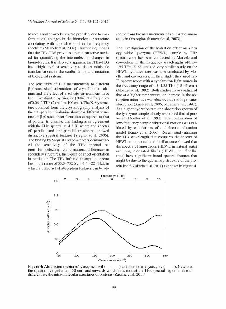
The investigation of the hydration effect on a hen egg white lysozyme (HEWL) sample by THz spectroscopy has been conducted by Markelz and co-workers in the frequency wavelengths of0.15–1.95 THz (5–65 cm-1). A very similar study on the HEWL hydration rate was also conducted by Mo-eller and co-workers. In their study, they used far-IR spectroscopy with a synchrotron light source in the frequency range of 0.5–1.35 THz (15–45 cm-1) (Moeller et al, 1992). Both studies have confirmed that at a higher temperature, an increase in the ab-sorption intensities was observed due to high water absorption (Knab et al, 2006; Moeller et al, 1992). At a higher hydration rate, the absorption spectra of the lysozyme sample closely resembled that of pure water (Moeller et al, 1992). The confirmation of low-frequency sample vibrational motions was val-idated by calculations of a dielectric relaxation model (Knab et al, 2006). Recent study utilizing the THz wavelength that compares the spectra of HEWL at its natural and fibrillar state showed that the spectra of amorphous (HEWL in natural state) and long, elongated fibrils (HEWL in fibrillar state) have significant broad spectral features that might be due to the quaternary structure of the pro-tein itself (Zakaria et al, 2011) as shown in Figure 4.
Figure 4: Absorption spectra of lysozyme fibril (— — —) and monomeric lysozyme ( _____ ). Note that the spectra diverged after 150 cm-1 and onwards which indicate that the THz spectral region is able to differentiate the intra-molecular structures of proteins (Zakaria et al, 2011)
50 100 150 200 250 300 3500
0.3
0.6
0.9
1.2
Frequency (THz)
Abs
orpt
ion
coef
ficie
nt (
cm-1
)
Wavenumber (cm-1)
2 3 4 5 6 7 8 9 101.5
Malaysian Journal of Science 34 (1) : 93-102 (2015)
99
CONCLUSIONS
Low frequency stretching and bending vibrations, crystalline phonon vibrations, hydrogen bond stretches and torsional vibrations of crystalline structures can be investigated in the THz wave-length (Beard et al, 2002). These attractive proper-ties of THz spectroscopy couples with the fact that the non-ionizing, able to transmit through almost any materials of THz rays enable intra-molecular structure analysis in the low frequency spectral re-gion, a spectral domain unachievable by other spec-troscopic methods. There is an increasing interest in analysing low- frequency collective vibrational modes in biomolecules since these modes may pro-vide information about their conformational state (Fischer et al, 2007). However there are several limitations in studying biomolecular structure in the THz range. The limitations are a stable and high en-ergy light source connected to the spectrometer is necessary to enable analysis of protein structure in the very low frequency region which is unachiev-able in a conventional bench-top spectrometer. This bright light source may provide a higher sig-nal-to-noise ratio and a wider dynamic range than traditional far-IR spectroscopy (Grischkowsky et al, 1990). The second factor to be considered was that the sample has to be rigorously characterized prior to spectroscopic measurements and it is best to measure the sample in pressed pellet form to ensure consistency in the resulting spectra. Protein samples immersed in water or solutions will induce interfer-ence to the spectra in the form of emergence of ro-tational water lines and poor signal-to-noise ratio.
ACKNOWLEDGEMENTS
I would like to thank Professor Anton Middelberg, Dr. Robert Falconer and Associate Professor An-drew Bradley for fruitful discussions towards the completion of this manuscript.
REFERENCES1. Aymard P, Nicolai T, Durand D, Clark A
(1999) Static and dynamic scattering of beta-lactoglobulin aggregates formed after heat-induced denaturation at pH 2. Macromolecules 32: 2542-2552
2. Bea S, Teich M (1974) Fundamentals of photonics, New York.
3. Beard MC, Turner GM, Schmuttenmaer CA (2002) Terahertz spectroscopy. Journal of Physical Chemistry B106: 7146-7159
4. Brandt NN, Chikishev AY, Kargovsky AV, Nazarov MM, Parashchuk OD, Sapozhnikov DA, Smirnova IN, Shkurinov AP, Sumbatyan NV (2008) Terahertz time-domain and Raman spectroscopy of the sulfur- containing peptide dimers: Low-frequency markers of disulfide bridges. Vibrational Spectroscopy 47: 53-58
5. Brown ER, Bjarnason JE, Chan TLJ, Lee AWM, Celis MA (2004) Optical attenuation signatures of Bacillus subtillis in the THz region. Applied Physics Letters 84: 3438-3440
6. Chirgadze YN, Nevskaya NA (1976) Infrared-spectra and resonance interaction of amide-one vibration of parallel-chain pleated sheet. Biopolymers 15: 627-636
7. Clark AH, Saunderson DHP, Suggett A (1981) Infrared and laser-Raman spectroscopic studies of thermally-induced globular protein gels. International Journal of Peptide and Protein Research 17: 353-364
8. Davies AG, Linfield EH, Johnston MB (2002) The development of terahertz sources and their applications. Physics in Medicine and Biology 47: 3679-3689
9. Doria A, Gallerano GP, Giovenale E, Messina G, Lai A, Ramundo-Orlando A, Sposato V, D’Arienzo M, Perrotta A, Romano M, Sarti M, Scarfi MR, Spassovsky I, Zeni O (2004) THz radiation studies on biological systems at the ENEA FEL facility. Infrared Physics & Technology 45: 339-347
10. Dyakonov M, Shur M (1996) Detection, mixing, and frequency multiplication of terahertz radiation by two-dimensional electronic fluid. IEEE Trans Electron Devices 43: 380-387
11. Falconer RJ, Zakaria HA, Fan YY, Bradley AP, Middelberg AP (2010) Far-infrared spectroscopy of protein higher-order structures. Applied spectroscopy 64: 1259-1264
12. Fattinger C, Grischkowsky D (1989) TERAHERTZ BEAMS. Applied Physics
Malaysian Journal of Science 34 (1) : 93-102 (2015)
100

Letters 54: 490-492
13. Ferguson B, Zhang XC (2002) Materials for terahertz science and technology. Nat Mater 1: 26-33
14. Fischer BM (2005a) Broadband THz Time-Domain Spectroscopy of Biomolecules, A Comprehensive Study of the Dielectric Properties of Biomaterials in the Far-Infrared, Doctor of Philosophy Thesis, Fakultat fur Mathematik und Physik, Universität Freiburg, Freiburg
15. Fischer BM, Helm H, Jepsen PU (2007) Chemical recognition with broadband THz spectroscopy. Proceedings of the Ieee 95: 1592-1604
16. Fischer BM, Hoffmann M, Helm H, Wilk R, Rutz F, Kleine-Ostmann T, Koch M, Jepsen PU (2005) Terahertz time-domain spectroscopy and imaging of artificial RNA. Optics Express 13: 5205-5215
17. Giraud G, Karolin J, Wynne K (2003) Low-frequency modes of peptides and globular proteins in solution observed by ultrafast OHD-RIKES Spectroscopy. Biophys J 85: 1903-1913
18. Grischkowsky D, Keiding S, Vanexter M, Fattinger C (1990) Far-infrared time-domain spectroscopy with terahertz beams of dielectrics and semiconductors. Journal of the Optical Society of America B-Optical Physics 7: 2006-2015
19. Hagen E, Rubens H (1902) The reflection ability of some metals for ultraviolet and infrared radiation Ann Phys-Berlin 8: 1-21
20. He Y, Chen JY, Knab JR, Zheng W, Markelz AG (2011a) Evidence of Protein Collective Motions on the Picosecond Timescale. Biophysical Journal 100: 1058-1065
21. Jackson M, Mantsch HH (1995) The use and misuse of FTIR spectroscopy in the determination of protein-structure. Critical Reviews in Biochemistry and Molecular Biology 30: 95-120
22. Kachorovskii VY, Shur MS (2008) Field effect
transistor as ultrafast detector of modulated terahertz radiation. Solid-State Electron 52: 182-185
23. Knab J, Chen JY, Markelz A (2006) Hydration dependence of conformational dielectric relaxation of lysozyme. Biophys J 90: 2576-2581
24. Knap W, Dyakonov MI (2013) 5 - Field effect transistors for terahertz applications. In Handbook of Terahertz Technology for Imaging, Sensing and Communications, Saeedkia D (ed), pp 121-155. Woodhead Publishing
25. Kutteruf MR, Brown CM, Iwaki LK, Campbell MB, Korter TM, Heilweil EJ (2003) Terahertz spectroscopy of short-chain polypeptides. Chemical Physics Letters 375: 337-343
26. Liu HB, Zhang XC (2006) Dehydration kinetics of D-glucose monohydrate studied using THz time-domain spectroscopy. Chemical Physics Letters 429: 229-233
27. Lu JQ, Shur MS, Hesler JL, Sun LQ, Weikle R (1998) Terahertz detector utilizing two-dimensional electronic fluid. IEEE Electron Device Lett 19: 373-375
28. Markelz A, Whitmire S, Hillebrecht J, Birge R (2002) THz time domain spectroscopy of biomolecular conformational modes. Physics in Medicine and Biology 47: 3797-3805
29. Markelz AG (2008) Terahertz dielectric sensitivity to biomolecular structure and function. Ieee Journal of Selected Topics in Quantum Electronics 14: 180-190
30. Mickan SP, Menikh A, Liu HB, Mannella CA, MacColl R, Abbott D, Munch J, Zhang XC (2002) Label-free bioaffinity detection using terahertz technology. Physics in Medicine and Biology 47: 3789-3795
31. Moeller KD, Williams GP, Steinhauser S, Hirschmugl C, Smith JC (1992) HYDRATION-DEPENDENT FAR- INFRARED ABSORPTION IN LYSOZYME DETECTED USING SYNCHROTRON RADIATION. Biophys J 61: 276-280
Malaysian Journal of Science 34 (1) : 93-102 (2015)
101
32. Pickwell E, Cole BE, Fitzgerald AJ, Wallace VP, Pepper M (2004) Simulation of terahertz pulse propagation in biological systems. Applied Physics Letters 84: 2190-2192
33. Picometrix. (2005) T-RAY 2000 ANALYTICAL SYSTEM HARDWARE MANUAL. Picometrix,Inc., Michigan,US. Plusquellic DF, Siegrist K, Heilweil EJ, Esenturk M (2007) Applications of terahertz spectroscopy in biosystems. Chemphyschem 8: 2412-2431
34. Png GM, Falconer RJ, Fischer BM, Zakaria HA, Mickan SP, Middelberg AP, Abbott D (2009) Terahertz spectroscopic differentiation of microstructures in protein gels. Optics express 17: 13102-13115
35. Sauer K (1995) WHY SPECTROSCOPY - WHICH SPECTROSCOPY. In Biochemical Spectroscopy Vol. 246, pp 1-10. San Diego: Academic Press Inc
36. Shen YC, Upadhya PC, Linfield EH, Beere HE, Davies AG (2003) Ultrabroadband terahertz radiation from low-temperature-grown GaAs photoconductive emitters. Applied Physics Letters 83: 3117-3119
37. Siegrist K, Bucher CR, Mandelbaum I, Walker ARH, Balu R, Gregurick SK, Plusquellic DF (2006) High- resolution terahertz spectroscopy of crystalline trialanine: Extreme sensitivity to beta-sheet structure and cocrystallized water. J Am Chem Soc 128: 5764-5775
38. Smye SW, Chamberlain JM, Fitzgerald AJ, Berry E (2001) The interaction between Terahertz radiation and biological tissue. Physics in Medicine and Biology 46: R101-R112
39. Trzebiatowska-Gusowska M, Plinski EF, Baran J, Walczakowski MJ, Jarzab PP, Nowak K, Fuglewicz B, Mikulics M, Palka N, Szustakowski M (2014) Terahertz and Raman spectra of non-centrosymmetrical organic molecular crystals. Optical Materials 37: 28-35
40. Ueno Y, Ajito K (2008) Analytical terahertz spectroscopy. Analytical Sciences 24: 185-192
41. Vanexter M, Grischkowsky D (1990) Optical and Electronic-Properties of Doped Silicon From 0.1 To 2 Thz. Applied Physics Letters 56: 1694-1696
42. Walther M, Fischer B, Schall M, Helm H, Jepsen PU (2000) Far-infrared vibrational spectra of all-trans, 9- cis and 13-cis retinal measured by THz time-domain spectroscopy. Chemical Physics Letters 332: 389-395
43. Walther M, Plochocka P, Fischer B, Helm H, Jepsen PU (2002) Collective vibrational modes in biological molecules investigated by terahertz time-domain spectroscopy. Biopolymers 67: 310-313
44. Woodward RM, Wallace VP, Arnone DD, Linfield EH, Pepper M (2003) Terahertz pulsed imaging of skin cancer in the time and frequency domain. Journal of Biological Physics 29: 257-261
45. Yamaguchi M, Miyamaru F, Yamamoto K, Tani M, Hangyo M (2005) Terahertz absorption spectra of L-, D-, and DL-alanine and their application to determination of enantiometric composition. Applied Physics Letters 86
46. Yin XX, Kong KM, Lim JW, Ng BWH, Ferguson B, Mickan SP, Abbott D (2007) Enhanced T-ray signal classification using wavelet preprocessing. Medical & Biological Engineering & Computing 45: 611-616
47. Zakaria HA, Fischer BM, Bradley AP, Jones I, Abbott D, Middelberg AP, Falconer RJ (2011) Low-frequency spectroscopic analysis of monomeric and fibrillar lysozyme. Applied spectroscopy 65: 260-264
48. Zhang CF, Durbin SM (2006) Hydration-induced far-infrared absorption increase in myoglobin. Journal of Physical Chemistry B 110: 23607-2361
Malaysian Journal of Science 34 (1) : 93-102 (2015)
102