Embed Size (px)
Citation preview
REVIEW PAPER
A Review of the Antioxidant Mechanisms of PolyphenolCompounds Related to Iron Binding
Nathan R. Perron Æ Julia L. Brumaghim
Published online: 28 January 2009
� Humana Press Inc. 2009
Abstract In this review, primary attention is given to the
antioxidant (and prooxidant) activity of polyphenols aris-
ing from their interactions with iron both in vitro and in
vivo. In addition, an overview of oxidative stress and the
Fenton reaction is provided, as well as a discussion of the
chemistry of iron binding by catecholate, gallate, and
semiquinone ligands along with their stability constants,
UV–vis spectra, stoichiometries in solution as a function of
pH, rates of iron oxidation by O2 upon polyphenol binding,
and the published crystal structures for iron–polyphenol
complexes. Radical scavenging mechanisms of polyphe-
nols unrelated to iron binding, their interactions with
copper, and the prooxidant activity of iron–polyphenol
complexes are briefly discussed.
Keywords Polyphenol antioxidants � Iron binding �Catecholate � Gallate � Flavonoids � Catechins � Tannins �Proanthocyanidins � EGCG � Stability constant �Autooxidation � DNA damage � Fenton reaction �Cytotoxicity � Cytoprotection � Alzheimer’s � Parkinson’s �Blood and plasma � Preservatives � Radical scavenging �Reactive oxygen species
Abbreviations
Ab Amyloid-beta
EC (–)-Epicatechin
ECG (–)-Epicatechin-3-gallate
EGC (–)-Epigallocatechin
EGCG (–)-Epigallocatechin-3-gallate
GA Gallic acid
Q Quercetin
MEGA Gallic acid methyl ester
MEPCA Protocatechuic acid methyl ester
PCA Protocatechuic acid
PrEGA Gallic acid propyl ester
RNS Reactive nitrogen species
ROS Reactive oxygen species
Rut Rutin
TA Tannic acid
VA Vanillic acid
EPR Electron paramagnetic resonance
TEAC Trolox-equivalent antioxidant activity
ORAC Oxygen radical absorbance capacity
DPPH 2,2-Diphenylpicrylhydrazyl
Polyphenol Compounds: Sources, Structures,
and General Biological Activities
Polyphenol compounds are widely studied for their anti-
oxidant properties, although the term antioxidant has a
broad range of meanings. For the purposes of this review,
antioxidant activity refers to both the ability of polyphenol
compounds to prevent damage from reactive oxygen spe-
cies (ROS) (such as through radical scavenging) or to
prevent generation of these species (by binding iron). As
described in the title, the primary focus will be on poly-
phenol–iron interactions as a mechanism of antioxidant
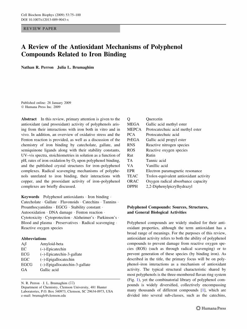
activity. The typical structural characteristic shared by
most polyphenols is the three-membered flavan ring system
(Fig. 1), yet the combinatorial library of polyphenol com-
pounds is widely diversified, collectively encompassing
many thousands of different compounds [1], which are
divided into several sub-classes, such as the catechins,
N. R. Perron � J. L. Brumaghim (&)
Department of Chemistry, Clemson University, 481 Hunter
Laboratories, P.O. Box 340973, Clemson, SC 29634-0973, USA
e-mail: [email protected]
Cell Biochem Biophys (2009) 53:75–100
DOI 10.1007/s12013-009-9043-x
flavonols, flavanols, flavones, anthocyanins, proanthocy-
anidins, and phenolic acids, just to name a few (Fig. 1).
Polyphenols are found in green [2, 3] and black teas [4, 5],
coffee [6], fruits [7, 8], fruit juices [9–11], vegetables [12,
13], olive oil [14, 15], red and white wines [16, 17], and
chocolate [18], and are found in medium to high milligram
quantities per serving for all of these foods (Figs. 2, 3),
although measurements of the precise concentrations of
polyphenol compounds in each type of food often vary [7,
11, 12, 17–22]. Thus, people with diets rich in fruits and
vegetables may consume one or more grams per day of
these compounds, based on the recommendation of
5 servings/day of colorful fruits and vegetables by the
Centers for Disease Control and Prevention [23]. Because
polyphenols are such a large and integral part of the human
diet, it is highly desirable to understand their biological
functions and modes of activity.
Green tea leaf is particularly abundant in the group of
polyphenols collectively referred to as catechins (Fig. 1),
which constitute up to 30% of the plant’s dry leaf weight
[24]. Within just 2 h after consumption of one cup of green
or black tea (350–600 ml) [25–28], catechins have been
found in concentrations of 0.3–1 lM in human plasma and
may even approach 10 lM with higher doses [29]. Flavo-
nols (Fig. 1) such as quercetin are reportedly less
bioavailable than catechins; however, they may reach
similar plasma concentrations (high nanomolar to low
micromolar) in people who eat large amounts of fruits and
vegetables or intentionally supplement their diets with
polyphenols [30, 31].
While polyphenols are primarily recognized for their
antioxidant functions, they also have many other biological
activities, such as anti-histamine [32], anti-inflammatory
[33], antibacterial [34], and antiviral activities [35]. Car-
diovascular effects such as vasodilation have been
observed in tea drinkers [36, 37], and this property has
been attributed to the ability of polyphenols to increase
endothelial nitric oxide synthase (eNOS) activity by over
400% [36]. They have also been shown to bind many
different proteins such as caseins [36], and inhibit telo-
merase [38], a-amylase, pepsin, trypsin, and lipase [39],
among many other enzymes. Furthermore, polyphenols are
implicated in the prevention of neurodegenerative diseases
[2, 40] and cancer [41]. They also induce apoptosis in
cancer cells, implicating them in cancer senescence as well
[42–46].
Because a full discussion of the biological activity of
polyphenols would be prohibitively long, this review will
focus on the antioxidant mechanisms of polyphenols spe-
cifically related to iron-binding, with brief mention made
of other mechanisms such as radical scavenging, prooxi-
dant activity, and interactions between polyphenols and
copper.
Oxidative Stress, DNA Damage, and the Fenton
Reaction
Reactive oxygen species and reactive nitrogen species
(RNS), such as hydroxyl radical (•OH), hydrogen perox-
ide (H2O2), superoxide (O2•-), nitric oxide (NO•),
peroxynitrite (ONOO-), and others, are major sources of
oxidative stress in cells, damaging proteins, lipids, and
DNA [47]. Oxidative DNA damage has been implicated
as a cause of cancer [48, 49], aging and neurodegenera-
tive diseases such as Alzheimer’s and Parkinson’s
[50–54], cardiovascular diseases such as arteriosclerosis
[55, 56], and is the primary cause of cell death and tissue
damage resulting from heart attack and stroke [57, 58].
Therefore, prevention of oxidative stress caused by ROS
and RNS has important implications for the prevention
and treatment of disease.
O
A C
B1
2
3
45
6
78 1'
2'3'
4'
5'
6'OH
OH
catechol
OH
OH
OH
gallol
O
O
OH
flavonols
R2
OH
HO O
OH
OH
catechins
OH
HO
R1
R2
R3R1
flavan
O
O
flavones
OH
HO
R1
R2
R3
O
O
anthocyaninsOH
HO
R1
OH
R2
sugar
Fig. 1 Flavan general structure,
showing the ring labeling and
numbering system. Structures of
catechol, gallol, and general
structures of catechins,
flavonols, flavones, and
anthocyanins. R groups are
typically H, OH, OCH3, galloyl
esters, or carbohydrate groups,
depending on the compound
76 Cell Biochem Biophys (2009) 53:75–100
Radical Scavenging Pathways of Polyphenol
Antioxidant Activity
Many mechanisms have been proposed for polyphenol
prevention of oxidative stress and ROS/RNS generation
both in vitro and in vivo. Radical scavenging by poly-
phenols is the most widely published mechanism for their
antioxidant activity, with over 700 papers since 1995 alone
[59]. In this radical scavenging mechanism, polyphenols
sacrificially reduce ROS/RNS, such as •OH, O2•-, NO•, or
OONO- after generation, preventing damage to biomole-
cules or formation of more reactive ROS [15, 60–68].
Several assays, such as the trolox-equivalent antioxidant
activity (TEAC) and oxygen radical absorbance capacity
(ORAC) assays as well as 2,2-diphenylpicrylhydrazyl
(DPPH) scavenging, are commonly used to study the rad-
ical-scavenging ability of polyphenols [69–72] These
assays provide a relative measure of antioxidant activity,
but often the radicals scavenged have little relevance to
those present in biological systems. In addition, radical
scavenging assays do not account for the iron-binding
properties of polyphenol antioxidants.
It is clear that polyphenols have many different bio-
logical activities; among them are enzyme regulation and
antioxidant behavior. Radical scavenging is a probable
mechanism for reduction of oxidative stress by these
compounds, but as it does not involve iron binding, it is
therefore outside the scope of this review.
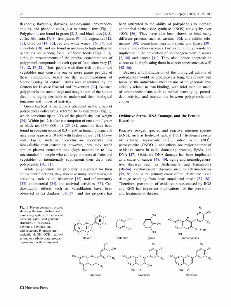
Fig. 2 A chart showing the phenolic content of selected beverages,
vegetables, and chocolate in milligrams per serving. Serving size is
based on a typical beverage size (240 ml), piece of chocolate (40 g),
or serving of vegetables. Values are taken or calculated from data in
the references [5, 6, 11, 12, 17, 18]. Reported polyphenol content
varies
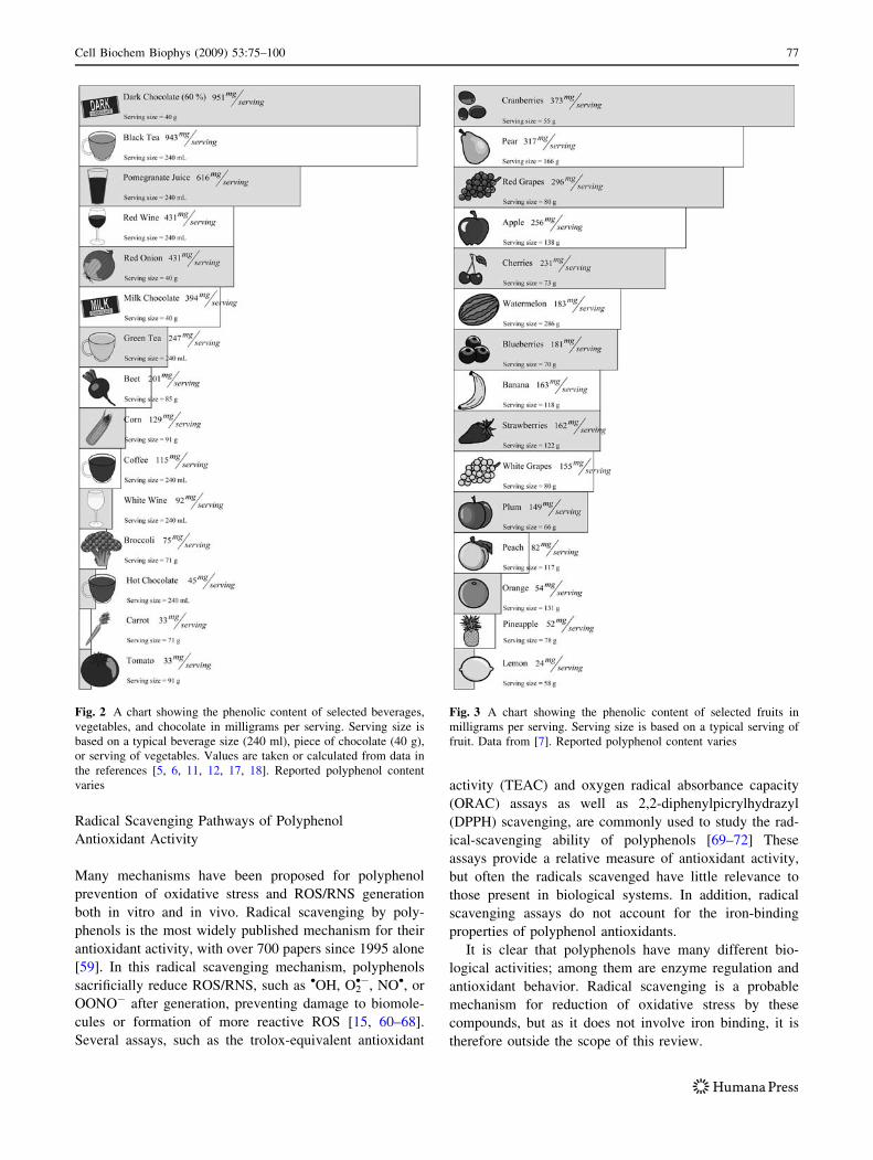
Fig. 3 A chart showing the phenolic content of selected fruits in
milligrams per serving. Serving size is based on a typical serving of
fruit. Data from [7]. Reported polyphenol content varies
Cell Biochem Biophys (2009) 53:75–100 77
The Role of Iron in ROS Generation
Hydroxyl radical, the most reactive ROS known, abstracts
a hydrogen atom from biological substrates at diffusion-
limited rates [67]. Multiple pathways generate •OH,
including the decomposition of peroxynitrous acid [73], or
the metal-mediated reduction of peroxides. Formation of
biological peroxides, such as H2O2, is a process that occurs
naturally during cellular respiration [74], and cell signaling
mechanisms often involve ROS or RNS such as H2O2, O2•-,
and NO•. These species can also form more potent oxidants
if not closely regulated, leading to cellular damage and
oxidative stress [75–80]. For example, O2•- and NO• can
react to form OONO- [81, 82], which can then decompose
into •OH [73]. H2O2 is commonly reduced in vivo by either
Fe2? or Cu?, resulting in the formation of •OH via Fenton-
type reactions (reaction 1). Superoxide forms H2O2 upon
protonation in aqueous solution (reaction 2) [83]. DNA
damage is observed directly from •OH [84–86], and indi-
rectly from O2•- oxidation of [4Fe–4S] iron–sulfur clusters
(reaction 3) to form H2O2 [87]. In addition to H2O2 for-
mation, O2•- also releases Fe2? from enzymes, such as
ferritin [88] and the [4Fe–4S]-containing dehydratases by
reducing Fe3?, generating an unstable iron–sulfur cluster,
and releasing Fe2? (reaction 4) [83, 87, 89]. Superoxide
can also reduce aqueous Fe3? or Cu2? (reaction 5), making
these metal ions available to react with H2O2, although the
rate of iron reduction is slow (10 h is the proposed half-
time for this reaction in vivo), and it is generally assumed
that more abundant cellular reductants such as NADH
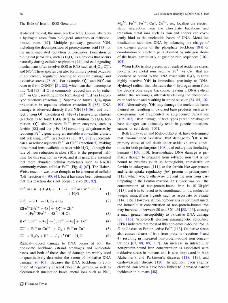
commonly reduce cellular Fe3? (Fig. 4) [67]. The Haber–
Weiss reaction was once thought to be a source of cellular•OH (reaction 6) [90, 91], but it has since been determined
that this reaction does not occur in vivo [91, 92].
Fe2þor Cuþ þ H2O2 þ Hþ ! Fe3þor Cu2þ þ� OH
þ H2O ð1Þ
2O��2 þ 2Hþ ! H2O2 þ O2 ð2Þ
2Fe2þ2Fe3þ � 4S� �
þ O��2 þ 2Hþ
! Fe2þ3Fe3þ � 4S� �
þ H2O2 ð3Þ
Fe2þ3Fe3þ � 4S� �
! 3Fe3þ � 4S� �
þ Fe2þ ð4Þ
O��2 þ Fe3þor Cu2þ ! O2 þ Fe2þor Cuþ ð5Þ
O��2 þ H2O2 þ Hþ ! O2 þ� OHþ H2O ð6Þ
Radical-induced damage to DNA occurs at both the
phosphate backbone (strand breakage) and nucleotide
bases, and both of these sites of damage are widely used
to quantitatively determine the extent of oxidative DNA
damage [93–101]. Because the DNA backbone is com-
posed of negatively charged phosphate groups, as well as
electron-rich nucleotide bases, metal ions such as Na?,
Mg2?, Fe2?, Fe3?, Cu?, Cu2?, etc. localize via electro-
static interaction near the phosphate backbone and
transition metal ions such as iron and copper can cova-
lently bind to the nucleotide bases of DNA. Metal ion
localization stabilizes DNA by balancing the charge of
the oxygen atoms of the phosphate backbone [84] or
coordination to electron pairs donated by nitrogen atoms
of the bases, particularly at guanine-rich sequences [102–
104].
When H2O2 is also present as a result of oxidative stress,
redox active metal ions such as Fe2? or Cu? that are
localized or bound to the DNA react with H2O2 to form
highly reactive •OH in immediate proximity to DNA.
Hydroxyl radical then abstracts the 40 hydrogen atom from
the deoxyribose sugar backbone, leaving a DNA radical
adduct that rearranges, ultimately cleaving the phosphodi-
ester backbone and resulting in strand scission [84, 85, 102,
104]. Alternatively, •OH may damage the nucleotide bases
themselves, resulting in oxidized base products such as 8-
oxo-guanine and fragmented or ring-opened derivatives
[105–107]. DNA damage of both types (strand breakage or
base damage) can ultimately result in genetic mutations,
cancer, or cell death [105].
Both Imlay et al. and Mello-Filho et al. have determined
that iron-mediated oxidative DNA damage by •OH is the
primary cause of cell death under oxidative stress condi-
tions for both prokaryotes [108], and eukaryotes (including
humans) [109, 110]. Iron-mediated DNA damage is pri-
marily thought to originate from solvated iron that is not
bound to proteins (such as hemoglobin, transferrin, or
ferritin in eukaryotes [111], or the ferritin-like dpr protein
and ferric uptake regulatory (fur) protein of prokaryotes)
[112], which would otherwise prevent the iron from par-
ticipating in the Fenton reaction. In Escherichia coli, the
concentration of non-protein-bound iron is 10–30 lM
[113], and it is believed to be coordinated to low molecular
weight intracellular ligands such as ascorbate or citrate
[114, 115]. However, if iron homeostasis is not maintained,
the intracellular concentration of non-protein-bound iron
may increase to between 80 and 320 lM [88, 113], causing
a much greater susceptibility to oxidative DNA damage
[88, 116]. Whole-cell electron paramagnetic resonance
(EPR) indicates that most of this non-protein-bound iron in
E. coli exists as Fenton-active Fe2? [113]. Oxidative stress
also causes release of iron from proteins (reactions 3 and
4), resulting in increased non-protein-bound iron concen-
trations [67, 88, 89, 117]. An increase in intracellular
non-protein-bound iron concentration is associated with
oxidative stress in humans and is also implicated in both
Alzheimer’s and Parkinson’s diseases [118, 119], and
cardiovascular disease [120]. In addition, even slightly
elevated iron levels have been linked to increased cancer
incidence in humans [48].
78 Cell Biochem Biophys (2009) 53:75–100
Damage to both nuclear and mitochondrial DNA occurs
in cancer and other diseases linked to iron mis-regulation
[121, 122]. Although nuclear DNA is packaged with his-
tone proteins in chromatin, several studies have shown that
oxidative damage to nuclear DNA occurs even in the
presence of histone proteins; in fact, histone proteins can
increase metal-mediated oxidative DNA damage because
redox-active metal ions are associated with these proteins
[123–126]. Mitochondrial DNA is particularly at risk for
oxidative damage due to its proximity to respiratory pro-
cesses that produce O2•-, H2O2, and other ROS [127–129].
In fact, oxidative damage to mitochondrial DNA may
actually be a more relevant cause of cell death than nuclear
DNA damage because of this higher risk of damage [130,
131].
Because iron is a primary cause of ROS generation in
vivo and because it plays such a pivotal role in contributing
to oxidative stress, DNA damage, and cell death, iron has
been the target of many antioxidant therapies. Due to their
ability to coordinate iron, polyphenols are one large class
of antioxidants that has been extensively examined for
treatment and prevention of conditions associated with
iron-generated ROS and oxidative stress.
Iron Binding by Catecholate, Gallate, and Semiquinone
Ligands
Stability Constants for Iron–Polyphenol Complexes
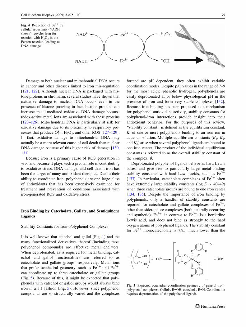
It is well known that catechol and gallol (Fig. 1) and the
many functionalized derivatives thereof (including most
polyphenol compounds) are effective metal chelators.
When deprotonated, as is required for metal binding, cat-
echol and gallol functionalities are referred to as
catecholate and gallate groups, respectively. Metal ions
that prefer octahedral geometry, such as Fe2? and Fe3?,
can coordinate up to three catecholate or gallate groups
(Fig. 5). Because of this, it might be expected that poly-
phenols with catechol or gallol groups would always bind
iron in a 3:1 fashion (Fig. 5). However, since polyphenol
compounds are so structurally varied and the complexes
formed are pH dependent, they often exhibit variable
coordination modes. Despite pKa values in the range of 7–9
for the most acidic phenolic hydrogen, polyphenols are
easily deprotonated at or below physiological pH in the
presence of iron and form very stable complexes [132].
Because iron binding has been proposed as a mechanism
for polyphenol antioxidant activity, stability constants for
polyphenol–iron interactions provide insight into their
antioxidant behavior. For the purposes of this review,
‘‘stability constant’’ is defined as the equilibrium constant,
K, of one or more polyphenols binding to an iron ion in
aqueous solution. Multiple equilibrium constants (K1, K2,
and K3) arise when several polyphenol ligands are bound to
one iron center. The product of the individual equilibrium
constants is referred to as the overall stability constant of
the complex, b.
Deprotonated polyphenol ligands behave as hard Lewis
bases, and give rise to particularly large metal-binding
stability constants with hard Lewis acids, such as Fe3?
[133]. In particular, catecholate complexes of Fe3? often
have extremely large stability constants (log b * 40–49)
when three catecholate groups are bound to one iron center
[134, 135]. Despite the importance of iron binding by
polyphenols, only a handful of stability constants are
reported for catecholate and gallate complexes of Fe3?,
other than siderophore complexes (both naturally occurring
and synthetic). Fe2?, in contrast to Fe3?, is a borderline
Lewis acid, and does not bind as strongly to the hard
oxygen atoms of polyphenol ligands. The stability constant
for Fe2? monocatecholate is 7.95, much lower than the
Fe3+
Fe2+ H2O2
OH
NAD+
NADH
Fig. 4 Reduction of Fe3? by
cellular reductants (NADH
shown) recycles iron for
reaction with H2O2 in the
Fenton reaction, leading to
DNA damage
OH
OHR
+ Fe3+
O
OR
Fe
3-
+ 6H+
OO
R
OO
R
3
Fig. 5 Expected octahedral coordination geometry of general iron–
polyphenol complexes. Gallols, R=OH; catechols, R=H. Coordination
requires deprotonation of the polyphenol ligands
Cell Biochem Biophys (2009) 53:75–100 79
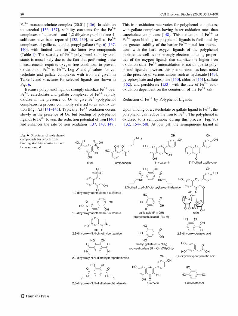
Fe3? monocatecholate complex (20.01) [136]. In addition
to catechol [136, 137], stability constants for the Fe2?
complexes of quercetin and 1,2-dihydroxynaphthalene-4-
sulfonate have been reported [138, 139], as well as Fe2?
complexes of gallic acid and n-propyl gallate (Fig. 6) [137,
140], with limited data for the latter two compounds
(Table 1). The scarcity of Fe2?-polyphenol stability con-
stants is most likely due to the fact that performing these
measurements requires oxygen-free conditions to prevent
oxidation of Fe2? to Fe3?. Log K and b values for ca-
techolate and gallate complexes with iron are given in
Table 1, and structures for selected ligands are shown in
Fig. 6.
Because polyphenol ligands strongly stabilize Fe3? over
Fe2?, catecholate and gallate complexes of Fe2? rapidly
oxidize in the presence of O2 to give Fe3?-polyphenol
complexes, a process commonly referred to as autooxida-
tion (Fig. 7a) [141–145]. Typically, Fe2? oxidation occurs
slowly in the presence of O2, but binding of polyphenol
ligands to Fe2? lowers the reduction potential of iron [146]
and enhances the rate of iron oxidation [137, 143, 147].
This iron oxidation rate varies for polyphenol complexes,
with gallate complexes having faster oxidation rates than
catecholate complexes [148]. This oxidation of Fe2? to
Fe3? upon binding to polyphenol ligands is facilitated by
the greater stability of the harder Fe3? metal ion interac-
tions with the hard oxygen ligands of the polyphenol
moieties as well as the strongly electron-donating proper-
ties of the oxygen ligands that stabilize the higher iron
oxidation state. Fe2? autooxidation is not unique to poly-
phenol ligands; however, this phenomenon has been noted
in the presence of various anions such as hydroxide [149],
pyrophosphate and phosphate [150], chloride [151], sulfate
[152], and perchlorate [153], with the rate of Fe2? auto-
oxidation dependent on the counterion of the Fe2? salt.
Reduction of Fe3? by Polyphenol Ligands
Upon binding of a catecholate or gallate ligand to Fe3?, the
polyphenol can reduce the iron to Fe2?. The polyphenol is
oxidized to a semiquinone during this process (Fig. 7b)
[132, 154–158]. At low pH, the semiquinone ligand is
S OOOH
OHOH
1,2-dihydroxynaphthalene-4-sulfonate
SO
OHO
OH
OH1,2-dihydroxynaphthalene-6-sulfonate
O
OHHO OH
2,3-dihydroxybenzoic acidO
NHO
OH2,3-dihydroxy-N,N-dimethylbenzamide
O
HN
O
NH
OHHO
2,3-dihydroxy-N,N'-dimethylterephthalamide
O
HN
O
NH
HO OH
2,3-dihydroxy-N,N'-diethylterephthalamide
O
NH
O
HN
OHHO
2,3-dihydroxy-N,N'-dipropylterephthalamide
O
OH
HO
HO
3,4-dihydroxyphenylacetic acid
O
O
OHOH
3',4'-dihydroxyflavone
HO
HO
NO2
4-nitrocatechol
OHOH
HO O
OHOH
(+)-catechin
R
HO
HOO
OH
gallic acid (R = OH)protocatechuic acid (R = H)
HO
HO
HO
O
OR
methyl gallate (R = CH3)n-propyl gallate (R = CH2CH2CH3)
OHOOH
HO O
OHOH
quercetin
SO
O OHS
O
O
HO
OHOH
tiron
OOOH
HO O
OHOH
OO
OOH
OHOH OH
OH
OHrutin
O
NH
OHHO
NH2
aminochelin
Fig. 6 Structures of polyphenol
compounds for which iron-
binding stability constants have
been measured
80 Cell Biochem Biophys (2009) 53:75–100
protonated and is therefore a neutral ligand [132].
According to Basolo and Pearson [159], Fe2? is stabilized
relative to Fe3? by neutral unsaturated ligands due to the
greater crystal field stabilization of a d6 electronic config-
uration (Fe2?) than that of a d5 system (Fe3?). These Fe2?-
semiquinone complexes are green in color, likely due to
stabilization of the semiquinone radical by the aromatic
ring, and may often be mistaken for mono(polyphenol)
Fe3? complexes at low pH. However, the presence of Fe2?
in these semiquinone complexes has been confirmed by
both Mossbauer spectroscopy [160] and magnetic moment
measurements [161].
Once the semiquinone form of the polyphenol is gen-
erated, it is capable of reducing another equivalent of Fe3?,
simultaneously oxidizing the semiquinone to the quinone
(Fig. 7b) [132, 154–158]. The studies investigating this
Fe3? reduction behavior are performed at very low pH
[1–3], which may be relevant for processes occurring
during digestion in the stomach. At higher pH, the for-
mation of bis- and tris-polyphenol complexes with iron
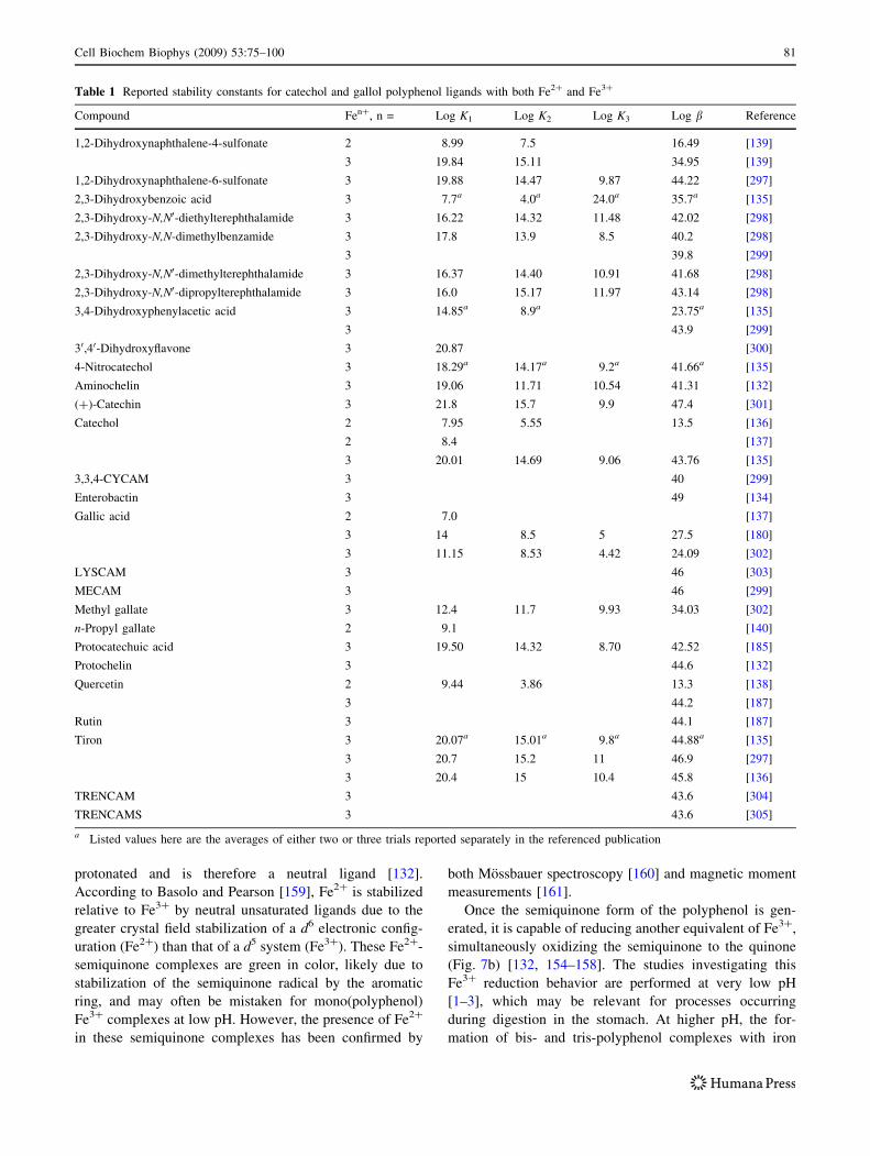
Table 1 Reported stability constants for catechol and gallol polyphenol ligands with both Fe2? and Fe3?
Compound Fen?, n = Log K1 Log K2 Log K3 Log b Reference
1,2-Dihydroxynaphthalene-4-sulfonate 2 8.99 7.5 16.49 [139]
3 19.84 15.11 34.95 [139]
1,2-Dihydroxynaphthalene-6-sulfonate 3 19.88 14.47 9.87 44.22 [297]
2,3-Dihydroxybenzoic acid 3 7.7a 4.0a 24.0a 35.7a [135]
2,3-Dihydroxy-N,N0-diethylterephthalamide 3 16.22 14.32 11.48 42.02 [298]
2,3-Dihydroxy-N,N-dimethylbenzamide 3 17.8 13.9 8.5 40.2 [298]
3 39.8 [299]
2,3-Dihydroxy-N,N0-dimethylterephthalamide 3 16.37 14.40 10.91 41.68 [298]
2,3-Dihydroxy-N,N0-dipropylterephthalamide 3 16.0 15.17 11.97 43.14 [298]
3,4-Dihydroxyphenylacetic acid 3 14.85a 8.9a 23.75a [135]
3 43.9 [299]
30,40-Dihydroxyflavone 3 20.87 [300]
4-Nitrocatechol 3 18.29a 14.17a 9.2a 41.66a [135]
Aminochelin 3 19.06 11.71 10.54 41.31 [132]
(?)-Catechin 3 21.8 15.7 9.9 47.4 [301]
Catechol 2 7.95 5.55 13.5 [136]
2 8.4 [137]
3 20.01 14.69 9.06 43.76 [135]
3,3,4-CYCAM 3 40 [299]
Enterobactin 3 49 [134]
Gallic acid 2 7.0 [137]
3 14 8.5 5 27.5 [180]
3 11.15 8.53 4.42 24.09 [302]
LYSCAM 3 46 [303]
MECAM 3 46 [299]
Methyl gallate 3 12.4 11.7 9.93 34.03 [302]
n-Propyl gallate 2 9.1 [140]
Protocatechuic acid 3 19.50 14.32 8.70 42.52 [185]
Protochelin 3 44.6 [132]
Quercetin 2 9.44 3.86 13.3 [138]
3 44.2 [187]
Rutin 3 44.1 [187]
Tiron 3 20.07a 15.01a 9.8a 44.88a [135]
3 20.7 15.2 11 46.9 [297]
3 20.4 15 10.4 45.8 [136]
TRENCAM 3 43.6 [304]
TRENCAMS 3 43.6 [305]
a Listed values here are the averages of either two or three trials reported separately in the referenced publication
Cell Biochem Biophys (2009) 53:75–100 81
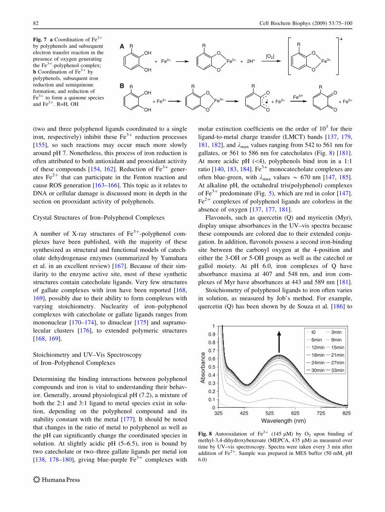
(two and three polyphenol ligands coordinated to a single
iron, respectively) inhibit these Fe3? reduction processes
[155], so such reactions may occur much more slowly
around pH 7. Nonetheless, this process of iron reduction is
often attributed to both antioxidant and prooxidant activity
of these compounds [154, 162]. Reduction of Fe3? gener-
ates Fe2? that can participate in the Fenton reaction and
cause ROS generation [163–166]. This topic as it relates to
DNA or cellular damage is discussed more in depth in the
section on prooxidant activity of polyphenols.
Crystal Structures of Iron–Polyphenol Complexes
A number of X-ray structures of Fe3?-polyphenol com-
plexes have been published, with the majority of these
synthesized as structural and functional models of catech-
olate dehydrogenase enzymes (summarized by Yamahara
et al. in an excellent review) [167]. Because of their sim-
ilarity to the enzyme active site, most of these synthetic
structures contain catecholate ligands. Very few structures
of gallate complexes with iron have been reported [168,
169], possibly due to their ability to form complexes with
varying stoichiometry. Nuclearity of iron–polyphenol
complexes with catecholate or gallate ligands ranges from
mononuclear [170–174], to dinuclear [175] and supramo-
lecular clusters [176], to extended polymeric structures
[168, 169].
Stoichiometry and UV–Vis Spectroscopy
of Iron–Polyphenol Complexes
Determining the binding interactions between polyphenol
compounds and iron is vital to understanding their behav-
ior. Generally, around physiological pH (7.2), a mixture of
both the 2:1 and 3:1 ligand to metal species exist in solu-
tion, depending on the polyphenol compound and its
stability constant with the metal [177]. It should be noted
that changes in the ratio of metal to polyphenol as well as
the pH can significantly change the coordinated species in
solution. At slightly acidic pH (5–6.5), iron is bound by
two catecholate or two–three gallate ligands per metal ion
[138, 178–180], giving blue-purple Fe3? complexes with
molar extinction coefficients on the order of 103 for their
ligand-to-metal charge transfer (LMCT) bands [137, 179,
181, 182], and kmax values ranging from 542 to 561 nm for
gallates, or 561 to 586 nm for catecholates (Fig. 8) [181].
At more acidic pH (\4), polyphenols bind iron in a 1:1
ratio [140, 183, 184]. Fe3? monocatecholate complexes are
often blue-green, with kmax values * 670 nm [147, 185].
At alkaline pH, the octahedral tris(polyphenol) complexes
of Fe3? predominate (Fig. 5), which are red in color [147].
Fe2? complexes of polyphenol ligands are colorless in the
absence of oxygen [137, 177, 181].
Flavonols, such as quercetin (Q) and myricetin (Myr),
display unique absorbances in the UV–vis spectra because
these compounds are colored due to their extended conju-
gation. In addition, flavonols possess a second iron-binding
site between the carbonyl oxygen at the 4-position and
either the 3-OH or 5-OH groups as well as the catechol or
gallol moiety. At pH 6.0, iron complexes of Q have
absorbance maxima at 407 and 548 nm, and iron com-
plexes of Myr have absorbances at 443 and 589 nm [181].
Stoichiometry of polyphenol ligands to iron often varies
in solution, as measured by Job’s method. For example,
quercetin (Q) has been shown by de Souza et al. [186] to
0
0.1
0.2
0.3
0.4
0.5
0.6
0.7
0.8
0.9
1
325 425 525 625 725 825
Wavelength (nm)
Abs
orba
nce
t0 3min
6min 9min
12min 15min
18min 21min
24min 27min
30min 33min
Fig. 8 Autooxidation of Fe2? (145 lM) by O2 upon binding of
methyl-3,4-dihydroxybenzoate (MEPCA, 435 lM) as measured over
time by UV–vis spectroscopy. Spectra were taken every 3 min after
addition of Fe2?. Sample was prepared in MES buffer (50 mM, pH
6.0)
OH
OH
+ Fe3+
O
O
Fe3+
O
O
O
O
- + Fe2+Fe3+
+ Fe2+
OH
OH
R
+ Fe2+
O
O
R
Fe2+ + 2H+[O2]
O
O
R
Fe3+
R R R R
A
B
Fig. 7 a Coordination of Fe2?
by polyphenols and subsequent
electron transfer reaction in the
presence of oxygen generating
the Fe3?-polyphenol complex;
b Coordination of Fe3? by
polyphenols, subsequent iron
reduction and semiquinone
formation, and reduction of
Fe3? to form a quinone species
and Fe2?. R=H, OH
82 Cell Biochem Biophys (2009) 53:75–100
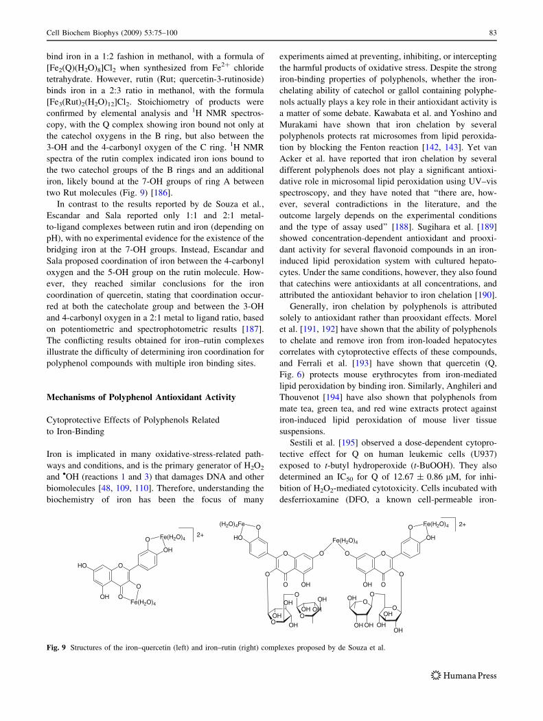
bind iron in a 1:2 fashion in methanol, with a formula of
[Fe2(Q)(H2O)8]Cl2 when synthesized from Fe2? chloride
tetrahydrate. However, rutin (Rut; quercetin-3-rutinoside)
binds iron in a 2:3 ratio in methanol, with the formula
[Fe3(Rut)2(H2O)12]Cl2. Stoichiometry of products were
confirmed by elemental analysis and 1H NMR spectros-
copy, with the Q complex showing iron bound not only at
the catechol oxygens in the B ring, but also between the
3-OH and the 4-carbonyl oxygen of the C ring. 1H NMR
spectra of the rutin complex indicated iron ions bound to
the two catechol groups of the B rings and an additional
iron, likely bound at the 7-OH groups of ring A between
two Rut molecules (Fig. 9) [186].
In contrast to the results reported by de Souza et al.,
Escandar and Sala reported only 1:1 and 2:1 metal-
to-ligand complexes between rutin and iron (depending on
pH), with no experimental evidence for the existence of the
bridging iron at the 7-OH groups. Instead, Escandar and
Sala proposed coordination of iron between the 4-carbonyl
oxygen and the 5-OH group on the rutin molecule. How-
ever, they reached similar conclusions for the iron
coordination of quercetin, stating that coordination occur-
red at both the catecholate group and between the 3-OH
and 4-carbonyl oxygen in a 2:1 metal to ligand ratio, based
on potentiometric and spectrophotometric results [187].
The conflicting results obtained for iron–rutin complexes
illustrate the difficulty of determining iron coordination for
polyphenol compounds with multiple iron binding sites.
Mechanisms of Polyphenol Antioxidant Activity
Cytoprotective Effects of Polyphenols Related
to Iron-Binding
Iron is implicated in many oxidative-stress-related path-
ways and conditions, and is the primary generator of H2O2
and •OH (reactions 1 and 3) that damages DNA and other
biomolecules [48, 109, 110]. Therefore, understanding the
biochemistry of iron has been the focus of many
experiments aimed at preventing, inhibiting, or intercepting
the harmful products of oxidative stress. Despite the strong
iron-binding properties of polyphenols, whether the iron-
chelating ability of catechol or gallol containing polyphe-
nols actually plays a key role in their antioxidant activity is
a matter of some debate. Kawabata et al. and Yoshino and
Murakami have shown that iron chelation by several
polyphenols protects rat microsomes from lipid peroxida-
tion by blocking the Fenton reaction [142, 143]. Yet van
Acker et al. have reported that iron chelation by several
different polyphenols does not play a significant antioxi-
dative role in microsomal lipid peroxidation using UV–vis
spectroscopy, and they have noted that ‘‘there are, how-
ever, several contradictions in the literature, and the
outcome largely depends on the experimental conditions
and the type of assay used’’ [188]. Sugihara et al. [189]
showed concentration-dependent antioxidant and prooxi-
dant activity for several flavonoid compounds in an iron-
induced lipid peroxidation system with cultured hepato-
cytes. Under the same conditions, however, they also found
that catechins were antioxidants at all concentrations, and
attributed the antioxidant behavior to iron chelation [190].
Generally, iron chelation by polyphenols is attributed
solely to antioxidant rather than prooxidant effects. Morel
et al. [191, 192] have shown that the ability of polyphenols
to chelate and remove iron from iron-loaded hepatocytes
correlates with cytoprotective effects of these compounds,
and Ferrali et al. [193] have shown that quercetin (Q,
Fig. 6) protects mouse erythrocytes from iron-mediated
lipid peroxidation by binding iron. Similarly, Anghileri and
Thouvenot [194] have also shown that polyphenols from
mate tea, green tea, and red wine extracts protect against
iron-induced lipid peroxidation of mouse liver tissue
suspensions.
Sestili et al. [195] observed a dose-dependent cytopro-
tective effect for Q on human leukemic cells (U937)
exposed to t-butyl hydroperoxide (t-BuOOH). They also
determined an IC50 for Q of 12.67 ± 0.86 lM, for inhi-
bition of H2O2-mediated cytotoxicity. Cells incubated with
desferrioxamine (DFO, a known cell-permeable iron-
O
OOH
HO O
O
OH
Fe(H2O)4
Fe(H2O)4
O
O OH
OO
O
HO
OO
O
OOH
O O
O
OH
OO
OOH
OH OH OH
OH
OH
O
OH
OH
OHOH OH
OH
Fe(H2O)4
Fe(H2O)4(H2O)4Fe
2+
2+
Fig. 9 Structures of the iron–quercetin (left) and iron–rutin (right) complexes proposed by de Souza et al.
Cell Biochem Biophys (2009) 53:75–100 83
chelator) showed the same inhibitory effect on DNA strand
scission as Q, so Sestili et al. [195] inferred that iron-
chelation was responsible for prevention of nuclear DNA
damage by Q. Using similar methods, Sestili et al. [196]
tested a variety of polyphenols and reported that only those
compounds with catechol groups displayed both cytopro-
tective effects and DNA damage inhibition. Once again,
these results suggested that iron binding contributes to the
antioxidant activity of these compounds. Antioxidant
activity of these compounds also correlated to their lipo-
philicity (ClogP), suggesting that a combination of iron-
binding ability and the ability to cross cell membranes
contribute to the antioxidant activity of polyphenol com-
pounds [196]. Similar correlations between polyphenol
iron-chelating ability and lipophilicity on prevention of
DNA damage in H2O2-treated Jurkat cells were also noted
by Melidou et al. [197].
Recently, Garcia-Alonso et al. [10] also studied the
ability of a polyphenol-rich fruit juice blend to protect
U937 cells from cell death by t-BuOOH. The blend of
grape, cherry, and berry juices inhibited DNA damage
similarly to the iron chelator ortho-phenanthroline so iron-
chelation by the juice polyphenols was inferred to be the
cause of antioxidant activity [10]. Boato et al. [198]
observed that fruit juices high in polyphenols ([1 mg/ml),
such as red grape and prune juices, limited bioavailability
of iron for human colon cancer cells (Caco-2) in iron-
enriched medium. Because juices low in polyphenols
(\1 mg/ml) increased iron bioavailability, this reduced
bioavailability was attributed to iron–polyphenol coordi-
nation preventing absorption by the cells. The increase in
iron bioavailability was correlated to high ascorbate/poly-
phenol ratio, since ascorbate is known to reduce Fe3? to the
more water-soluble Fe2?. Thus, Boato et al. [198] sug-
gested a dietary balance of juices containing high ascorbate
concentrations (for proper iron uptake) and high polyphe-
nol content (for their cancer preventative properties).
Several other groups have shown that polyphenols have a
significant inhibitory effect on the bioavailability of iron
not only in vitro [199–201], but also in rats [202], and in
humans [203, 204]. This effect is usually assumed to occur
either pre-ingestion (i.e., due iron-binding in the food or
beverage) or in the gut, not after iron and polyphenols are
separately absorbed or metabolized. The general bioavail-
ability of polyphenol compounds, not related to iron, is an
active research area [205, 206], but is beyond the scope of
this review.
Kuo et al. [207] also examined the effects of polyphe-
nols on Caco-2 cells but from a different perspective:
metallothionein (MT) expression. MT is a sulfur-rich pro-
tein, which helps to control oxidative stress and heavy
metal toxicity in vivo by chelating metal ions, including
lead, mercury, cadmium, copper, and zinc. Kuo et al.
proposed that polyphenols might upregulate or downregu-
late MT expression, since polyphenol–metal chelation
might prevent metal-mediated damage to biomolecules,
resulting in lower MT levels. If the polyphenols themselves
upregulate MT expression, increased MT levels could bind
potentially damaging metal ions before they disrupt cel-
lular function [207]. Using a 109Cd-binding assay to
determine MT expression, Kuo and coworkers observed
that upon addition of the flavonoid quercetin, expression of
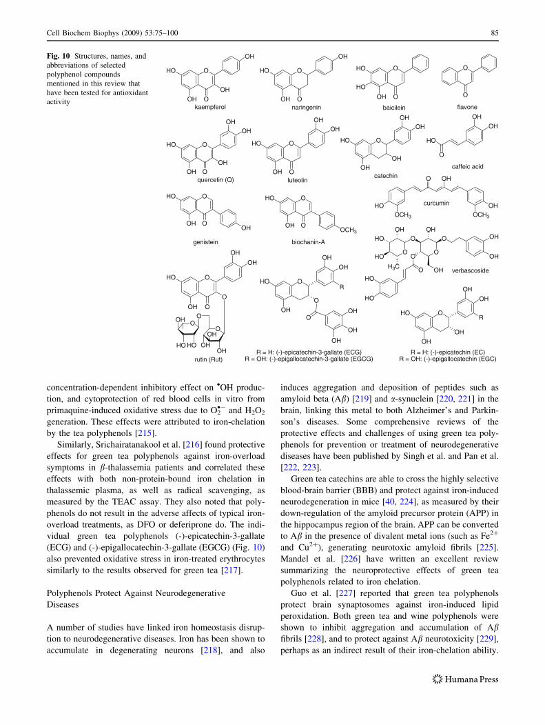
MT was decreased in a dose- and time-dependent manner.
On the other hand, kaempferol, genistein and biochanin-A-
increased expression of MT, and flavone, catechin, and
rutin had no effect on MT levels (Fig. 10). Although these
results were primarily attributed to the ability of these
compounds to bind either Cu2? or Zn2? (two metals that
increase MT expression) [208–210], UV–vis spectroscopy
also indicated interactions with Mn2?, Fe2?, and Fe3? for
some of these polyphenols. Interestingly, none of the pol-
yphenol compounds that increased MT expression were
shown to bind to metal ions under their experimental
conditions [207].
In addition, Anghileri and Thouvenot reported that mate
tea and green tea extracts and, to a lesser extent, red wine
polyphenols, prevented iron-dependent calcium uptake in
mouse liver tissue suspensions. They reasoned that iron
binding by polyphenols limited the bioavailability of iron,
thus inhibiting iron-dependent calcium uptake [194].
It is clear that polyphenols have varied cellular effects
due to their metal-binding properties. A substantial body of
work suggests that iron-binding by polyphenol compounds
results in cytoprotective effects, and that both iron binding
and lipophilicity are important factors contributing to
overall antioxidant activity. However, definitive correla-
tions between results from cell studies and prevention of
oxidative stress-related diseases in animal or human clini-
cal trials have not been established. Further investigation of
indirect modes of polyphenol antioxidant activity, such as
limitation of iron bioavailability and gene regulation, in
animals or humans is also warranted.
Protective Effects of Polyphenols in Blood and Plasma
Although iron homeostasis in the blood is tightly regulated,
red blood cells may become a target of oxidative damage in
diseases disrupting normal hemoglobin function, such as
b-thalassemia, sickle cell anemia, and other hemoglobin-
opathies, due in large part to their high non-protein-bound
iron content [211–214]. Since plasma concentrations of
polyphenols in humans commonly reach 1–10 lM [25–29],
these compounds might be expected to have a protective
effect on red blood cells. Grinberg, et al. have reported
such protective effects for polyphenols in green and black
tea against red blood cell lipid peroxidation, noting a
84 Cell Biochem Biophys (2009) 53:75–100
concentration-dependent inhibitory effect on •OH produc-
tion, and cytoprotection of red blood cells in vitro from
primaquine-induced oxidative stress due to O2•- and H2O2
generation. These effects were attributed to iron-chelation
by the tea polyphenols [215].
Similarly, Srichairatanakool et al. [216] found protective
effects for green tea polyphenols against iron-overload
symptoms in b-thalassemia patients and correlated these
effects with both non-protein-bound iron chelation in
thalassemic plasma, as well as radical scavenging, as
measured by the TEAC assay. They also noted that poly-
phenols do not result in the adverse affects of typical iron-
overload treatments, as DFO or deferiprone do. The indi-
vidual green tea polyphenols (-)-epicatechin-3-gallate
(ECG) and (-)-epigallocatechin-3-gallate (EGCG) (Fig. 10)
also prevented oxidative stress in iron-treated erythrocytes
similarly to the results observed for green tea [217].
Polyphenols Protect Against Neurodegenerative
Diseases
A number of studies have linked iron homeostasis disrup-
tion to neurodegenerative diseases. Iron has been shown to
accumulate in degenerating neurons [218], and also
induces aggregation and deposition of peptides such as
amyloid beta (Ab) [219] and a-synuclein [220, 221] in the
brain, linking this metal to both Alzheimer’s and Parkin-
son’s diseases. Some comprehensive reviews of the
protective effects and challenges of using green tea poly-
phenols for prevention or treatment of neurodegenerative
diseases have been published by Singh et al. and Pan et al.
[222, 223].
Green tea catechins are able to cross the highly selective
blood-brain barrier (BBB) and protect against iron-induced
neurodegeneration in mice [40, 224], as measured by their
down-regulation of the amyloid precursor protein (APP) in
the hippocampus region of the brain. APP can be converted
to Ab in the presence of divalent metal ions (such as Fe2?
and Cu2?), generating neurotoxic amyloid fibrils [225].
Mandel et al. [226] have written an excellent review
summarizing the neuroprotective effects of green tea
polyphenols related to iron chelation.
Guo et al. [227] reported that green tea polyphenols
protect brain synaptosomes against iron-induced lipid
peroxidation. Both green tea and wine polyphenols were
shown to inhibit aggregation and accumulation of Abfibrils [228], and to protect against Ab neurotoxicity [229],
perhaps as an indirect result of their iron-chelation ability.
OH
OOH
HO O
OH
OHOOH
HO O
OHOH
O
O
OHOH
HO O
OHOH
OOH
HO O
OH
O
OOH
HO O
OH
OH
OO
OOH
HO HO OH
OH
OH
OOH
HO O
OCH3
flavonekaempferol
catechinquercetin (Q)
A-ninahcoibnietsineg
rutin (Rut)
OOH
HO
HO O
OOH
HO O
OH
OOH
HO O
OHOH
OH
OH
O
HO
caffeic acid
luteolin
baicileinnaringenin
O
OH
HO O
OH
OH
OH
OH
O
R = H: (-)-epicatechin-3-gallate (ECG)R = OH: (-)-epigallocatechin-3-gallate (EGCG)
OH
R
OH
OH
HO O
OH
OH
R = H: (-)-epicatechin (EC)R = OH: (-)-epigallocatechin (EGC)
O OH
OHOCH3OCH3
HO curcumin
R
HO
HO
OH
OH
O
O
O
O
OH
O
OHO
HOOH OH
H3Cverbascoside
Fig. 10 Structures, names, and
abbreviations of selected
polyphenol compounds
mentioned in this review that
have been tested for antioxidant
activity
Cell Biochem Biophys (2009) 53:75–100 85
Curcumin (Fig. 10), a polyphenol from turmeric, inhibits
neurodegeneration in a mouse model, likely through a
similar mechanism to green tea catechins [230, 231].
Additional work in the area of iron-mediated neurode-
generation prevention comes from Oboh and Rocha [13],
who have used the thiobarbituric acid reactive species
(TBARS) assay on homogenates of brain and liver cells of
rats. TBARS is a common antioxidant assay that uses UV–
vis spectroscopy to monitor the products of lipid peroxi-
dation as they react with thiobarbituric acid and generate a
colored species [232]. Oboh and Rocha’s results show that
polyphenols isolated from red pepper inhibit Fe2?-medi-
ated lipid peroxidation in both brain and liver cells by an
iron-chelating mechanism, although iron chelation by the
polyphenols was measured separately by UV–vis spec-
troscopy. In addition to iron binding, •OH and NO•
scavenging were implicated as additional protective
mechanisms for pepper polyphenols [233].
The interest in using antioxidant properties of poly-
phenols to prevent and treat neurodegenerative diseases is
relatively new, and only a small number of compounds
have been tested for this activity. Additional polyphenols
should be tested for their effects on prevention of oxidative
stress-induced neurodegeneration by chelating iron and
copper ions as well as their ability to cross the BBB. These
compounds should also be examined for their ability to
inhibit APP aggregation. Identifying specific polyphenol
compounds that have BBB permeability and metal che-
lating activity may lead to new treatments for
neurodegenerative diseases.
Assays Quantifying the Inhibition of Iron-Mediated
DNA Damage by Polyphenols
Whole-cell assays involve many variables and can make it
difficult to definitively attribute polyphenol antioxidant
activity to metal binding. For this reason, many in vitro
methods have been used to examine the iron-binding
antioxidant mechanism of polyphenols in order to correlate
the results to those observed in biological systems. Two
common methods for assessing inhibition of iron-mediated
DNA damage by polyphenols are DNA gel electrophoresis
and the deoxyribose assay, an assay that uses UV–vis
spectroscopy to quantify malonaldehyde (a product formed
from •OH degradation of 2-deoxyribose) by its condensa-
tion reaction with thiobarbituric acid [182, 234, 235]. The
major benefit of the deoxyribose assay is that it allows for
faster screening of compounds, but its conditions do not
closely resemble those of biological systems: the substrate
is not truly DNA, but only 2-deoxyribose. In addition,
many Fe3?-polyphenol complexes absorb at or near the
wavelength of the deoxyribose degradation product
(532 nm), making this technique inherently problematic for
measurements of polyphenol inhibition of iron-mediated
damage. Therefore, gel electrophoresis is perhaps the best
technique because it both directly determines DNA damage
and closely simulates biologically relevant conditions
(such as pH, buffering capacity, and ionic strength).
Romanova et al. [236] have shown prevention of iron-
mediated DNA damage for three flavonoid compounds
(including quercetin), citing their iron-chelation ability,
measured by UV–vis spectroscopy, as the most likely cause
for DNA protection. In addition, Lopes et al. [182] used the
deoxyribose assay to show that tannic acid prevents the
Fenton reaction and protects 2-deoxyribose from oxidation
by chelating Fe2?. In contrast, Moran et al. observed pro-
oxidant activity for polyphenol compounds, including
gallic acid and methyl gallate (Fig. 6), using a modification
of the deoxyribose assay. However, they used Fe3?-EDTA
in place of aqueous Fe2?, and cited both the ability of
polyphenols to reduce Fe3? and EDTA blocking polyphe-
nol chelation as likely causes of this prooxidant activity
[237]. Prooxidant activity was also observed for polyphe-
nols in a DNA damage assay using the Fe3?-bleomycin
complex as the iron source. Again, reduction of Fe3?-ble-
omycin by polyphenols was described as the mechanism
for prooxidant activity [237]. Laughtin et al. [165] also
reported prooxidant activity for polyphenols in the pres-
ence of Fe3?-EDTA/H2O2 and with the Fe3?-bleomycin
complex.
Using gel electrophoresis, both Zhao et al. and Perron
et al. [181, 238] have studied the effects of polyphenols on
plasmid DNA damage resulting from Fe2? and H2O2. Zhao
et al. [238] observed that verbascoside (Fig. 10) inhibited
DNA damage from the Fenton reaction in a dose-depen-
dent manner, and attributed this antioxidant activity to iron
binding by the polyphenol. In an extension of this work,
Perron et al. [181] assayed 12 different phenolic com-
pounds, showing that all of the compounds with catechol or
gallol groups inhibited 50% of the DNA damage from Fe2?
and H2O2 (IC50) at concentrations between 1–59 lM.
Notably, the IC50 value for quercetin (10.8 ± 1 lM) was
identical within error to the IC50 reported by Sestili et al.
for inhibition of H2O2-mediated cytotoxicity in U937 cells.
Perron et al. [181] also directly confirmed the necessity of
iron binding for DNA damage prevention by showing that
EGCG (Fig. 10) did not inhibit DNA damage when Fe2?
was coordinated with EDTA prior to addition of the pol-
yphenol, although the Fe2? EDTA complex generated
hydroxyl radical in the presence of H2O2.
Generally, polyphenols protect DNA from damage in
systems involving Fe2? at biologically relevant, low
micromolar concentrations. While some experiments with
Fe3? have shown prooxidant activity of polyphenol com-
pounds, it is expected that most non-protein-bound iron in
cells is in the Fe2? oxidation state [113]. Because research
86 Cell Biochem Biophys (2009) 53:75–100
in vitro is extremely promising, it will be important to
confirm iron binding as a primary mechanism of antioxi-
dant activity in animal models. The next logical step would
be testing of polyphenol compounds in clinical trials for
treatments or prevention of diseases attributed to iron-
mediated DNA damage and other oxidative damage, such
as cancer, cardiovascular diseases, and neurodegenerative
diseases. One such clinical trial by the Mayo Clinic, in
collaboration with the National Cancer Institute, is cur-
rently recruiting participants; their goal is to test green tea
extract with high EGCG content in patients with chronic
lymphocytic leukemia to observe the effects on this form of
cancer [239].
In Vitro Iron-Binding Structure–Activity Relationships
of Polyphenols
Because of the immense variety and many different classes
of polyphenol compounds, determining structure–activity
relationships (SARs) for antioxidant properties of poly-
phenols is a challenging undertaking. For identification of
effective polyphenol antioxidants, determining SARs for
these compounds is required to realize their potential to
treat and prevent diseases caused by oxidative damage.
Cheng and Breen have used cyclic voltammetry to show
that four polyphenol compounds, baicilein, naringenin,
luteolin, and quercetin (Fig. 10) effectively suppress
reduction of H2O2 by the Fe2?-ATP complex. The two
compounds with catechol moieties on the B ring (luteolin
and quercetin) are more potent inhibitors of the Fenton
reaction than the two compounds without catechols
(baicilein and naringenin) [240]. Based on the large stability
constants for iron–catecholate complexes, it would seem,
therefore, that iron binding at the catecholate group may be
responsible for the greater antioxidant activity observed for
luteolin and quercetin. However, baicilein has a gallol
group on the A ring rather than the B ring, which could also
bind iron. Since baicilein was a weaker antioxidant in this
system, Cheng and Breen concluded that substituents on the
B ring more significantly affect antioxidant activity.
The higher antioxidant activity of phenol substituents on
the B ring as compared to the A ring was confirmed by
Jovanovich et al. [241], as well as Arora et al. [242], who
showed that a catechol group on the B ring gives rise to
iron binding and antioxidant activity. In addition to
reporting that compounds with no phenol groups had
negligible antioxidant activity in a lipid peroxidation model
evaluating antioxidant activities of polyphenols, Arora
et al. stated that phenol substituents on the A ring con-
tribute little to the antioxidant activity of polyphenols.
However, in this study they also tested one compound (7,8-
dihydroxyflavone) with a catechol substituent on the A ring
that had similar antioxidant activity to compounds with B
ring catechol substituents. Thus, the lower antioxidant
activity of A ring polyphenols compared to B ring poly-
phenols may not apply universally.
Khokar and Apenten [243] compared polyphenol com-
pounds with catechol and gallol moieties on the B ring (as
well as tannic acid), and concluded that these structural
motifs are optimal for iron binding and antioxidant activity
in vitro. They also proposed that the presence of a hydroxy-
keto group (a 3-OH or 5-OH plus a 4-C=O), as well as a
large number of catechol/gallol groups (as in the case of
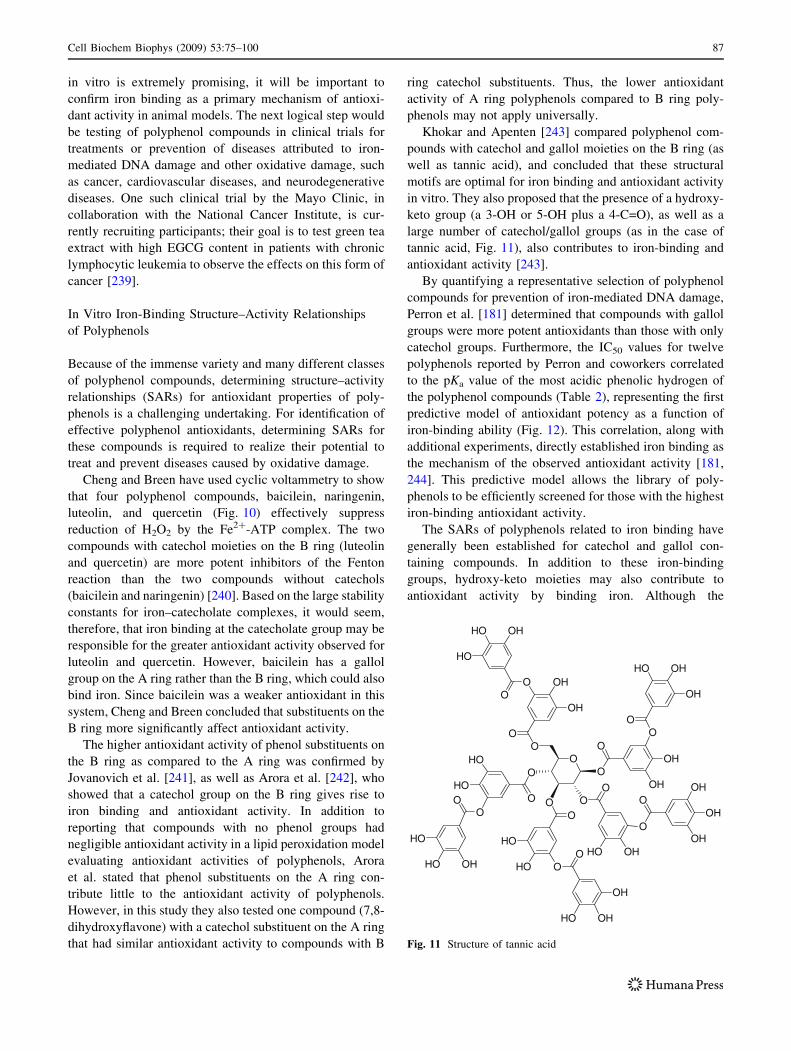
tannic acid, Fig. 11), also contributes to iron-binding and
antioxidant activity [243].
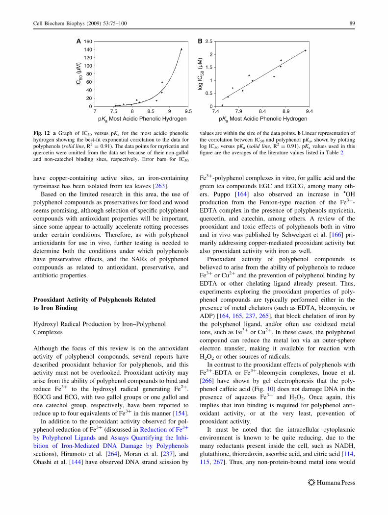
By quantifying a representative selection of polyphenol
compounds for prevention of iron-mediated DNA damage,
Perron et al. [181] determined that compounds with gallol
groups were more potent antioxidants than those with only
catechol groups. Furthermore, the IC50 values for twelve
polyphenols reported by Perron and coworkers correlated
to the pKa value of the most acidic phenolic hydrogen of
the polyphenol compounds (Table 2), representing the first
predictive model of antioxidant potency as a function of
iron-binding ability (Fig. 12). This correlation, along with
additional experiments, directly established iron binding as
the mechanism of the observed antioxidant activity [181,
244]. This predictive model allows the library of poly-
phenols to be efficiently screened for those with the highest
iron-binding antioxidant activity.
The SARs of polyphenols related to iron binding have
generally been established for catechol and gallol con-
taining compounds. In addition to these iron-binding
groups, hydroxy-keto moieties may also contribute to
antioxidant activity by binding iron. Although the
O
OH
OH
OO
OHHO
HO
OO
HO
HO
OO
HO OH
OH
OO
OH
OHO
O
OH
OHHO
OO
HO OH
OO
OH
OH
OHO
O
HO
HOO
O
OO
HO
HO OH
Fig. 11 Structure of tannic acid
Cell Biochem Biophys (2009) 53:75–100 87
important iron-binding functional groups are known, better
predictive models must now be developed to screen the
enormous number of polyphenol compounds based on their
physical and chemical properties (lipophilicity/bioavail-
ability, molecular weight, shape, p-acidity, etc.) for those
with optimal antioxidant activity in vitro. After selecting
the best candidates, the in vitro experiments should be
followed by cell and animal studies, and eventually clinical
trials for the most promising polyphenol antioxidants.
Polyphenols as Preservatives
Iron-mediated oxidative damage is not limited to living
organisms. Due to the presence of iron in the environment,
iron generated •OH is also responsible for food spoilage
and wood rotting [245, 246], and conflicting reports exist
about whether polyphenols are suitable for use as preser-
vatives of food and wood. Contreras et al. [247] have
reported increased degradation of wood after treatment
with several catechol polyphenols, including catechol,
protocatechuic acid, and 2,3-dihydroxybenzoic acid
(Fig. 6), and similar to compounds that have been isolated
from brown-rot fungi [248]. Under mildly acidic condi-
tions, these compounds were able to reduce Fe3? to Fe2? in
their mono (catecholate) complex forms [247]. Tiron
(Fig. 6), however, was not shown to participate in the
redox-cycling mechanism, as this catechol compound
cannot reduce Fe3? [249]. In contrast, Binbuga et al. [140]
have shown preservation of wood using n-propyl gallate
(Fig. 6), citing iron chelation as a likely mechanism even
under the mildly acidic conditions present in wood. This
preservative effect of n-propyl gallate was attributed pri-
marily to its antifungal properties, since it may interfere
with the Fenton reaction and redox cycles that wood-
decaying brown rot fungi use to decompose wood [248,
250, 251].
In addition to wood preservation, polyphenol com-
pounds have been widely studied for use as preservatives
for food, cosmetics, and pharmaceuticals, with many pat-
ents for these applications both nationally and
internationally related to their antioxidant properties [252–
258]. The FDA has also approved n-propyl gallate for use
as a food preservative [259]. However, most patents dis-
cussing polyphenol preservative mechanisms do not cite
metal chelation as a cause of their antioxidant activity,
instead claiming the radical scavenging ability often
attributed to these compounds.
The formation of iron–polyphenol complexes has been
attributed to negative aspects in foods as well, such as off-
color development or browning of bruised or sliced fruits
[260]. Similar polyphenol oxidation pathways occur in
polyphenol oxidase and tyrosinase, two enzymes that uti-
lize both a metal ion and O2 to oxidize phenolic
compounds [261, 262]. While these enzymes typically
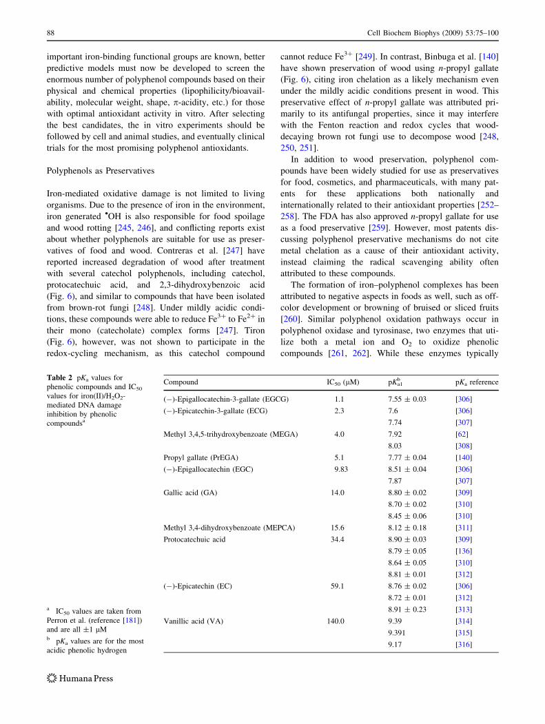
Table 2 pKa values for
phenolic compounds and IC50
values for iron(II)/H2O2-
mediated DNA damage
inhibition by phenolic
compoundsa
a IC50 values are taken from
Perron et al. (reference [181])
and are all ±1 lMb pKa values are for the most
acidic phenolic hydrogen
Compound IC50 (lM) pKa1b pKa reference
(-)-Epigallocatechin-3-gallate (EGCG) 1.1 7.55 ± 0.03 [306]
(-)-Epicatechin-3-gallate (ECG) 2.3 7.6 [306]
7.74 [307]
Methyl 3,4,5-trihydroxybenzoate (MEGA) 4.0 7.92 [62]
8.03 [308]
Propyl gallate (PrEGA) 5.1 7.77 ± 0.04 [140]
(-)-Epigallocatechin (EGC) 9.83 8.51 ± 0.04 [306]
7.87 [307]
Gallic acid (GA) 14.0 8.80 ± 0.02 [309]
8.70 ± 0.02 [310]
8.45 ± 0.06 [310]
Methyl 3,4-dihydroxybenzoate (MEPCA) 15.6 8.12 ± 0.18 [311]
Protocatechuic acid 34.4 8.90 ± 0.03 [309]
8.79 ± 0.05 [136]
8.64 ± 0.05 [310]
8.81 ± 0.01 [312]
(-)-Epicatechin (EC) 59.1 8.76 ± 0.02 [306]
8.72 ± 0.01 [312]
8.91 ± 0.23 [313]
Vanillic acid (VA) 140.0 9.39 [314]
9.391 [315]
9.17 [316]
88 Cell Biochem Biophys (2009) 53:75–100
have copper-containing active sites, an iron-containing
tyrosinase has been isolated from tea leaves [263].
Based on the limited research in this area, the use of
polyphenol compounds as preservatives for food and wood
seems promising, although selection of specific polyphenol
compounds with antioxidant properties will be important,
since some appear to actually accelerate rotting processes
under certain conditions. Therefore, as with polyphenol
antioxidants for use in vivo, further testing is needed to
determine both the conditions under which polyphenols
have preservative effects, and the SARs of polyphenol
compounds as related to antioxidant, preservative, and
antibiotic properties.
Prooxidant Activity of Polyphenols Related
to Iron Binding
Hydroxyl Radical Production by Iron–Polyphenol
Complexes
Although the focus of this review is on the antioxidant
activity of polyphenol compounds, several reports have
described prooxidant behavior for polyphenols, and this
activity must not be overlooked. Prooxidant activity may
arise from the ability of polyphenol compounds to bind and
reduce Fe3? to the hydroxyl radical generating Fe2?.
EGCG and ECG, with two gallol groups or one gallol and
one catechol group, respectively, have been reported to
reduce up to four equivalents of Fe3? in this manner [154].
In addition to the prooxidant activity observed for pol-
yphenol reduction of Fe3? (discussed in Reduction of Fe3?
by Polyphenol Ligands and Assays Quantifying the Inhi-
bition of Iron-Mediated DNA Damage by Polyphenols
sections), Hiramoto et al. [264], Moran et al. [237], and
Ohashi et al. [144] have observed DNA strand scission by
Fe3?-polyphenol complexes in vitro, for gallic acid and the
green tea compounds EGC and EGCG, among many oth-
ers. Puppo [164] also observed an increase in •OH
production from the Fenton-type reaction of the Fe3?-
EDTA complex in the presence of polyphenols myricetin,
quercetin, and catechin, among others. A review of the
prooxidant and toxic effects of polyphenols both in vitro
and in vivo was published by Schweigert et al. [166] pri-
marily addressing copper-mediated prooxidant activity but
also prooxidant activity with iron as well.
Prooxidant activity of polyphenol compounds is
believed to arise from the ability of polyphenols to reduce
Fe3? or Cu2? and the prevention of polyphenol binding by
EDTA or other chelating ligand already present. Thus,
experiments exploring the prooxidant properties of poly-
phenol compounds are typically performed either in the
presence of metal chelators (such as EDTA, bleomycin, or
ADP) [164, 165, 237, 265], that block chelation of iron by
the polyphenol ligand, and/or often use oxidized metal
ions, such as Fe3? or Cu2?. In these cases, the polyphenol
compound can reduce the metal ion via an outer-sphere
electron transfer, making it available for reaction with
H2O2 or other sources of radicals.
In contrast to the prooxidant effects of polyphenols with
Fe3?-EDTA or Fe3?-bleomycin complexes, Inoue et al.
[266] have shown by gel electrophoresis that the poly-
phenol caffeic acid (Fig. 10) does not damage DNA in the
presence of aqueous Fe3? and H2O2. Once again, this
implies that iron binding is required for polyphenol anti-
oxidant activity, or at the very least, prevention of
prooxidant activity.
It must be noted that the intracellular cytoplasmic
environment is known to be quite reducing, due to the
many reductants present inside the cell, such as NADH,
glutathione, thioredoxin, ascorbic acid, and citric acid [114,
115, 267]. Thus, any non-protein-bound metal ions would
0
20
40
60
80
100
120
140
160
pKa Most Acidic Phenolic Hydrogen
IC50
(µM
)
A
0
0.5
1
1.5
2
2.5
7 7.5 8 8.5 9 9.5 7.4 7.9 8.4 8.9 9.4
pKa Most Acidic Phenolic Hydrogen
log
IC50
(µM
)
B
Fig. 12 a Graph of IC50 versus pKa for the most acidic phenolic
hydrogen showing the best-fit exponential correlation to the data for
polyphenols (solid line, R2 = 0.91). The data points for myricetin and
quercetin were omitted from the data set because of their non-gallol
and non-catechol binding sites, respectively. Error bars for IC50
values are within the size of the data points. b Linear representation of
the correlation between IC50 and polyphenol pKa, shown by plotting
log IC50 versus pKa (solid line, R2 = 0.91). pKa values used in this
figure are the averages of the literature values listed in Table 2
Cell Biochem Biophys (2009) 53:75–100 89
most likely be present in their reduced forms in vivo [113].
Due to the deleterious effects of prooxidant activity,
however, it is essential to understand both the antioxidant
and prooxidant behavior of polyphenol compounds.
It is interesting that polyphenol compounds can display
both antioxidant and prooxidant activity under very similar
conditions. Often these conditions are quite similar to those
in biological systems, however, there are not usually
ligands as strongly chelating as EDTA other than proteins
in cells, and these are highly specialized and prevent iron-
mediated prooxidant activity. Plus, iron is not usually
found as Fe3? in any appreciable intracellular concentra-
tion. Therefore, the conditions for polyphenol prooxidant
activity are actually quite limited, often not biologically
relevant, and therefore may be of less concern to humans
who ingest these compounds in food sources. However,
this body of work showing prooxidant activity for poly-
phenols when they cannot directly chelate iron does stress
the essentiality of iron-binding as a viable antioxidant
mechanism for these compounds.
Superoxide Dismutase-Like Activity of Polyphenols
and Iron-Polyphenol Complexes
Polyphenol compounds and their complexes with Fe2? are
reported to react with superoxide (O2•-) to form H2O2 and a
semiquinone radical, resulting in superoxide dismutase
(SOD)-like behavior [66, 186, 259, 268, 269]. The reac-
tivity is similar to SOD, an enzyme present in all aerobic
organisms, which catalyzes the dismutation of two O2•-
anions under physiological pH into H2O2 and O2 (reac-
tion 7) [270].
2O��2 þ 2Hþ !SODH2O2 þ O2 ð7Þ
Since these reactions form H2O2, polyphenol compounds
may contribute to cellular oxidative stress. An example of
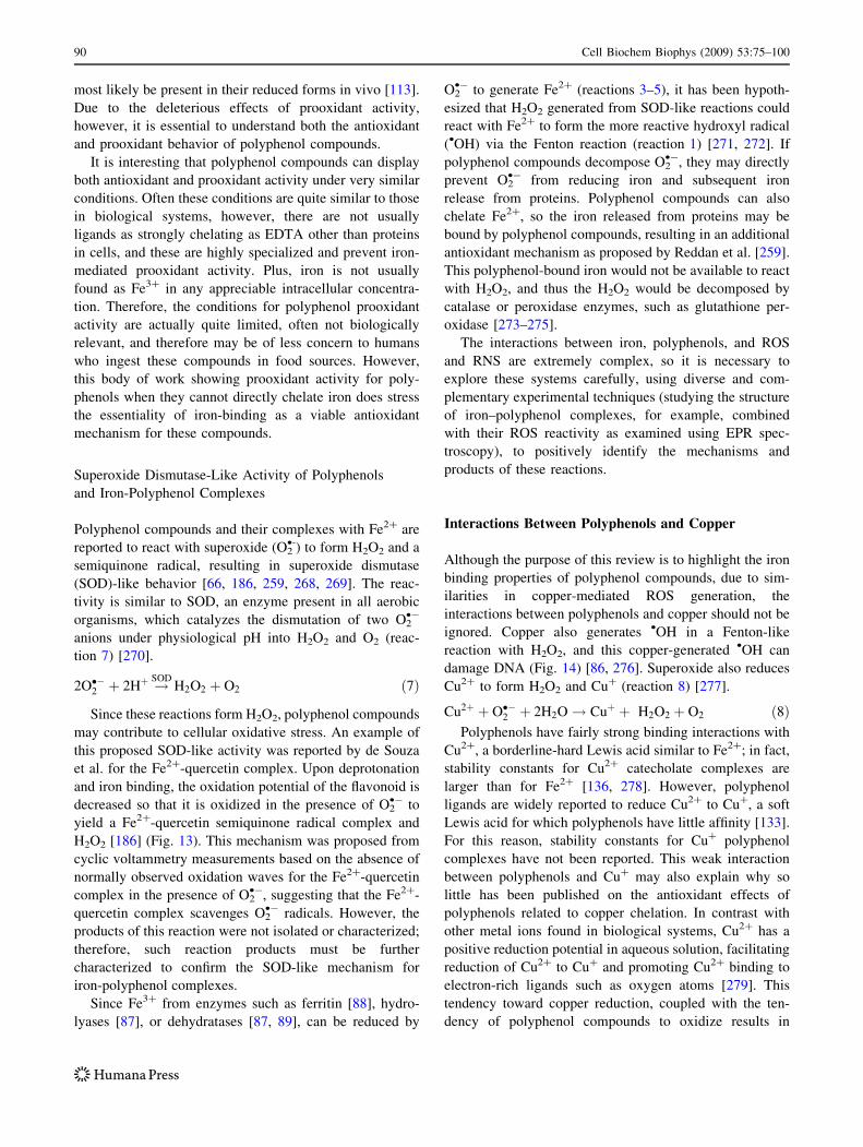
this proposed SOD-like activity was reported by de Souza
et al. for the Fe2?-quercetin complex. Upon deprotonation
and iron binding, the oxidation potential of the flavonoid is
decreased so that it is oxidized in the presence of O2•- to
yield a Fe2?-quercetin semiquinone radical complex and
H2O2 [186] (Fig. 13). This mechanism was proposed from
cyclic voltammetry measurements based on the absence of
normally observed oxidation waves for the Fe2?-quercetin
complex in the presence of O2•-, suggesting that the Fe2?-
quercetin complex scavenges O2•- radicals. However, the
products of this reaction were not isolated or characterized;
therefore, such reaction products must be further
characterized to confirm the SOD-like mechanism for
iron-polyphenol complexes.
Since Fe3? from enzymes such as ferritin [88], hydro-
lyases [87], or dehydratases [87, 89], can be reduced by
O2•- to generate Fe2? (reactions 3–5), it has been hypoth-
esized that H2O2 generated from SOD-like reactions could
react with Fe2? to form the more reactive hydroxyl radical
(•OH) via the Fenton reaction (reaction 1) [271, 272]. If
polyphenol compounds decompose O2•-, they may directly
prevent O2•- from reducing iron and subsequent iron
release from proteins. Polyphenol compounds can also
chelate Fe2?, so the iron released from proteins may be
bound by polyphenol compounds, resulting in an additional
antioxidant mechanism as proposed by Reddan et al. [259].
This polyphenol-bound iron would not be available to react
with H2O2, and thus the H2O2 would be decomposed by
catalase or peroxidase enzymes, such as glutathione per-
oxidase [273–275].
The interactions between iron, polyphenols, and ROS
and RNS are extremely complex, so it is necessary to
explore these systems carefully, using diverse and com-
plementary experimental techniques (studying the structure
of iron–polyphenol complexes, for example, combined
with their ROS reactivity as examined using EPR spec-
troscopy), to positively identify the mechanisms and
products of these reactions.
Interactions Between Polyphenols and Copper
Although the purpose of this review is to highlight the iron
binding properties of polyphenol compounds, due to sim-
ilarities in copper-mediated ROS generation, the
interactions between polyphenols and copper should not be
ignored. Copper also generates •OH in a Fenton-like
reaction with H2O2, and this copper-generated •OH can
damage DNA (Fig. 14) [86, 276]. Superoxide also reduces
Cu2? to form H2O2 and Cu? (reaction 8) [277].
Cu2þ þ O��2 þ 2H2O! Cuþ þ H2O2 þ O2 ð8ÞPolyphenols have fairly strong binding interactions with
Cu2?, a borderline-hard Lewis acid similar to Fe2?; in fact,
stability constants for Cu2? catecholate complexes are
larger than for Fe2? [136, 278]. However, polyphenol
ligands are widely reported to reduce Cu2? to Cu?, a soft
Lewis acid for which polyphenols have little affinity [133].
For this reason, stability constants for Cu? polyphenol
complexes have not been reported. This weak interaction
between polyphenols and Cu? may also explain why so
little has been published on the antioxidant effects of
polyphenols related to copper chelation. In contrast with
other metal ions found in biological systems, Cu2? has a
positive reduction potential in aqueous solution, facilitating
reduction of Cu2? to Cu? and promoting Cu2? binding to
electron-rich ligands such as oxygen atoms [279]. This
tendency toward copper reduction, coupled with the ten-
dency of polyphenol compounds to oxidize results in
90 Cell Biochem Biophys (2009) 53:75–100
complex copper–polyphenol interactions, especially in the
presence of ROS [280–282].
To the best of our knowledge, only Andrade et al. and
Perron, et al. [283, 284] have reported antioxidant effects
for protection of copper-induced DNA damage by poly-
phenols. Andrade et al. examined the antioxidant activity
of tannic acid (Fig. 10) using the deoxyribose assay with
Cu?/H2O2 (see the Assays Quantifying the Inhibition of
Iron-Mediated DNA Damage by Polyphenols section).
From their results, they calculated an IC50 value of
5.3 ± 0.8 lM for tannic acid [283].
Using gel electrophoresis, Perron et al. tested 12 phe-
nolic compounds for their effects on Cu?/H2O2-mediated
DNA damage and observed both antioxidant and prooxi-
dant behaviors. They showed inhibition of copper-
mediated DNA damage for EGCG (Fig. 10) resulting in an
IC50 of 249 ± 1 lM. In contrast, EC and EGC displayed
prooxidant activity under the same conditions, whereas
ECG displayed both prooxidant activity and antioxidant
activity at low (0.1–4 lM) and high (10–1,000 lM) con-
centrations, respectively. Very similar experiments with
Fe2? instead of Cu? resulted in an IC50 of 1.1 ± 1 lM for
inhibition of iron-mediated DNA damage: nearly 250 times
more potent. The greatly diminished antioxidant potency
for polyphenols in the copper system was attributed to the
weak interactions between polyphenols and Cu?. Based on
these experiments, a copper redox-cycling mechanism was
proposed for the prooxidant activity observed for some
polyphenols under these conditions [132, 276, 285, 286].
The prooxidant activity for polyphenol compounds
under certain conditions has prompted some cautionary
advice on consuming large amounts of these compounds
[287, 288], but copper homeostasis is even more tightly
regulated than iron; it has been estimated that the intra-
cellular concentration of non-protein-bound Cu? is less
than 10-18 M in unstressed yeast cells, corresponding to
less than one non-protein-bound Cu? ion per cell [289].
Furthermore, compounds such as glutathione (1–15 mM)
[122, 290] are present at much higher concentrations in
vivo than those attainable by polyphenols (*1–10 lM),
and have much higher affinity for Cu? than polyphenols.
Recently, however, non-protein-bound copper pools
have been found in the mitochondria and neuronal cells of
higher organisms [291, 292], and mis-regulation of copper
homeostasis results in higher cellular concentrations and
increased oxidative stress [277, 293–296]. Therefore, elu-
cidating the antioxidant and prooxidant mechanisms of
polyphenol–copper interactions will be important for a
complete understanding of polyphenol activity in vivo.
Conclusions and Future Directions
Although both Fe2? and Cu? perform Fenton-like reactions
with H2O2, polyphenol compounds containing metal
binding catechol and gallol groups have very different
activities, depending on the metal ion. Antioxidant activity
is commonly observed for polyphenols in Fe2?/H2O2 sys-
tems using a number of different assays, including cell
studies and in vitro DNA damage inhibition experiments.
In contrast, when testing polyphenols in Cu?/H2O2 sys-
tems, greatly diminished antioxidant potency or even
prooxidant activity is often observed from the interactions
between polyphenols and copper. These findings stress the
need for caution in experimental design: in addition to the
polyphenol compounds, the metal ion must be specifically
chosen to examine the metal–antioxidant interactions.
Since results are highly dependent on experimental con-
ditions, it should be the goal of researchers to design the
most biologically relevant experiments possible.
Because iron-mediated damage to biomolecules such as
lipids and DNA is implicated in disease development, the
iron-chelating mechanism of polyphenol antioxidant
activity must be fully explored in addition to radical
scavenging to understand polyphenol antioxidant behavior.
Polyphenols with gallol or catechol groups are generally
the most potent antioxidants, primarily because of the large
iron-binding stability constants for these groups. Also,
compounds with the hydroxy-keto moiety can chelate iron,
giving rise to antioxidant activity. As compared to radical
O
OOH
HO
OH
OH
OH
+ 2 Fe2+ + O2- O
OOH
HO
O
O
O
Fe2+
Fe2+
+ H2O2
3+Fig. 13 SOD-like reactions of
the Fe2?-quercetin complex, as
proposed by de Souza et al.
Cu2+
Cu+ H2O2
OH
Ascorbate Radical (AH -)
Ascorbate (AH-)
Fig. 14 The Fenton-like reaction with copper
Cell Biochem Biophys (2009) 53:75–100 91
scavenging, however, the iron-binding mechanism for
polyphenol antioxidant activity is relatively underdevel-
oped. Therefore, additional research is needed in several
areas, including stability constant measurements for iron
binding, pKa measurements of polyphenols, and additional
cell studies and DNA damage prevention experiments to
correlate in vitro and in vivo antioxidant activity of these
compounds. The goal of this work should be to develop
biologically relevant predictive models and SARs for pol-
yphenol compounds, as well as high-throughput screening
methods for determining antioxidant (or prooxidant)
behavior. These further studies will enable identification of
highly effective polyphenol antioxidants for clinical trials
to prevent or treat diseases caused by oxidative stress.
References
1. Kuhnau, J. (1976). The flavonoids: A class of semi-essential
food components: Their role in human nutrition. World Reviewof Nutrition and Dietetics, 24, 117–191.
2. Sutherland, B. A., Rahman, R. M. A., & Appleton, I. (2006).
Mechanisms of action of green tea catechins, with a focus on
ischemia-induced neurodegeneration. The Journal of NutritionalBiochemistry, 17, 291–306.
3. Cabrera, C., Artacho, R., & Gimenez, R. (2006). Beneficial
effects of green tea—A review. Journal of the American Collegeof Nutrition, 25, 79–99.
4. Gardner, E. J., Ruxton, C. H. S., & Leeds, A. R. (2007). Black
tea—Helpful or harmful? A review of the evidence. EuropeanJournal of Clinical Nutrition, 61, 3–18.
5. Vinson, J. A. (1998). Flavonoids in foods as in vitro and in vivo
antioxidants. Advances in Experimental Medicine and Biology,439, 151–164.
6. Nardini, M., Cirillo, E., Natella, F., & Scaccini, C. (2002).
Absorption of phenolic acids in humans after coffee consump-
tion. Journal of Agricultural and Food Chemistry, 50,
5735–5741.
7. Vinson, J. A., Su, X., Zubik, L., & Bose, P. (2001). Phenol
antioxidant quantity and quality in foods: Fruits. Journal ofAgricultural and Food Chemistry, 49, 5315–5321.
8. Mertens-Talcott, S. U., Jilma-Stohlawetz, P., Rios, J., Hingorani,
L., & Derendorf, H. (2006). Absorption, metabolism, and anti-
oxidant effects of pomegranate (Punica granatum L.)
polyphenols after ingestion of a standardized extract in healthy
human volunteers. Journal of Agricultural and Food Chemistry,54, 8956–8961.
9. Seeram, N. P., Aviram, M., Zhang, Y., Henning, S. M., Feng, L.,
Dreher, M., et al. (2008). Comparison of antioxidant potency of
commonly consumed polyphenol-rich beverages in the United
States. Journal of Agricultural and Food Chemistry, 56,
1415–1422.
10. Garcia-Alonso, F. J., Guidarelli, A., & Periago, M. J. (2007).
Phenolic-rich juice prevents DNA single-strand breakage and
cytotoxicity caused by tert-butylhydroperoxide in U937 cells:
The role of iron chelation. The Journal of Nutritional Bio-chemistry, 18, 457–466.
11. Gil, M. I., Tomas-Barberan, F. A., Hess-Pierce, B., Holcroft, D.
M., & Kader, A. A. (2000). Antioxidant activity of pomegranate
juice and its relationship with phenolic composition and
processing. Journal of Agricultural and Food Chemistry, 48,
4581–4589.
12. Vinson, J. A., Hao, Y., Su, X., & Zubik, L. (1998). Phenol
antioxidant quantity and quality in foods: Vegetables. Journal ofAgricultural and Food Chemistry, 46, 3630–3634.
13. Oboh, G., & Rocha, J. B. T. (2007). Polyphenols in red pepper
[Capsicum annuum var. aviculare (Tepin)] and their protective
effect on some pro-oxidants induced lipid peroxidation in brain
and liver. European Food Research and Technology, 225,
239–247.
14. Gutierrez, F., Arnaud, T., & Garrido, A. (2001). Contribution of
polyphenols to the oxidative stability of virgin olive oil. Journalof the Science of Food and Agriculture, 81, 1463–1470.
15. Visioli, F., Bellomo, G., & Galli, C. (1998). Free radical-scav-
enging properties of olive oil polyphenols. Biochemical andBiophysical Research Communications, 247, 60–64.
16. Lodovici, M., Guglielmi, F., Casalini, C., Meoni, M., Cheynier,
V., & Dolara, P. (2001). Antioxidant and radical scavenging
properties in vitro of polyphenolic extracts from red wine.
European Journal of Nutrition, 40, 74–77.
17. Makris, D. P., Psarra, E., Kallithraka, S., & Kefalas, P. (2003).
The effect of polyphenolic composition as related to antioxidant
capacity in white wines. Food Research International (Ottawa,Ont.), 36, 805–814.
18. Vinson, J. A., Proch, J., & Zubik, L. (1999). Phenol antioxidant
quantity and quality in foods: Cocoa, dark chocolate, and milk
chocolate. Journal of Agricultural and Food Chemistry, 47,
4821–4824.
19. Arts, I. C. W., Van de Putte, B., & Hollman, P. C. H. (2000).
Catechin contents of foods commonly consumed in the Neth-
erlands. Part 2. Tea, wine, fruit juices, and chocolate milk.
Journal of Agricultural and Food Chemistry, 48, 1752–1757.
20. Arts, I. C. W., Van de Putte, B., & Hollman, P. C. H. (2000).
Catechin contents of foods commonly consumed in the Neth-
erlands. Part 1. Fruits, vegetables, staple foods, and processed
foods. Journal of Agricultural and Food Chemistry, 48,
1746–1751.
21. de Pascual-Teresa, S., Santos-Buelga, C., & Rivas-Gonzalo, J.
C. (2000). Quantitative analysis of flavan-3-ols in Spanish
foodstuffs and beverages. Journal of Agricultural and FoodChemistry, 48, 5331–5337.
22. USDA. (2004). USDA database for the proanthocyanidin con-tent of selected foods. Accessed December 18, 2008, from
http://www.nal.usda.gov/fnic/foodcomp/Data/PA/PA.pdf.
23. ‘‘5 A Day’’. (2008). Accessed April 9, 2008, from www.cdc.
gov/nccdphp/dnpa/5aday/index.htm.
24. Graham, H. N. (1992). Green tea composition, consumption, and
polyphenol chemistry. Preventive Medicine, 21, 334–350.
25. Reddy, V. C., Sagar, G. V. V., Sreeramulu, D., Venu, L., &
Raghunath, M. (2005). Addition of milk does not alter the
antioxidant activity of black tea. Annals of Nutrition andMetabolism, 49, 189–195.
26. Sugisawa, A., & Umegaki, K. (2002). Physiological concentra-
tions of (-)-epigallocatechin-3-O-gallate (EGCg) prevent
chromosomal damage induced by reactive oxygen species in
WIL2-NS cells. Journal of Nutrition, 132, 1836–1839.
27. Van het Hof, K. H., Kivits, G. A. A., Weststrate, J. A., & Tijburg,
L. B. M. (1998). Bioavailability of catechins from tea: The effect
of milk. European Journal of Clinical Nutrition, 52, 356–359.
28. Yang, C. S., Chen, L., Lee, M.-J., Balentine, D., Kuo, M. C., &
Schantz, S. P. (1998). Blood and urine levels of tea catechins
after ingestion of different amounts of green tea by human
volunteers. Cancer Epidemiology, Biomarkers and Prevention,7, 351–354.
29. Yamamoto, T., Hsu, S., Lewis, J., Wataha, J., Dickinson, D.,
Singh, B., et al. (2003). Green tea polyphenol causes differential
92 Cell Biochem Biophys (2009) 53:75–100
oxidative environments in tumor versus normal epithelial cells.
The Journal of Pharmacology and Experimental Therapeutics,307, 230–236.
30. Erlund, I., Silaste, M. L., Alfthan, G., Rantala, M., Kesaniemi,
Y. A., & Aro, A. (2002). Plasma concentrations of the flavo-
noids hesperetin, naringenin and quercetin in human subjects
following their habitual diets, and diets high or low in fruit and
vegetables. European Journal of Clinical Nutrition, 56,
891–898.
31. Hollman, P. C. H., Gaag, M. V. D., Mengelers, M. J. B., van Trijp,
J. M. P., de Vries, J. H. M., & Katan, M. B. (1996). Absorption
and disposition kinetics of the dietary antioxidant quercetin in
man. Free Radical Biology and Medicine, 21, 703–707.
32. Nitta, Y., Kikuzaki, H., & Ueno, H. (2007). Food components
inhibiting recombinant human histidine decarboxylase activity.
Journal of Agricultural and Food Chemistry, 55, 299–304.
33. Macheix, J.-J., Fleuriet, A., & Billot, J. (1990). Fruit Phenolics(pp. 272–273). Boca Raton: CRC Press, Inc.
34. Romero, C., Medina, E., Vargas, J., Brenes, M., & De Castro, A.
(2007). In vitro activity of olive oil polyphenols against Heli-cobacter pylori. Journal of Agricultural and Food Chemistry,55, 680–686.
35. Song, J.-M., Lee, K.-H., & Seong, B.-L. (2005). Antiviral effect
of catechins in green tea on influenza virus. Antiviral Research,68, 66–74.
36. Lorenz, M., Jochmann, N., von Krosigk, A., Martus, P., Bau-
mann, G., Stangl, K., et al. (2007). Addition of milk prevents
vascular protective effects of tea. European Heart Journal, 28,
219–223.
37. Hertog, M. G. L., Feskens, E. J. M., Hollman, P. C. H., Katan,
M. B., & Kromhout, D. (1993). Dietary antioxidant flavonoids
and risk of coronary heart disease. The Zutphen elderly study.
Lancet, 342, 1007–1011.
38. Naasani, I., Oh-hashi, F., Oh-hara, T., Feng, W. Y., Johnston, J.,
Chan, K., et al. (2003). Blocking telomerase by dietary poly-
phenols is a major mechanism for limiting the growth of human
cancer cells in vitro and in vivo. Cancer Research, 63, 824–830.
39. He, Q., Lv, Y., & Yao, K. (2006). Effects of tea polyphenols on
the activities of a-amylase, pepsin, trypsin and lipase. FoodChemistry, 101, 1178–1182.
40. Mandel, S. A., Amit, T., Zheng, H., Weinreb, O., & Youdim, M.
B. H. (2006). The essentiality of iron chelation in neuroprotec-
tion: A potential role of green tea catechins. Oxidative Stressand Disease, 22, 277–299.
41. Lambert, J. D., & Yang, C. S. (2003). Mechanisms of cancer
prevention by tea constituents. Journal of Nutrition, 133(Suppl),
3262S–3267S.
42. Fresco, P., Borges, F., Diniz, C., & Marques, M. P. M. (2006).
New insights on the anticancer properties of dietary polyphe-
nols. Medicinal Research Reviews, 26, 747–766.
43. Garg, A. K., Buchholz, T. A., & Aggarwal, B. B. (2005).
Chemosensitization and radiosensitization of tumors by plant
polyphenols. Antioxidants and Redox Signaling, 7, 1630–1647.
44. Stoner, G. D., & Casto, B. C. (2004). Chemoprevention by fruit
phenolic compounds. In G. J. Kelloff, E. T. Hawk, & C. C.
Sigman (Eds.), Cancer chemoprevention (pp. 419–435). Tot-
owa, NJ: Humana Press, Inc.
45. Horvathova, K., Novotny, L., Tothova, D., & Vachalkova, A.
(2004). Determination of free radical scavenging activity of
quercetin, rutin, luteolin and apigenin in H2O2-treated human
ML cells K562. Neoplasma, 51, 395–399.
46. Inoue, M., Suzuki, R., Koide, T., Sakaguchi, N., Ogihara, Y., &
Yabu, Y. (1994). Antioxidant, gallic acid, induces apoptosis in
HL-60RG cells. Biochemical and Biophysical Research Com-munications, 204, 898–904.
47. Orrenius, S., Gogvadze, V., & Zhivotovsky, B. (2007). Mito-
chondrial oxidative stress: Implications for cell death. AnnualReview of Pharmacology and Toxicology, 47, 143–183.
48. Huang, X. (2003). Iron overload and its association with cancer
risk in humans: Evidence for iron as a carcinogenic metal.
Mutation Research, 533, 153–171.
49. Hajiliadis, N. D. (Ed.). (1997). Cytotoxic, mutagenic, and car-cinogenic potential of heavy metals related to humanenvironment. Netherlands: Kluwer Academic Press.
50. Markesbery, W. R., & Lovell, M. A. (2006). DNA oxidation in
Alzheimer’s disease. Antioxidants and Redox Signaling, 8,
2039–2045.
51. Halliwell, B. (2001). Role of free radicals in the neurodegen-
erative diseases. Drugs and Aging, 18, 685–716.
52. Markesbery, W. R. (1997). Oxidative stress hypothesis in Alz-
heimer’s disease. Free Radical Biology and Medicine, 23,
134–147.
53. Markesbery, W. R. (1999). Oxidative alterations in Alzheimer’s
disease. Brain Pathology, 9, 133–146.
54. Drew, B., & Leeuwenburgh, C. (2002). Aging and the role of
reactive nitrogen species. Annals of the New York Academy ofSciences, 959, 66–81.
55. Vokurkova, M., Xu, S., & Touyz, R. M. (2007). Reactive oxy-
gen species, cell growth, cell cycle progression and vascular
remodeling in hypertension. Future Cardiology, 3, 53–63.
56. Steinberg, D. (1997). Low density lipoprotein oxidation and its
pathobiological significance. The Journal of Biological Chem-istry, 272, 20963–20966.
57. Ide, T., Tsutsui, H., Hayashidani, S., Kang, D., Suematsu, N.,
Nakamura, K.-I., et al. (2001). Mitochondrial DNA damage and
dysfunction associated with oxidative stress in failing hearts
after myocardial infarction. Circulation Research, 88, 529–535.
58. Chevion, M., Berenshtein, E., & Zhu, B.-Z. (1999). The role of
transition metal ions in free radical-mediated damage. In D. L.
Gilbert & C. A. Colton (Eds.), Reactive oxygen species in bio-logical systems (pp. 103–131). New York: Plenum Publishers.
59. SciFinder search on May 5, 2008 using the terms ‘‘polyphenol’’
and ‘‘radical scavenging’’, refined by year 1995–current
(duplicate entries removed).
60. Hanasaki, Y., Ogawa, S., & Fukui, S. (1994). The correlation
between active oxygens scavenging and antioxidative effects of
flavonoids. Free Radical Biology and Medicine, 16, 845–850.
61. Bors, W., Heller, W., Michel, C., & Saran, M. (1990). Flavo-
noids as antioxidants: Determination of radical-scavenging
efficiencies. Methods in Enzymology, 186, 343–355.
62. Muzolf, M., Szymusiak, H., Swiglo, A. G., Rietjens, I. M. C. M.,
& Tyrakowska, B. (2008). pH-dependent radical scavenging
capacity of green tea catechins. Journal of Agricultural andFood Chemistry, 56, 816–823.
63. Nanjo, F., Goto, K., Seto, R., Suzuki, M., Sakai, M., & Hara, Y.
(1996). Scavenging effects of tea catechins and their derivatives
on 1, 1-diphenyl-2-picrylhydrazyl radical. Free Radical Biologyand Medicine, 21, 895–902.
64. Borkowski, T., Szymusiak, H., Gliszczynska-Swiglo, A., Rietj-
ens, I. M. C. M., & Tyrakowska, B. (2005). Radical scavenging
capacity of wine anthocyanins is strongly pH-dependent. Jour-nal of Agricultural and Food Chemistry, 53, 5526–5534.
65. van Acker, S. A. B. E., Tromp, M. N. J. L., Haenen, G. R. M.
M., van der Vijgh, W. J. F., & Bast, A. (1995). Flavonoids as
scavengers of nitric oxide radical. Biochemical and BiophysicalResearch Communications, 214, 755–759.
66. Furuno, K., Akasako, T., & Sugihara, N. (2002). The contribu-
tion of the pyrogallol moiety to the superoxide radical
scavenging activity of flavonoids. Biological and Pharmaceu-tical Bulletin, 25, 19–23.
Cell Biochem Biophys (2009) 53:75–100 93
67. Keyer, K., Gort, A. S., & Imlay, J. A. (1995). Superoxide and
the production of oxidative DNA damage. Journal of Bacteri-ology, 177, 6782–6790.
68. Cos, P., Ying, L., Calomme, M., Hu, J. P., Cimanga, K., Poel, B.
V., et al. (1998). Structure–activity relationship and classifica-
tion of flavonoids as inhibitors of xanthine oxidase and
superoxide scavengers. Journal of Natural Products, 61, 71–76.
69. Arts, M. J. T. J., Dallinga, J. S., Voss, H.-P., Haenen, G. R. M. M.,
& Bast, A. (2004). A new approach to asses the total antioxidant
capacity using the TEAC assay. Food Chemistry, 88, 567–570.
70. Prior, R. L., Wu, X., & Schaich, K. (2005). Standardized
methods for the determination of antioxidant capacity of phen-
olics in foods and dietary supplements. Journal of Agriculturaland Food Chemistry, 53, 4290–4302.
71. Sharma, A., Bhardwaj, S., Mann, A. S., Jain, A., & Kharya, M.
D. (2007). Screening methods of antioxidant activity: An
overview. Pharmacognosy Reviews, 1, 232–238.
72. Huang, D., Ou, B., & Prior, R. L. (2005). The chemistry behind
antioxidant capacity assays. Journal of Agricultural and FoodChemistry, 53, 1841–1856.
73. Beckman, J. S., Beckman, T. W., Chen, J., Marshall, P. A., &
Freeman, B. A. (1990). Apparent hydroxyl radical production by
peroxynitrite: Implications for endothelial injury from nitric
oxide and superoxide. Proceedings of the National Academy ofSciences of the United States of America, 87, 1620–1624.
74. Gilbert, D. L., & Colton, C. A. (Eds.). (1999). Reactive oxygenspecies in biological systems. New York: Plenum Publishers.
75. Adler, V., Yin, Z., Tew, K. D., & Ronai, Z. (1999). Role of
redox potential and reactive oxygen species in stress signaling.
Oncogene, 18, 6104–6111.
76. Forman, H. J., & Torres, M. (2002). Reactive oxygen species
and cell signaling. American Journal of Respiratory and CriticalCare Medicine, 166, 54–58.
77. Bredt, D. S., & Snyder, S. H. (1994). Nitric oxide: A physiologic
messenger molecule. Annual Review of Biochemistry, 63,
175–195.
78. Suzuki, Y. J., Forman, H. J., & Sevanian, A. (1997). Oxidants as
stimulators of signal transduction. Free Radical Biology andMedicine, 22, 269–285.
79. Rhee, S. G. (1999). Redox signaling: Hydrogen peroxide as
intracellular messenger. Experimental and Molecular Medicine,31, 53–59.
80. Droge, W. (2002). Free radicals in the physiological control of
cell function. Physiological Reviews, 82, 47–95.
81. Koppenol, W. H., Moreno, J. J., Pryor, W. A., Ischiropoulos, H.,
& Beckman, J. S. (1992). Peroxynitrite, a cloaked oxidant
formed by nitric oxide and superoxide. Chemical Research inToxicology, 5, 834–842.
82. Squadrito, G. L., & Pryor, W. A. (1995). The formation of
peroxynitrite in vivo from nitric oxide and superoxide. Chemico-Biological Interactions, 96, 203–206.
83. Henle, E. S., & Linn, S. (1997). Formation, prevention, and
repair of DNA damage by iron/hydrogen peroxide. The Journalof Biological Chemistry, 272, 19095–19098.
84. Imlay, J. A., & Linn, S. (1988). DNA damage and oxygen
radical toxicity. Science, 240, 1302–1309.
85. Henle, E. S., Han, Z., Tang, N., Rai, P., Luo, Y., & Linn, S.
(1999). Sequence-specific DNA cleavage by Fe2?-mediated
Fenton reaction has possible biological implications. The Jour-nal of Biological Chemistry, 274, 962–971.
86. Battin, E. E., Perron, N. R., & Brumaghim, J. L. (2006). The
central role of metal coordination in selenium antioxidant
activity. Inorganic Chemistry, 45, 499–501.
87. Flint, D. H., Tuminello, J. F., & Emptage, M. H. (1993). The
inactivation of Fe–S cluster containing hydro-lyases by super-
oxide. The Journal of Biological Chemistry, 268, 22369–22376.
88. Keyer, K., & Imlay, J. A. (1996). Superoxide accelerates DNA
damage by elevating free-iron levels. Proceedings of theNational Academy of Sciences of the United States of America,93, 13635–13640.
89. Benov, L. (2001). How superoxide radical damages the cell.
Protoplasma, 217, 33–36.
90. Haber, F., & Weiss, J. (1932). Uber die katalyse des hydroper-
oxydes. Naturwiss, 51, 948–950.
91. Koppenol, W. H. (2001). The Haber–Weiss cycle—70 years
later. Redox Report, 6, 229–234.
92. George, P. (1947). Some experiments on the reactions of
potassium superoxide in aqueous solutions. Discussions of theFaraday Society, 2, 196–205.
93. Nakagawa, O., Ono, S., Tsujimoto, A., Li, Z., & Sasaki, S.
(2007). Selective fluorescence detection of 8-oxoguanosine with
8-oxoG-CLAMP. Nucleosides, Nucleotides, and Nucleic Acids,26, 645–649.
94. Weimann, A., Belling, D., & Poulsen, H. E. (2002). Quantifi-
cation of 8-oxo-guanine and guanine as the nucleobase,
nucleoside and deoxynucleoside forms in human urine by high-
performance liquid chromatography-electrospray tandem mass
spectrometry. Nucleic Acids Research, 30, e7/1–e7/8.
95. Shigenaga, M. K., Aboujaoude, E. N., Chen, Q., & Ames, B.
N. (1994). Assays of oxidative DNA damage biomarkers 8-
oxo-20-deoxyguanosine and 8-oxoguanine in nuclear DNA and
biological fluids by high-performance liquid chromatography
with electrochemical detection. Methods in Enzymology, 234,
16–33.
96. Shigenaga, M. K., Gimeno, C. J., & Ames, B. N. (1989). Urinary
8-hydroxy-20-deoxyguanosine as a biological marker of in vivo
oxidative DNA damage. Proceedings of the National Academyof Sciences of the United States of America, 86, 9697–9701.
97. Zheng, L.-F., Dai, F., Zhou, B., Yang, L., & Liu, Z.-L. (2008).
Prooxidant activity of hydroxycinnamic acids on DNA damage
in the presence of Cu(II) ions: Mechanism and structure-activity
relationship. Food and Chemical Toxicology, 46, 149–156.
98. Ohshima, H., Gilibert, I., & Bianchini, F. (1999). Induction of
DNA strand breakage and base oxidation by nitroxyl anion
through hydroxyl radical production. Free Radical Biology andMedicine, 26, 1305–1313.
99. Fisher, G. R., & Gutierrez, P. L. (1991). Free radical formation
and DNA strand breakage during metabolism of diaziquone by
NAD(P)H quinone-acceptor oxidoreductase (DT-diaphorase)
and NADPH cytochrome c reductase. Free Radical Biology andMedicine, 11, 597–607.
100. Kashige, N., Yamaguchi, T., Ohtakara, A., Mitsutomi, M.,
Brimacombe, J. S., Miake, F., et al. (1994). Structure-activity
relationships in the induction of single-strand breakage in
plasmid pBR322 DNA by amino sugars and derivatives. Car-bohydrate Research, 257, 285–291.
101. Bhat, R., & Hadi, S. M. (1994). DNA breakage by tannic acid
and Cu(II): Sequence specificity of the reaction and involvement
of active oxygen species. Mutation Research, 313, 39–48.
102. Rai, P., Cole, T. D., Wemmer, D. E., & Linn, S. (2001).
Localization of Fe2? at an RTGR sequence within a DNA
duplex explains preferential cleavage by Fe2? and hydrogen
peroxide. Journal of Molecular Biology, 312, 1089–1101.
103. Gao, Y. G., Sriram, M., & Wang, A. H. (1993). Crystallographic
studies of metal ion–DNA interactions: Different binding modes
of cobalt(II), copper(II) and barium(II) to N7 of guanines in Z-
DNA and a drug–DNA complex. Nucleic Acids Research, 21,
4093–4101.
104. Rai, P., Wemmer, D. E., & Linn, S. (2005). Preferential binding
and structural distortion by Fe2? at RGGG-containing DNA
sequences correlates with enhanced oxidative cleavage at such
sequences. Nucleic Acids Research, 33, 497–510.
94 Cell Biochem Biophys (2009) 53:75–100
105. Lu, A.-L., Li, X., Gu, Y., Wright, P. M., & Chang, D.-Y. (2001).
Repair of oxidative DNA damage. Cell Biochemistry and Bio-physics, 35, 141–170.
106. Kennedy, L. J., Moore, K., Jr., Caulfield, J. L., Tannenbaum, S.
R., & Dedon, P. C. (1997). Quantitaion of 8-oxoguanine and
strand breaks produced by four oxidizing agents. ChemicalResearch in Toxicology, 10, 386–392.
107. Aruoma, O. I., Halliwell, B., Gajewski, E., & Dizdaroglu, M.
(1991). Copper-ion-dependent damage to the bases in DNA in
the presence of hydrogen peroxide. The Biochemical Journal,273, 601–604.
108. Macomber, L., Rensing, C., & Imlay, J. A. (2007). Intracellular
copper does not catalyze the formation of oxidative DNA damage
in Escherichia coli. Journal of Bacteriology, 189, 1616–1626.
109. Mello-Filho, A. C., & Meneghini, R. (1991). Iron is the intra-
cellular metal involved in the production of DNA damage by
oxygen radicals. Mutation Research, 251, 109–113.
110. Hoffmann, M. E., Mello-Filho, A. C., & Meneghini, R. (1984).
Correlation between cytotoxic effect of hydrogen peroxide and
the yield of DNA strand breaks in cells of different species.
Biochimica et Biophysica Acta, 781, 234–238.
111. Andrews, N. C. (2004). Probing the iron pool. Focus on
‘‘detection of intracellular iron by its regulatory effect’’. Ameri-can Journal of Physiology. Cell Physiology, 287, C1537–C1538.
112. Yamamoto, Y., Fukui, K., Koujin, N., Ohya, H., Kimura, K., &
Kamio, Y. (2004). Regulation of the intracellular free iron pool
by Dpr provides oxygen tolerance to Streptococcus mutans.
Journal of Bacteriology, 186, 5997–6002.
113. Woodmansee, A. N., & Imlay, J. A. (2002). Quantitation of
intracellular free iron by electron paramagnetic resonance
spectroscopy. Methods in Enzymology, 349, 3–9.
114. Jacobs, A. (1977). Low molecular weight intracellular iron
transport compounds. Blood, 50, 433–439.
115. Miller, J. P. G., & Perkins, D. J. (1969). Model experiments for
the study of iron transfer from transferrin to ferritin. EuropeanJournal of Biochemistry, 10, 146–151.
116. Touati, D., Jacques, M., Tardat, B., Bouchard, L., & Despied, S.
(1995). Lethal oxidative damage and mutagenesis are generated
by iron in Dfur mutants of Escherichia coli: Protective role of
superoxide dismutase. Journal of Bacteriology, 177, 2305–2314.
117. Biemond, P., Swaak, A. J. G., van Eijk, H. G., & Koster, J. F.
(1988). Superoxide dependent iron release from ferritin in
inflammatory diseases. Free Radical Biology and Medicine, 4,
185–198.
118. Ke, Y., & Qian, Z. M. (2003). Iron misregulation in the brain: a
primary cause of neurodegenerative disorders. Lancet Neurol-ogy, 2, 246–253.
119. Selima, M. H., & Ratan, R. R. (2004). The role of iron neuro-
toxicity in ischemic stroke. Ageing Research Reviews, 3,
345–353.
120. Wood, R. J. (2004). The iron–heart disease connection: Is it
dead or just hiding? Ageing Research Reviews, 3, 355–367.
121. Brewer, G. J. (2007). Iron and copper toxicity in diseases of
aging, particularly atherosclerosis and Alzheimer’s disease.
Experimental Biology and Medicine, 232, 323–335.
122. Valko, M., Rhodes, C. J., Moncol, J., Izakovic, M., & Mazur, M.
(2006). Free radicals, metals and antioxidants in oxidative stress-
induced cancer. Chemico-Biological Interactions, 160, 1–40.
123. Luxford, C., Dean, R. T., & Davies, M. J. (2000). Radicals
derived from histone hydroperoxides damage nucleobases inRNA and DNA. Chemical Research in Toxicology, 13, 665–672.
124. Liang, Q., & Dedon, P. C. (2001). Cu(II)/H2O2-induced DNA
damage is enhanced by packaging of DNA as a nucleosome.
Chemical Research in Toxicology, 14, 416–422.
125. Midorikawa, K., Murata, M., & Kawanishi, S. (2005). Histone
peptide AKRHRK enhances H2O2-induced DNA damage and
alters its site specificity. Biochemical and Biophysical ResearchCommunications, 333, 1073–1077.
126. Turro, N. J. (2002). Damage control of DNA in nucleosome core
particles: When a histone’s loving embrace is just not good
enough. Chemistry and Biology, 9, 399–401.
127. Weissman, L., de Souza-Pinto, N. C., Stevnsner, T., & Bohr, V.
A. (2007). DNA repair, mitochondria, and neurodegeneration.
Neuroscience, 145, 1318–1329.
128. Berneburg, M., Kamenisch, M., Krutmann, J., & Roecken, M.
(2006). ‘To repair or not to repair—No longer a question’:
Repair of mitochondrial DNA shielding against age and cancer.
Experimental Dermatology, 15, 1005–1015.
129. Birch-Machin, M. A. (2005). Using mitochondrial DNA as a
biosensor of early cancer development. British Journal ofCancer, 93, 271–272.
130. Chatterjee, A., Mambo, E., Zhang, Y., DeWeese, T., & Si-
dransky, D. (2006). Targeting of mutant hogg1 in mammalian
mitochondria and nucleus: effect on cellular survival upon
oxidative stress. BMC Cancer, 6, 235.
131. de Grey, A. D. N. J. (1997). A proposed refinement of the mito-
chondrial free radical theory of aging. BioEssays, 19, 161–166.
132. Hider, R. C., Liu, Z. D., & Khodr, H. H. (2001). Metal chelation
of polyphenols. Methods in Enzymology, 335, 190–203.
133. Pearson, R. G. (1963). Hard and soft acids and bases. Journal ofthe American Chemical Society, 85, 3533–3539.
134. Loomis, L. D., & Raymond, K. N. (1991). Solution equilibria of
enterobactin and metal–enterobactin complexes. InorganicChemistry, 30, 906–911.
135. Avdeef, A., Sofen, S. R., Bregante, T. L., & Raymond, K. N.
(1978). Coordination chemistry of microbial iron transport
compounds. 9. Stability constants for catechol models of ente-
robactin. Journal of the American Chemical Society, 100,
5362–5370.
136. Martell, A. E., & Smith, R. M. (1977). Critical stability con-stants (Vol. 3, pp. 200–201). New York: Plenum Press.
137. Kipton, H., Powell, J., & Taylor, M. C. (1982). Interactions of
iron(II) and iron(III) with gallic acid and its homologues: A
potentiometric and spectrophotometric study. Australian Jour-nal of Chemistry, 35, 739–756.
138. Erdogan, G., Karadag, R., & Dolen, E. (2005). Potentiometric
and spectrophotometric determination of the stability constants
of quercetin (3, 3040, 5, 7-pentahydroxyflavone) complexes with
aluminium(III) and iron(II). Reviews in Analytical Chemistry,24, 247–261.
139. Buffle, J., & Martell, A. E. (1977). Metal ion catalyzed oxidation
of o-dihydroxy aromatic compounds by oxygen. 2. Complexes
of 1, 2-dihydroxynaphthalene-4-sulfonate with iron(III) and
iron(II). Inorganic Chemistry, 16, 2225–2229.
140. Binbuga, N., Chambers, K., Henry, W. P., & Schultz, T. P.
(2005). Metal chelation studies relevant to wood preservation. 1.
Complexation of propyl gallate with Fe2?. Holzforschung, 59,
205–209.
141. Chvatalova, K., Slaninova, I., Brezinova, L., & Slanina, J.
(2008). Influence of dietary phenolic acids on redox status of
iron: Ferrous iron autoxidation and ferric iron reduction. FoodChemistry, 106, 650–660.
142. Yoshino, M., & Murakami, K. (1998). Interaction of iron with
polyphenolic compounds: Application to antioxidant character-
ization. Analytical Biochemistry, 257, 40–44.
143. Kawabata, T., Schepkin, V., Haramaki, N., Phadke, R. S., &
Packer, L. (1996). Iron coordination by catechol derivative
antioxidants. Biochemical Pharmacology, 51, 1569–1577.
144. Ohashi, Y., Yoshinaga, K., Yoshioka, H., & Yoshioka, H.
(2002). Kinetic analysis of the effect of (-)-epigallocatechin
gallate on the DNA strand scission induced by Fe(II). Biosci-ence, Biotechnology, and Biochemistry, 66, 770–776.
Cell Biochem Biophys (2009) 53:75–100 95
145. Hajji, H. E., Nkhili, E., Tomao, V., & Dangles, O. (2006).
Interactions of quercetin with iron and copper ions: Complex-
ation and autoxidation. Free Radical Research, 40, 303–320.
146. Cooper, S. R., McArdle, J. V., & Raymond, K. N. (1978).
Siderophore electrochemistry: Relation to intracellular iron
release mechanism. Proceedings of the National Academy ofSciences of the United States of America, 75, 3551–3554.
147. McBryde, W. A. E. (1964). A spectrophotometric reexamination
of the spectra and stabilities of the iron (III)—Tiron complexes.
Canadian Journal of Chemistry, 42, 1917–1927.
148. Perron, N. R., DeGuire, S. M., & Brumaghim, J. L. (2008).
Kinetics of iron oxidation upon polyphenol binding (in
preparation).
149. Stumm, W., & Lee, G. F. (1961). Oxygenation of ferrous iron.
Industrial and Engineering Chemistry, 53, 143–146.
150. King, J., & Davidson, N. (1958). Kinetics of the ferrous iron-
oxygen reaction in acidic phosphate–pyrophosphate solutions.
Journal of the American Chemical Society, 80, 1542–1545.
151. Posner, A. M. (1953). The kinetics of autoxidation of ferrous
ions in concentrated HCl solutions. Transactions of FaradaySociety, 49, 382–388.
152. Huffman, R. E., & Davidson, N. (1956). Kinetics of the ferrous
iron-oxygen reaction in sulfuric acid solution. Journal of theAmerican Chemical Society, 78, 4836–4842.
153. George, P. (1954). The oxidation of ferrous perchlorate by
molecular oxygen. Journal of the Chemical Society, 4349–4359.
154. Ryan, P., & Hynes, M. J. (2007). The kinetics and mechanisms
of the complex formation and antioxidant behaviour of the
polyphenols EGCg and ECG with iron(III). Journal of InorganicBiochemistry, 101, 585–593.
155. Jameson, G. N. L., & Linert, W. (2001). The oxidation of 6-
hydroxydopamine in aqueous solution. Part 3. Kinetics and
mechanism of the oxidation with iron(III). Journal of theChemical Society. Perkin Transactions, 2, 569–575.
156. Hynes, M. J., & Coinceanainn, M. O. (2001). The kinetics and
mechanisms of the reaction of iron(III) with gallic acid, gallic
acid methyl ester and catechin. Journal of Inorganic Biochem-istry, 85, 131–142.
157. Ryan, P., & Hynes, M. J. (2008). The kinetics and mechanisms
of the reactions of iron(III) with quercetin and morin. Journal ofInorganic Biochemistry, 102, 127–136.
158. El-Ayaan, U., Herlinger, E., Jameson, R. F., & Linert, W.
(1997). Anaerobic oxidation of dopamine by iron(III). Journalof the Chemical Society, Dalton Transactions, 2813–2818.
159. Basolo, F., & Pearson, R. G. (1967). Mechanism of inorganicreactions, a study of metal complexes in solution (2nd ed.). New
York: Wiley.
160. Hider, R. C., Mohr-Nor, A. R., Silver, J., Morrison, I. E. G., &
Rees, L. V. C. (1981). Model compounds for microbial iron-
transport compounds. Part 1. Solution chemistry and Mossbauer
study of iron(II) and iron(III) complexes from phenolic and
catecholic systems. Journal of the Chemical Society, DaltonTransactions, 609–622.
161. Hider, R. C., Howlin, B., Miller, J. R., Mohr-Nor, A. R., &
Silver, J. (1983). Model compounds for microbial iron-transport
compounds. Part IV. Further solution chemistry and Mossbauer
studies on iron(II) and iron(III) catechol complexes. InorganicaChimica Acta, 80, 51–56.
162. Pulido, R., Bravo, L., & Saura-Calixto, F. (2000). Antioxidant
activity of dietary polyphenols as determined by a modified
ferric reducing/antioxidant power assay. Journal of Agriculturaland Food Chemistry, 48, 3396–3402.
163. Zhang, L., Bandy, B., & Davison, A. J. (1996). Effects of
metals, ligands and antioxidants on the reaction of oxygen with
1, 2, 4-benzenetriol. Free Radical Biology and Medicine, 20,
495–505.
164. Puppo, A. (1992). Effect of flavonoids on hydroxyl radical
formation by Fenton-type reactions; influence of the iron che-
lator. Phytochemistry, 31, 85–88.
165. Laughton, M. J., Halliwell, B., Evans, P. J., & Hoult, J. R. S.
(1989). Antioxidant and pro-oxidant actions of the plant phen-
olics quercetin, gossypol and myricetin. Effects on lipid
peroxidation, hydroxyl radical generation and bleomycin-
dependent damage to DNA. Biochemical Pharmacology, 38,
2859–2865.
166. Schweigert, N., Zehnder, A. J. B., & Eggen, R. I. L. (2001).
Chemical properties of catechols and their molecular modes of
toxic action in cells, from microorganisms to mammals. Envi-ronmental Microbiology, 3, 81–91.
167. Yamahara, R., Ogo, S., Masuda, H., & Watanabe, Y. (2002).
(Catecholato)iron(III) complexes: Structural and functional
models for the catechol-bound iron(III) form of catechol diox-
ygenases. Journal of Inorganic Biochemistry, 88, 284–294.
168. Wunderlich, C. H., Weber, R., & Bergerhoff, G. (1991). On iron
gallic ink. Zeitschrift fur Anorganische Und Allgemeine Chemie,598(599), 371–376.
169. Feller, R. K., & Cheetham, A. K. (2006). Fe(III), Mn(II), Co(II),
and Ni(II) 3, 4, 5-trihydroxybenzoate (gallate) dihydrates; a new
family of hybrid framework materials. Solid State Sciences, 8,
1121–1125.
170. Higuchi, M., Hitomi, Y., Minami, H., Tanaka, T., & Funabiki, T.
(2005). Correlation of spin states and spin delocalization with
the dioxygen reactivity of catecholatoiron (III) complexes.
Inorganic Chemistry, 44, 8810–8821.
171. Floquet, S., Simaan, A. J., Riviere, E., Nierlich, M., Thuery, P.,
Ensling, J., Gutlich, P., Girerd, J.-J., Boillot, M.-L. (2005). Spin
crossover of ferric complexes with catecholate derivatives.
Single-crystal X-ray structure, magnetic and Mossbauer inves-
tigations. Dalton Transactions, 1734–1742.
172. Chiou, Y.-M., & Que, L. (1995). Structure of a mononuclear
iron(II)-catecholate complex and its relevance to the extradiol-
cleaving catechol dioxygenases. Inorganic Chemistry, 34,
3577–3578.
173. Velusamy, M., Mayilmurugan, R., & Palaniadavar, M. (2004).
Iron(III) complexes of sterically hindered tetradentate mono-
phenolate ligands as functional models for catechol 1, 2-
dioxygenases: The role of ligands stereoelectronic properties.
Inorganic Chemistry, 43, 6284–6293.
174. Jo, D.-H., Chiou, Y.-M., & Que, J. L. (2001). Models of
extradiol cleaving catechol dioxygenases: Syntheses, structures,
and reactivities of iron(II)-monoanionic catecholate complexes.
Inorganic Chemistry, 40, 3181–3190.
175. Grillo, V. A., Hanson, G. R., Wang, D., Hambley, T. W., Gahan,
L. R., Murray, K. S., et al. (1996). Synthesis, X-ray structural
determination, and magnetic susceptibility, Mossbauer, and
EPR studies of (Ph4P)2[Fe2(Cat)4(H2O)2]�6H2O, a catecholato-
bridged dimer of iron(III). Inorganic Chemistry, 35, 3568–3576.
176. Caulder, D. L., Powers, R. E., Parac, T. N., & Raymond, K. N.
(1998). The self-assembly of a predesigned tetrahedral M4L6
supramolecular cluster. Angewandte Chemie (International ed.in English), 37, 1840–1843.
177. Jewett, S. L., Eggling, S., & Geller, L. (1997). Novel method to
examine the formation of unstable 2:1 and 3:1 complexes of
catecholamines and iron(III). Journal of Inorganic Biochemis-try, 66, 165–173.
178. Jovanovic, S. V., Simic, M. G., Steenken, S., & Hara, Y. (1998).
Iron complexes of gallocatechins. Antioxidant action or iron
regulation? Journal of the Chemical Society. Perkin Transac-tions, 2, 2365–2369.
179. Ackermann, V. G., & Hesse, D. (1970). Uber eisen(III)-ko-
mplexe mit phenolen. III. Zeitschrift fur Anorganische UndAllgemeine Chemie, 375, 77–86.
96 Cell Biochem Biophys (2009) 53:75–100
180. Maqsood, Z. A. T., & Kazmi, S. A. (1993). Formation of iron
gallic acid complexes at different pH and determination of their
stability constants. Pakistan Journal of Scientific and IndustrialResearch, 6, 511–516.
181. Perron, N. R., Hodges, J. N., Jenkins, M., & Brumaghim, J. L.
(2008). Predicting how polyphenol antioxidants prevent DNA
damage by binding to iron. Inorganic Chemistry, 47,
6153–6161.
182. Lopes, G. K. B., Schulman, H. M., & Hermes-Lima, M. (1999).
Polyphenol tannic acid inhibits hydroxyl radical formation from
Fenton reaction by complexing ferrous ions. Biochimica et Bi-ophysica Acta, 1472, 142–152.
183. Mentasti, E., Pelizzetti, E., & Saini, G. (1976). Interactions of
Fe(III) with adrenaline, L-Dopa, and other catechol derivatives.
Journal of Inorganic and Nuclear Chemistry, 38, 785–788.
184. Mentasti, E., & Pelizzetti, E. (1973). Reactions between iron(III)
and catechol (o-dihydroxybenzene). Part I. Equilibria and
kinetics of complex formation in aqueous acid solution. Journalof the Chemical Society. Dalton Transactions, 2605–2608.
185. Kennedy, J. A., & Powell, H. K. J. (1985). Aluminium(III) and
iron(III) 1, 2-diphenolato complexes: A potentiometric study.
Australian Journal of Chemistry, 38, 659–667.
186. de Souza, R. F. V., Sussuchi, E. M., & De Giovani, W. F.
(2003). Synthesis, electrochemical, spectral, and antioxidant
properties of complexes of flavonoids with metal ions. Synthesisand Reactivity in Inorganic and Metal-Organic Chem, 33,
1125–1144.
187. Escandar, G. M., & Sala, L. F. (1991). Complexing behavior of
rutin and quercetin. Canadian Journal of Chemistry, 69,
1994–2001.
188. van Acker, S. A. B. E., van Balen, G. P., van den Berg, D.-J.,
Bast, A., & van der Vijgh, W. J. F. (1998). Influence of iron
chelation on the antioxidant activity of flavonoids. BiochemicalPharmacology, 56, 935–943.
189. Sugihara, N., Arakawa, T., Ohnishi, M., & Furuno, K. (1999).
Anti- and pro-oxidative effects of flavonoids on metal-induced
lipid hydroperoxide-dependent lipid peroxidation in cultured
hepatocytes loaded with a-linolenic acid. Free Radical Biologyand Medicine, 27, 1313–1323.
190. Sugihara, N., Ohnishi, M., Imamura, M., & Furuno, K. (2001).
Differences in antioxidative efficiency of catechins in various
metal-induced lipid peroxidations in cultured hepatocytes.
Journal of Health Science, 47, 99–106.
191. Morel, I., Lescoat, G., Cogrel, P., Sergent, O., Pasdeloup, N.,
Brissot, P., et al. (1993). Antioxidant and iron-chelating activi-
ties of the flavonoids catechin, quercetin and diosmetin on iron-
loaded rat hepatocyte cultures. Biochemical Pharmacology, 45,
13–19.
192. Morel, I., Lescoat, G., Cillard, P., & Cillard, J. (1994). Role of
flavonoids and iron chelation in antioxidant action. Methods inEnzymology, 234, 437–443.
193. Ferrali, M., Signorini, C., Caciotti, B., Sugherini, L., Ciccoli, L.,
Giachetti, D., et al. (1997). Protection against oxidative damage
of erythrocyte membrane by the flavonoid quercetin and its
relation to iron chelating activity. FEBS Letters, 416, 123–129.
194. Anghileri, L. J., & Thouvenot, P. (2000). Natural polyphenols–
iron interaction. Biological Trace Element Research, 73,
251–258.
195. Sestili, P., Guidarelli, A., Dacha, M., & Cantoni, O. (1998).
Quercetin prevents DNA single strand breakage and cytotoxicity
caused by tert-butylhydroperoxide: Free radical scavenging
versus iron chelating mechanism. Free Radical Biology andMedicine, 25, 196–200.
196. Sestili, P., Diamantini, G., Bedini, A., Cerioni, L., Tommasini,
I., Tarzia, G., et al. (2002). Plant-derived phenolic compounds
prevent the DNA single-strand breakage and cytotoxicity by
tert-butylhydroperoxide via an iron-chelating mechanism. TheBiochemical Journal, 364, 121–128.
197. Melidou, M., Riganakos, K., & Galaris, D. (2005). Protection
against nuclear DNA damage offered by flavonoids in cells
exposed to hydrogen peroxide: The role of iron chelation. FreeRadical Biology and Medicine, 39, 1591–1600.
198. Boato, F., Wortley, G. M., Liu, R. H., & Glahn, R. P. (2002).
Red grape juice inhibits iron availability: Application of an in
vitro digestion/Caco-2 cell model. Journal of Agricultural andFood Chemistry, 50, 6935–6938.
199. Matuschek, E., & Svanberg, U. (2002). Oxidation of polyphe-
nols and the effect on in vitro iron accessibility in a model food
system. Journal of Food Science, 67, 420–424.
200. Matuschek, E., Towo, E., & Svanberg, U. (2001). Oxidation of
polyphenols in phytate-reduced high-tannin cereals: Effect on
different phenolic groups and on in vitro accessible iron. Journalof Agricultural and Food Chemistry, 49, 5630–5638.
201. Gaffney, S., Williams, V., Flynn, P., Carlino, R., Mowry, C.,
Dierenfeld, E., et al. (2004). Tannin/polyphenol effects on iron
solubilization in vitro. Bios, 75, 43–52.
202. Brown, R., Klein, A., & Hurrell, R. F. (1989). Effect of poly-
phenols on iron bioavailability in rats. Special Publication(Royal Society of Chemistry), 72, 152–154.
203. Das, P., Raghuramulu, N., & Rao, K. C. (2003). Effect of
organic acids and polyphenols on in vitro available iron from
foods. Journal of Food Science and Technology, 40, 677–681.
204. Tuntawiroon, M., Sritongkul, N., Brune, M., Rossander-Hulten,
L., Pleehachinda, R., Suwanik, R., et al. (1991). Dose-dependent
inhibitory effect of phenolic compounds in foods on nonheme-
iron absorption in men. The American Journal of ClinicalNutrition, 53, 554–557.
205. Manach, C., Williamson, G., Morand, C., Scalbert, A., &
Remesy, C. (2005). Bioavailability and bioefficacy of polyphe-
nols in humans. I. Review of 97 bioavailability studies. TheAmerican Journal of Clinical Nutrition, 81, 230S–242S.
206. Williamson, G., & Manach, C. (2005). Bioavailability and
bioefficacy of polyphenols in humans. II. Review of 93 inter-
vention studies. The American Journal of Clinical Nutrition, 81,
243S–255S.
207. Kuo, S.-M., Leavitt, P. S., & Lin, C.-P. (1998). Dietary flavo-
noids interact with trace metals and affect metallothionein level
in human intestinal cells. Biological Trace Element Research,62, 135–153.
208. Davis, S. R., & Cousins, R. J. (2000). Metallothionein expres-
sion in animals: A physiological perspective on function. TheJournal of Nutrition, 130, 1085–1088.
209. Riggio, M., Filosa, S., Parisi, E., & Scudiero, R. (2003).
Changes in zinc, copper and metallothionein contents during
oocyte growth and early development of the teleost Danio rerio(zebrafish). Comparitive Biochemistry and Physiology C. Com-paritive Pharmacology and Toxicology, 135, 191–196.
210. Eiichi, T., Shin-Ichi, O., Kumiko, I., Akira, N., Yoshihisa, I., &
Takashi, S. (2007). Metallothionein proteins expression, copper
and zinc concentrations, and lipid peroxidation level in a rodent
model for amyotrophic lateral sclerosis. Toxicology, 229, 33–41.
211. Rachmilewitz, E. A., Lubin, B. H., & Shohet, S. B. (1976). Lipid
membrane peroxidation in b-thalassemia major. Blood, 47,
495–505.
212. Hebbel, R. P., Eaton, J. W., Balasingam, M., & Steinber, M. H.
(1982). Spontaneous oxygen radical generation by sickle eryth-
rocytes. The Journal of Clinical Investigation, 70, 1253–1259.
213. Scott, M. D., van den Berg, J. J. M., Repka, T., Rouyer-Fessard,
P., Hebbel, R. P., Beuzard, Y., et al. (1993). Effect of excess a-
hemoglobin chains on cellular and membrane oxidation in
model b-thalassemic erythrocytes. The Journal of ClinicalInvestigation, 91, 1706–1712.
Cell Biochem Biophys (2009) 53:75–100 97
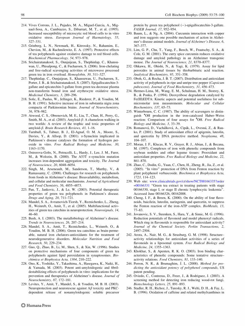
214. Vives Corrons, J. L., Pujades, M. A., Miguel-Garcia, A., Mig-
uael-Sosa, A., Cambiazzo, S., Dibarrart, M. T., et al. (1995).
Increased susceptibility of microcytic red blood cells to in vitro
oxidative stress. European Journal of Haematology, 55,
327–331.
215. Grinberg, L. N., Newmark, H., Kitrossky, N., Rahamim, E.,
Chevion, M., & Rachmilewitz, E. A. (1997). Protective effects
of tea polyphenols against oxidative damage to red blood cells.
Biochemical Pharmacology, 54, 973–978.
216. Srichairatanakool, S., Ounjaijean, S., Thephinlap, C., Khansu-
wan, U., Phisalpong, C., & Fucharoen, S. (2006). Iron-chelating
and free-radical scavenging activities of microwave processed
green tea in iron overload. Hemoglobin, 30, 311–327.
217. Thephinlap, C., Ounjaijean, S., Khansuwan, U., Fucharoen, S.,
Porter, J. B., & Srichairatanakool, S. (2007). Epigallocatechin-3-
gallate and epicatechin-3-gallate from green tea decrease plasma
non-transferrin bound iron and erythrocyte oxidative stress.
Medicinal Chemistry, 3, 289–296.
218. Sofic, E., Paulus, W., Jellinger, K., Riederer, P., & Youdim, M.
B. H. (1991). Selective increase of iron in substantia nigra zona
compacta of Parkinsonian brains. Journal of Neurochemistry,56, 978–982.
219. Atwood, C. S., Obrenovich, M. E., Liu, T., Chan, H., Perry, G.,
Smith, M. A., et al. (2003). Amyloid-b: A chameleon walking in
two worlds: A review of the trophic and toxic properties of
amyloid-b. Brain Research Reviews, 43, 1–16.
220. Turnbull, S., Tabner, B. J., El-Agnaf, O. M. A., Moore, S.,
Davies, Y., & Allsop, D. (2001). a-Synuclein implicated in
Parkinson’s disease catalyses the formation of hydrogen per-
oxide in vitro. Free Radical Biology and Medicine, 30,
1163–1170.
221. Ostrerova-Golts, N., Petrucelli, L., Hardy, J., Lee, J. M., Farer,
M., & Wolozin, B. (2000). The A53T a-synuclein mutation
increases iron-dependent aggregation and toxicity. The Journalof Neuroscience, 20, 6048–6054.
222. Singh, M., Arseneault, M., Sanderson, T., Murthy, V., &
Ramassamy, C. (2008). Challenges for research on polyphenols
from foods in Alzheimer’s disease: Bioavailability, metabolism,
and cellular and molecular mechanisms. Journal of Agriculturaland Food Chemistry, 56, 4855–4873.
223. Pan, T., Jankovic, J., & Le, W. (2003). Potential therapeutic
properties of green tea polyphenols in Parkinson’s disease.
Drugs and Aging, 20, 711–721.
224. Mandel, S. A., Avramovich-Tiorsh, Y., Reznichenko, L., Zheng,
H., Weinreb, O., Amit, T., et al. (2005). Multifunctional activ-
ities of green tea catechins in neuroprotection. Neurosignals, 14,
46–60.
225. Bush, A. I. (2003). The metallobiology of Alzheimer’s disease.
Trends in Neurosciences, 26, 207–214.
226. Mandel, S. A., Amit, T., Reznichenko, L., Weinreb, O., &
Youdim, M. B. H. (2006). Green tea catechins as brain-perme-
able, natural iron chelators-antioxidants for the treatment of
neurodegenerative disorders. Molecular Nutrition and FoodResearch, 50, 229–234.
227. Guo, Q., Zhao, B., Li, M., Shen, S., & Xin, W. (1996). Studies
on protective mechanisms of four components of green tea
polyphenols against lipid peroxidation in synaptosomes. Bio-chimica et Biophysica Acta, 1304, 210–222.
228. Ono, K., Yoshiike, Y., Takashima, A., Hasegawa, K., Naiki, H.,
& Yamada, M. (2003). Potent anti-amyloidogenic and fibril-
destabilizing effects of polyphenols in vitro: implications for the
prevention and therapeutics of Alzheimer’s disease. Journal ofNeurochemistry, 87, 172–181.
229. Levites, Y., Amit, T., Mandel, S., & Youdim, M. B. H. (2003).
Neuroprotection and neurorescue against Ab toxicity and PKC-
dependent release of nonamyloidogenic soluble precursor
protein by green tea polyphenol (-)-epigallocatechin-3-gallate.
FASEB Journal, 17, 952–954.
230. Baum, L., & Ng, A. (2004). Curcumin interaction with copper
and iron suggests one possible mechanism of action in Alzhei-
mer’s disease animal models. Journal of Alzheimer’s Disease, 6,
367–377.
231. Lim, G. P., Chu, T., Yang, F., Beech, W., Frautschy, S. A., &
Cole, G. M. (2001). The curry spice curcumin reduces oxidative
damage and amyloid pathology in an Alzheimer transgenic
mouse. The Journal of Neuroscience, 21, 8370–8377.
232. Ohkawa, H., Ohishi, N., & Yagi, K. (1979). Assay for lipid
peroxides in animal tissues by thiobarbituric acid reaction.
Analytical Biochemistry, 95, 351–358.
233. Oboh, G., & Rocha, J. B. T. (2007). Distribution and antioxidant
activity of polyphenols in ripe and unripe tree pepper (Capsicumpubescens). Journal of Food Biochemistry, 31, 456–473.
234. Hermes-Lima, M., Wang, E. M., Schulman, H. M., Storrey, K.
B., & Ponka, P. (1994). Deoxyribose degradation catalyzed by
Fe(III)EDTA: Kinetic aspects and potential usefulness for sub-
micromolar iron measurements. Molecular and CellularBiochemistry, 137, 65–73.
235. Winterbourn, C. C. (1987). The ability of scavengers to distin-
guish •OH production in the iron-catalyzed Haber–Weiss
reaction: Comparison of four assays for •OH. Free RadicalBiology and Medicine, 3, 33–39.
236. Romanova, D., Vachalkova, A., Cipak, L., Ovesna, Z., & Rau-
ko, P. (2001). Study of antioxidant effect of apigenin, luteolin,
and quercetin by DNA protective method. Neoplasma, 48,
104–107.
237. Moran, J. F., Klucas, R. V., Grayer, R. J., Abian, J., & Becana,
M. (1997). Complexes of iron with phenolic compounds from
soybean nodules and other legume tissues: Prooxidant and
antioxidant properties. Free Radical Biology and Medicine, 22,
861–870.
238. Zhao, C., Dodin, G., Yuan, C., Chen, H., Zheng, R., Jia, Z., et al.
(2005). ‘‘In vitro’’ protection of DNA from Fenton reaction by
plant polyphenol verbascoside. Biochimica et Biophysica Acta,1723, 114–123.
239. Web site: www.clinicaltrials.gov/ct/show/NCT00166335?order
=00166331 ‘‘Green tea extract in treating patients with stage
00166330, stage I, or stage II chronic lymphocytic leukemia’’.
Accessed June 00166326, 00162008.
240. Cheng, I. F., & Breen, K. (2000). On the ability of four flavo-
noids, baicilein, luteolin, naringenin, and quercetin, to suppress
the Fenton reaction of the iron-ATP complex. BioMetals, 13,
77–83.
241. Jovanovic, S. V., Steenken, S., Hara, Y., & Simic, M. G. (1996).
Reduction potentials of flavonoid and model phenoxyl radicals.
Which ring in flavonoids is responsible for antioxidant activity?
Journal of the Chemical Society. Perkin Transactions, 2,
2497–2504.
242. Arora, A., Nair, M. G., & Strasburg, G. M. (1998). Structure–
activity relationships for antioxidant activities of a series of
flavonoids in a liposomal system. Free Radical Biology andMedicine, 24, 1355–1363.
243. Khokhar, S., & Apenten, R. K. O. (2003). Iron binding char-
acteristics of phenolic compounds: Some tentative structure–
activity relations. Food Chemistry, 81, 133–140.
244. Perron, N. R., & Brumaghim, J. L. (2008). Method for pre-dicting the antioxidant potency of polyphenol compounds, US
patent pending.
245. Oviedo, C., Contreras, D., Freer, J., & Rodriguez, J. (2003). A
screening method for detecting iron reducing wood-rot fungi.
Biotechnology Letters, 25, 891–893.
246. Stadler, R. H., Richoz, J., Turesky, R. J., Welti, D. H., & Fay, L.
B. (1996). Oxidation of caffeine and related methylxanthines in
98 Cell Biochem Biophys (2009) 53:75–100
ascorbate and polyphenol-driven Fenton-type oxidations. FreeRadical Research, 24, 225–240.
247. Contreras, D., Rodrıguez, J., Freer, J., Schwederski, B., & Kaim,
W. (2007). Enhanced hydroxyl radical production by dihy-
droxybenzene-driven Fenton reactions: Implications for wood
biodegradation. Journal of Biological Inorganic Chemistry, 12,
1055–1061.
248. Paszczynski, A., Crawford, R., Funk, D., & Goodell, B. (1999).
De novo synthesis of 4, 5-dimethoxycatechol and 2, 5-dimeth-
oxyhydroquinone by the brown rot fungus Gloeophyllumtrabeum. Applied and Environmental Microbiology, 65, 674–679.
249. Kennedy, J. A., & Powell, H. K. J. (1985). Polyphenol inter-
actions with aluminium(III) and iron(III): Their possible
involvement in the podalization process. Australian Journal ofChemistry, 38, 879–888.
250. Kerem, Z., Jensen, K. A., & Hammel, K. E. (1999). Biodegra-
dative mechanism of the brown rot basidiomycete Gloeophyllumtradbeum: Evidence for an extracellular hydroquinone-driven
Fenton reaction. FEBS Letters, 446, 49–54.
251. Wang, W., & Gao, P. J. (2003). Function and mechanism of a
low-molecular-weight peptide produced by Gloeophyllumtrabeum in biodegradation of cellulose. Journal of Biotechnol-ogy, 101, 119–130.
252. Richards, M. P., Lee, C.-H., & Reed, J. D. (2005). Methods andlipophilic antioxidant compositions to inhibit lipid oxidation infood products. US patent 2005175762.
253. Furukawa, J., Shibuta, T., & Takamine, K. (2003). Food addi-tives for improving shelf life. Japan patent 2003088345.
254. Richards, G. N. (1999). Polyphenol-containing wood extract asantibacterial for food. US patent 19970924.
255. Schur, J. P. (1998). Preservative for foods and cosmetics. Ger-
many patent 97-19726429 19970623.
256. Levy, M.-C., & Andry, M.-C. (1995). Microcapsules with wallsmade of cross-linked plant polyphenols, for foods, pharmaceu-ticals or cosmetics. PCT Int. Appl. patent 94-1146 19940202.
257. Hara, M., & Ishigami, T. (1990). Complex of tea polyphenolsand protein for uses in medicine and food industries. Japan
patent 02202900.
258. Mai, J., Chambers, L. J., & McDonald, R. E. (1985). Tea extractused for food preservation. Brit. UK Pat. Appl. patent 2151123.
259. Reddan, J. R., Giblin, F. J., Sevilla, M., Padgaonkar, V., Dziedzic,
D. C., Leverenz, V. R., et al. (2003). Propyl gallate is a superoxide
dismutase mimic and protects cultured lens epithelial cells from
H2O2 insult. Experimental Eye Research, 76, 49–59.
260. Mellican, R. I., Li, J., Mehansho, H., & Nielsen, S. S. (2003).
The role of iron and the factors affecting off-color development
of polyphenols. Journal of Agricultural and Food Chemistry, 51,
2304–2316.
261. Martinez, M. V., & Whitaker, J. R. (1995). The biochemistry
and control of enzymatic browning. Trends in Food Science andTechnology, 6, 195–200.
262. Mayer, A. M. (2006). Polyphenol oxidases in plants and fungi:
Going places? A review. Phytochemistry, 67, 2318–2331.
263. Singh, D. V., & Mukherjee, P. P. (1973). Tyrosinase, an iron(II)-
containing enzyme from tea leaves. Current Science, 42, 391.
264. Hiramoto, K., Ojima, N., Sako, K., & Kikugawa, K. (1996).
Effect of plant phenolics on the formation of spin-adduct of
hydroxyl radical and the DNA strand breaking of hydroxyl
radical. Biological and Pharmaceutical Bulletin, 19, 558–563.
265. Kawanishi, S., Oikawa, S., & Murata, M. (2005). Evaluation for
safety of antioxidant chemopreventive agents. Antioxidants andRedox Signaling, 7, 1728–1739.
266. Inoue, S., Ito, K., Yamamoto, K., & Kawanishi, S. (1992).
Caffeic acid causes metal-dependent damage to cellular and
isolated DNA through H2O2 formation. Carcinogenesis, 13,
1497–1502.
267. Malaisse, W. J., Hutton, J. C., Kawazu, S., Herchuelz, A.,
Valverde, I., & Sener, A. (1979). The stimulus-secretion cou-
pling of glucose-induced insulin release. Diabetologia, 16,
331–341.
268. Kostyuk, V. A., Potapovich, A. I., Strigunova, E. N., Kostyuk, T.
V., & Afanas’ev, I. B. (2004). Experimental evidence that flavo-
noid metal complexes may act as mimics of superoxide dismutase.
Archives of Biochemistry and Biophysics, 428, 204–208.
269. Afanas’ev, I. B., Ostrakhovitch, E. A., Mikhal’chik, E. V., I-
bragimova, G. A., & Korkina, L. G. (2001). Enhancement of
antioxidant and anti-inflammatory activities of bioflavonoid
rutin by complexation with transition metals. BiochemicalPharmacology, 61, 677–684.
270. McCord, J. M., Keele, J. B. B., & Fridovich, I. (1971). An
enzyme-based theory of obligate anaerobiosis: The physiologi-
cal function of superoxide dismutase. Proceedings of theNational Academy of Sciences of the United States of America,68, 1024–1027.
271. Fenton, H. J. H. (1894). Oxidation of tartaric acid in presence of
iron. Journal of the Chemical Society, Transactions, 65, 899–910.
272. Fenton, H. J. H., & Jones, H. O. (1900). The oxidation of
organic acids in presence of ferrous iron. Part I. Journal of theChemical Society, Transactions, 77, 69–76.
273. Deisseroth, A., & Dounce, A. L. (1970). Catalase: Physical and
chemical properties, mechanism of catalysis, and physiological
role. Physiological Reviews, 50, 319–369.
274. Rotruck, J. T., Pope, A. L., Ganther, H. E., Swanson, A. B.,
Hafeman, D. G., & Hoekstra, W. G. (1972). Selenium: Bio-
chemical role as a component of glutathione peroxidase.
Science, 179, 588–590.
275. Mugesh, G., & du Mont, W.-W. (2001). Structure-activity cor-
relation between natural glutathione peroxidase (GPx) and
mimics: A biomimetic concept for the design and synthesis of
more efficient GPx mimics. Chemistry—A European Journal, 7,
1365–1370.
276. Gunther, M. R., Hanna, P. M., Mason, R. P., & Cohen, M. S.
(1995). Hydroxyl radical formation from cuprous ion and
hydrogen peroxide: A spin-trapping study. Archives of Bio-chemistry and Biophysics, 316, 515–522.
277. Kadiiska, M. B., & Mason, R. P. (2002). In vivo copper-medi-
ated free radical production: an ESR spin-trapping study.
Spectrochimica Acta. Part A: Molecular Spectroscopy, 58,
1227–1239.
278. Bhattacharya, P. K., & Patel, V. K. (1985). Effect of substitution
on the catecholate ring on ternary complex stability. Proceed-ings of the Indiana Academy of Science (Chemical Science), 94,
495–500.
279. Blanco, C. A., & Hynes, M. J. (1992). Catalysis of the depro-
tonation of b-diketones during formation of the 1:1 metal
complexes. Canadian Journal of Chemistry, 70, 2285–2289.
280. Esparza, I., Salinas, I., Santamaria, C., Garcia-Mina, J. M., &
Fernandez, J. M. (2005). Electrochemical and theoretical com-
plexation studies for Zn and Cu with individual polyphenols.
Analytica Chimica Acta, 543, 267–274.
281. Mahal, H. S., Kapoor, S., Satpati, A. K., & Mukherjee, T.
(2005). Radical scavenging and catalytic activity of metal–
phenolic complexes. The Journal of Physical Chemistry B, 109,
24197–24202.
282. Bendini, A., Cerretani, L., Vecchi, S., Carrasco-Pancorbo, A., &
Lercker, G. (2006). Protective effects of extra virgin olive oil
phenolics on oxidative stability in the presence or absence of
copper ions. Journal of Agricultural and Food Chemistry, 54,
4880–4887.
283. Andrade, R. G., Jr., Dalvi, L. T., Silva, J. J. M. C., Lopes, G. K.
B., Alonso, A., & Hermes-Lima, M. (2005). The antioxidant
effect of tannic acid on the in vitro copper-mediated formation
Cell Biochem Biophys (2009) 53:75–100 99
of free radicals. Archives of Biochemistry and Biophysics, 437,
1–9.
284. Perron, N. R., Chaur, M., Echegoyen, L., & Brumaghim, J. L.
(2008). Antioxidant and prooxidant effects of polyphenols on
copper-mediated DNA damage (submitted).
285. Li, Y., & Cao, Z. (2002). The neuroprotectant ebselen inhibits
oxidative DNA damage induced by dopamine in the presence of
copper ions. Neuroscience Letters, 330, 69–73.
286. Rahman, A., Shahabuddin, Hadi, S. M., Parish, J. H., & Ainley,
K. (1989). Strand scission in DNA induced by quercetin and
Cu(II): Role of Cu(I) and oxygen free radicals. Carcinogenesis,10, 1833–1839.
287. Elbling, L., Weiss, R.-M., Teufelhofer, O., Uhl, M., Knasmueller,
S., Schulte-Hermann, R., et al. (2005). Green tea extract and (-)-
epigallocatechin-3-gallate, the major tea catechin, exert oxidant
but lack antioxidant activities. FASEB Journal, 19, 807–809.
288. Lambert, J. D., Sang, S., & Yang, C. S. (2007). Possible con-
troversy over dietary polyphenols: Benefits vs risks. ChemicalResearch in Toxicology, 20, 583–585.
289. Rae, T. D., Schmidt, P. J., Pufahl, R. A., Culotta, V. C., &
O’Halloran, T. V. (1999). Undetectable intracellular free copper:
The requirement of a copper chaperone for superoxide dismu-
tase. Science, 284, 805–808.
290. Masella, R., Benedetto, R. D., Vari, R., Filesi, C., & Giovannini,
C. (2005). Novel mechanisms of natural antioxidant compounds
in biological systems: Involvement of glutathione and glutathi-
one-related enzymes. The Journal of Nutritional Biochemistry,16, 577–586.
291. Yang, L., McRae, R., Henary, M. M., Patel, R., Lai, B., Vogt, S.,
et al. (2005). Imaging of the intracellular topography of copper
with a fluorescent sensor and by synchrotron X-ray fluorescence
microscopy. Proceedings of the National Academy of Sciencesof the United States of America, 102, 11179–11184.
292. Que, E. L., Domaille, D. W., & Chang, C. J. (2008). Metals in
neurobiology: Probing their chemistry and biology with
molecular imaging. Chemical Reviews, 108, 1517–1549.
293. Turnlund, J. R., Jacob, R. A., Keen, C. L., Strain, J. J., Kelley,
D. S., Domek, J. M., et al. (2004). Long-term high copper
intake: Effects on indexes of copper status, antioxidant status,
and immune function in young men. The American Journal ofClinical Nutrition, 79, 1037–1044.
294. Bremner, I. (1998). Manifestations of copper excess. TheAmerican Journal of Clinical Nutrition, 67, 1069S–1073S.
295. Koizumi, M., Fujii, J., Suzuki, K., Inoue, T., Inoue, T., Gut-
teridge, J. M. C., et al. (1998). A marked increase in free copper
levels in the plasma and liver of LEC rats: An animal model for
Wilson disease and liver cancer. Free Radical Research, 28,
441–450.
296. Sarkar, B. (1999). Treatment of Wilson and Menkes diseases.
Chemical Reviews, 99, 2535–2544.
297. Heller, J., & Schwarzenbach, G. (1952). 102. Metallindikatoren
V. Die eisenkomplexe der 2, 3-dioxy-naphtalin-6-sulfonsaure.
Helvetica Chimica Acta, 35, 812–817.
298. Raymond, K. N., McMurry, T. J., & Garrett, T. M. (1988).
Macrocyclic catechol-containing ligands. Pure and AppliedChemistry, 60, 545–548.
299. Harris, W. R., & Raymond, K. N. (1979). Ferric ion sequestering
agents. 3. The spectrophotometric and potentiometric evaluation
of two new enterobactin analogues: 1, 5, 9-N, N0, N0 0-Tris(2, 3-
dihydroxybenzoyl)-cyclotriazatridecane and 1, 3, 5-N, N0, N0 0-tris(2, 3-dihydroxybenzoyl)triaminomethylbenzene. Journal ofthe American Chemical Society, 101, 6534–6541.
300. Engelmann, M. D., Hutcheson, R., & Cheng, I. F. (2005). Sta-
bility of ferric complexes with 3-hydroxyflavone (flavonol), 5,
7-dihyroxyflavone (chrysin), and 30, 40-dihydroxyflavone. Jour-nal of Agricultural and Food Chemistry, 53, 2953–2960.
301. Elhabiri, M., Carrer, C., Marmolle, F., & Traboulsi, H. (2007).
Complexation of iron(III) by catecholate-type polyphenols. In-organica Chimica Acta, 360, 353–359.
302. Maqsood, Z. T., & Kazmi, S. A. (1993). Determination and
comparison of stability constants, enthalpy and entropy of for-
mation of iron(III) complexes of gallic acid and methyl ester of
gallic acid. Journal of the Chemical Society of Pakistan, 15,
30–35.
303. Tsobuchi, A., Kanno, C., & Akiyama, M. (1997). Enterobactin
model compounds (LYSCAMs): Iron transport to microorgan-
isms and iron removal from transferrin. Journal of InorganicBiochemistry, 67, 31.
304. Rodgers, S. J., Lee, C.-W., Ng, C. Y., & Raymond, K. N. (1987).
Ferric ion sequestering agents. 15. Synthesis, solution chemistry,
and electrochemistry of a new cationic analogue of enterobactin.
Inorganic Chemistry, 26, 1622–1625.
305. Thomas, F., Beguin, C., Pierre, J.-L., & Serratrice, G. (1999).
Thermodynamic and kinetic studies of the sulfonated derivative
of the iron chelator TRENCAM, an analog of enterobactin. In-organica Chimica Acta, 291, 148–157.
306. Inoue, M. B., Inoue, M., Fernando, Q., Valcic, S., & Timmer-
mann, B. N. (2002). Potentiometric and 1H NMR studies of
complexation of Al3? with (-)-epigallocatechin gallate, a major
active constituent of green tea. Journal of Inorganic Biochem-istry, 88, 7–13.
307. Kumamoto, M., Sonda, T., Nagyama, K., & Tabata, M. (2001).
Effects of pH and metal ions on antioxidative activities of cat-
echins. Bioscience, Biotechnology, and Biochemistry, 65,
126–132.
308. Jovanovic, S. V., Hara, Y., Steenken, S., & Simic, M. G. (1995).
Antioxidant potential of gallocatechins. A pulse radiolysis and
laser photolysis study. Journal of the American ChemicalSociety, 117, 9881–9888.
309. Beltran, J. L., Sanli, N., Fonrodona, G., Barron, D., Ozkan, G., &
Barbosa, J. (2003). Spectrophotometric, potentiometric and
chromatographic pKa values of polyphenolic acids in water and
acetonitrile-water media. Analytica Chimica Acta, 484, 253–264.
310. IUPAC. (1979). IUPAC stability constants of metal–ion com-plexes, part B, organic ligands. Oxford, UK: Pergamon Press.
311. Calculated using Advanced Chemistry Development (ACD/
Labs) Software V8.14 for Solaris.
312. Kennedy, J. A., Munro, M. H. G., Powell, H. K. J., Porter, L. J.,
& Foo, L. Y. (1984). The protonation reactions of catechin,
epicatechin and related compounds. Australian Journal ofChemistry, 37, 885–892.
313. Herrero-Martınez, J. M., Sanmartin, M., Roses, M., Bosch, E., &
Rafols, C. (2005). Determination of dissociation constants of
flavonoids by capillary electrophoresis. Electrophoresis, 26,
1886–1895.
314. Ragnar, M., Lindgren, C. T., & Nilvebrant, N.-O. (2000). pKa-
values of guaiacyl and syringyl phenols related to lignin. Jour-nal of Wood Chemistry and Technology, 20, 277–305.
315. Nordstrom, C.-G., Lindberg, J. J., & Karumaa, L. J. (1963).
Thermodynamic ionization constants of phenolic carboxylic
acids related to guaiacol. Soumen Kem B, 36B, 105–109.
316. Ermakova, M. I., Kiryushina, M. F., & Zarubin, M. Y. (1985).
OH-acidity of phenols related to lignin in dimethyl sulfoxide,
dioxane and their mixtures with water. Koksnes Kimija, 6, 61–64.
100 Cell Biochem Biophys (2009) 53:75–100