Embed Size (px)
Citation preview
1
A Review of the Analysis of Biomarkers of Exposure to Tobacco and Vaping Products
Habibagahi et al, 2020
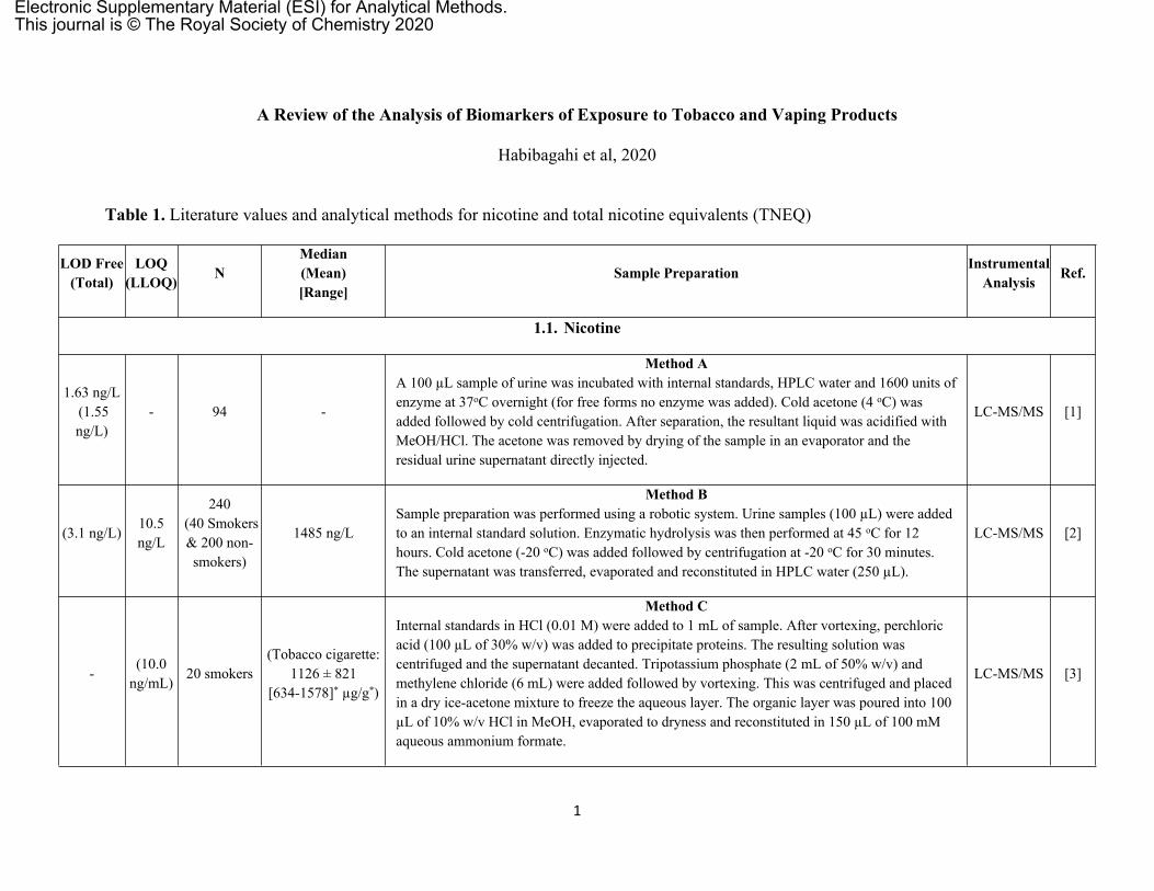
Table 1. Literature values and analytical methods for nicotine and total nicotine equivalents (TNEQ)
LOD Free(Total)
LOQ (LLOQ) N
Median(Mean)[Range]
Sample Preparation InstrumentalAnalysis Ref.
1.1. Nicotine
1.63 ng/L (1.55 ng/L)
- 94 -
Method AA 100 µL sample of urine was incubated with internal standards, HPLC water and 1600 units of enzyme at 37oC overnight (for free forms no enzyme was added). Cold acetone (4 oC) was added followed by cold centrifugation. After separation, the resultant liquid was acidified with MeOH/HCl. The acetone was removed by drying of the sample in an evaporator and the residual urine supernatant directly injected.
LC-MS/MS [1]
(3.1 ng/L)10.5 ng/L
240 (40 Smokers & 200 non-smokers)
1485 ng/L
Method BSample preparation was performed using a robotic system. Urine samples (100 µL) were added to an internal standard solution. Enzymatic hydrolysis was then performed at 45 oC for 12 hours. Cold acetone (-20 oC) was added followed by centrifugation at -20 oC for 30 minutes. The supernatant was transferred, evaporated and reconstituted in HPLC water (250 µL).
LC-MS/MS [2]
-(10.0
ng/mL)20 smokers
(Tobacco cigarette: 1126 ± 821
[634-1578]* µg/g*)
Method CInternal standards in HCl (0.01 M) were added to 1 mL of sample. After vortexing, perchloric acid (100 µL of 30% w/v) was added to precipitate proteins. The resulting solution was centrifuged and the supernatant decanted. Tripotassium phosphate (2 mL of 50% w/v) and methylene chloride (6 mL) were added followed by vortexing. This was centrifuged and placed in a dry ice-acetone mixture to freeze the aqueous layer. The organic layer was poured into 100 µL of 10% w/v HCl in MeOH, evaporated to dryness and reconstituted in 150 µL of 100 mM aqueous ammonium formate.
LC-MS/MS [3]
Electronic Supplementary Material (ESI) for Analytical Methods.This journal is © The Royal Society of Chemistry 2020
2
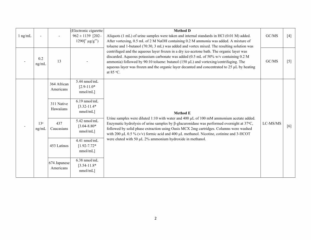
1 ng/mL - -(Electronic cigarette:
962 ± 1139 [202–1290]* µg/g**)
GC/MS [4]
-0.2
ng/mL13 -
Method DAliquots (1 mL) of urine samples were taken and internal standards in HCl (0.01 M) added. After vortexing, 0.5 mL of 2 M NaOH containing 0.2 M ammonia was added. A mixture of toluene and 1-butanol (70:30, 3 mL) was added and vortex mixed. The resulting solution was centrifuged and the aqueous layer frozen in a dry ice-acetone bath. The organic layer was discarded. Aqueous potassium carbonate was added (0.5 mL of 50% w/v containing 0.2 M ammonia) followed by 90:10 toluene: butanol (150 µL) and vortexing/centrifuging. The aqueous layer was frozen and the organic layer decanted and concentrated to 25 µL by heating at 85 oC.
GC/MS [5]
364 African Americans
5.44 nmol/mL[2.9-11.0* nmol/mL]
311 Native Hawaiians
6.19 nmol/mL[3.32-11.4* nmol/mL]
437 Caucasians
5.42 nmol/mL[3.04-8.80* nmol/mL]
453 Latinos4.41 nmol/mL
[1.92-7.72* nmol/mL]
-13a
ng/mL
674 Japanese Americans
6.38 nmol/mL[3.54-11.8* nmol/mL]
Method EUrine samples were diluted 1:10 with water and 400 µL of 100 mM ammonium acetate added. Enzymatic hydrolysis of urine samples by β-glucuronidase was performed overnight at 37oC, followed by solid phase extraction using Oasis MCX 2mg cartridges. Columns were washed with 200 µL 0.5 % (v/v) formic acid and 400 µL methanol. Nicotine, cotinine and 3-HCOT were eluted with 50 µL 2% ammonium hydroxide in methanol.
LC-MS/MS[6]
3
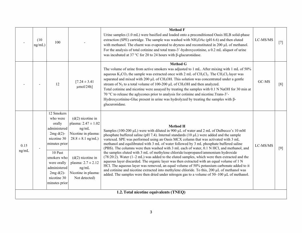
-(10
ng/mL)100 -
Method FUrine samples (1.0 mL) were basified and loaded onto a preconditioned Oasis HLB solid-phase extraction (SPE) cartridge. The sample was washed with NH4OAc (pH 6.6) and then eluted with methanol. The eluent was evaporated to dryness and reconstituted in 200 µL of methanol. For the analysis of total cotinine and total trans-3´-hydroxycotinine, a 0.2 mL aliquot of urine was incubated at 37 °C for 20 to 24 hours with β-glucuronidase.
LC-MS/MS[7]
- - 12[7.24 ± 3.41 µmol/24h]
Method GThe volume of urine from active smokers was adjusted to 1 mL. After mixing with 1 mL of 50% aqueous K2CO3 the sample was extracted once with 2 mL of CH2Cl2. The CH2Cl2 layer was separated and mixed with 200 µL of CH3OH. This solution was concentrated under a gentle stream of N2 to a total volume of 100-200 µL of CH3OH and then analyzed.Total cotinine and nicotine were assayed by treating the samples with 0.1 N NaOH for 30 min at 70 °C to release the aglycones prior to analysis for cotinine and nicotine.Trans-3’-Hydroxycotinine-Gluc present in urine was hydrolyzed by treating the samples with β-glucuronidase.
GC-MS[8]
12 Smokers who were
orally administered
2mg d(2)-nicotine 30
minutes prior
(d(2) nicotine in plasma: 2.47 ± 1.02
ng/mLNicotine in plasma: 28.8 ± 8.1 ng/mL)
0.15 ng/mL
-10 Past
smokers who were orally
administered 2mg d(2)-nicotine 30
minutes prior
(d(2) nicotine in plasma: 2.7 ± 2.12
ng/mLNicotine in plasma:
Not detected)
Method HSamples (100-200 µL) were with diluted in 900 µL of water and 2 mL of Dulbecco’s 10 mM phosphate buffered saline (pH 7.4). Internal standards (10 µL) were added and the sample vortexed. SPE was performed using an Oasis MCX column that was activated with 3 mL methanol and equilibrated with 3 mL of water followed by 3 mL phosphate buffered saline (PBS). The columns were then washed with 3 mL each of water, 0.1 N HCl, and methanol, and the samples eluted with 3 mL of methylene chloride/isopropanol/ammonium hydroxide (78:20:2). Water (1–2 mL) was added to the eluted samples, which were then extracted and the aqueous layer discarded. The organic layer was then extracted with an equal volume of 1 N HCl. The aqueous layer was removed, an equal volume of 50% potassium carbonate added to it and cotinine and nicotine extracted into methylene chloride. To this, 200 µL of methanol was added. The samples were then dried under nitrogen gas to a volume of 50–100 µL of methanol.
LC-MS/MS[9]
1.2. Total nicotine equivalents (TNEQ)
4
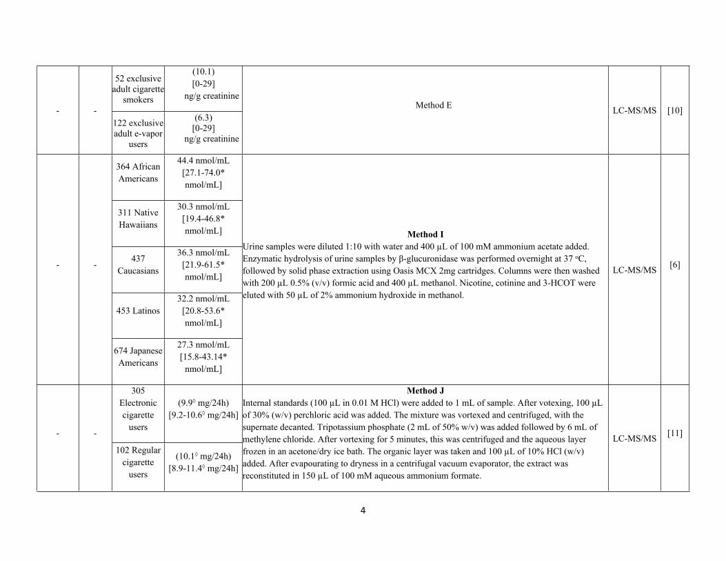
52 exclusive adult cigarette
smokers
(10.1)[0-29]
ng/g creatinine- -
122 exclusive adult e-vapor
users
(6.3)[0-29]
ng/g creatinine
Method E LC-MS/MS [10]
364 African Americans
44.4 nmol/mL[27.1-74.0* nmol/mL]
311 Native Hawaiians
30.3 nmol/mL[19.4-46.8* nmol/mL]
437 Caucasians
36.3 nmol/mL[21.9-61.5* nmol/mL]
453 Latinos32.2 nmol/mL
[20.8-53.6* nmol/mL]
- -
674 Japanese Americans
27.3 nmol/mL[15.8-43.14*
nmol/mL]
Method IUrine samples were diluted 1:10 with water and 400 µL of 100 mM ammonium acetate added. Enzymatic hydrolysis of urine samples by β-glucuronidase was performed overnight at 37 oC, followed by solid phase extraction using Oasis MCX 2mg cartridges. Columns were then washed with 200 µL 0.5% (v/v) formic acid and 400 µL methanol. Nicotine, cotinine and 3-HCOT were eluted with 50 µL of 2% ammonium hydroxide in methanol.
LC-MS/MS [6]
305 Electronic cigarette
users
(9.9◊ mg/24h)[9.2-10.6◊ mg/24h]
- -
102 Regular cigarette
users
(10.1◊ mg/24h)[8.9-11.4◊ mg/24h]
Method JInternal standards (100 µL in 0.01 M HCl) were added to 1 mL of sample. After votexing, 100 µL of 30% (w/v) perchloric acid was added. The mixture was vortexed and centrifuged, with the supernate decanted. Tripotassium phosphate (2 mL of 50% w/v) was added followed by 6 mL of methylene chloride. After vortexing for 5 minutes, this was centrifuged and the aqueous layer frozen in an acetone/dry ice bath. The organic layer was taken and 100 µL of 10% HCl (w/v) added. After evapourating to dryness in a centrifugal vacuum evaporator, the extract was reconstituted in 150 µL of 100 mM aqueous ammonium formate.
LC-MS/MS [11]
5
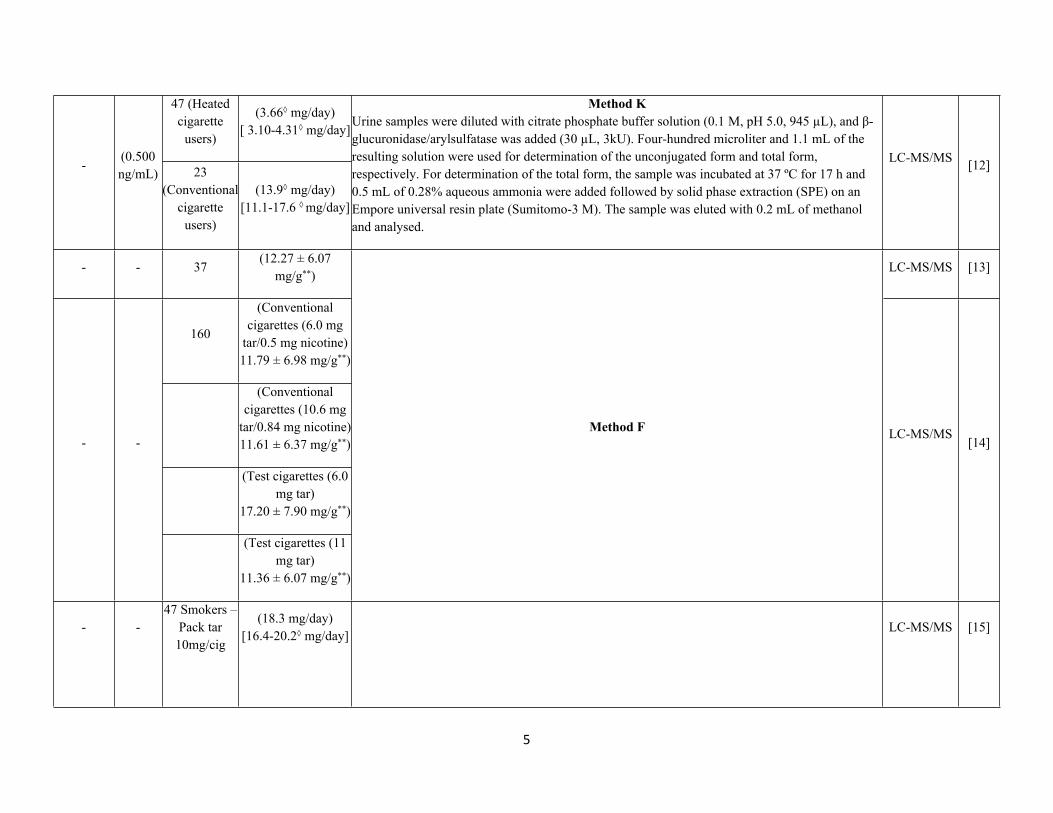
47 (Heated cigarette
users)
(3.66◊ mg/day)[ 3.10-4.31◊ mg/day]
-(0.500 ng/mL) 23
(Conventional cigarette
users)
(13.9◊ mg/day)[11.1-17.6 ◊ mg/day]
Method KUrine samples were diluted with citrate phosphate buffer solution (0.1 M, pH 5.0, 945 µL), and β-glucuronidase/arylsulfatase was added (30 µL, 3kU). Four-hundred microliter and 1.1 mL of the resulting solution were used for determination of the unconjugated form and total form, respectively. For determination of the total form, the sample was incubated at 37 ºC for 17 h and 0.5 mL of 0.28% aqueous ammonia were added followed by solid phase extraction (SPE) on an Empore universal resin plate (Sumitomo-3 M). The sample was eluted with 0.2 mL of methanol and analysed.
LC-MS/MS [12]
- - 37(12.27 ± 6.07
mg/g**)LC-MS/MS [13]
160
(Conventional cigarettes (6.0 mg
tar/0.5 mg nicotine)11.79 ± 6.98 mg/g**)
(Conventional cigarettes (10.6 mg
tar/0.84 mg nicotine)11.61 ± 6.37 mg/g**)
(Test cigarettes (6.0 mg tar)
17.20 ± 7.90 mg/g**)
- -
(Test cigarettes (11 mg tar)
11.36 ± 6.07 mg/g**)
Method F LC-MS/MS[14]
- -47 Smokers –
Pack tar 10mg/cig
(18.3 mg/day)[16.4-20.2◊ mg/day]
LC-MS/MS [15]
6
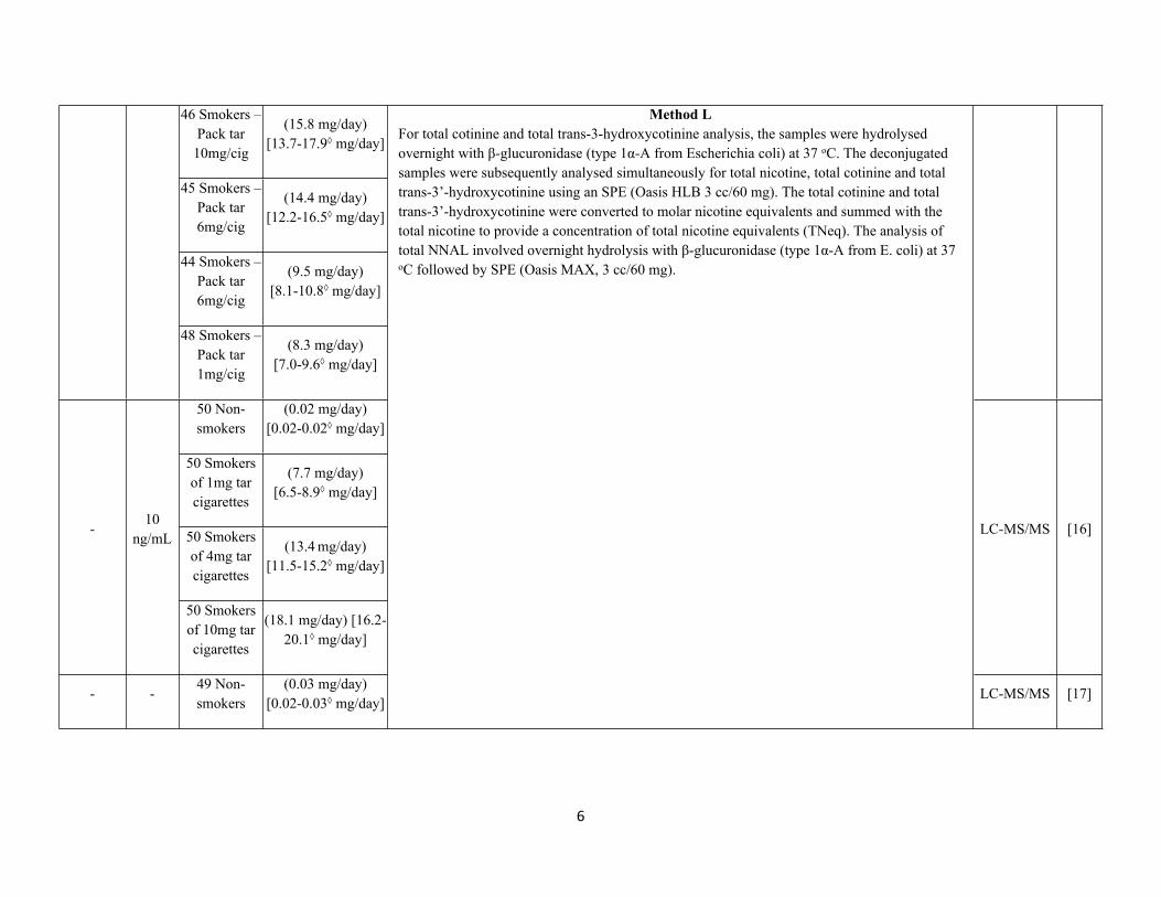
46 Smokers – Pack tar
10mg/cig
(15.8 mg/day)[13.7-17.9◊ mg/day]
45 Smokers – Pack tar 6mg/cig
(14.4 mg/day) [12.2-16.5◊ mg/day]
44 Smokers – Pack tar 6mg/cig
(9.5 mg/day)[8.1-10.8◊ mg/day]
48 Smokers – Pack tar 1mg/cig
(8.3 mg/day)[7.0-9.6◊ mg/day]
50 Non-smokers
(0.02 mg/day)[0.02-0.02◊ mg/day]
50 Smokers of 1mg tar cigarettes
(7.7 mg/day)[6.5-8.9◊ mg/day]
50 Smokers of 4mg tar cigarettes
(13.4 mg/day)[11.5-15.2◊ mg/day]
-10
ng/mL
50 Smokers of 10mg tar cigarettes
(18.1 mg/day) [16.2-20.1◊ mg/day]
LC-MS/MS [16]
- -49 Non-smokers
(0.03 mg/day)[0.02-0.03◊ mg/day]
Method LFor total cotinine and total trans-3-hydroxycotinine analysis, the samples were hydrolysed overnight with β-glucuronidase (type 1α-A from Escherichia coli) at 37 oC. The deconjugated samples were subsequently analysed simultaneously for total nicotine, total cotinine and total trans-3’-hydroxycotinine using an SPE (Oasis HLB 3 cc/60 mg). The total cotinine and total trans-3’-hydroxycotinine were converted to molar nicotine equivalents and summed with the total nicotine to provide a concentration of total nicotine equivalents (TNeq). The analysis of total NNAL involved overnight hydrolysis with β-glucuronidase (type 1α-A from E. coli) at 37 oC followed by SPE (Oasis MAX, 3 cc/60 mg).
LC-MS/MS [17]
7
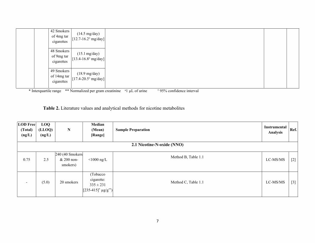
42 Smokers of 4mg tar cigarettes
(14.5 mg/day)[12.7-16.2◊ mg/day]
48 Smokers of 9mg tar cigarettes
(15.1 mg/day)[13.4-16.8◊ mg/day]
49 Smokers of 14mg tar cigarettes
(18.9 mg/day)[17.4-20.5◊ mg/day]
* Interquartile range ** Normalized per gram creatinine a1 µL of urine ◊ 95% confidence interval
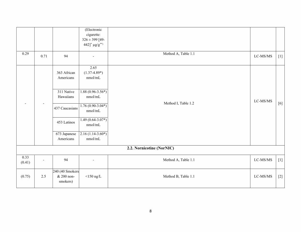
Table 2. Literature values and analytical methods for nicotine metabolites
LOD Free (Total) (ng/L)
LOQ (LLOQ)(ng/L)
NMedian(Mean)[Range]
Sample Preparation InstrumentalAnalysis Ref.
2.1 Nicotine-N-oxide (NNO)
0.75 2.5240 (40 Smokers
& 200 non-smokers)
<1000 ng/LMethod B, Table 1.1
LC-MS/MS [2]
- (5.0) 20 smokers
(Tobacco cigarette:335 ± 231
[235-415]* µg/g**)
Method C, Table 1.1 LC-MS/MS [3]
8
(Electronic cigarette:
326 ± 399 [49-442]* µg/g**)
0.29 0.71 94 -
Method A, Table 1.1LC-MS/MS [1]
363 African Americans
2.65(1.37-4.89*)
nmol/mL
311 Native Hawaiians
1.88 (0.96-3.56*) nmol/mL
437 Caucasians1.76 (0.90-3.04*)
nmol/mL
453 Latinos1.49 (0.64-3.07*)
nmol/mL
- -
673 Japanese Americans
2.16 (1.14-3.60*) nmol/mL
Method I, Table 1.2 LC-MS/MS [6]
2.2. Nornicotine (NorNIC)
0.33(0.41)
- 94 - Method A, Table 1.1 LC-MS/MS [1]
(0.75) 2.5240 (40 Smokers
& 200 non-smokers)
<150 ng/L Method B, Table 1.1 LC-MS/MS [2]
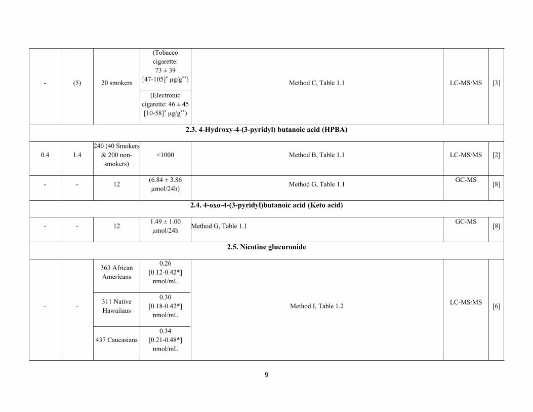
9
(Tobacco cigarette: 73 ± 39
[47-105]* µg/g**)- (5) 20 smokers
(Electronic cigarette: 46 ± 45[10-58]* µg/g**)
Method C, Table 1.1 LC-MS/MS [3]
2.3. 4-Hydroxy-4-(3-pyridyl) butanoic acid (HPBA)
0.4 1.4240 (40 Smokers
& 200 non-smokers)
<1000 Method B, Table 1.1 LC-MS/MS [2]
- - 12(6.84 ± 3.86 µmol/24h)
Method G, Table 1.1GC-MS
[8]
2.4. 4-oxo-4-(3-pyridyl)butanoic acid (Keto acid)
- - 121.49 ± 1.00 µmol/24h
Method G, Table 1.1GC-MS
[8]
2.5. Nicotine glucuronide
363 African Americans
0.26 [0.12-0.42*]
nmol/mL
311 Native Hawaiians
0.30 [0.18-0.42*]
nmol/mL- -
437 Caucasians0.34
[0.21-0.48*] nmol/mL
Method I, Table 1.2 LC-MS/MS [6]
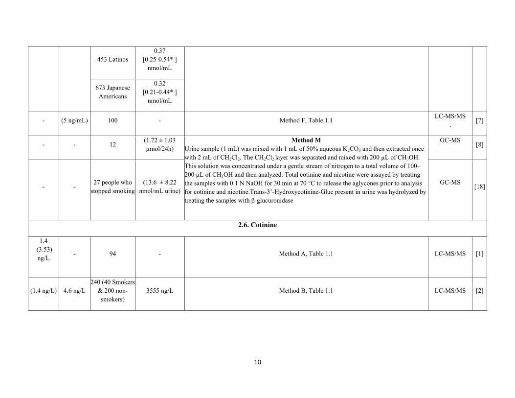
10
453 Latinos0.37
[0.25-0.54* ] nmol/mL
673 Japanese Americans
0.32 [0.21-0.44* ]
nmol/mL
- (5 ng/mL) 100 - Method F, Table 1.1LC-MS/MS
.[7]
- - 12(1.72 ± 1.03 µmol/24h)
GC-MS[8]
- -27 people who
stopped smoking(13.6 ± 8.22
nmol/mL urine)
Method MUrine sample (1 mL) was mixed with 1 mL of 50% aqueous K2CO3 and then extracted once with 2 mL of CH2Cl2. The CH2Cl2 layer was separated and mixed with 200 µL of CH3OH. This solution was concentrated under a gentle stream of nitrogen to a total volume of 100–200 µL of CH3OH and then analyzed. Total cotinine and nicotine were assayed by treating the samples with 0.1 N NaOH for 30 min at 70 °C to release the aglycones prior to analysis for cotinine and nicotine.Trans-3’-Hydroxycotinine-Gluc present in urine was hydrolyzed by treating the samples with β-glucuronidase
GC-MS[18]
2.6. Cotinine
1.4 (3.53) ng/L
- 94 - Method A, Table 1.1 LC-MS/MS [1]
(1.4 ng/L) 4.6 ng/L240 (40 Smokers
& 200 non-smokers)
3555 ng/L Method B, Table 1.1 LC-MS/MS [2]
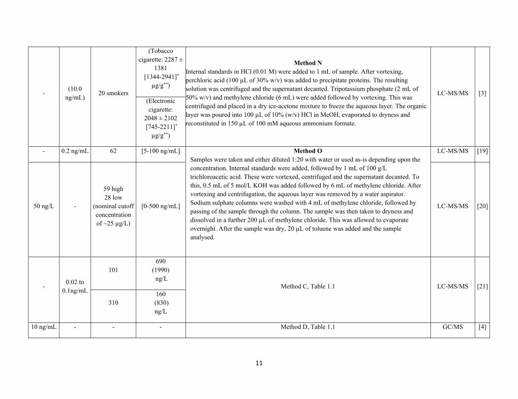
11
(Tobacco cigarette: 2287 ±
1381 [1344-2941]*
µg/g**)-
(10.0 ng/mL)
20 smokers(Electronic cigarette:
2048 ± 2102 [745-2211]*
µg/g**)
Method NInternal standards in HCl (0.01 M) were added to 1 mL of sample. After vortexing, perchloric acid (100 µL of 30% w/v) was added to precipitate proteins. The resulting solution was centrifuged and the supernatant decanted. Tripotassium phosphate (2 mL of 50% w/v) and methylene chloride (6 mL) were added followed by vortexing. This was centrifuged and placed in a dry ice-acetone mixture to freeze the aqueous layer. The organic layer was poured into 100 µL of 10% (w/v) HCl in MeOH, evaporated to dryness and reconstituted in 150 µL of 100 mM aqueous ammonium formate.
LC-MS/MS [3]
- 0.2 ng/mL 62 [5-100 ng/mL] LC-MS/MS [19]
50 ng/L -
59 high28 low
(nominal cutoff concentrationof ~25 µg/L)
[0-500 ng/mL]
Method OSamples were taken and either diluted 1:20 with water or used as-is depending upon the concentration. Internal standards were added, followed by 1 mL of 100 g/L trichloroacetic acid. These were vortexed, centrifuged and the supernatant decanted. To this, 0.5 mL of 5 mol/L KOH was added followed by 6 mL of methylene chloride. After vortexing and centrifugation, the aqueous layer was removed by a water aspirator. Sodium sulphate columns were washed with 4 mL of methylene chloride, followed by passing of the sample through the column. The sample was then taken to dryness and dissolved in a further 200 µL of methylene chloride. This was allowed to evaporate overnight. After the sample was dry, 20 µL of toluene was added and the sample analysed.
LC-MS/MS [20]
101690
(1990) ng/L
-0.02 to
0.1ng/mL
310160
(830) ng/L
Method C, Table 1.1 LC-MS/MS [21]
10 ng/mL - - - Method D, Table 1.1 GC/MS [4]
12
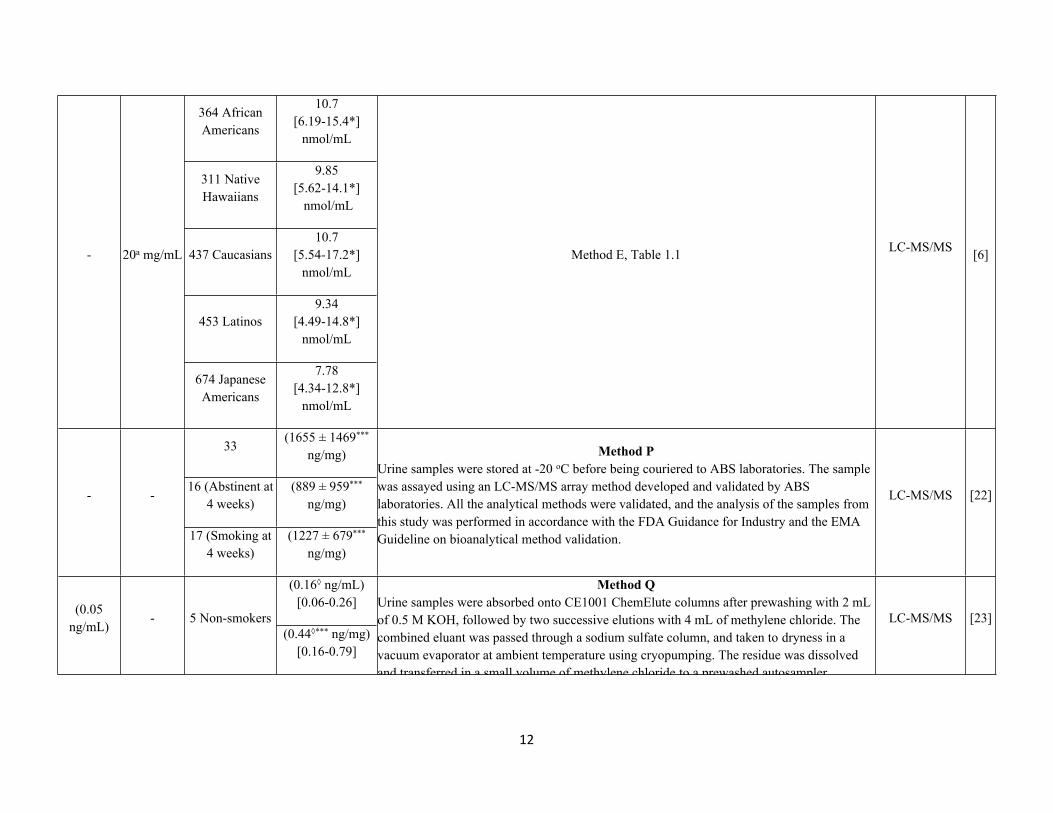
364 African Americans
10.7 [6.19-15.4*]
nmol/mL
311 Native Hawaiians
9.85[5.62-14.1*]
nmol/mL
437 Caucasians10.7
[5.54-17.2*]nmol/mL
453 Latinos9.34
[4.49-14.8*]nmol/mL
- 20a mg/mL
674 Japanese Americans
7.78[4.34-12.8*]
nmol/mL
Method E, Table 1.1 LC-MS/MS [6]
33(1655 ± 1469***
ng/mg)
16 (Abstinent at 4 weeks)
(889 ± 959*** ng/mg)
- -
17 (Smoking at 4 weeks)
(1227 ± 679*** ng/mg)
Method PUrine samples were stored at -20 oC before being couriered to ABS laboratories. The sample was assayed using an LC-MS/MS array method developed and validated by ABS laboratories. All the analytical methods were validated, and the analysis of the samples from this study was performed in accordance with the FDA Guidance for Industry and the EMA Guideline on bioanalytical method validation.
LC-MS/MS [22]
(0.16◊ ng/mL)[0.06-0.26](0.05
ng/mL)- 5 Non-smokers
(0.44◊*** ng/mg)[0.16-0.79]
Method QUrine samples were absorbed onto CE1001 ChemElute columns after prewashing with 2 mL of 0.5 M KOH, followed by two successive elutions with 4 mL of methylene chloride. The combined eluant was passed through a sodium sulfate column, and taken to dryness in a vacuum evaporator at ambient temperature using cryopumping. The residue was dissolved and transferred in a small volume of methylene chloride to a prewashed autosampler microvial, and the solvent was allowed to evaporate at room temperature.
LC-MS/MS [23]
13
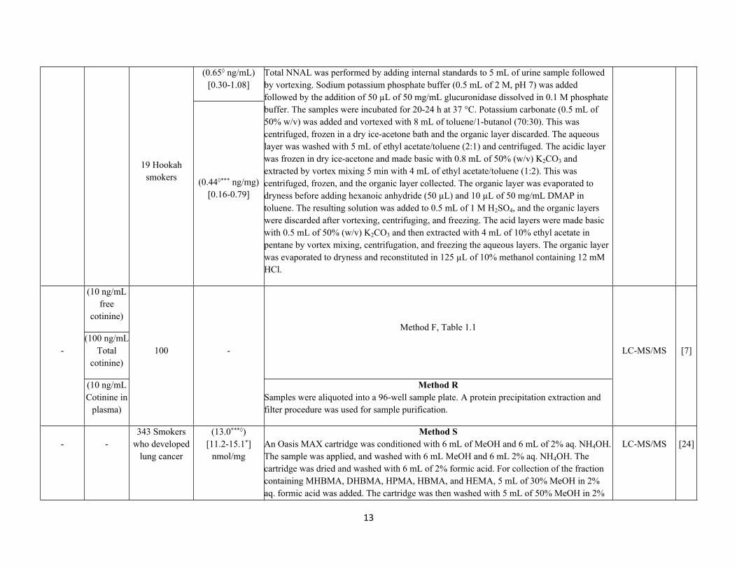
(0.65◊ ng/mL)[0.30-1.08]
19 Hookah smokers (0.44◊*** ng/mg)
[0.16-0.79]
Total NNAL was performed by adding internal standards to 5 mL of urine sample followed by vortexing. Sodium potassium phosphate buffer (0.5 mL of 2 M, pH 7) was added followed by the addition of 50 µL of 50 mg/mL glucuronidase dissolved in 0.1 M phosphate buffer. The samples were incubated for 20-24 h at 37 °C. Potassium carbonate (0.5 mL of 50% w/v) was added and vortexed with 8 mL of toluene/1-butanol (70:30). This was centrifuged, frozen in a dry ice-acetone bath and the organic layer discarded. The aqueous layer was washed with 5 mL of ethyl acetate/toluene (2:1) and centrifuged. The acidic layer was frozen in dry ice-acetone and made basic with 0.8 mL of 50% (w/v) K2CO3 and extracted by vortex mixing 5 min with 4 mL of ethyl acetate/toluene (1:2). This was centrifuged, frozen, and the organic layer collected. The organic layer was evaporated to dryness before adding hexanoic anhydride (50 µL) and 10 µL of 50 mg/mL DMAP in toluene. The resulting solution was added to 0.5 mL of 1 M H2SO4, and the organic layers were discarded after vortexing, centrifuging, and freezing. The acid layers were made basic with 0.5 mL of 50% (w/v) K2CO3 and then extracted with 4 mL of 10% ethyl acetate in pentane by vortex mixing, centrifugation, and freezing the aqueous layers. The organic layer was evaporated to dryness and reconstituted in 125 µL of 10% methanol containing 12 mM HCl.
(10 ng/mL free
cotinine)
(100 ng/mL Total
cotinine)
Method F, Table 1.1
-
(10 ng/mL Cotinine in
plasma)
100 -
Method RSamples were aliquoted into a 96-well sample plate. A protein precipitation extraction and filter procedure was used for sample purification.
LC-MS/MS [7]
- -343 Smokers
who developed lung cancer
(13.0***◊)[11.2-15.1*]
nmol/mg
Method SAn Oasis MAX cartridge was conditioned with 6 mL of MeOH and 6 mL of 2% aq. NH4OH. The sample was applied, and washed with 6 mL MeOH and 6 mL 2% aq. NH4OH. The cartridge was dried and washed with 6 mL of 2% formic acid. For collection of the fraction containing MHBMA, DHBMA, HPMA, HBMA, and HEMA, 5 mL of 30% MeOH in 2% aq. formic acid was added. The cartridge was then washed with 5 mL of 50% MeOH in 2%
LC-MS/MS [24]
14
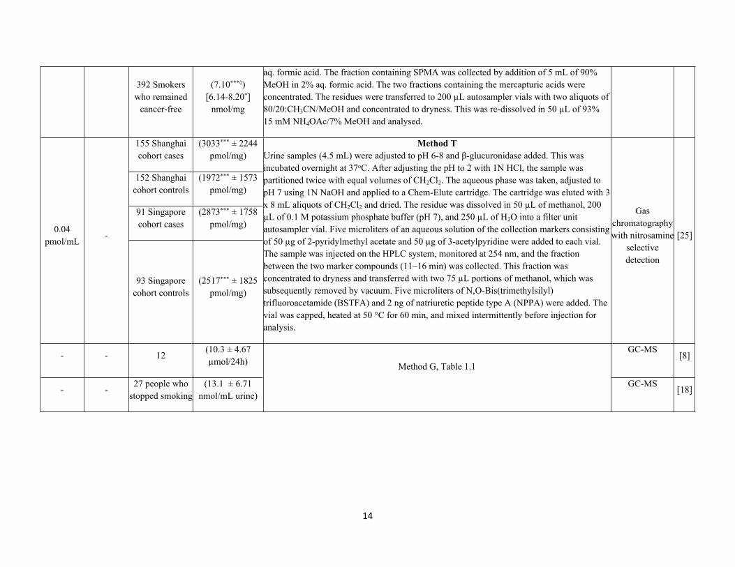
392 Smokers who remained
cancer-free
(7.10***◊)[6.14-8.20*]
nmol/mg
aq. formic acid. The fraction containing SPMA was collected by addition of 5 mL of 90% MeOH in 2% aq. formic acid. The two fractions containing the mercapturic acids were concentrated. The residues were transferred to 200 µL autosampler vials with two aliquots of 80/20:CH3CN/MeOH and concentrated to dryness. This was re-dissolved in 50 µL of 93% 15 mM NH4OAc/7% MeOH and analysed.
155 Shanghai cohort cases
(3033*** ± 2244pmol/mg)
152 Shanghai cohort controls
(1972*** ± 1573pmol/mg)
91 Singapore cohort cases
(2873*** ± 1758pmol/mg)0.04
pmol/mL-
93 Singapore cohort controls
(2517*** ± 1825pmol/mg)
Method TUrine samples (4.5 mL) were adjusted to pH 6-8 and β-glucuronidase added. This was incubated overnight at 37oC. After adjusting the pH to 2 with 1N HCl, the sample was partitioned twice with equal volumes of CH2Cl2. The aqueous phase was taken, adjusted to pH 7 using 1N NaOH and applied to a Chem-Elute cartridge. The cartridge was eluted with 3 x 8 mL aliquots of CH2Cl2 and dried. The residue was dissolved in 50 µL of methanol, 200 µL of 0.1 M potassium phosphate buffer (pH 7), and 250 µL of H2O into a filter unit autosampler vial. Five microliters of an aqueous solution of the collection markers consisting of 50 µg of 2-pyridylmethyl acetate and 50 µg of 3-acetylpyridine were added to each vial. The sample was injected on the HPLC system, monitored at 254 nm, and the fraction between the two marker compounds (11–16 min) was collected. This fraction was concentrated to dryness and transferred with two 75 µL portions of methanol, which was subsequently removed by vacuum. Five microliters of N,O-Bis(trimethylsilyl) trifluoroacetamide (BSTFA) and 2 ng of natriuretic peptide type A (NPPA) were added. The vial was capped, heated at 50 °C for 60 min, and mixed intermittently before injection for analysis.
Gas chromatography with nitrosamine
selective detection
[25]
- - 12(10.3 ± 4.67 µmol/24h)
GC-MS[8]
- -27 people who
stopped smoking(13.1 ± 6.71
nmol/mL urine)
Method G, Table 1.1
GC-MS[18]
15
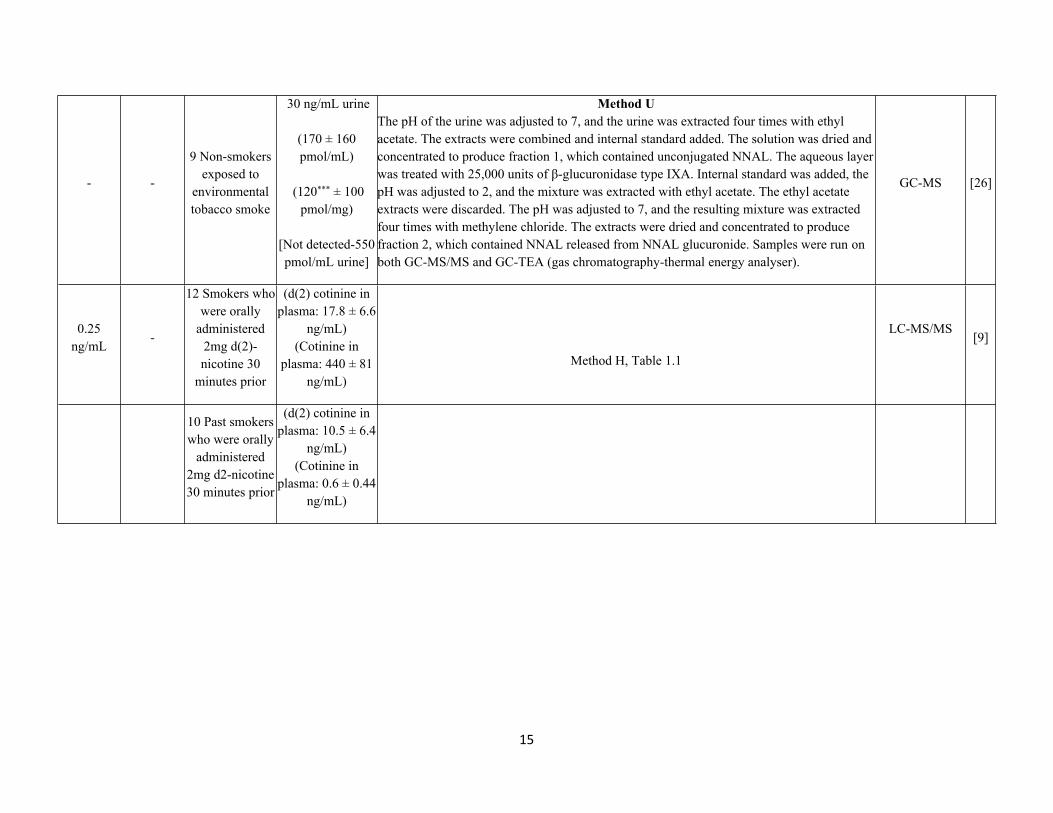
- -
9 Non-smokers exposed to
environmental tobacco smoke
30 ng/mL urine
(170 ± 160 pmol/mL)
(120*** ± 100 pmol/mg)
[Not detected-550 pmol/mL urine]
Method UThe pH of the urine was adjusted to 7, and the urine was extracted four times with ethyl acetate. The extracts were combined and internal standard added. The solution was dried and concentrated to produce fraction 1, which contained unconjugated NNAL. The aqueous layer was treated with 25,000 units of β-glucuronidase type IXA. Internal standard was added, the pH was adjusted to 2, and the mixture was extracted with ethyl acetate. The ethyl acetate extracts were discarded. The pH was adjusted to 7, and the resulting mixture was extracted four times with methylene chloride. The extracts were dried and concentrated to produce fraction 2, which contained NNAL released from NNAL glucuronide. Samples were run on both GC-MS/MS and GC-TEA (gas chromatography-thermal energy analyser).
GC-MS [26]
0.25 ng/mL
-
12 Smokers who were orally
administered 2mg d(2)-nicotine 30
minutes prior
(d(2) cotinine in plasma: 17.8 ± 6.6
ng/mL)(Cotinine in
plasma: 440 ± 81 ng/mL)
Method H, Table 1.1
LC-MS/MS[9]
10 Past smokers who were orally
administered 2mg d2-nicotine 30 minutes prior
(d(2) cotinine in plasma: 10.5 ± 6.4
ng/mL)(Cotinine in
plasma: 0.6 ± 0.44 ng/mL)
16
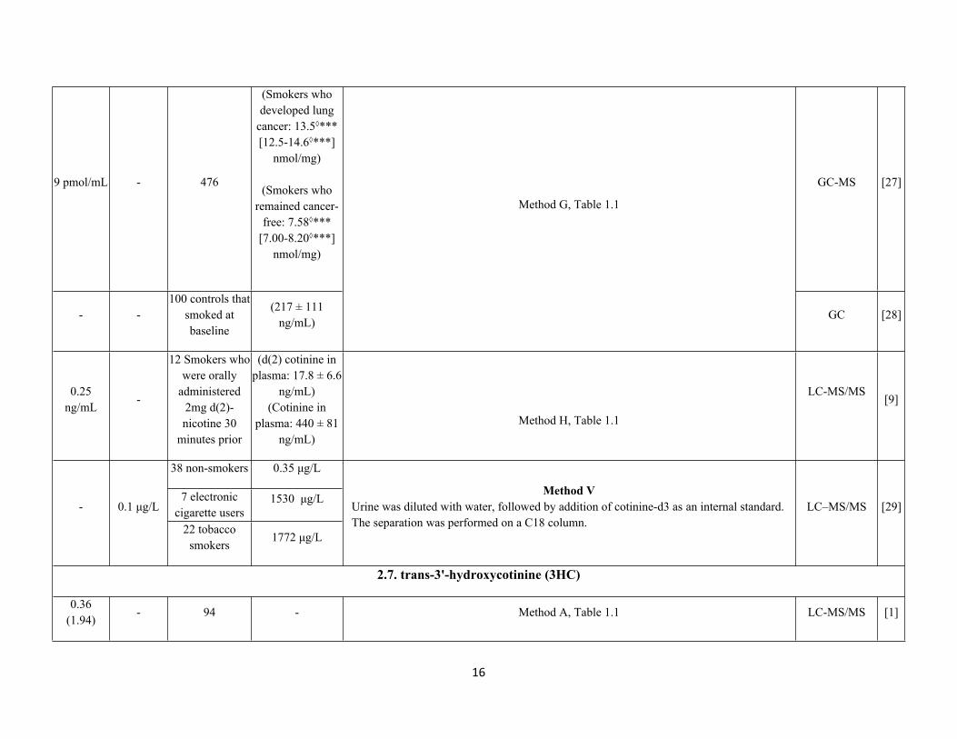
9 pmol/mL - 476
(Smokers who developed lung
cancer: 13.5◊*** [12.5-14.6◊***]
nmol/mg)
(Smokers who remained cancer-
free: 7.58◊*** [7.00-8.20◊***]
nmol/mg)
GC-MS [27]
- -100 controls that
smoked at baseline
(217 ± 111 ng/mL)
Method G, Table 1.1
GC [28]
0.25 ng/mL
-
12 Smokers who were orally
administered 2mg d(2)-nicotine 30
minutes prior
(d(2) cotinine in plasma: 17.8 ± 6.6
ng/mL)(Cotinine in
plasma: 440 ± 81 ng/mL)
Method H, Table 1.1
LC-MS/MS[9]
38 non-smokers 0.35 μg/L
7 electronic cigarette users
1530 μg/L- 0.1 μg/L
22 tobacco smokers
1772 μg/L
Method VUrine was diluted with water, followed by addition of cotinine-d3 as an internal standard. The separation was performed on a C18 column.
LC–MS/MS [29]
2.7. trans-3'-hydroxycotinine (3HC)
0.36(1.94)
- 94 - Method A, Table 1.1 LC-MS/MS [1]
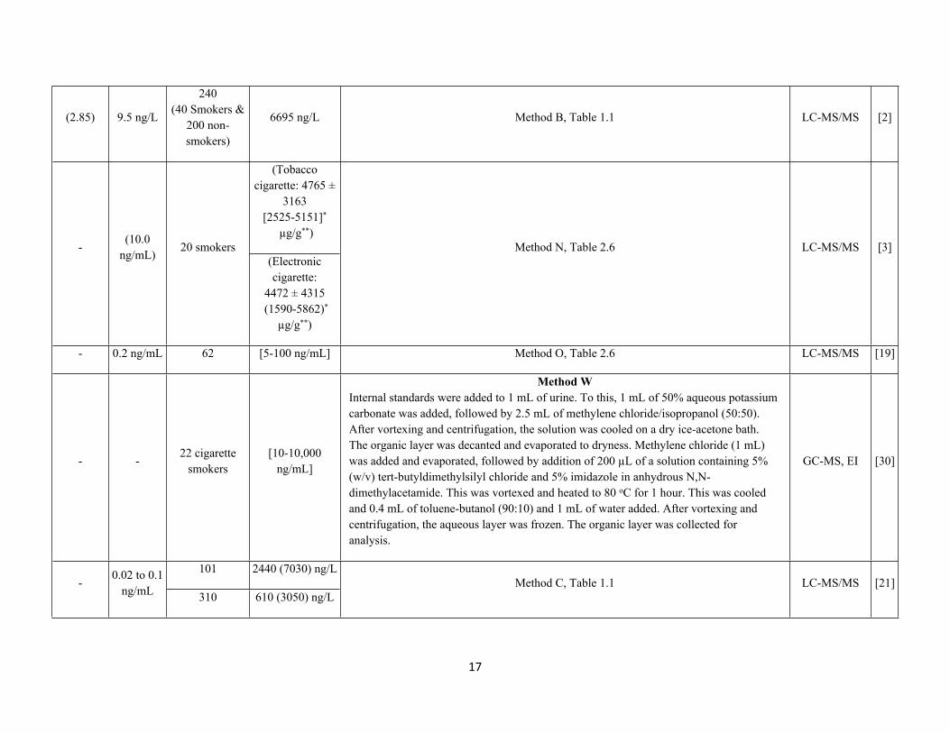
17
(2.85) 9.5 ng/L
240 (40 Smokers &
200 non-smokers)
6695 ng/L Method B, Table 1.1 LC-MS/MS [2]
(Tobacco cigarette: 4765 ±
3163 [2525-5151]*
µg/g**)-
(10.0 ng/mL)
20 smokers(Electronic cigarette:
4472 ± 4315 (1590-5862)*
µg/g**)
Method N, Table 2.6 LC-MS/MS [3]
- 0.2 ng/mL 62 [5-100 ng/mL] Method O, Table 2.6 LC-MS/MS [19]
- -22 cigarette
smokers[10-10,000
ng/mL]
Method WInternal standards were added to 1 mL of urine. To this, 1 mL of 50% aqueous potassium carbonate was added, followed by 2.5 mL of methylene chloride/isopropanol (50:50). After vortexing and centrifugation, the solution was cooled on a dry ice-acetone bath. The organic layer was decanted and evaporated to dryness. Methylene chloride (1 mL) was added and evaporated, followed by addition of 200 µL of a solution containing 5% (w/v) tert-butyldimethylsilyl chloride and 5% imidazole in anhydrous N,N-dimethylacetamide. This was vortexed and heated to 80 oC for 1 hour. This was cooled and 0.4 mL of toluene-butanol (90:10) and 1 mL of water added. After vortexing and centrifugation, the aqueous layer was frozen. The organic layer was collected for analysis.
GC-MS, EI [30]
101 2440 (7030) ng/L-
0.02 to 0.1 ng/mL 310 610 (3050) ng/L
Method C, Table 1.1 LC-MS/MS [21]
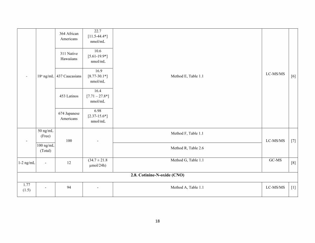
18
364 African Americans
22.7 [11.5-44.4*]
nmol/mL
311 Native Hawaiians
10.6[5.61-19.9*]
nmol/mL
437 Caucasians 16.9
[8.77-30.1*]nmol/mL
453 Latinos16.4
[7.71 – 27.8*]nmol/mL
- 18a ng/mL
674 Japanese Americans
6.98[2.37-15.6*]
nmol/mL
Method E, Table 1.1 LC-MS/MS [6]
50 ng/mL (Free)
Method F, Table 1.1
-100 ng/mL
(Total)
100 -
Method R, Table 2.6
LC-MS/MS [7]
1-2 ng/mL - 12(34.7 ± 21.8 µmol/24h)
Method G, Table 1.1 GC-MS[8]
2.8. Cotinine-N-oxide (CNO)
1.77(1.5)
- 94 - Method A, Table 1.1 LC-MS/MS [1]
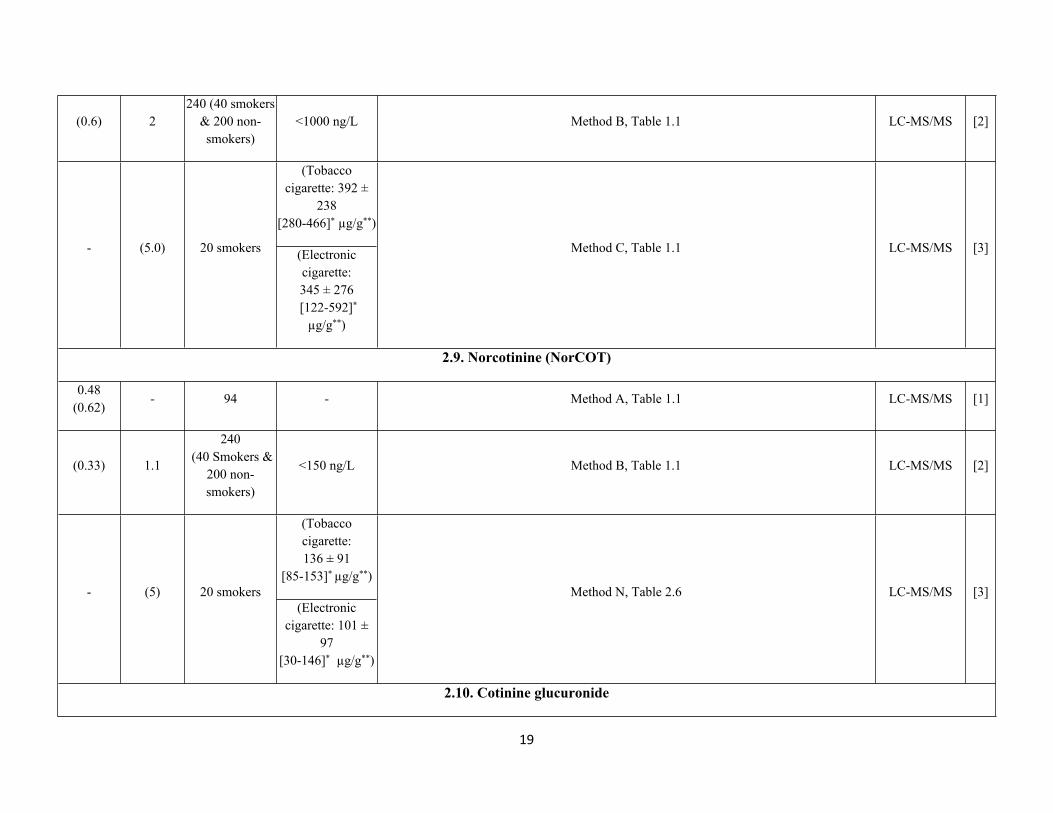
19
(0.6) 2240 (40 smokers
& 200 non-smokers)
<1000 ng/L Method B, Table 1.1 LC-MS/MS [2]
(Tobacco cigarette: 392 ±
238 [280-466]* µg/g**)
- (5.0) 20 smokers (Electronic cigarette: 345 ± 276
[122-592]* µg/g**)
Method C, Table 1.1 LC-MS/MS [3]
2.9. Norcotinine (NorCOT)
0.48(0.62)
- 94 - Method A, Table 1.1 LC-MS/MS [1]
(0.33) 1.1
240 (40 Smokers &
200 non-smokers)
<150 ng/L Method B, Table 1.1 LC-MS/MS [2]
(Tobacco cigarette:136 ± 91
[85-153]* µg/g**)- (5) 20 smokers
(Electronic cigarette: 101 ±
97[30-146]* µg/g**)
Method N, Table 2.6 LC-MS/MS [3]
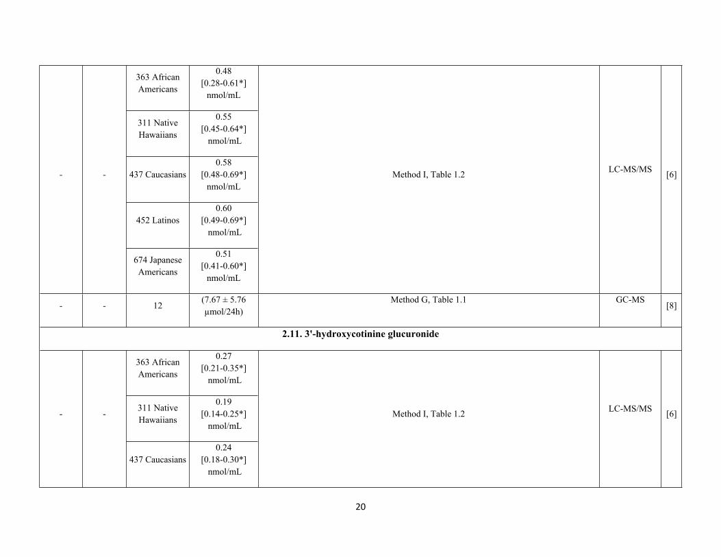
2.10. Cotinine glucuronide
20
363 African Americans
0.48 [0.28-0.61*]
nmol/mL
311 Native Hawaiians
0.55[0.45-0.64*]
nmol/mL
437 Caucasians0.58
[0.48-0.69*]nmol/mL
452 Latinos0.60
[0.49-0.69*] nmol/mL
- -
674 Japanese Americans
0.51 [0.41-0.60*]
nmol/mL
Method I, Table 1.2 LC-MS/MS [6]
- - 12(7.67 ± 5.76 µmol/24h)
Method G, Table 1.1 GC-MS[8]
2.11. 3'-hydroxycotinine glucuronide
363 African Americans
0.27[0.21-0.35*]
nmol/mL
311 Native Hawaiians
0.19[0.14-0.25*]
nmol/mL- -
437 Caucasians0.24
[0.18-0.30*] nmol/mL
Method I, Table 1.2 LC-MS/MS [6]
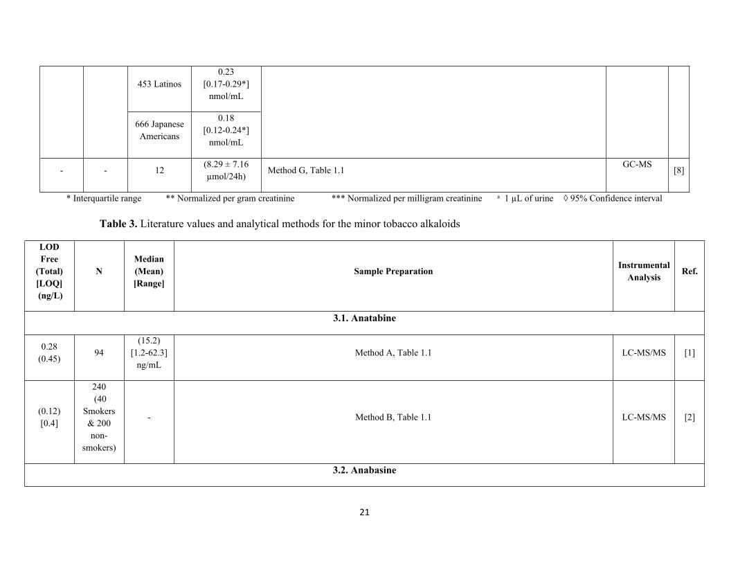
21
453 Latinos0.23
[0.17-0.29*]nmol/mL
666 Japanese Americans
0.18 [0.12-0.24*]
nmol/mL
- - 12(8.29 ± 7.16 µmol/24h)
Method G, Table 1.1GC-MS
[8]
* Interquartile range ** Normalized per gram creatinine *** Normalized per milligram creatinine a 1 µL of urine ◊ 95% Confidence interval
Table 3. Literature values and analytical methods for the minor tobacco alkaloids
LOD Free
(Total) [LOQ] (ng/L)
NMedian(Mean) [Range]
Sample Preparation InstrumentalAnalysis Ref.
3.1. Anatabine
0.28 (0.45)
94(15.2)
[1.2-62.3] ng/mL
Method A, Table 1.1 LC-MS/MS [1]
(0.12)[0.4]
240 (40
Smokers & 200 non-
smokers)
- Method B, Table 1.1 LC-MS/MS [2]
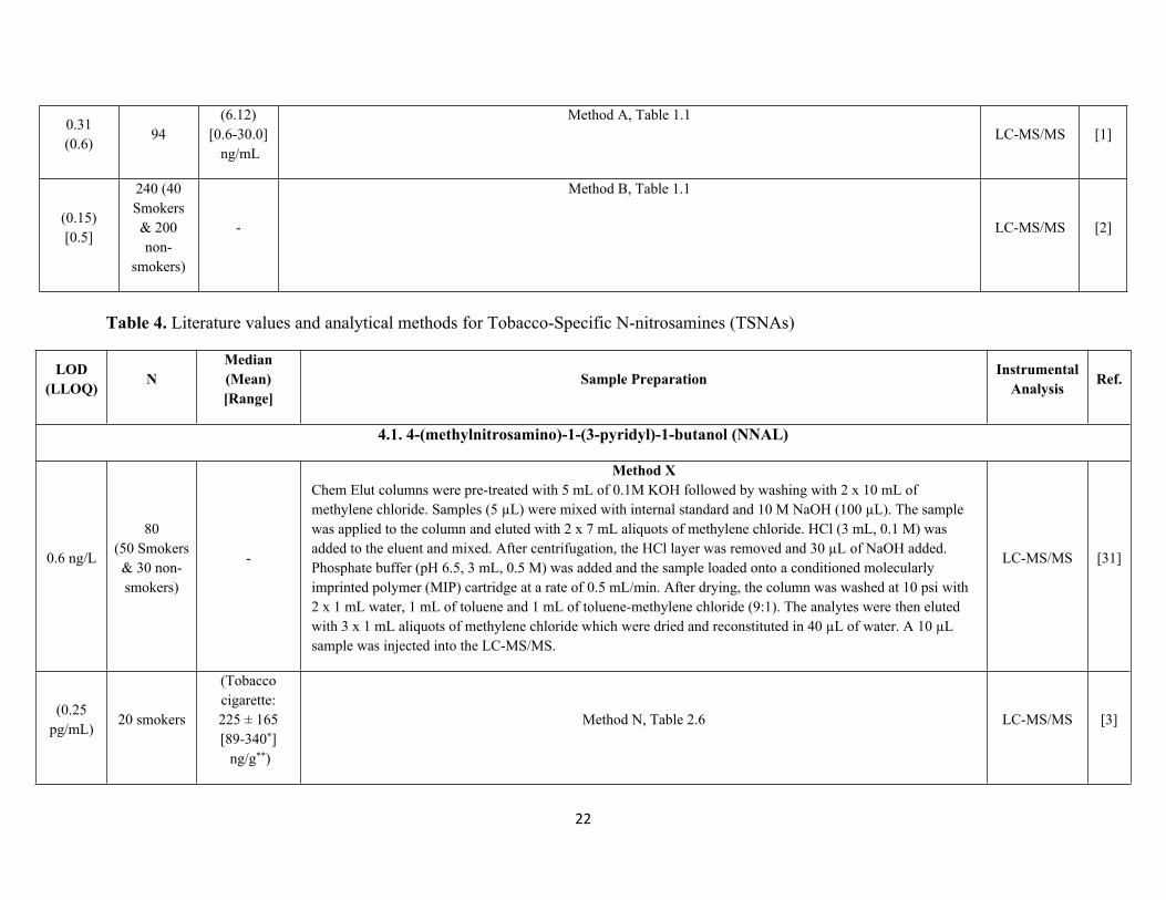
3.2. Anabasine
22
0.31(0.6)
94(6.12)
[0.6-30.0] ng/mL
Method A, Table 1.1LC-MS/MS [1]
(0.15)[0.5]
240 (40 Smokers & 200 non-
smokers)
-
Method B, Table 1.1
LC-MS/MS [2]
Table 4. Literature values and analytical methods for Tobacco-Specific N-nitrosamines (TSNAs)
LOD (LLOQ) N
Median (Mean)[Range]
Sample Preparation InstrumentalAnalysis Ref.
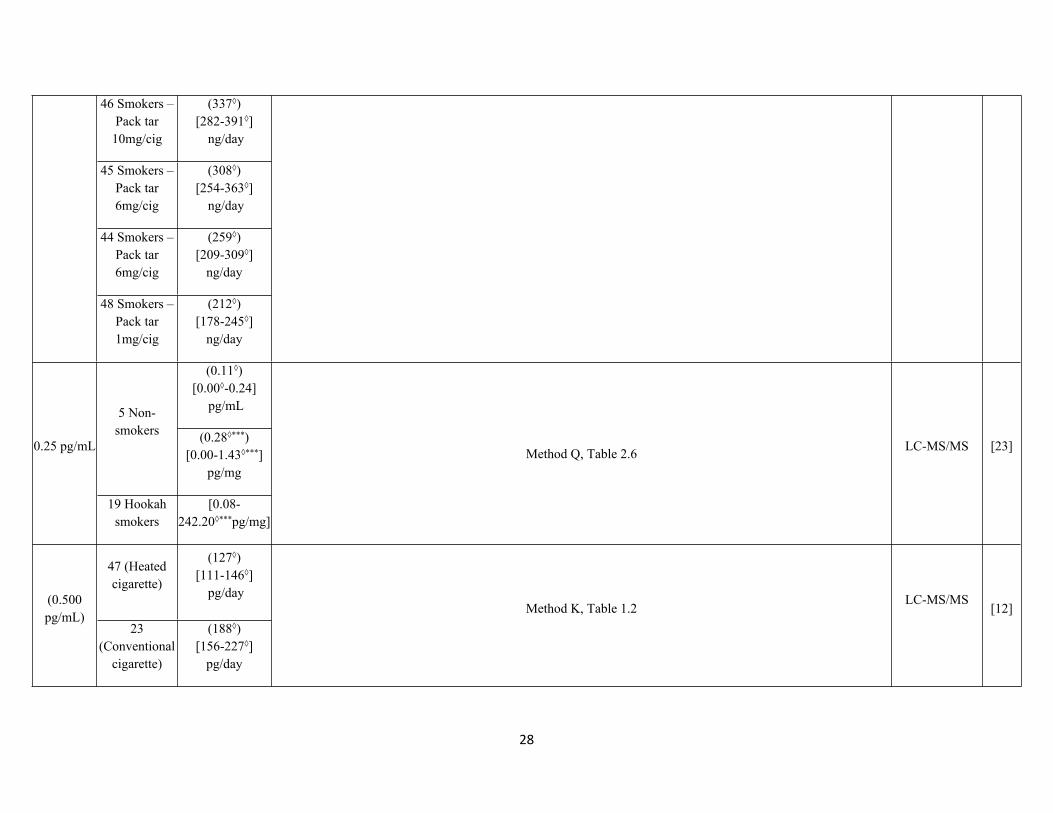
4.1. 4-(methylnitrosamino)-1-(3-pyridyl)-1-butanol (NNAL)
0.6 ng/L
80 (50 Smokers & 30 non-smokers)
-
Method XChem Elut columns were pre-treated with 5 mL of 0.1M KOH followed by washing with 2 x 10 mL of methylene chloride. Samples (5 µL) were mixed with internal standard and 10 M NaOH (100 µL). The sample was applied to the column and eluted with 2 x 7 mL aliquots of methylene chloride. HCl (3 mL, 0.1 M) was added to the eluent and mixed. After centrifugation, the HCl layer was removed and 30 µL of NaOH added. Phosphate buffer (pH 6.5, 3 mL, 0.5 M) was added and the sample loaded onto a conditioned molecularly imprinted polymer (MIP) cartridge at a rate of 0.5 mL/min. After drying, the column was washed at 10 psi with 2 x 1 mL water, 1 mL of toluene and 1 mL of toluene-methylene chloride (9:1). The analytes were then eluted with 3 x 1 mL aliquots of methylene chloride which were dried and reconstituted in 40 µL of water. A 10 µL sample was injected into the LC-MS/MS.
LC-MS/MS [31]
(0.25 pg/mL)
20 smokers
(Tobacco cigarette:225 ± 165[89-340*] ng/g**)
Method N, Table 2.6 LC-MS/MS [3]
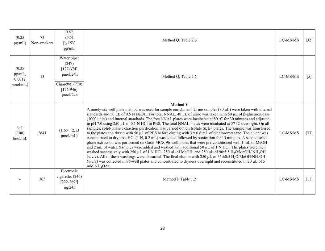
23
(0.25pg/mL)
73Non-smokers
0.87 (5.5)
[≤ 153]pg/mL
Method Q, Table 2.6 LC-MS/MS [32]
Water pipe:(247)
[127-374] pmol/24h
(0.25 pg/mL, 0.0012
pmol/mL)
13
Cigarette: (770)[176-946] pmol/24h
Method Q, Table 2.6 LC-MS/MS [5]
0.4 (100)
fmol/mL2641
(1.65 ± 2.13 pmol/mL)
Method YA ninety-six well plate method was used for sample enrichment. Urine samples (80 µL) were taken with internal standards and 50 µL of 0.5 N NaOH. For total NNAL, 40 µL of urine was taken with 50 µL of β-glucuronidase (1000 units) and internal standards. The free NNAL plates were incubated at 80 oC for 30 minutes and adjusted to pH 7.0 using 250 µL of 0.1 N HCl in PBS. The total NNAL plates were incubated at 37 oC overnight. On all samples, solid-phase extraction purification was carried out on Isolute SLE+ plates. The sample was transferred to the plates and rinsed with 50 µL of PBS before eluting with 3 x 0.6 mL of dichloromethane. The eluent was concentrated to dryness. HCl (1 N, 0.2 mL) was added followed by sonication for 15 minutes. A second solid-phase extraction was performed on Oasis MCX 96-well plates that were pre-conditioned with 1 mL of MeOH and 2 mL of water. Samples were added and washed with additional 50 µL of 1 N HCl. The plates were then washed successively with 250 µL of 1 N HCl, 250 µL of MeOH, and 250 µL of 90:5:5 H2O/MeOH/ NH4OH (v/v/v). All of these washings were discarded. The final elution with 250 µL of 35:60:5 H2O/MeOH/NH4OH (v/v/v) was collected in 96-well plates and concentrated to dryness overnight and reconstituted in 20 µL of 5 mM NH4OAc.
LC-MS/MS [33]
- 305
Electronic cigarette: (246)
[222-269◊] ng/24h
Method J, Table 1.2 LC-MS/MS [11]
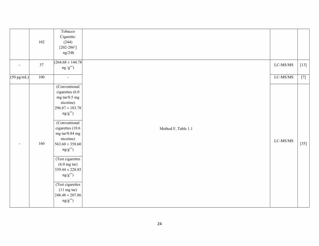
24
102
Tobacco Cigarette:
(244)[202-286◊]
ng/24h
- 37(264.68 ± 144.78
ng /g**)LC-MS/MS [13]
(50 pg/mL) 100 - LC-MS/MS [7]
(Conventional cigarettes (6.0 mg tar/0.5 mg
nicotine)296.87 ± 183.78
ng/g**)
(Conventional cigarettes (10.6 mg tar/0.84 mg
nicotine)563.60 ± 358.60
ng/g**)
(Test cigarettes (6.0 mg tar)
339.44 ± 228.85 ng/g**)
- 160
(Test cigarettes (11 mg tar)
248.48 ± 207.86 ng/g**)
Method F, Table 1.1
LC-MS/MS[35]
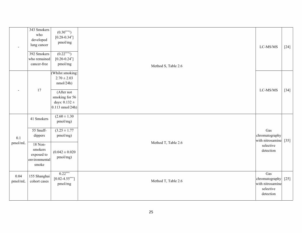
25
343 Smokers who
developed lung cancer
(0.30***◊)[0.28-0.34*] pmol/mg
-
392 Smokers who remained
cancer-free
(0.22***◊)[0.20-0.24*]
pmol/mg
LC-MS/MS [24]
(Whilst smoking: 2.70 ± 2.03 nmol/24h)
- 17 (After not smoking for 56 days: 0.132 ±
0.113 nmol/24h)
Method S, Table 2.6
LC-MS/MS [34]
41 Smokers(2.60 ± 1.30 pmol/mg)
55 Snuff-dippers
(3.25 ± 1.77 pmol/mg)
0.1 pmol/mL 18 Non-
smokers exposed to
environmental smoke
(0.042 ± 0.020 pmol/mg)
Method T, Table 2.6
Gas chromatography with nitrosamine
selective detection
[35]
0.04 pmol/mL
155 Shanghai cohort cases
0.22*** [0.02-4.55***]
pmol/mgMethod T, Table 2.6
Gas chromatography with nitrosamine
selective detection
[25]
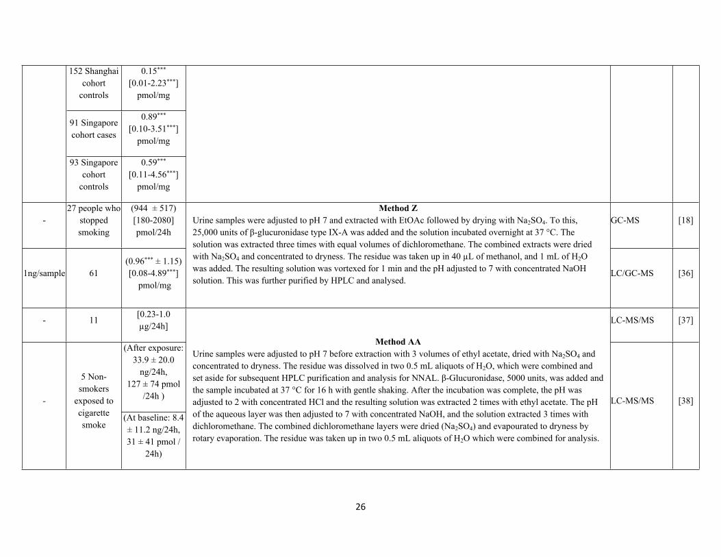
26
152 Shanghai cohort
controls
0.15*** [0.01-2.23***]
pmol/mg
91 Singapore cohort cases
0.89*** [0.10-3.51***]
pmol/mg
93 Singapore cohort
controls
0.59*** [0.11-4.56***]
pmol/mg
-27 people who
stopped smoking
(944 ± 517)[180-2080]pmol/24h
GC-MS [18]
1ng/sample 61(0.96*** ± 1.15)[0.08-4.89***]
pmol/mg
Method ZUrine samples were adjusted to pH 7 and extracted with EtOAc followed by drying with Na2SO4. To this, 25,000 units of β-glucuronidase type IX-A was added and the solution incubated overnight at 37 °C. The solution was extracted three times with equal volumes of dichloromethane. The combined extracts were dried with Na2SO4 and concentrated to dryness. The residue was taken up in 40 µL of methanol, and 1 mL of H2O was added. The resulting solution was vortexed for 1 min and the pH adjusted to 7 with concentrated NaOH solution. This was further purified by HPLC and analysed.
LC/GC-MS [36]
- 11[0.23-1.0 µg/24h]
LC-MS/MS [37]
(After exposure: 33.9 ± 20.0
ng/24h,127 ± 74 pmol
/24h )-
5 Non-smokers
exposed to cigarette smoke
(At baseline: 8.4 ± 11.2 ng/24h,31 ± 41 pmol /
24h)
Method AAUrine samples were adjusted to pH 7 before extraction with 3 volumes of ethyl acetate, dried with Na2SO4 and concentrated to dryness. The residue was dissolved in two 0.5 mL aliquots of H2O, which were combined and set aside for subsequent HPLC purification and analysis for NNAL. β-Glucuronidase, 5000 units, was added and the sample incubated at 37 °C for 16 h with gentle shaking. After the incubation was complete, the pH was adjusted to 2 with concentrated HCl and the resulting solution was extracted 2 times with ethyl acetate. The pH of the aqueous layer was then adjusted to 7 with concentrated NaOH, and the solution extracted 3 times with dichloromethane. The combined dichloromethane layers were dried (Na2SO4) and evapourated to dryness by rotary evaporation. The residue was taken up in two 0.5 mL aliquots of H2O which were combined for analysis.
LC-MS/MS [38]
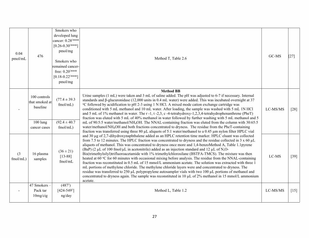
27
0.04 pmol/mL
476
Smokers who developed lung
cancer: 0.28◊*** [0.26-0.30◊***]
pmol/mg
Smokers who remained cancer-
free: 0.20◊*** [0.18-0.22◊***]
pmol/mg
Method T, Table 2.6 GC-MS [27]
100 controls that smoked at
baseline
(77.4 ± 39.3 fmol/mL)
-
100 lung cancer cases
(92.4 ± 40.7 fmol/mL)
LC-MS/MS [28]
(3 fmol/mL)
16 plasma samples
(36 ± 21)[13-88]
fmol/mL
Method BBUrine samples (1 mL) were taken and 3 mL of saline added. The pH was adjusted to 6-7 if necessary. Internal standards and β-glucuronidase (12,000 units in 0.4 mL water) were added. This was incubated overnight at 37 oC followed by acidification to pH 2-3 using 1 N HCl. A mixed mode cation exchange cartridge was conditioned with 5 mL methanol and 10 mL water. After loading, the sample was washed with 5 mL 1N HCl and 5 mL of 1% methanol in water. The r -1, t -2,3, c -4-tetrahydroxy-1,2,3,4-tetrahydrophenanthrene (PheT) fraction was eluted with 5 mL of 40% methanol in water followed by further washing with 5 mL methanol and 5 mL of 90:5:5 water/methanol/NH4OH. The NNAL-containing fraction was eluted from the column with 30:65:5 water/methanol/NH4OH and both fractions concentrated to dryness. The residue from the PheT-containing fraction was transferred using three 80 µL aliquots of 5:1 water/methanol to a 0.45 µm nylon filter HPLC vial and 30 µg of 2,7-dihydroxynaphthalene added as an HPLC retention time marker. HPLC eluant was collected from 7.5 to 12 minutes. The HPLC fraction was concentrated to dryness and the residue collected in 3 x 60 µL aliquots of methanol. This was concentrated to dryness once more and 1,4-benzoMethod A, Table 1.1pyrene (BaP) (2 µL of 100 fmol/µL in acetonitrile) added as an injection standard and 12 µL of N,O-Bis(trimethylsilyl)trifluoroacetamide with 1% trimethylchlorosilane (BSTFA-TMCS). The mixture was then heated at 60 oC for 60 minutes with occasional mixing before analysis. The residue from the NNAL-containing fraction was reconstituted in 0.5 mL of 15 mmol/L ammonium acetate. The solution was extracted with three 1 mL portions of methylene chloride. The methylene chloride layers were and concentrated to dryness. The residue was transferred to 250 µL polypropylene autosampler vials with two 100 µL portions of methanol and concentrated to dryness again. The sample was reconstituted in 10 µL of 2% methanol in 15 mmol/L ammonium acetate.
LC-MS [39]
-47 Smokers –
Pack tar 10mg/cig
(487◊)[424-549◊]
ng/dayMethod L, Table 1.2 LC-MS/MS [15]
28
46 Smokers – Pack tar
10mg/cig
(337◊)[282-391◊]
ng/day
45 Smokers – Pack tar 6mg/cig
(308◊)[254-363◊]
ng/day
44 Smokers – Pack tar 6mg/cig
(259◊)[209-309◊]
ng/day
48 Smokers – Pack tar 1mg/cig
(212◊)[178-245◊]
ng/day
(0.11◊)[0.00◊-0.24]
pg/mL5 Non-smokers (0.28◊***)
[0.00-1.43◊***]pg/mg
0.25 pg/mL
19 Hookah smokers
[0.08-242.20◊***pg/mg]
Method Q, Table 2.6 LC-MS/MS [23]
47 (Heated cigarette)
(127◊)[111-146◊]
pg/day(0.500 pg/mL)
23 (Conventional
cigarette)
(188◊) [156-227◊]
pg/day
Method K, Table 1.2LC-MS/MS
[12]
29
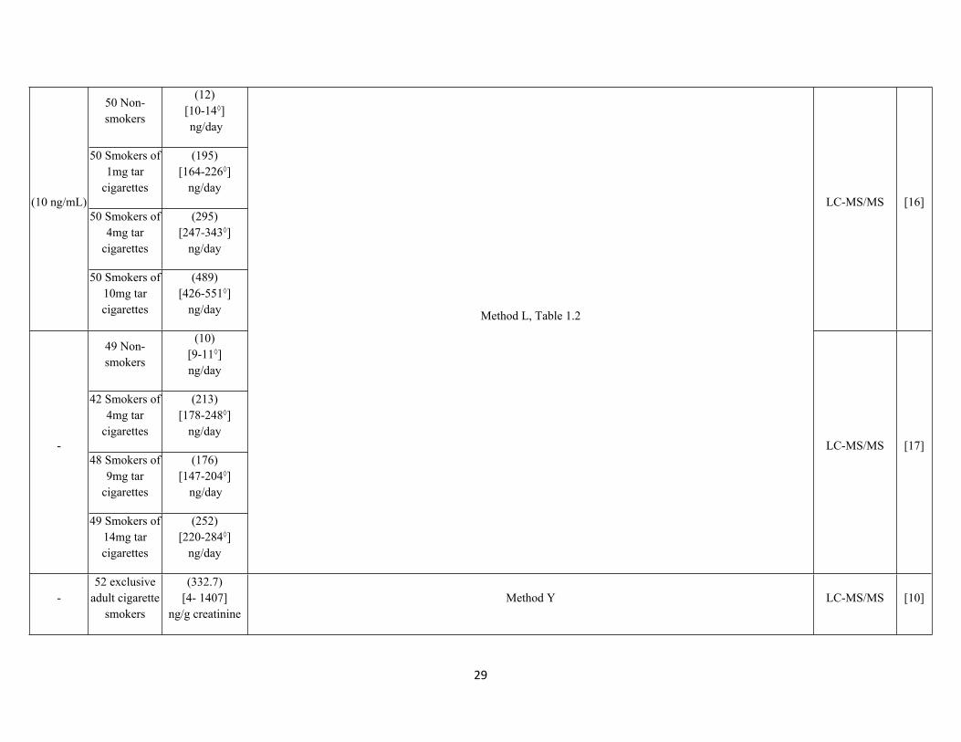
50 Non-smokers
(12)[10-14◊] ng/day
50 Smokers of 1mg tar
cigarettes
(195)[164-226◊]
ng/day
50 Smokers of 4mg tar
cigarettes
(295)[247-343◊]
ng/day
(10 ng/mL)
50 Smokers of 10mg tar cigarettes
(489)[426-551◊]
ng/day
LC-MS/MS [16]
49 Non-smokers
(10)[9-11◊]ng/day
42 Smokers of 4mg tar
cigarettes
(213)[178-248◊]
ng/day
48 Smokers of 9mg tar
cigarettes
(176)[147-204◊]
ng/day
-
49 Smokers of 14mg tar cigarettes
(252)[220-284◊]
ng/day
Method L, Table 1.2
LC-MS/MS [17]
-52 exclusive
adult cigarette smokers
(332.7)[4- 1407]
ng/g creatinineMethod Y LC-MS/MS [10]
30
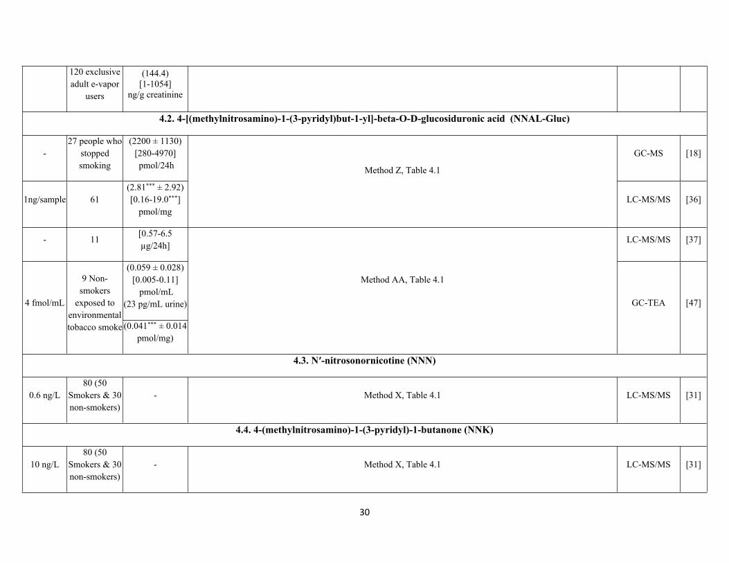
120 exclusive adult e-vapor
users
(144.4)[1-1054]
ng/g creatinine
4.2. 4-[(methylnitrosamino)-1-(3-pyridyl)but-1-yl]-beta-O-D-glucosiduronic acid (NNAL-Gluc)
-27 people who
stopped smoking
(2200 ± 1130)[280-4970] pmol/24h
GC-MS [18]
1ng/sample 61(2.81*** ± 2.92)[0.16-19.0***]
pmol/mg
Method Z, Table 4.1
LC-MS/MS [36]
- 11[0.57-6.5 µg/24h]
LC-MS/MS [37]
(0.059 ± 0.028)[0.005-0.11]
pmol/mL (23 pg/mL urine)4 fmol/mL
9 Non-smokers
exposed to environmental tobacco smoke (0.041*** ± 0.014
pmol/mg)
Method AA, Table 4.1
GC-TEA [47]
4.3. N′-nitrosonornicotine (NNN)
0.6 ng/L80 (50
Smokers & 30 non-smokers)
- Method X, Table 4.1 LC-MS/MS [31]
4.4. 4-(methylnitrosamino)-1-(3-pyridyl)-1-butanone (NNK)
10 ng/L80 (50
Smokers & 30 non-smokers)
- Method X, Table 4.1 LC-MS/MS [31]
31
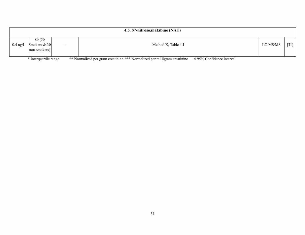
4.5. N′-nitrosoanatabine (NAT)
0.4 ng/L80 (50
Smokers & 30 non-smokers)
- Method X, Table 4.1 LC-MS/MS [31]
* Interquartile range ** Normalized per gram creatinine *** Normalized per milligram creatinine ◊ 95% Confidence interval
32
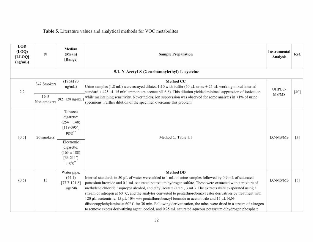
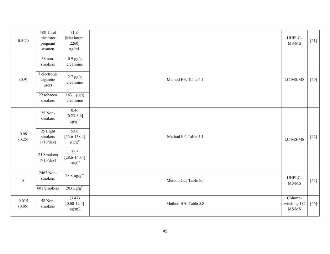
Table 5. Literature values and analytical methods for VOC metabolites
LOD(LOQ)
[LLOQ] (ng/mL)
NMedian (Mean)[Range]
Sample Preparation InstrumentalAnalysis Ref.
5.1. N-Acetyl-S-(2-carbamoylethyl)-L-cysteine
347 Smokers(196±180 ng/mL)
2.21203
Non-smokers(82±128 ng/mL)
Method CCUrine samples (1.8 mL) were assayed diluted 1:10 with buffer (50 µL urine + 25 µL working mixed internal standard + 425 µL 15 mM ammonium acetate pH 6.8). This dilution yielded minimal suppression of ionization while maintaining sensitivity. Nevertheless, ion suppression was observed for some analytes in <1% of urine specimens. Further dilution of the specimen overcame this problem.
UHPLC-MS/MS
[40]
Tobacco cigarette:
(254 ± 148) [119-395*]
µg/g**
[0.5] 20 smokersElectronic cigarette:
(163 ± 188)[66-211*]
µg/g**
Method C, Table 1.1 LC-MS/MS [3]
(0.5) 13
Water pipe:(44.1)
[77.7-121.8] µg/24h
Method DDInternal standards in 50 µL of water were added to 1 mL of urine samples followed by 0.9 mL of saturated potassium bromide and 0.1 mL saturated potassium hydrogen sulfate. These were extracted with a mixture of methylene chloride, isopropyl alcohol, and ethyl acetate (1:1:1, 3 mL). The extracts were evaporated using a stream of nitrogen at 60 °C, and the analytes converted to pentafluorobenzyl ester derivatives by treatment with 120 µL acetonitrile, 15 µL 10% w/v pentafluorobenzyl bromide in acetonitrile and 15 µL N,N-diisopropylethylamine at 60° C for 30 min. Following derivatization, the tubes were dried in a stream of nitrogen to remove excess derivatizing agent, cooled, and 0.25 mL saturated aqueous potassium dihydrogen phosphate
LC-MS/MS [5]
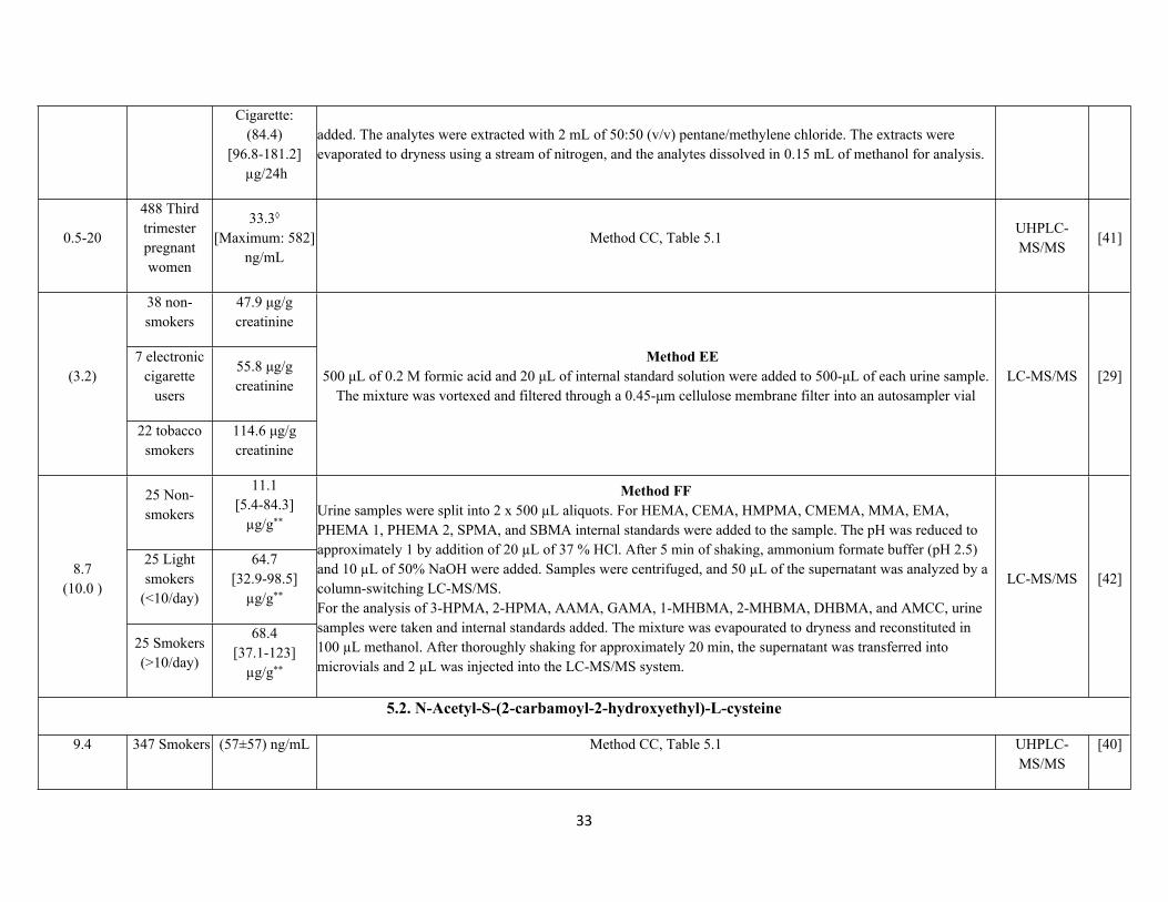
33
Cigarette:(84.4)
[96.8-181.2] µg/24h
added. The analytes were extracted with 2 mL of 50:50 (v/v) pentane/methylene chloride. The extracts were evaporated to dryness using a stream of nitrogen, and the analytes dissolved in 0.15 mL of methanol for analysis.
0.5-20
488 Third trimester pregnant women
33.3◊
[Maximum: 582]ng/mL
Method CC, Table 5.1UHPLC-MS/MS
[41]
38 non-smokers
47.9 μg/g creatinine
7 electronic cigarette
users
55.8 μg/g creatinine
(3.2)
22 tobacco smokers
114.6 μg/g creatinine
Method EE500 μL of 0.2 M formic acid and 20 μL of internal standard solution were added to 500-μL of each urine sample.
The mixture was vortexed and filtered through a 0.45-μm cellulose membrane filter into an autosampler vialLC-MS/MS [29]
25 Non-smokers
11.1[5.4-84.3]
µg/g**
25 Light smokers
(<10/day)
64.7[32.9-98.5]
µg/g**
8.7 (10.0 )
25 Smokers (>10/day)
68.4[37.1-123]
µg/g**
Method FFUrine samples were split into 2 x 500 µL aliquots. For HEMA, CEMA, HMPMA, CMEMA, MMA, EMA, PHEMA 1, PHEMA 2, SPMA, and SBMA internal standards were added to the sample. The pH was reduced to approximately 1 by addition of 20 µL of 37 % HCl. After 5 min of shaking, ammonium formate buffer (pH 2.5) and 10 µL of 50% NaOH were added. Samples were centrifuged, and 50 µL of the supernatant was analyzed by a column-switching LC-MS/MS.For the analysis of 3-HPMA, 2-HPMA, AAMA, GAMA, 1-MHBMA, 2-MHBMA, DHBMA, and AMCC, urine samples were taken and internal standards added. The mixture was evapourated to dryness and reconstituted in 100 µL methanol. After thoroughly shaking for approximately 20 min, the supernatant was transferred into microvials and 2 µL was injected into the LC-MS/MS system.
LC-MS/MS [42]
5.2. N-Acetyl-S-(2-carbamoyl-2-hydroxyethyl)-L-cysteine
9.4 347 Smokers (57±57) ng/mL Method CC, Table 5.1 UHPLC-MS/MS
[40]
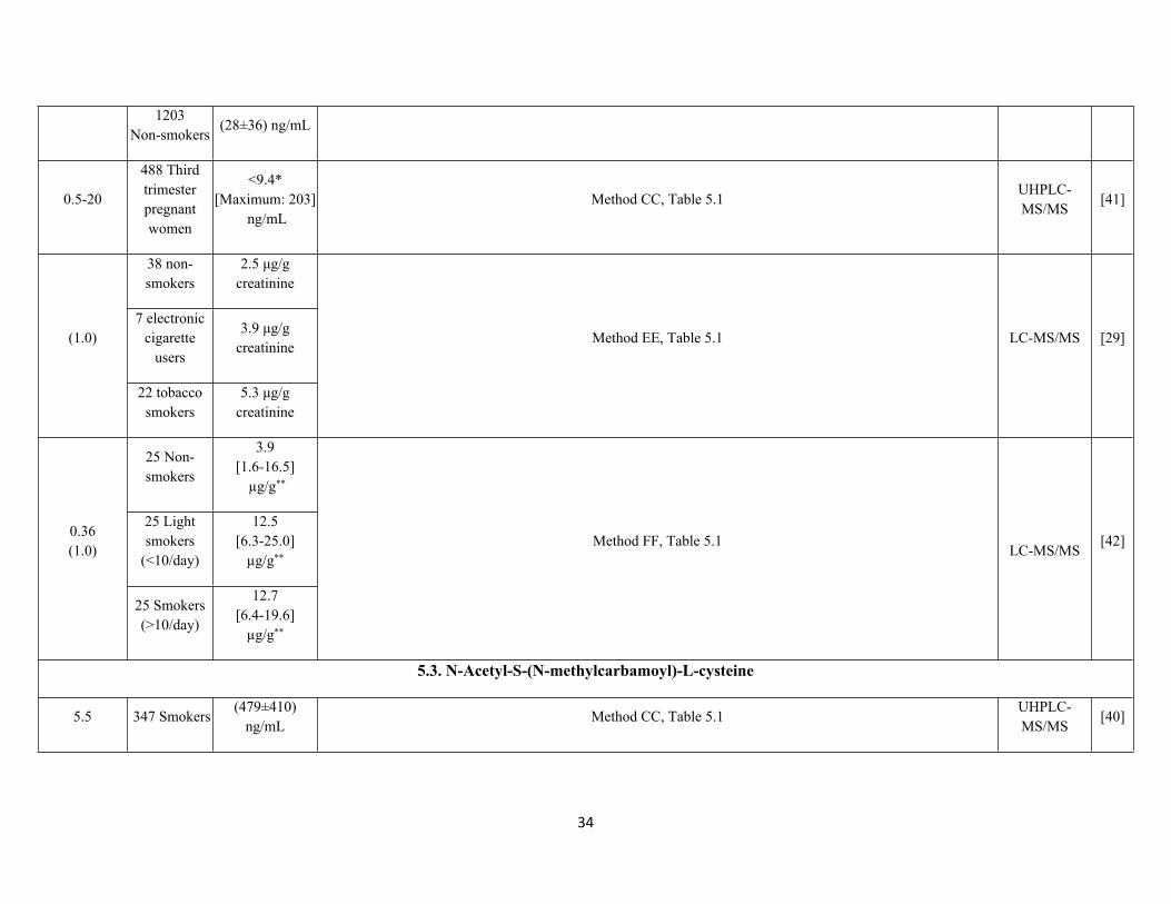
34
1203Non-smokers
(28±36) ng/mL
0.5-20
488 Third trimester pregnant women
<9.4*[Maximum: 203]
ng/mLMethod CC, Table 5.1
UHPLC-MS/MS
[41]
38 non-smokers
2.5 μg/g creatinine
7 electronic cigarette
users
3.9 μg/g creatinine
(1.0)
22 tobacco smokers
5.3 μg/g creatinine
Method EE, Table 5.1 LC-MS/MS [29]
25 Non-smokers
3.9[1.6-16.5]
µg/g**
25 Light smokers
(<10/day)
12.5 [6.3-25.0]
µg/g**
0.36(1.0)
25 Smokers (>10/day)
12.7[6.4-19.6]
µg/g**
Method FF, Table 5.1LC-MS/MS
[42]
5.3. N-Acetyl-S-(N-methylcarbamoyl)-L-cysteine
5.5 347 Smokers(479±410)
ng/mLMethod CC, Table 5.1
UHPLC-MS/MS
[40]
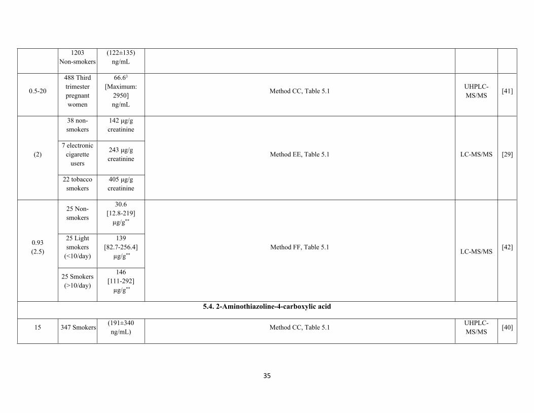
35
1203Non-smokers
(122±135) ng/mL
0.5-20
488 Third trimester pregnant women
66.6◊
[Maximum: 2950]ng/mL
Method CC, Table 5.1UHPLC-MS/MS
[41]
38 non-smokers
142 μg/g creatinine
7 electronic cigarette
users
243 μg/g creatinine
(2)
22 tobacco smokers
405 μg/g creatinine
Method EE, Table 5.1 LC-MS/MS [29]
25 Non-smokers
30.6[12.8-219]
µg/g**
25 Light smokers
(<10/day)
139[82.7-256.4]
µg/g**
0.93 (2.5)
25 Smokers (>10/day)
146[111-292]
µg/g**
Method FF, Table 5.1LC-MS/MS
[42]
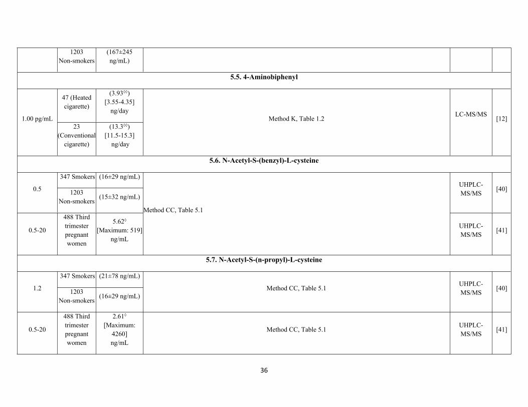
5.4. 2-Aminothiazoline-4-carboxylic acid
15 347 Smokers(191±340 ng/mL)
Method CC, Table 5.1UHPLC-MS/MS
[40]
36
1203Non-smokers
(167±245 ng/mL)
5.5. 4-Aminobiphenyl
47 (Heated cigarette)
(3.93◊◊)[3.55-4.35]
ng/day1.00 pg/mL
23 (Conventional
cigarette)
(13.3◊◊)[11.5-15.3]
ng/day
Method K, Table 1.2LC-MS/MS
[12]
5.6. N-Acetyl-S-(benzyl)-L-cysteine
347 Smokers (16±29 ng/mL)
0.5 1203Non-smokers
(15±32 ng/mL)
UHPLC-MS/MS
[40]
0.5-20
488 Third trimester pregnant women
5.62◊
[Maximum: 519]ng/mL
Method CC, Table 5.1
UHPLC-MS/MS
[41]
5.7. N-Acetyl-S-(n-propyl)-L-cysteine
347 Smokers (21±78 ng/mL)
1.2 1203Non-smokers
(16±29 ng/mL)Method CC, Table 5.1
UHPLC-MS/MS
[40]
0.5-20
488 Third trimester pregnant women
2.61◊
[Maximum: 4260]ng/mL
Method CC, Table 5.1UHPLC-MS/MS
[41]

37
5.8. N-Acetyl-S-(3-hydroxypropyl)-L-cysteine
347 Smokers(1546±1643
ng/mL)1.3 ng/mL
1203Non-smokers
(406±487 ng/mL)
Method CC, Table 5.1UHPLC-MS/MS
[40]
Tobacco cigarette:
(937 ± 700)[433-1118*]
µg/g** [1.0 ng/mL] 20 smokers
Electronic cigarette:
(492 ± 455)[162-680*]
µg/g**
Method C, Table 1.1 LC-MS/MS [3]
Water pipe:(152.6)
[337.6-490.2] µg/24h
(1 ng/mL) 13Cigarette:
(388.6)[425.3-814]
µg/24h
Method DD, Table 5.1 LC-MS/MS [5]
(20 pmol/mL) 81(6.60□)
[5.80–7.48□] nmol/mg**
Method GGUrine samples (0.4 mL) were placed in 96-well plates and internal standards added. After vortexing and heating to 50 oC, sample clean-up was performed using a pre-conditioned (0.7 mL methanol and 0.7 mL 2% NH4OH) solid-phase extraction 96-well plate. This was washed with 0.7 mL 2% NH4OH and 0.7 mL methanol and dried. After washing with 0.7 mL of 2% aqueous formic acid, the eluants were collected using 0.7 mL of 30% methanol in 2% aqueous formic acid.
LC/MS-MS [43]
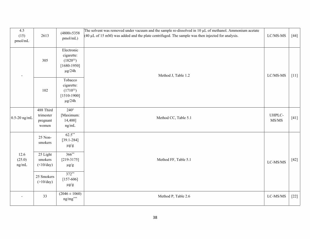
38
4.5 (15)
pmol/mL2613
(4800±5358 pmol/mL)
The solvent was removed under vacuum and the sample re-dissolved in 10 µL of methanol. Ammonium acetate (40 µL of 15 mM) was added and the plate centrifuged. The sample was then injected for analysis. LC/MS-MS [44]
305
Electronic cigarette: (1820◊◊)
[1680-1950]µg/24h
-
102
Tobacco cigarette:(1710◊◊)
[1510-1900]µg/24h
Method J, Table 1.2 LC/MS-MS [11]
0.5-20 ng/mL
488 Third trimester pregnant women
240◊
[Maximum: 14,400]ng/mL
Method CC, Table 5.1UHPLC-MS/MS
[41]
25 Non-smokers
62.5** [39.1-284]
µg/g
25 Light smokers
(<10/day)
366** [219-3175]
µg/g
12.6(25.0)ng/mL
25 Smokers (>10/day)
372** [157-606]
µg/g
Method FF, Table 5.1LC-MS/MS
[42]
- 33(2046 ± 1060)
ng/mg*** Method P, Table 2.6 LC-MS/MS [22]
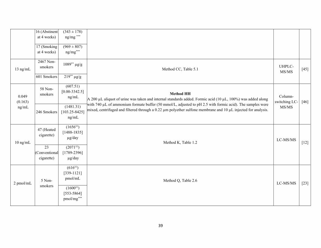
39
16 (Abstinent at 4 weeks)
(343 ± 178)ng/mg ***
17 (Smoking at 4 weeks)
(969 ± 807) ng/mg***
2467 Non-smokers
1089** µg/g13 ng/mL
601 Smokers 219** µg/g
Method CC, Table 5.1UHPLC-MS/MS
[45]
58 Non-smokers
(607.51)[0.00-3342.5]
ng/mL0.049(0.163) ng/mL
246 Smokers(1481.31)
[103.25-8425] ng/mL
Method HHA 200 µL aliquot of urine was taken and internal standards added. Formic acid (10 µL, 100%) was added along with 740 µL of ammonium formate buffer (50 mmol/L, adjusted to pH 2.5 with formic acid). The samples were mixed, centrifuged and filtered through a 0.22 µm polyether sulfone membrane and 10 µL injected for analysis.
Column-switching LC-
MS/MS[46]
47 (Heated cigarette)
(1656◊◊) [1488-1835]
µg/day10 ng/mL
23 (Conventional
cigarette)
(2071◊◊)[1789-2396]
µg/day
Method K, Table 1.2LC-MS/MS
[12]
(616◊◊) [339-1121]pmol/mL
2 pmol/mL5 Non-
smokers (1600◊◊) [553-5864]pmol/mg***
Method Q, Table 2.6LC-MS/MS [23]
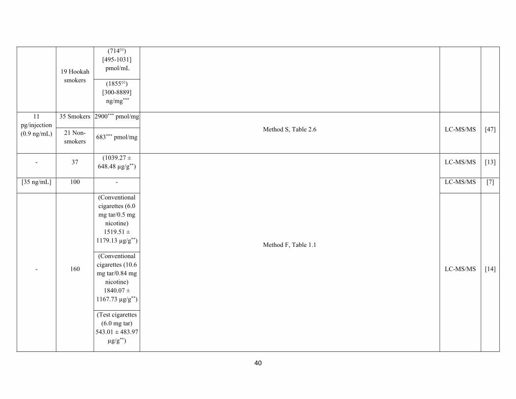
40
(714◊◊)[495-1031] pmol/mL19 Hookah
smokers (1855◊◊)[300-8889] ng/mg***
35 Smokers 2900*** pmol/mg11 pg/injection(0.9 ng/mL) 21 Non-
smokers683*** pmol/mg
Method S, Table 2.6 LC-MS/MS [47]
- 37(1039.27 ±
648.48 µg/g**)LC-MS/MS [13]
[35 ng/mL] 100 - LC-MS/MS [7]
(Conventional cigarettes (6.0 mg tar/0.5 mg
nicotine)1519.51 ±
1179.13 µg/g**)
(Conventional cigarettes (10.6 mg tar/0.84 mg
nicotine)1840.07 ±
1167.73 µg/g**)
- 160
(Test cigarettes (6.0 mg tar)
543.01 ± 483.97 µg/g**)
Method F, Table 1.1
LC-MS/MS [14]
41
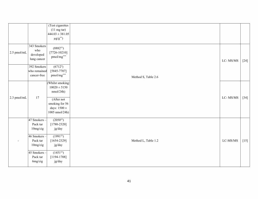
(Test cigarettes (11 mg tar)
444.03 ± 381.05 µg/g**)
2.5 pmol/mL
343 Smokers who
developed lung cancer
(8882◊◊)[7726-10210]
pmol/mg***
392 Smokers who remained
cancer-free
(6712◊)[5845-7707]pmol/mg***
LC- MS/MS [24]
(Whilst smoking: 10020 ± 5150
nmol/24h)
2.3 pmol/mL 17 (After not smoking for 56 days: 1500 ±
1005 nmol/24h)
Method S, Table 2.6
LC- MS/MS [34]
47 Smokers – Pack tar
10mg/cig
(2050◊◊) [1780-2320]
jg/day
46 Smokers – Pack tar
10mg/cig
(1991◊◊)[1654-2329]
jg/day-
45 Smokers – Pack tar 6mg/cig
(1451◊◊)[1194-1708]
jg/day
Method L, Table 1.2 LC-MS/MS [15]
42
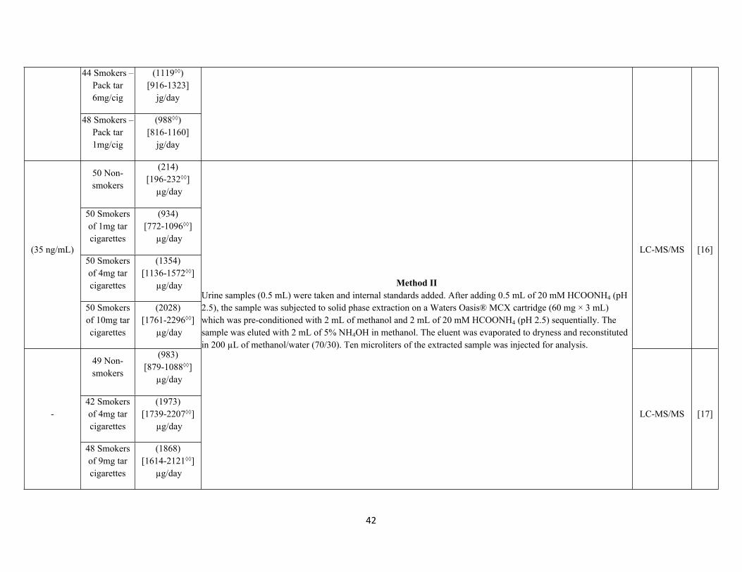
44 Smokers – Pack tar 6mg/cig
(1119◊◊)[916-1323]
jg/day
48 Smokers – Pack tar 1mg/cig
(988◊◊) [816-1160]
jg/day
50 Non-smokers
(214)[196-232◊◊]
µg/day
50 Smokers of 1mg tar cigarettes
(934)[772-1096◊◊]
µg/day
50 Smokers of 4mg tar cigarettes
(1354)[1136-1572◊◊]
µg/day
(35 ng/mL)
50 Smokers of 10mg tar cigarettes
(2028)[1761-2296◊◊]
µg/day
LC-MS/MS [16]
49 Non-smokers
(983)[879-1088◊◊]
µg/day
42 Smokers of 4mg tar cigarettes
(1973)[1739-2207◊◊]
µg/day-
48 Smokers of 9mg tar cigarettes
(1868)[1614-2121◊◊]
µg/day
Method IIUrine samples (0.5 mL) were taken and internal standards added. After adding 0.5 mL of 20 mM HCOONH4 (pH 2.5), the sample was subjected to solid phase extraction on a Waters Oasis® MCX cartridge (60 mg × 3 mL) which was pre-conditioned with 2 mL of methanol and 2 mL of 20 mM HCOONH4 (pH 2.5) sequentially. The sample was eluted with 2 mL of 5% NH4OH in methanol. The eluent was evaporated to dryness and reconstituted in 200 µL of methanol/water (70/30). Ten microliters of the extracted sample was injected for analysis.
LC-MS/MS [17]
43
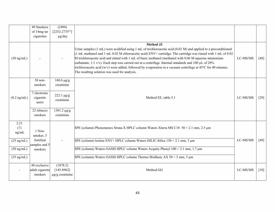
49 Smokers of 14mg tar cigarettes
(2494)[2252-2735◊◊]
µg/day
(50 ng/mL) - -
Method JJUrine samples (1 mL) were acidified using 1 mL of trichloroacetic acid (0.02 M) and applied to a preconditioned (1 mL methanol and 1 mL 0.02 M chloroacetic acid) ENV+ cartridge. The cartridge was rinsed with 1 mL of 0.02 M trichloroacetic acid and eluted with 1 mL of basic methanol (methanol with 0.06 M aqueous ammonium carbamate, 1:1 v/v). Each step was carried out in a centrifuge. Internal standards and 100 µL of 20% trichloroacetic acid (w/v) were added, followed by evaporation in a vacuum centrifuge at 45oC for 40 minutes. The resulting solution was used for analysis.
LC-MS/MS [48]
38 non-smokers
160.6 μg/g creatinine
7 electronic cigarette
users
222.1 μg/g creatinine
(0.2 ng/mL)
22 tobacco smokers
1301.2 μg/g creatinine
Method EE, table 5.1 LC-MS/MS [29]
2.21(7)
ng/mLSPE (column) Phenomenex Strata-X HPLC column Waters Xterra MS C18 50 × 2.1 mm, 2.5 μm
(25 ng/mL) SPE (column) Isolute ENV+ HPLC column Waters HILIC-Silica 150 × 2.1 mm, 3 μm
(50 ng/mL) SPE (column) Waters OASIS HPLC column Waters Acquity Phenyl 100 × 2.1 mm, 1.7 μm
(35 ng/mL)
1 Non-smoker, 3 fortified
samples and 5 smokers
-
SPE (column) Waters OASIS HPLC column Thermo BioBasic AX 50 × 3 mm, 5 μm
LC-MS/MS [49]
-48 exclusive
adult cigarette smokers
(1878.2)[145-8962]
µg/g creatinineMethod GG LC-MS/MS [10]
44
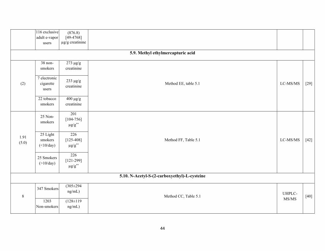
116 exclusive adult e-vapor
users
(876.8)[49-4768]
µg/g creatinine
5.9. Methyl ethylmercapturic acid
38 non-smokers
273 μg/g creatinine
7 electronic cigarette
users
233 μg/g creatinine
(2)
22 tobacco smokers
400 μg/g creatinine
Method EE, table 5.1 LC-MS/MS [29]
25 Non-smokers
201 [104-756]
µg/g**
25 Light smokers
(<10/day)
226[125-408]
µg/g**
1.91(5.0)
25 Smokers (>10/day)
226 [121-299]
µg/g**
Method FF, Table 5.1 LC-MS/MS [42]
5.10. N-Acetyl-S-(2-carboxyethyl)-L-cysteine
347 Smokers(305±294 ng/mL)
81203
Non-smokers(128±119 ng/mL)
Method CC, Table 5.1UHPLC-MS/MS
[40]
45
0.5-20
488 Third trimester pregnant women
71.8◊ [Maximum:
2260]ng/mL
UHPLC-MS/MS
[41]
38 non-smokers
0.9 μg/g creatinine
7 electronic cigarette
users
2.7 μg/g creatinine
(0.9)
22 tobacco smokers
163.1 μg/g creatinine
Method EE, Table 5.1 LC-MS/MS [29]
25 Non-smokers
0.46 [0.23-8.6]
µg/g**
25 Light smokers
(<10/day)
53.6[33.6-138.4]
µg/g**
0.08(0.25)
25 Smokers (>10/day)
72.5 [28.6-140.4]
µg/g**
Method FF, Table 5.1LC-MS/MS
[42]
2467 Non-smokers
78.8 µg/g**
8
601 Smokers 203 µg/g**
Method CC, Table 5.1UHPLC-MS/MS
[45]
0.015(0.05)
58 Non-smokers
(3.47)[0.00-12.4]
ng/mLMethod HH, Table 5.8
Column-switching LC-
MS/MS[46]
46
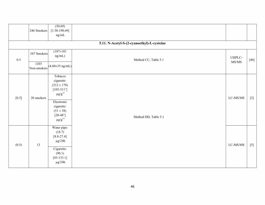
246 Smokers(50.69)
[1.58-198.69] ng/mL
5.11. N-Acetyl-S-(2-cyanoethyl)-L-cysteine
347 Smokers(187±181 ng/mL)
0.51203
Non-smokers(4.60±35 ng/mL)
Method CC, Table 5.1UHPLC-MS/MS
[40]
Tobacco cigarette:
(212 ± 178)[103-311*]
µg/g**
[0.5] 20 smokersElectronic cigarette:(51 ± 58)[20-48*] µg/g**
LC-MS/MS [3]
Water pipe:(18.7)
[8.8-27.4] µg/24h
(0.5) 13Cigarette:
(90.1)[43-133.1]
µg/24h
Method DD, Table 5.1
LC-MS/MS [5]
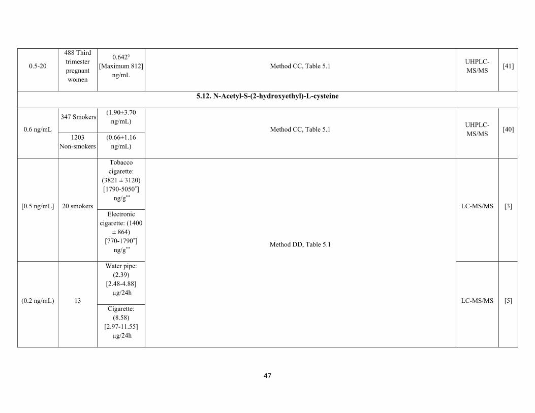
47
0.5-20
488 Third trimester pregnant women
0.642◊
[Maximum 812]ng/mL
Method CC, Table 5.1UHPLC-MS/MS
[41]
5.12. N-Acetyl-S-(2-hydroxyethyl)-L-cysteine
347 Smokers(1.90±3.70
ng/mL)0.6 ng/mL
1203Non-smokers
(0.66±1.16 ng/mL)
Method CC, Table 5.1UHPLC-MS/MS
[40]
Tobacco cigarette:
(3821 ± 3120)[1790-5050*]
ng/g**
[0.5 ng/mL] 20 smokersElectronic
cigarette: (1400 ± 864)
[770-1790*] ng/g**
LC-MS/MS [3]
Water pipe:(2.39)
[2.48-4.88] µg/24h
(0.2 ng/mL) 13Cigarette:
(8.58)[2.97-11.55]
µg/24h
Method DD, Table 5.1
LC-MS/MS [5]
48
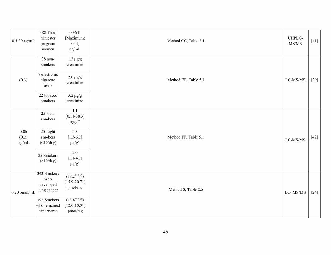
0.5-20 ng/mL
488 Third trimester pregnant women
0.963◊ [Maximum:
33.4]ng/mL
Method CC, Table 5.1UHPLC-MS/MS
[41]
38 non-smokers
1.3 μg/g creatinine
7 electronic cigarette
users
2.0 μg/g creatinine
(0.3)
22 tobacco smokers
3.2 μg/g creatinine
Method EE, Table 5.1 LC-MS/MS [29]
25 Non-smokers
1.1 [0.11-38.3]
µg/g**
25 Light smokers
(<10/day)
2.3[1.3-6.2] µg/g**
0.06(0.2)
ng/mL
25 Smokers (>10/day)
2.0[1.1-4.2] µg/g**
Method FF, Table 5.1LC-MS/MS
[42]
343 Smokers who
developed lung cancer
(18.2*** ◊◊)
[15.9-20.7a ]pmol/mg
0.20 pmol/mL
392 Smokers who remained
cancer-free
(13.6*** ◊◊)[12.0-15.5a ]
pmol/mg
Method S, Table 2.6LC- MS/MS [24]
49
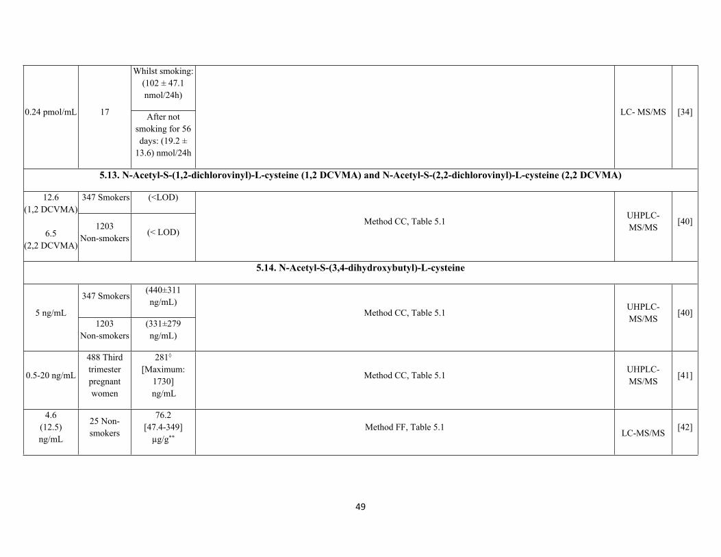
Whilst smoking: (102 ± 47.1 nmol/24h)
0.24 pmol/mL 17 After not smoking for 56 days: (19.2 ±
13.6) nmol/24h
LC- MS/MS [34]
5.13. N-Acetyl-S-(1,2-dichlorovinyl)-L-cysteine (1,2 DCVMA) and N-Acetyl-S-(2,2-dichlorovinyl)-L-cysteine (2,2 DCVMA)
347 Smokers (<LOD)12.6 (1,2 DCVMA)
6.5 (2,2 DCVMA)
1203Non-smokers
(< LOD)Method CC, Table 5.1
UHPLC-MS/MS
[40]
5.14. N-Acetyl-S-(3,4-dihydroxybutyl)-L-cysteine
347 Smokers(440±311 ng/mL)
5 ng/mL1203
Non-smokers(331±279 ng/mL)
Method CC, Table 5.1UHPLC-MS/MS
[40]
0.5-20 ng/mL
488 Third trimester pregnant women
281◊
[Maximum: 1730]
ng/mL
Method CC, Table 5.1UHPLC-MS/MS
[41]
4.6 (12.5)ng/mL
25 Non-smokers
76.2 [47.4-349]
µg/g**Method FF, Table 5.1
LC-MS/MS[42]

50
25 Light smokers
(<10/day)
112 [65.5-243]
µg/g**
25 Smokers (>10/day)
122[52.9-244]
µg/g**
58 Non-smokers
(184.61)[0.00-567.5]
ng/mL0.053 (0.177)ng/mL
246 Smokers(230.47)
[0.00-1345.0] ng/mL
Method HH, Table 5.8Column-
switching LC-MS/MS
[46]
38 non-smokers
247.5 μg/g creatinine
7 electronic cigarette
users
263.8 μg/g creatinine
(1.0)
22 tobacco smokers
479.1 μg/g creatinine
Method EE, Table 5.1 LC-MS/MS [29]
Whilst smoking: (1038 ± 514 nmol/24h)
12 pmol/mL 17 After not smoking for 56
days: (662 ± 248 nmol/24h)
Method S, Table 2.6 LC-MS/MS [34]
51
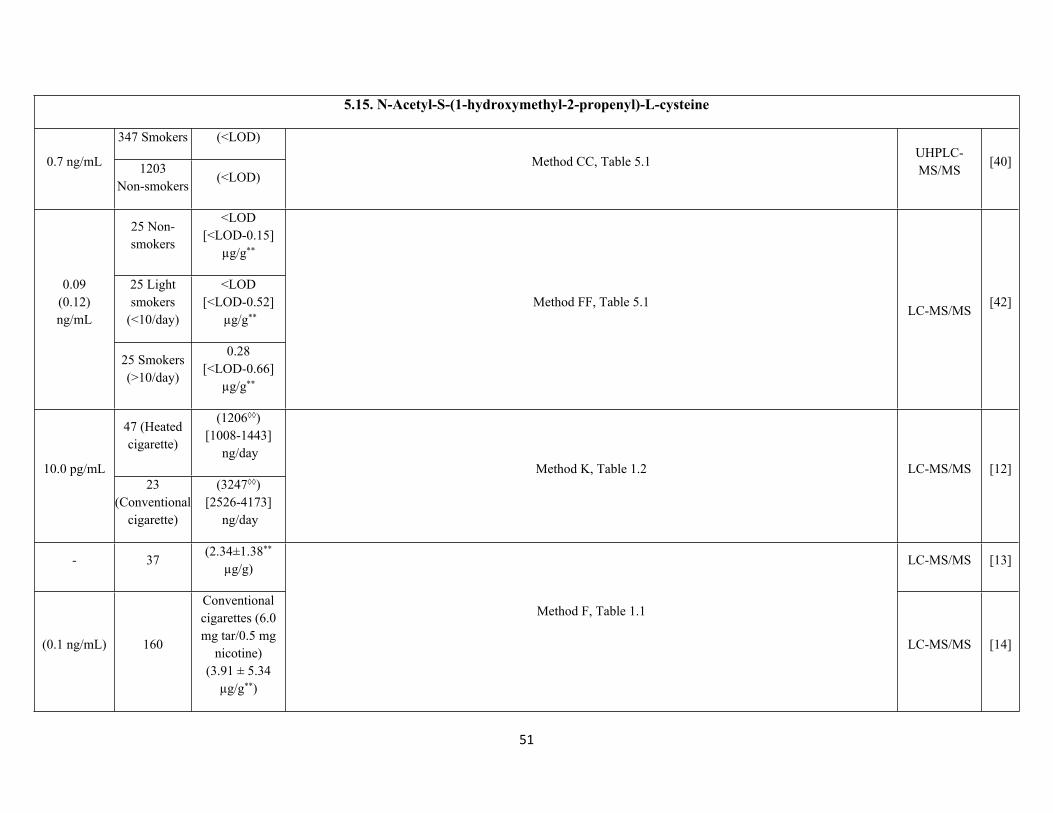
5.15. N-Acetyl-S-(1-hydroxymethyl-2-propenyl)-L-cysteine
347 Smokers (<LOD)
0.7 ng/mL 1203Non-smokers
(<LOD)Method CC, Table 5.1
UHPLC-MS/MS
[40]
25 Non-smokers
<LOD [<LOD-0.15]
µg/g**
25 Light smokers
(<10/day)
<LOD[<LOD-0.52]
µg/g**
0.09 (0.12)ng/mL
25 Smokers (>10/day)
0.28 [<LOD-0.66]
µg/g**
Method FF, Table 5.1LC-MS/MS
[42]
47 (Heated cigarette)
(1206◊◊)[1008-1443]
ng/day10.0 pg/mL
23 (Conventional
cigarette)
(3247◊◊)[2526-4173]
ng/day
Method K, Table 1.2 LC-MS/MS [12]
- 37(2.34±1.38**
µg/g)LC-MS/MS [13]
(0.1 ng/mL) 160
Conventional cigarettes (6.0 mg tar/0.5 mg
nicotine)(3.91 ± 5.34
µg/g**)
Method F, Table 1.1
LC-MS/MS [14]
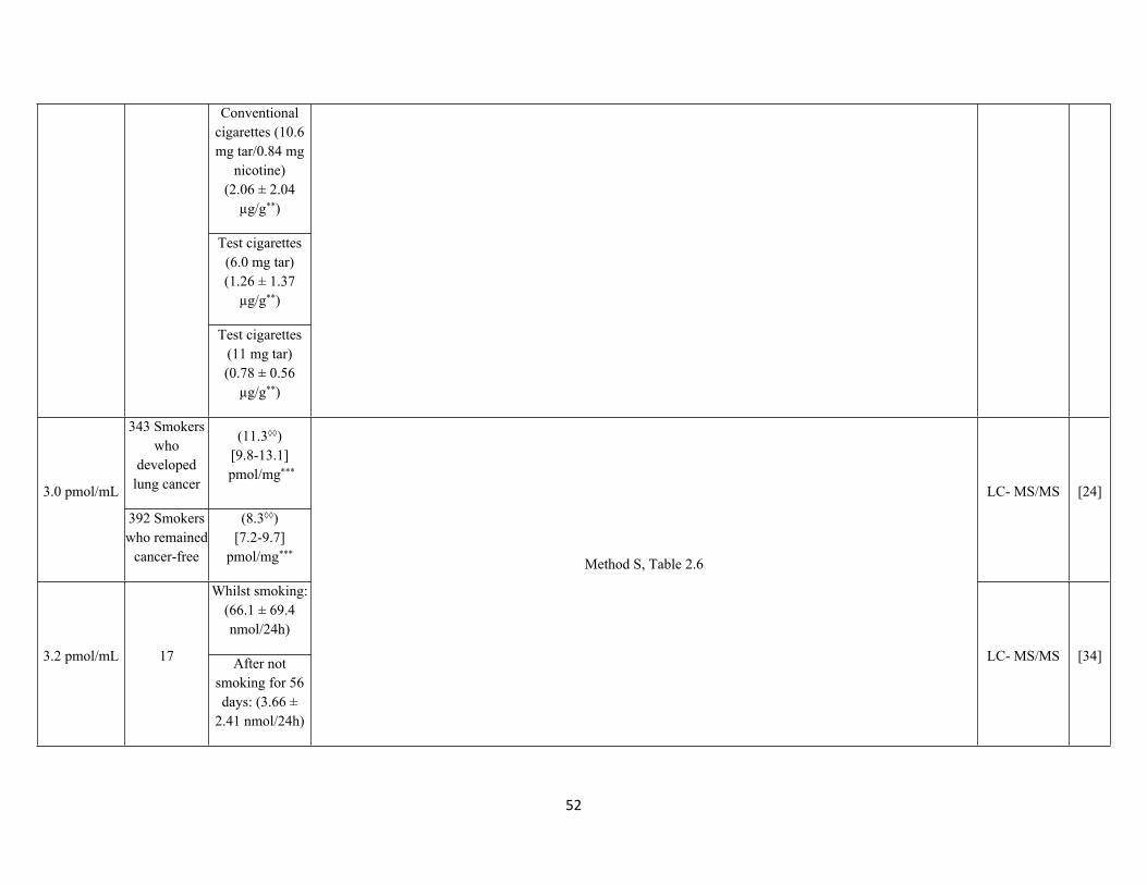
52
Conventional cigarettes (10.6 mg tar/0.84 mg
nicotine)(2.06 ± 2.04
µg/g**)
Test cigarettes (6.0 mg tar)(1.26 ± 1.37
µg/g**)
Test cigarettes (11 mg tar)(0.78 ± 0.56
µg/g**)
343 Smokers who
developed lung cancer
(11.3◊◊)[9.8-13.1]
pmol/mg***
3.0 pmol/mL
392 Smokers who remained
cancer-free
(8.3◊◊)[7.2-9.7]
pmol/mg***
LC- MS/MS [24]
Whilst smoking: (66.1 ± 69.4 nmol/24h)
3.2 pmol/mL 17 After not smoking for 56 days: (3.66 ±
2.41 nmol/24h)
Method S, Table 2.6
LC- MS/MS [34]
53
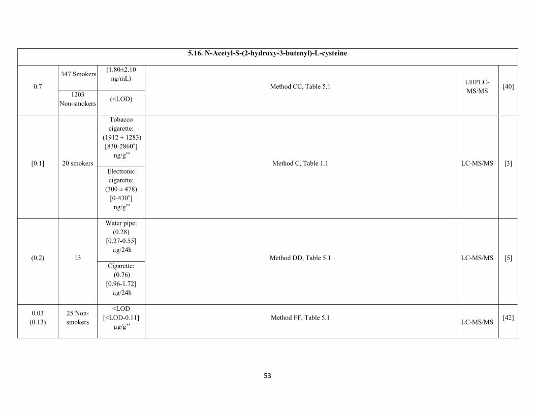
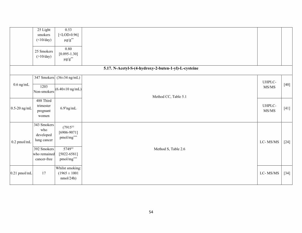
5.16. N-Acetyl-S-(2-hydroxy-3-butenyl)-L-cysteine
347 Smokers(1.80±2.10
ng/mL)0.7
1203Non-smokers
(<LOD)
Method CC, Table 5.1UHPLC-MS/MS
[40]
Tobacco cigarette:
(1912 ± 1283)[830-2860*]
ng/g**
[0.1] 20 smokersElectronic cigarette:
(300 ± 478)[0-430*] ng/g**
Method C, Table 1.1 LC-MS/MS [3]
Water pipe:(0.28)
[0.27-0.55] µg/24h
(0.2) 13Cigarette:
(0.76)[0.96-1.72]
µg/24h
Method DD, Table 5.1 LC-MS/MS [5]
0.03(0.13)
25 Non-smokers
<LOD[<LOD-0.11]
µg/g**Method FF, Table 5.1
LC-MS/MS[42]
54
25 Light smokers
(<10/day)
0.53[<LOD-0.96]
µg/g**
25 Smokers (>10/day)
0.80[0.095-1.30]
µg/g**
5.17. N-Acetyl-S-(4-hydroxy-2-buten-1-yl)-L-cysteine
347 Smokers (36±34 ng/mL)
0.6 ng/mL 1203Non-smokers
(6.40±10 ng/mL)
UHPLC-MS/MS
[40]
0.5-20 ng/mL
488 Third trimester pregnant women
6.9◊ng/mL
Method CC, Table 5.1
UHPLC-MS/MS
[41]
343 Smokers who
developed lung cancer
(7915◊◊
[6906-9071]pmol/mg***
0.2 pmol/mL
392 Smokers who remained
cancer-free
5749◊◊
[5022-6581]pmol/mg***
LC- MS/MS [24]
0.21 pmol/mL 17Whilst smoking:
(1965 ± 1001 nmol/24h)
Method S, Table 2.6
LC- MS/MS [34]
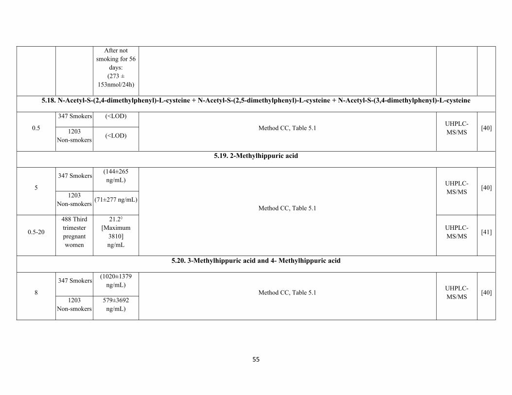
55
After not smoking for 56
days: (273 ±
153nmol/24h)
5.18. N-Acetyl-S-(2,4-dimethylphenyl)-L-cysteine + N-Acetyl-S-(2,5-dimethylphenyl)-L-cysteine + N-Acetyl-S-(3,4-dimethylphenyl)-L-cysteine
347 Smokers (<LOD)
0.5 1203Non-smokers
(<LOD)Method CC, Table 5.1
UHPLC-MS/MS
[40]
5.19. 2-Methylhippuric acid
347 Smokers(144±265 ng/mL)
51203
Non-smokers(71±277 ng/mL)
UHPLC-MS/MS
[40]
0.5-20
488 Third trimester pregnant women
21.2◊
[Maximum 3810]ng/mL
Method CC, Table 5.1
UHPLC-MS/MS
[41]
5.20. 3-Methylhippuric acid and 4- Methylhippuric acid
347 Smokers(1020±1379
ng/mL)8
1203Non-smokers
579±3692 ng/mL)
Method CC, Table 5.1UHPLC-MS/MS
[40]
56
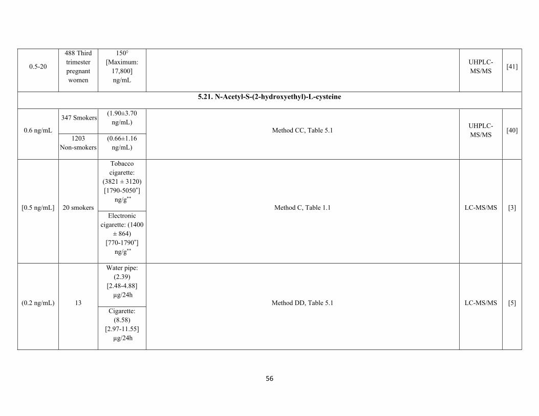
0.5-20
488 Third trimester pregnant women
150◊
[Maximum: 17,800]ng/mL
UHPLC-MS/MS
[41]
5.21. N-Acetyl-S-(2-hydroxyethyl)-L-cysteine
347 Smokers(1.90±3.70
ng/mL)0.6 ng/mL
1203Non-smokers
(0.66±1.16 ng/mL)
Method CC, Table 5.1UHPLC-MS/MS
[40]
Tobacco cigarette:
(3821 ± 3120)[1790-5050*]
ng/g**
[0.5 ng/mL] 20 smokersElectronic
cigarette: (1400 ± 864)
[770-1790*] ng/g**
Method C, Table 1.1 LC-MS/MS [3]
Water pipe:(2.39)
[2.48-4.88] µg/24h
(0.2 ng/mL) 13Cigarette:
(8.58)[2.97-11.55]
µg/24h
Method DD, Table 5.1 LC-MS/MS [5]
57
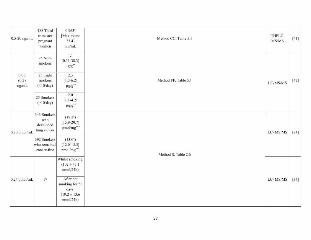
0.5-20 ng/mL
488 Third trimester pregnant women
0.963◊
[Maximum: 33.4]
nm/mL
Method CC, Table 5.1UHPLC-MS/MS
[41]
25 Non-smokers
1.1 [0.11-38.3]
µg/g**
25 Light smokers
(<10/day)
2.3[1.3-6.2] µg/g**
0.06(0.2)
ng/mL
25 Smokers (>10/day)
2.0[1.1-4.2] µg/g**
Method FF, Table 5.1LC-MS/MS
[42]
343 Smokers who
developed lung cancer
(18.2◊)[15.9-20.7] pmol/mg***
0.20 pmol/mL
392 Smokers who remained
cancer-free
(13.6◊)[12.0-15.5]pmol/mg***
LC- MS/MS [24]
Whilst smoking: (102 ± 47.1 nmol/24h)
0.24 pmol/mL 17 After not smoking for 56
days: (19.2 ± 13.6 nmol/24h)
Method S, Table 2.6
LC- MS/MS [34]
58
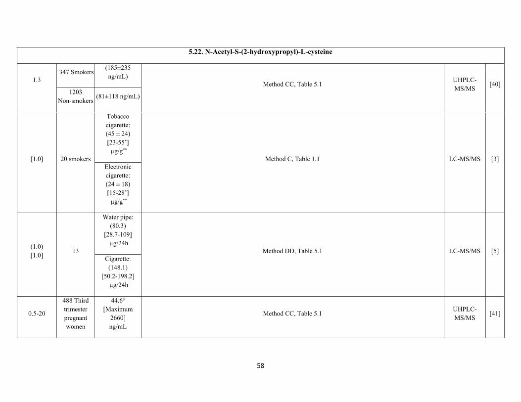
5.22. N-Acetyl-S-(2-hydroxypropyl)-L-cysteine
347 Smokers(185±235 ng/mL)1.3
1203Non-smokers
(81±118 ng/mL)
Method CC, Table 5.1UHPLC-MS/MS
[40]
Tobacco cigarette:(45 ± 24)[23-55*] µg/g**
[1.0] 20 smokersElectronic cigarette:(24 ± 18)[15-28*] µg/g**
Method C, Table 1.1 LC-MS/MS [3]
Water pipe:(80.3)
[28.7-109] µg/24h(1.0)
[1.0]13
Cigarette:(148.1)
[50.2-198.2] µg/24h
Method DD, Table 5.1 LC-MS/MS [5]
0.5-20
488 Third trimester pregnant women
44.6◊
[Maximum 2660]ng/mL
Method CC, Table 5.1UHPLC-MS/MS
[41]
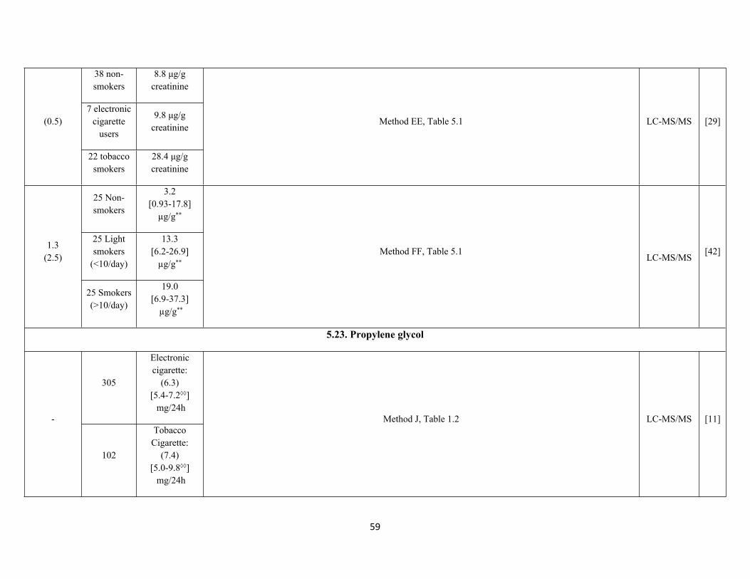
59
38 non-smokers
8.8 μg/g creatinine
7 electronic cigarette
users
9.8 μg/g creatinine
(0.5)
22 tobacco smokers
28.4 μg/g creatinine
Method EE, Table 5.1 LC-MS/MS [29]
25 Non-smokers
3.2[0.93-17.8]
µg/g**
25 Light smokers
(<10/day)
13.3 [6.2-26.9]
µg/g**
1.3(2.5)
25 Smokers (>10/day)
19.0[6.9-37.3]
µg/g**
Method FF, Table 5.1LC-MS/MS
[42]
5.23. Propylene glycol
305
Electronic cigarette:
(6.3)[5.4-7.2◊◊] mg/24h
-
102
Tobacco Cigarette:
(7.4)[5.0-9.8◊◊] mg/24h
Method J, Table 1.2 LC-MS/MS [11]
60
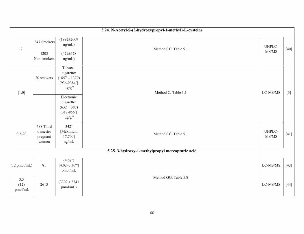
5.24. N-Acetyl-S-(3-hydroxypropyl-1-methyl)-L-cysteine
347 Smokers(1992±2009
ng/mL)2
1203Non-smokers
(429±478 ng/mL)
Method CC, Table 5.1UHPLC-MS/MS
[40]
20 smokers
Tobacco cigarette:
(1857 ± 1379)[936-2384*]
µg/g**
[1.0]Electronic cigarette:
(632 ± 387)[312-856*]
µg/g**
Method C, Table 1.1 LC-MS/MS [3]
0.5-20
488 Third trimester pregnant women
342◊
[Maximum 17,700]ng/mL
Method CC, Table 5.1UHPLC-MS/MS
[41]
5.25. 3-hydroxy-1-methylpropyl mercapturic acid
(12 pmol/mL) 81(4.62□)
[4.02–5.30◊◊]pmol/mL
LC-MS/MS [43]
3.5(12)
pmol/mL2613
(3302 ± 3341 pmol/mL)
Method GG, Table 5.8
LC-MS/MS [44]
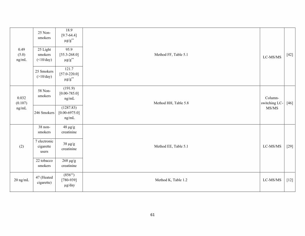
61
25 Non-smokers
18.9 [9.7-64.4]
µg/g**
25 Light smokers
(<10/day)
95.9 [55.3-268.0]
µg/g**
0.49 (5.0)
ng/mL
25 Smokers (>10/day)
121.7 [57.0-220.0]
µg/g**
Method FF, Table 5.1LC-MS/MS
[42]
58 Non-smokers
(191.9)[0.00-785.0]
ng/mL0.032 (0.107)ng/mL
246 Smokers(1287.83)
[0.00-6975.0] ng/mL
Method HH, Table 5.8Column-
switching LC-MS/MS
[46]
38 non-smokers
48 μg/g creatinine
7 electronic cigarette
users
38 μg/g creatinine
(2)
22 tobacco smokers
268 μg/g creatinine
Method EE, Table 5.1 LC-MS/MS [29]
20 ng/mL 47 (Heated cigarette)
(856◊◊)[780-939] µg/day
Method K, Table 1.2 LC-MS/MS [12]
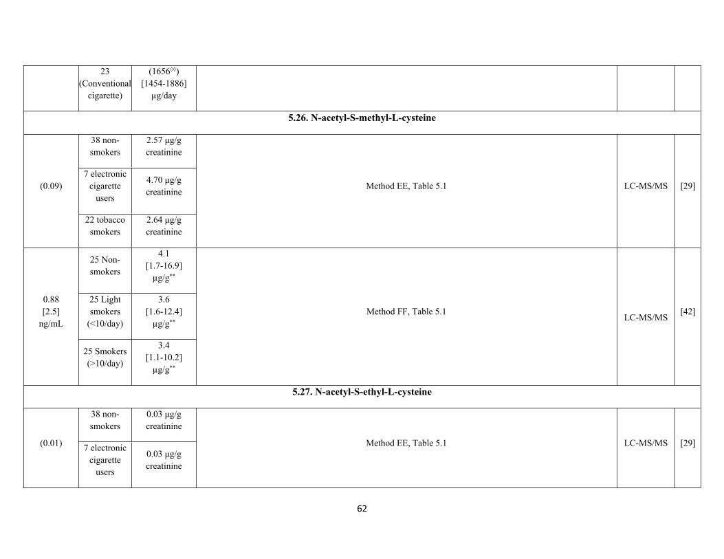
62
23 (Conventional
cigarette)
(1656◊◊)[1454-1886]
µg/day
5.26. N-acetyl-S-methyl-L-cysteine
38 non-smokers
2.57 μg/g creatinine
7 electronic cigarette
users
4.70 μg/g creatinine
(0.09)
22 tobacco smokers
2.64 μg/g creatinine
Method EE, Table 5.1 LC-MS/MS [29]
25 Non-smokers
4.1 [1.7-16.9]
µg/g**
25 Light smokers
(<10/day)
3.6[1.6-12.4]
µg/g**
0.88[2.5]
ng/mL
25 Smokers (>10/day)
3.4 [1.1-10.2]
µg/g**
Method FF, Table 5.1LC-MS/MS
[42]
5.27. N-acetyl-S-ethyl-L-cysteine
38 non-smokers
0.03 μg/g creatinine
(0.01) 7 electronic cigarette
users
0.03 μg/g creatinine
Method EE, Table 5.1 LC-MS/MS [29]
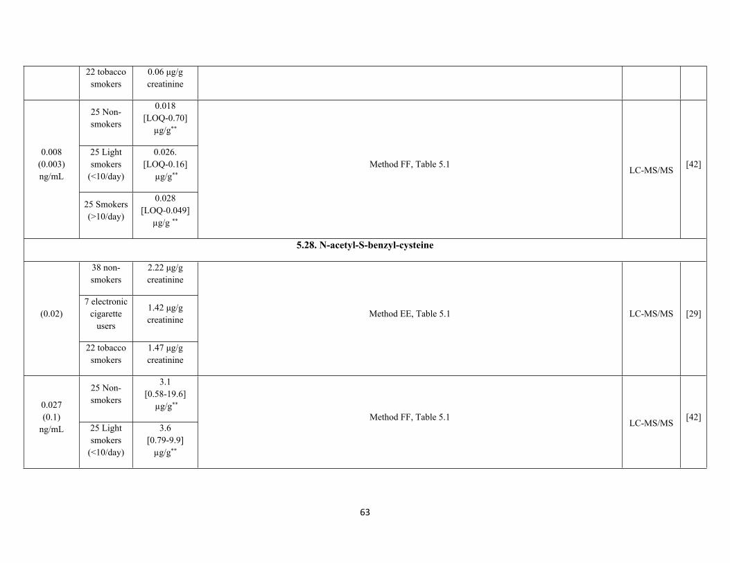
63
22 tobacco smokers
0.06 μg/g creatinine
25 Non-smokers
0.018 [LOQ-0.70]
µg/g**
25 Light smokers
(<10/day)
0.026.[LOQ-0.16]
µg/g**
0.008(0.003)ng/mL
25 Smokers (>10/day)
0.028[LOQ-0.049]
µg/g **
Method FF, Table 5.1LC-MS/MS
[42]
5.28. N-acetyl-S-benzyl-cysteine
38 non-smokers
2.22 μg/g creatinine
7 electronic cigarette
users
1.42 μg/g creatinine
(0.02)
22 tobacco smokers
1.47 μg/g creatinine
Method EE, Table 5.1 LC-MS/MS [29]
25 Non-smokers
3.1[0.58-19.6]
µg/g**0.027(0.1)
ng/mL 25 Light smokers
(<10/day)
3.6 [0.79-9.9]
µg/g**
Method FF, Table 5.1LC-MS/MS
[42]
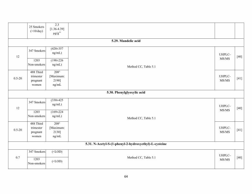
64
25 Smokers (>10/day)
2.3 [1.36-4.39]
µg/g**
5.29. Mandelic acid
347 Smokers(420±357 ng/mL)
121203
Non-smokers(198±226 ng/mL)
UHPLC-MS/MS
[40]
0.5-20
488 Third trimester pregnant women
208◊
[Maximum: 2190] ng/mL
Method CC, Table 5.1
UHPLC-MS/MS
[41]
5.30. Phenylglyoxylic acid
347 Smokers(330±425 ng/mL)
121203
Non-smokers(169±224 ng/mL)
UHPLC-MS/MS
[40]
0.5-20
488 Third trimester pregnant women
208◊
[Maximum: 2130]ng/mL
Method CC, Table 5.1
UHPLC-MS/MS
[41]
5.31. N-Acetyl-S-(1-phenyl-2-hydroxyethyl)-L-cysteine
347 Smokers (<LOD)
0.7 1203Non-smokers
(<LOD)Method CC, Table 5.1
UHPLC-MS/MS
[40]

65
0.5-20
488 Third trimester pregnant women
<LOD[Maximum: 9.84
ng/mL]
UHPLC-MS/MS
[41]
25 Non-smokers
<LOD[LOQ-0.11
µg/g**]
25 Light smokers
(<10/day)
<LOD[LOQ-0.38
µg/g**]
0.03(0.1)
(PHEMA 1)
25 Smokers (>10/day)
0.41[LOQ-0.98]
µg/g**
LC-MS/MS[42]
25 Non-smokers
<LOD [LOQ-0.71
µg/g**]
25 Light smokers
(<10/day)
<LOD[0.763 µg/g**]
0.13(0.4)
(PHEMA 2)
25 Smokers (>10/day)
0.42 [LOQ-0.66]
µg/g**
Method FF, Table 5.1
LC-MS/MS[42]
5.32. trans, trans-Muconic acid
347 Smokers(473±410 ng/mL)
201203
Non-smokers(358±291 ng/mL)
Method CC, Table 5.1UHPLC-MS/MS
[40]
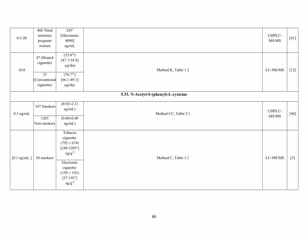
66
0.5-20
488 Third trimester pregnant women
245◊
[Maximum: 4090] ng/mL
UHPLC-MS/MS
[41]
47 (Heated cigarette)
(53.0◊◊)[47.7-58.9]
µg/day10.0
23 (Conventional
cigarette)
(76.7◊◊)[66.1-89.1]
µg/day
Method K, Table 1.2 LC-MS/MS [12]
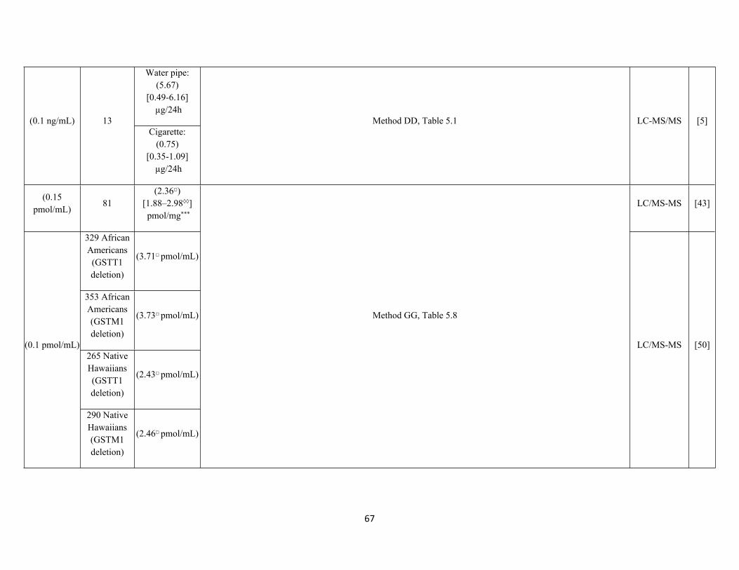
5.33. N-Acetyl-S-(phenyl)-L-cysteine
347 Smokers(0.92±2.11
ng/mL)0.3 ng/mL
1203Non-smokers
(0.60±0.40 ng/mL)
Method CC, Table 5.1UHPLC-MS/MS
[40]
Tobacco cigarette:
(792 ± 674)[249-1203*]
ng/g**
[0.1 ng/mL ] 20 smokersElectronic cigarette:
(159 ± 193)[37-193*]
ng/g**
Method C, Table 1.1 LC-MS/MS [3]
67
Water pipe:(5.67)
[0.49-6.16] µg/24h
(0.1 ng/mL) 13Cigarette:
(0.75)[0.35-1.09]
µg/24h
Method DD, Table 5.1 LC-MS/MS [5]
(0.15 pmol/mL)
81(2.36□)
[1.88–2.98◊◊] pmol/mg***
LC/MS-MS [43]
329 African Americans (GSTT1 deletion)
(3.71□ pmol/mL)
353 African Americans (GSTM1 deletion)
(3.73□ pmol/mL)
265 Native Hawaiians (GSTT1 deletion)
(2.43□ pmol/mL)
(0.1 pmol/mL)
290 Native Hawaiians (GSTM1 deletion)
(2.46□ pmol/mL)
Method GG, Table 5.8
LC/MS-MS [50]
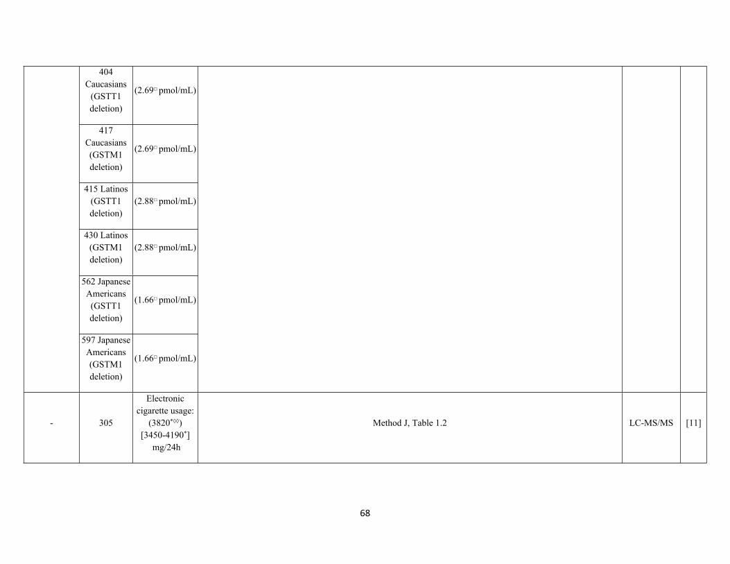
68
404 Caucasians
(GSTT1 deletion)
(2.69□ pmol/mL)
417 Caucasians (GSTM1 deletion)
(2.69□ pmol/mL)
415 Latinos (GSTT1 deletion)
(2.88□ pmol/mL)
430 Latinos (GSTM1 deletion)
(2.88□ pmol/mL)
562 Japanese Americans (GSTT1 deletion)
(1.66□ pmol/mL)
597 Japanese Americans (GSTM1 deletion)
(1.66□ pmol/mL)
- 305
Electronic cigarette usage:
(3820*◊◊)[3450-4190*]
mg/24h
Method J, Table 1.2 LC-MS/MS [11]

69
102
Regular cigarette usage:
(3660*◊◊)[3090-4220*]
mg/24h
0.5-20 ng/mL
488 Third trimester pregnant women
0.642◊
[Maximum: 12.3]
ng/mL
Method CC, Table 5.1UHPLC-MS/MS
[41]
25 Non-smokers
0.018 [LOQ-0.097]
µg/g**
25 Light smokers
(<10/day)
0.75 [0.21-1.6]
µg/g**
0.005 (0.02)ng/mL
25 Smokers (>10/day)
1.1[0.20-3.5]
µg/g**
Method FF, Table 5.1LC-MS/MS
[42]
58 Non-smokers
(0.36)[0.00-0.95]
ng/mL0.013(0.043) ng/mL
246 Smokers(0.2)
[0.00-4.17] ng/mL
Method HH, Table 5.8Column-
switching LC-MS/MS
[46]
(0.01)38 non-smokers
0.06 μg/g creatinine
Method EE, Table 5.1 LC-MS/MS [29]
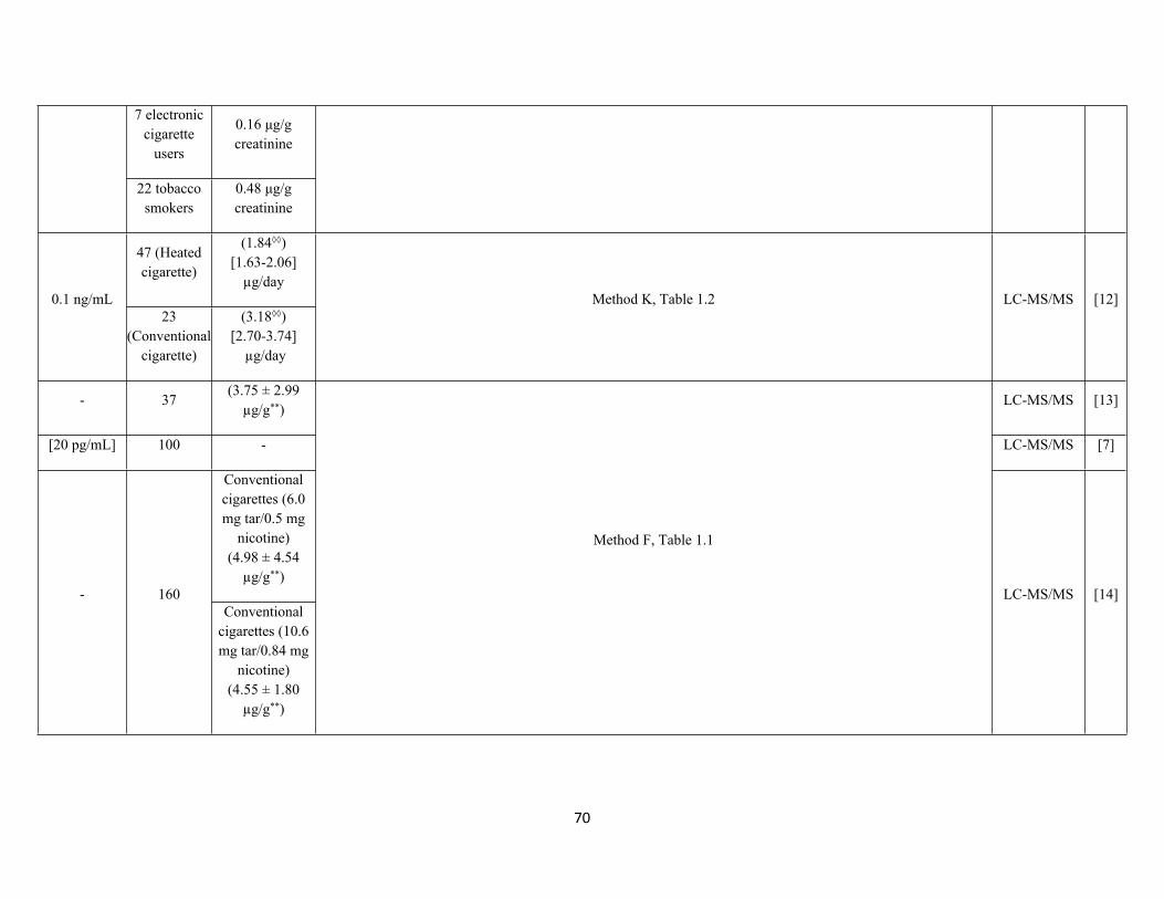
70
7 electronic cigarette
users
0.16 μg/g creatinine
22 tobacco smokers
0.48 μg/g creatinine
47 (Heated cigarette)
(1.84◊◊)[1.63-2.06]
µg/day0.1 ng/mL
23 (Conventional
cigarette)
(3.18◊◊)[2.70-3.74]
µg/day
Method K, Table 1.2 LC-MS/MS [12]
- 37(3.75 ± 2.99
µg/g**)LC-MS/MS [13]
[20 pg/mL] 100 - LC-MS/MS [7]
Conventional cigarettes (6.0 mg tar/0.5 mg
nicotine)(4.98 ± 4.54
µg/g**)- 160
Conventional cigarettes (10.6 mg tar/0.84 mg
nicotine)(4.55 ± 1.80
µg/g**)
Method F, Table 1.1
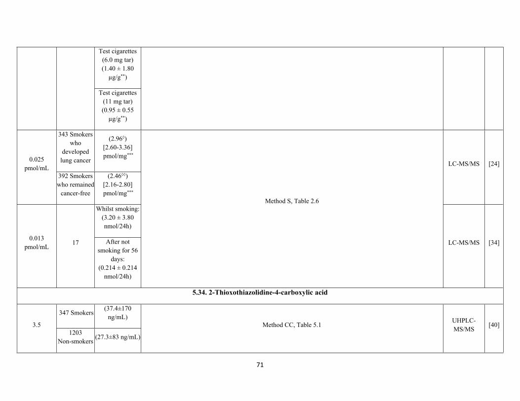
LC-MS/MS [14]
71
Test cigarettes (6.0 mg tar)(1.40 ± 1.80
µg/g**)
Test cigarettes (11 mg tar)(0.95 ± 0.55
µg/g**)
343 Smokers who
developed lung cancer
(2.96◊)[2.60-3.36] pmol/mg***
0.025 pmol/mL
392 Smokers who remained
cancer-free
(2.46◊◊)[2.16-2.80] pmol/mg***
LC-MS/MS [24]
Whilst smoking: (3.20 ± 3.80 nmol/24h)
0.013 pmol/mL
17 After not smoking for 56
days: (0.214 ± 0.214
nmol/24h)
Method S, Table 2.6
LC-MS/MS [34]
5.34. 2-Thioxothiazolidine-4-carboxylic acid
347 Smokers(37.4±170
ng/mL)3.5
1203Non-smokers
(27.3±83 ng/mL)
Method CC, Table 5.1UHPLC-MS/MS
[40]
72
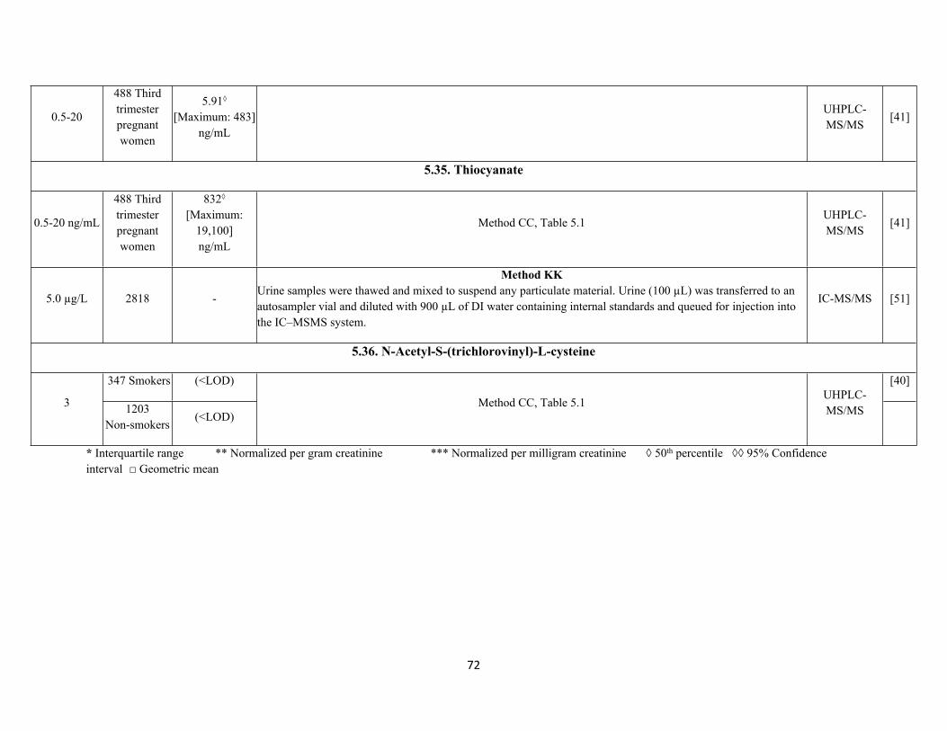
0.5-20
488 Third trimester pregnant women
5.91◊
[Maximum: 483]ng/mL
UHPLC-MS/MS
[41]
5.35. Thiocyanate
0.5-20 ng/mL
488 Third trimester pregnant women
832◊
[Maximum: 19,100]ng/mL
Method CC, Table 5.1UHPLC-MS/MS
[41]
5.0 µg/L 2818 -
Method KKUrine samples were thawed and mixed to suspend any particulate material. Urine (100 µL) was transferred to an autosampler vial and diluted with 900 µL of DI water containing internal standards and queued for injection into the IC–MSMS system.
IC-MS/MS [51]
5.36. N-Acetyl-S-(trichlorovinyl)-L-cysteine
347 Smokers (<LOD) [40]
3 1203Non-smokers
(<LOD)Method CC, Table 5.1
UHPLC-MS/MS
* Interquartile range ** Normalized per gram creatinine *** Normalized per milligram creatinine ◊ 50th percentile ◊◊ 95% Confidence interval □ Geometric mean
73
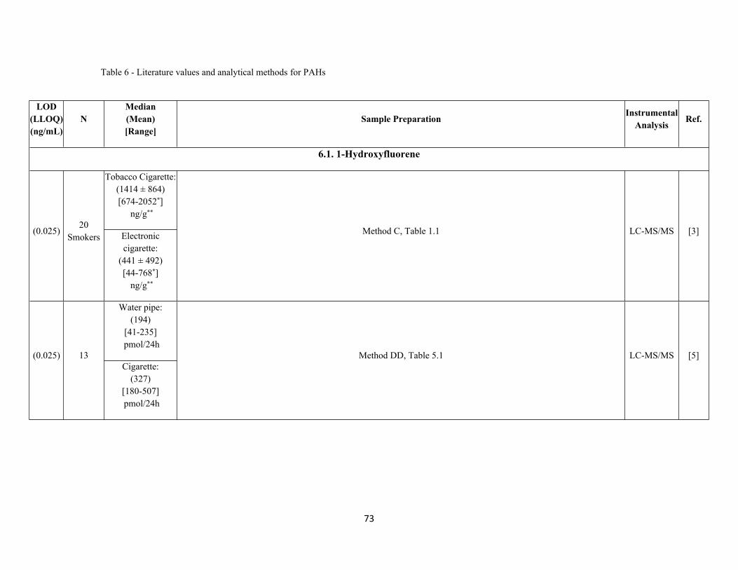
Table 6 - Literature values and analytical methods for PAHs
LOD(LLOQ) (ng/mL)
NMedian(Mean)[Range]
Sample Preparation InstrumentalAnalysis Ref.
6.1. 1-Hydroxyfluorene
Tobacco Cigarette: (1414 ± 864)[674-2052*]
ng/g**
(0.025)20
Smokers Electronic cigarette:
(441 ± 492)[44-768*]
ng/g**
Method C, Table 1.1 LC-MS/MS [3]
Water pipe:(194)
[41-235] pmol/24h
(0.025) 13Cigarette:
(327)[180-507] pmol/24h
Method DD, Table 5.1 LC-MS/MS [5]
74
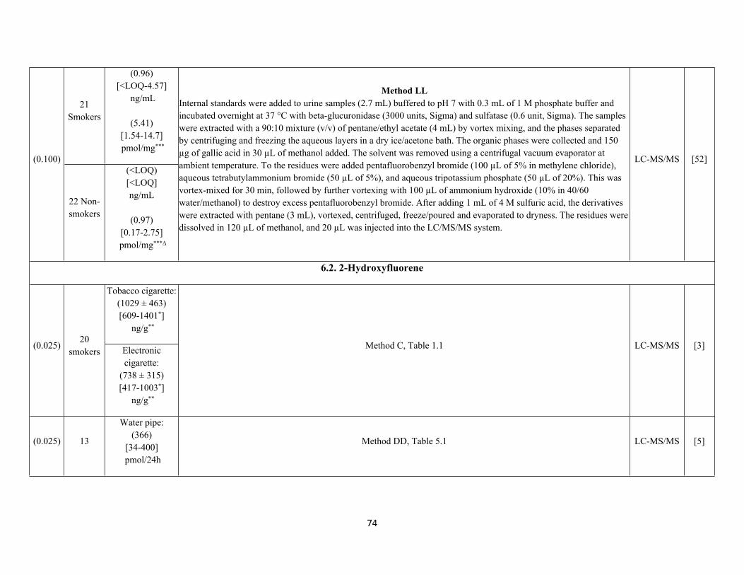
21 Smokers
(0.96)[<LOQ-4.57]
ng/mL
(5.41)[1.54-14.7] pmol/mg***
(0.100)
22 Non-smokers
(<LOQ)[<LOQ]ng/mL
(0.97)[0.17-2.75]
pmol/mg***Δ
Method LLInternal standards were added to urine samples (2.7 mL) buffered to pH 7 with 0.3 mL of 1 M phosphate buffer and incubated overnight at 37 °C with beta-glucuronidase (3000 units, Sigma) and sulfatase (0.6 unit, Sigma). The samples were extracted with a 90:10 mixture (v/v) of pentane/ethyl acetate (4 mL) by vortex mixing, and the phases separated by centrifuging and freezing the aqueous layers in a dry ice/acetone bath. The organic phases were collected and 150 µg of gallic acid in 30 µL of methanol added. The solvent was removed using a centrifugal vacuum evaporator at ambient temperature. To the residues were added pentafluorobenzyl bromide (100 µL of 5% in methylene chloride), aqueous tetrabutylammonium bromide (50 µL of 5%), and aqueous tripotassium phosphate (50 µL of 20%). This was vortex-mixed for 30 min, followed by further vortexing with 100 µL of ammonium hydroxide (10% in 40/60 water/methanol) to destroy excess pentafluorobenzyl bromide. After adding 1 mL of 4 M sulfuric acid, the derivatives were extracted with pentane (3 mL), vortexed, centrifuged, freeze/poured and evaporated to dryness. The residues were dissolved in 120 µL of methanol, and 20 µL was injected into the LC/MS/MS system.
LC-MS/MS [52]
6.2. 2-Hydroxyfluorene
Tobacco cigarette:(1029 ± 463)[609-1401*]
ng/g**
(0.025)20
smokers Electronic cigarette:
(738 ± 315)[417-1003*]
ng/g**
Method C, Table 1.1 LC-MS/MS [3]
(0.025) 13
Water pipe:(366)
[34-400] pmol/24h
Method DD, Table 5.1 LC-MS/MS [5]
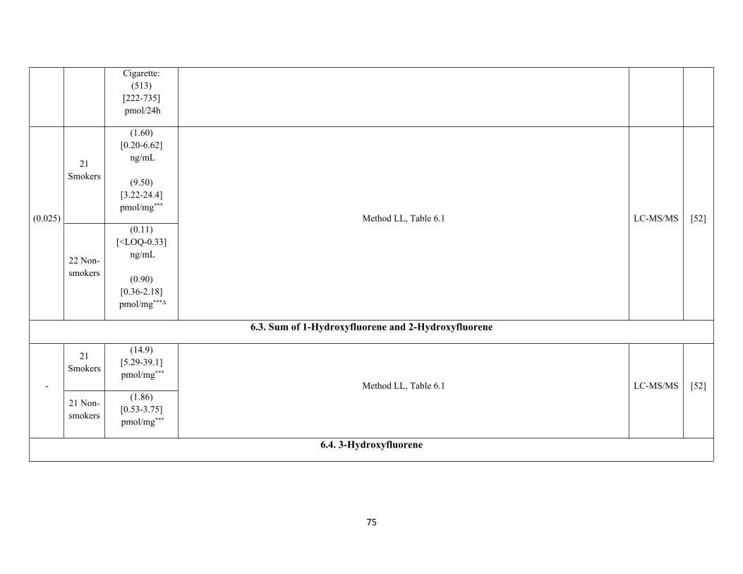
75
Cigarette:(513)
[222-735] pmol/24h
21 Smokers
(1.60)[0.20-6.62]
ng/mL
(9.50)[3.22-24.4] pmol/mg***
(0.025)
22 Non-smokers
(0.11)[<LOQ-0.33]
ng/mL
(0.90)[0.36-2.18]
pmol/mg***Δ
Method LL, Table 6.1 LC-MS/MS [52]
6.3. Sum of 1-Hydroxyfluorene and 2-Hydroxyfluorene
21 Smokers
(14.9)[5.29-39.1] pmol/mg***
-
21 Non-smokers
(1.86)[0.53-3.75] pmol/mg***
Method LL, Table 6.1 LC-MS/MS [52]
6.4. 3-Hydroxyfluorene
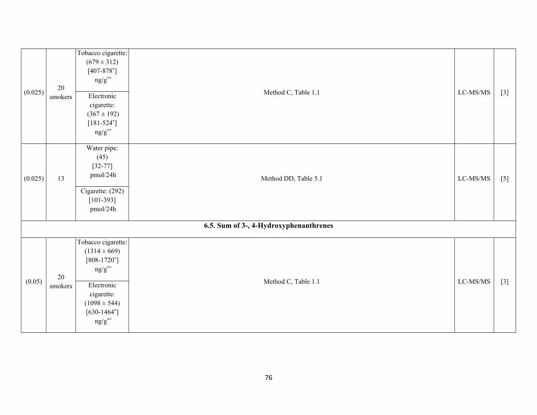
76
Tobacco cigarette:(679 ± 312)[407-878*]
ng/g**
(0.025)20
smokers Electronic cigarette:
(367 ± 192)[181-524*]
ng/g**
Method C, Table 1.1 LC-MS/MS [3]
Water pipe:(45)
[32-77] pmol/24h(0.025) 13
Cigarette: (292)[101-393] pmol/24h
Method DD, Table 5.1 LC-MS/MS [5]
6.5. Sum of 3-, 4-Hydroxyphenanthrenes
Tobacco cigarette:(1314 ± 669)[808-1720*]
ng/g**
(0.05)20
smokers Electronic cigarette:
(1098 ± 544)[630-1464*]
ng/g**
Method C, Table 1.1 LC-MS/MS [3]
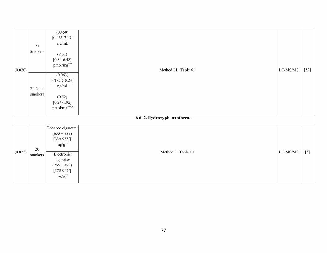
77
21 Smokers
(0.450)[0.066-2.13]
ng/mL
(2.31)[0.86-6.48] pmol/mg***
(0.020)
22 Non-smokers
(0.063)[<LOQ-0.23]
ng/mL
(0.52)[0.24-1.92]
pmol/mg***Δ
Method LL, Table 6.1 LC-MS/MS [52]
6.6. 2-Hydroxyphenanthrene
Tobacco cigarette:(655 ± 333)[339-933*]
ng/g**
(0.025)20
smokers Electronic cigarette:
(755 ± 492)[375-947*]
ng/g**
Method C, Table 1.1 LC-MS/MS [3]
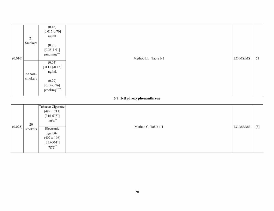
78
21 Smokers
(0.16)[0.017-0.70]
ng/mL
(0.85) [0.35-1.91]pmol/mg***
(0.010)
22 Non-smokers
(0.04)[<LOQ-0.15]
ng/mL
(0.29)[0.14-0.76]
pmol/mg***Δ
Method LL, Table 6.1 LC-MS/MS [52]
6.7. 1-Hydroxyphenanthrene
Tobacco Cigarette:(488 ± 211)[316-678*]
ng/g**
(0.025)20
smokers Electronic cigarette:
(407 ± 196)[235-561*]
ng/g**
Method C, Table 1.1 LC-MS/MS [3]
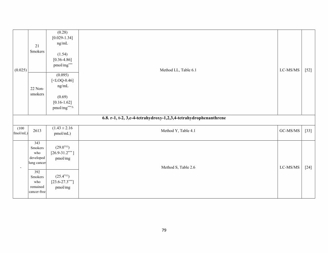
79
21 Smokers
(0.28) [0.029-1.34]
ng/mL
(1.54)[0.36-4.86] pmol/mg***
(0.025)
22 Non-smokers
(0.095)[<LOQ-0.46]
ng/mL
(0.69)[0.16-1.62]
pmol/mg***Δ
Method LL, Table 6.1 LC-MS/MS [52]
6.8. r-1, t-2, 3,c-4-tetrahydroxy-1,2,3,4-tetrahydrophenanthrene
(100 fmol/mL) 2613
(1.43 ± 2.16 pmol/mL)
Method Y, Table 4.1 GC-MS/MS [33]
343 Smokers
who developed
lung cancer
(29.0*◊◊)[26.9-31.2*** ]
pmol/mg
-392
Smokers who
remained cancer-free
(25.4*◊◊)[23.6-27.3***]
pmol/mg
Method S, Table 2.6 LC-MS/MS [24]
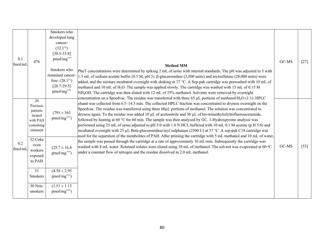
80
0.1 fmol/mL
476
Smokers who developed lung
cancer: (32.1◊◊)
[30.5-33.8] pmol/mg***
Smokers who remained cancer-
free: (28.1◊◊)[26.7-29.5] pmol/mg***
GC-MS [27]
20 Psoriasis patients treated
with PAH containing ointment
(791 ± 363 pmol/mg***)
32 Coke oven
workers exposed to PAH
(25.7 ± 16.8 pmol/mg***)
31 Smokers
(4.58 ± 2.95 pmol/mg***)
0.2 fmol/mL
30 Non-smokers
(1.51 ± 1.15 pmol/mg***)
Method MMPheT concentrations were determined by spiking 2 mL of urine with internal standards. The pH was adjusted to 5 with 1.5 mL of sodium acetate buffer (0.5 M, pH 5). β-glucuronidase (3,500 units) and arylsulfatase (28,000 units) were added, and the mixture incubated overnight with shaking at 37 °C. A Sep-pak cartridge was prewashed with 10 mL of methanol and 10 mL of H2O. The sample was applied slowly. The cartridge was washed with 15 mL of 0.15 M NH4OH. The cartridge was then eluted with 12 mL of 25% methanol. Solvents were removed by overnight concentration on a Speedvac. The residue was transferred with three 65 µL portions of methanol/H2O (1:1). HPLC eluant was collected from 6.5–14.5 min. The collected HPLC fraction was concentrated to dryness overnight on the Speedvac. The residue was transferred using three 60µL portions of methanol. The solution was concentrated to dryness again. To the residue was added 10 µL of acetonitrile and 30 µL of bis-trimethylsilyltrifluoroacetamide, followed by heating at 60 °C for 60 min. The sample was then analysed by GC. 1-Hydroxpyrene analysis was performed using 25 mL of urine adjusted to pH 5.0 with 1.0 N HCI, buffered with 10 mL 0.1 M acetate (p H 5.0) and incubated overnight with 25 µL Beta-glucuronidase/aryl sulphatase (2500 U) at 37 °C. A sep-pak C18 cartridge was used for the separation of the metabolites of PAH. After priming the cartridge with 5 mL methanol and 10 mL of water, the sample was passed through the cartridge at a rate of approximately 10 mL/min. Subsequently the cartridge was washed with 8 mL water. Retained solutes were eluted using 10 mL of methanol. The solvent was evaporated at 60 oC under a constant flow of nitrogen and the residue dissolved in 2.0 mL methanol.
GC-MS [53]
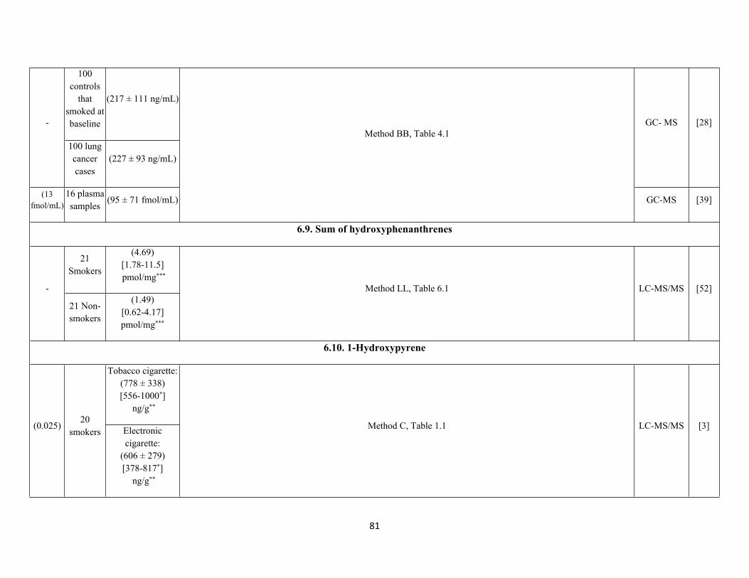
81
100 controls
that smoked at baseline
(217 ± 111 ng/mL)
-
100 lung cancer cases
(227 ± 93 ng/mL)
GC- MS [28]
(13 fmol/mL)
16 plasma samples
(95 ± 71 fmol/mL)
Method BB, Table 4.1
GC-MS [39]
6.9. Sum of hydroxyphenanthrenes
21 Smokers
(4.69)[1.78-11.5] pmol/mg***
-
21 Non-smokers
(1.49) [0.62-4.17]pmol/mg***
Method LL, Table 6.1 LC-MS/MS [52]
6.10. 1-Hydroxypyrene
Tobacco cigarette:(778 ± 338)[556-1000*]
ng/g**
(0.025)20
smokers Electronic cigarette:
(606 ± 279)[378-817*]
ng/g**
Method C, Table 1.1 LC-MS/MS [3]
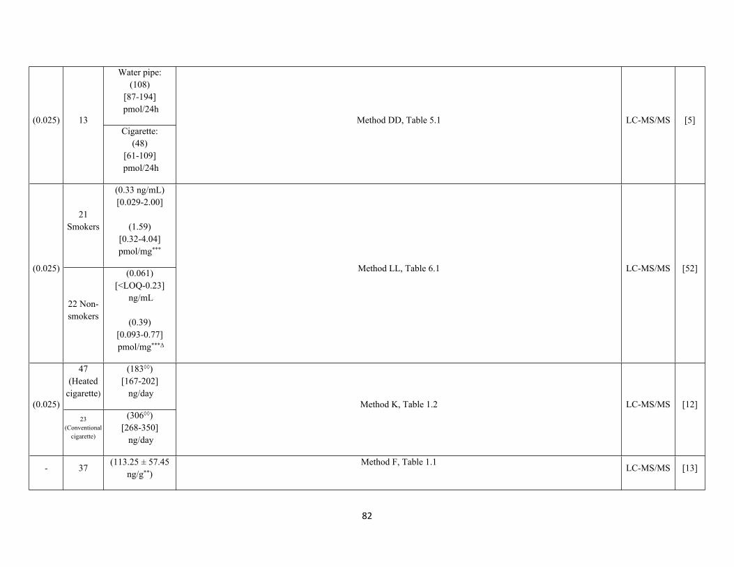
82
Water pipe:(108)
[87-194] pmol/24h
(0.025) 13Cigarette:
(48)[61-109]
pmol/24h
Method DD, Table 5.1 LC-MS/MS [5]
21 Smokers
(0.33 ng/mL)[0.029-2.00]
(1.59)[0.32-4.04] pmol/mg***
(0.025)
22 Non-smokers
(0.061)[<LOQ-0.23]
ng/mL
(0.39)[0.093-0.77] pmol/mg***Δ
Method LL, Table 6.1 LC-MS/MS [52]
47 (Heated
cigarette)
(183◊◊)[167-202]
ng/day(0.025)
23 (Conventional
cigarette)
(306◊◊)[268-350]
ng/day
Method K, Table 1.2 LC-MS/MS [12]
- 37(113.25 ± 57.45
ng/g**)Method F, Table 1.1
LC-MS/MS [13]
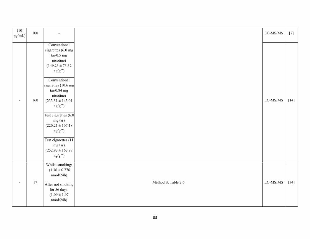
83
(10 pg/mL)
100 - LC-MS/MS [7]
Conventional cigarettes (6.0 mg
tar/0.5 mg nicotine)
(149.23 ± 73.32 ng/g**)
Conventional cigarettes (10.6 mg
tar/0.84 mg nicotine)
(233.51 ± 143.01 ng/g**)
Test cigarettes (6.0 mg tar)
(220.21 ± 107.18 ng/g**)
- 160
Test cigarettes (11 mg tar)
(252.93 ± 163.87 ng/g**)
LC-MS/MS [14]
Whilst smoking: (1.36 ± 0.776
nmol/24h)
- 17 After not smoking for 56 days:(1.09 ± 1.97 nmol/24h)
Method S, Table 2.6 LC-MS/MS [34]
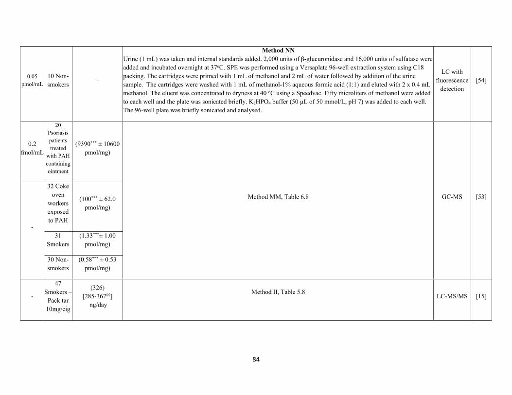
84
0.05 pmol/mL
10 Non-smokers
-
Method NNUrine (1 mL) was taken and internal standards added. 2,000 units of β-glucuronidase and 16,000 units of sulfatase were added and incubated overnight at 37oC. SPE was performed using a Versaplate 96-well extraction system using C18 packing. The cartridges were primed with 1 mL of methanol and 2 mL of water followed by addition of the urine sample. The cartridges were washed with 1 mL of methanol-1% aqueous formic acid (1:1) and eluted with 2 x 0.4 mL methanol. The eluent was concentrated to dryness at 40 oC using a Speedvac. Fifty microliters of methanol were added to each well and the plate was sonicated briefly. K2HPO4 buffer (50 µL of 50 mmol/L, pH 7) was added to each well. The 96-well plate was briefly sonicated and analysed.
LC with fluorescence
detection[54]
0.2 fmol/mL
20 Psoriasis patients treated
with PAH containing ointment
(9390*** ± 10600 pmol/mg)
32 Coke oven
workers exposed to PAH
(100*** ± 62.0 pmol/mg)
31 Smokers
(1.33***± 1.00 pmol/mg)
-
30 Non-smokers
(0.58*** ± 0.53 pmol/mg)
Method MM, Table 6.8 GC-MS [53]
-
47 Smokers –
Pack tar 10mg/cig
(326)[285-367◊◊]
ng/day
Method II, Table 5.8LC-MS/MS [15]
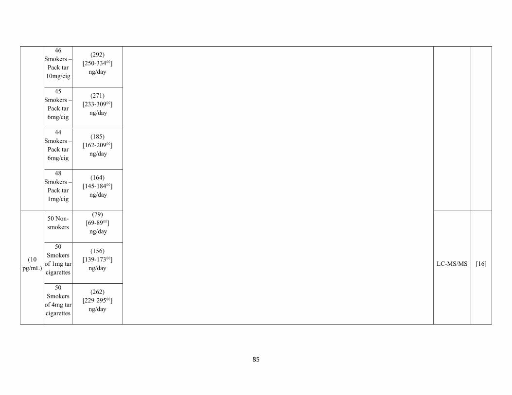
85
46 Smokers –
Pack tar 10mg/cig
(292)[250-334◊◊]
ng/day
45 Smokers –
Pack tar 6mg/cig
(271)[233-309◊◊]
ng/day
44 Smokers –
Pack tar 6mg/cig
(185)[162-209◊◊]
ng/day
48 Smokers –
Pack tar 1mg/cig
(164)[145-184◊◊]
ng/day
50 Non-smokers
(79)[69-89◊◊] ng/day
50 Smokers
of 1mg tar cigarettes
(156)[139-173◊◊]
ng/day(10
pg/mL)
50 Smokers
of 4mg tar cigarettes
(262) [229-295◊◊]
ng/day
LC-MS/MS [16]
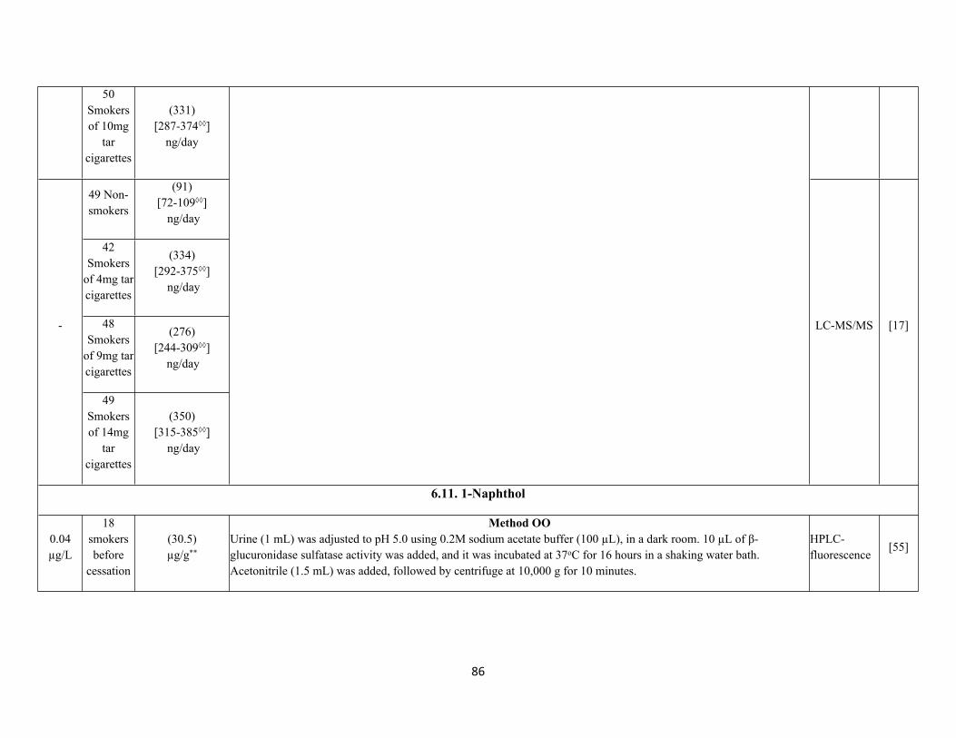
86
50 Smokers of 10mg
tar cigarettes
(331) [287-374◊◊]
ng/day
49 Non-smokers
(91)[72-109◊◊]
ng/day
42 Smokers
of 4mg tar cigarettes
(334)[292-375◊◊]
ng/day
48 Smokers
of 9mg tar cigarettes
(276)[244-309◊◊]
ng/day
-
49 Smokers of 14mg
tar cigarettes
(350)[315-385◊◊]
ng/day
LC-MS/MS [17]
6.11. 1-Naphthol
0.04 µg/L
18 smokers before
cessation
(30.5)µg/g**
Method OOUrine (1 mL) was adjusted to pH 5.0 using 0.2M sodium acetate buffer (100 µL), in a dark room. 10 µL of β-glucuronidase sulfatase activity was added, and it was incubated at 37oC for 16 hours in a shaking water bath. Acetonitrile (1.5 mL) was added, followed by centrifuge at 10,000 g for 10 minutes.
HPLC-fluorescence
[55]
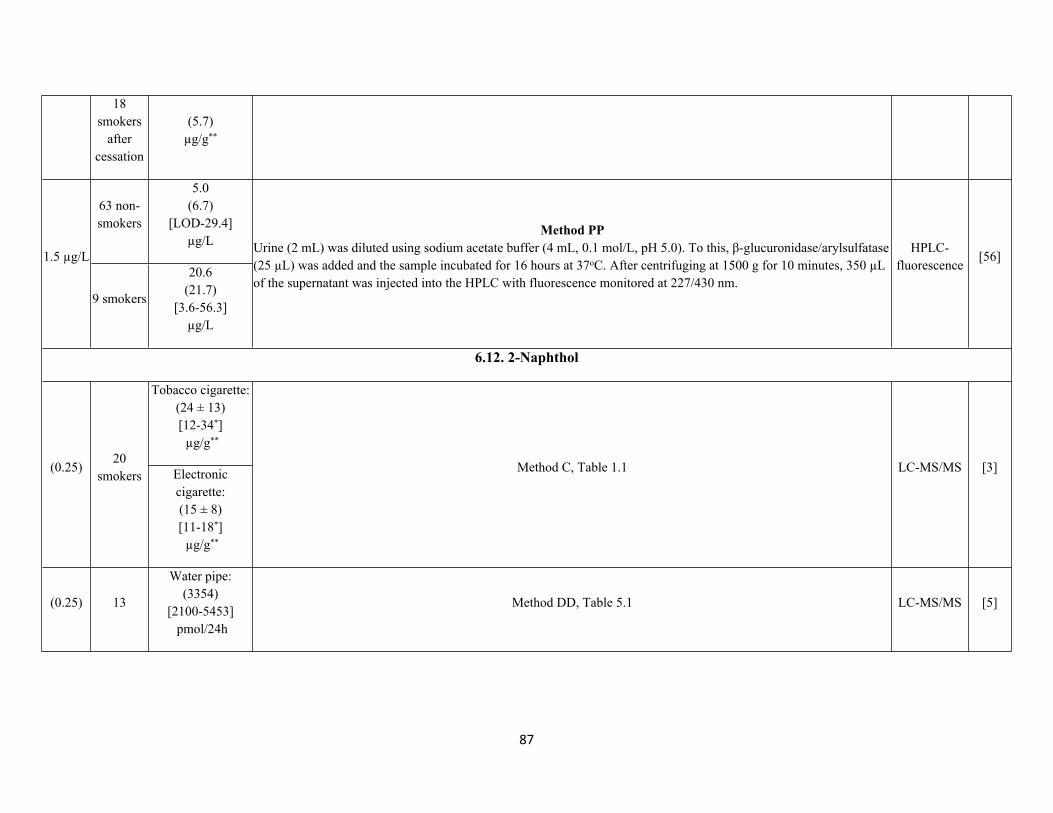
87
18 smokers
after cessation
(5.7)µg/g**
63 non-smokers
5.0(6.7)
[LOD-29.4]µg/L
1.5 µg/L
9 smokers
20.6(21.7)
[3.6-56.3]µg/L
Method PPUrine (2 mL) was diluted using sodium acetate buffer (4 mL, 0.1 mol/L, pH 5.0). To this, β-glucuronidase/arylsulfatase (25 µL) was added and the sample incubated for 16 hours at 37oC. After centrifuging at 1500 g for 10 minutes, 350 µL of the supernatant was injected into the HPLC with fluorescence monitored at 227/430 nm.
HPLC-fluorescence
[56]
6.12. 2-Naphthol
Tobacco cigarette:(24 ± 13)[12-34*] µg/g**
(0.25)20
smokers Electronic cigarette:(15 ± 8)[11-18*] µg/g**
Method C, Table 1.1 LC-MS/MS [3]
(0.25) 13
Water pipe:(3354)
[2100-5453] pmol/24h
Method DD, Table 5.1 LC-MS/MS [5]
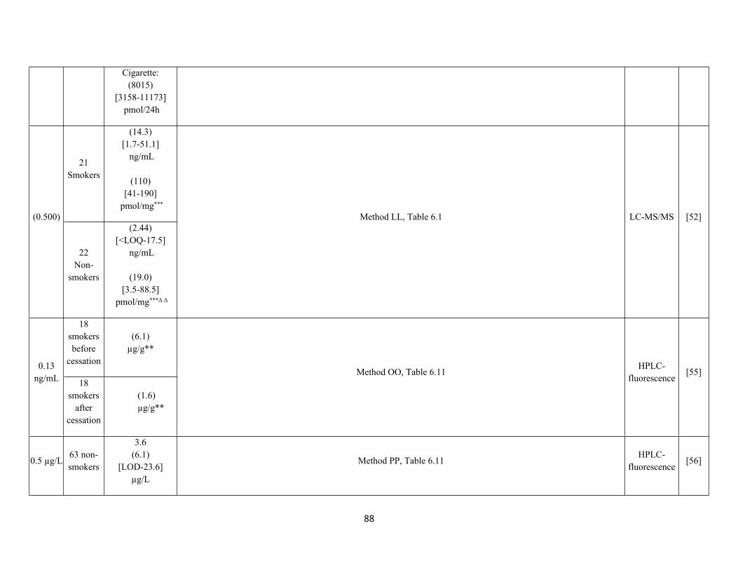
88
Cigarette:(8015)
[3158-11173] pmol/24h
21 Smokers
(14.3)[1.7-51.1]
ng/mL
(110)[41-190]
pmol/mg***
(0.500)
22Non-
smokers
(2.44)[<LOQ-17.5]
ng/mL
(19.0)[3.5-88.5]
pmol/mg***Δ Δ
Method LL, Table 6.1 LC-MS/MS [52]
18 smokers before
cessation
(6.1)µg/g**
0.13 ng/mL 18
smokers after
cessation
(1.6)µg/g**
Method OO, Table 6.11HPLC-
fluorescence[55]
0.5 µg/L63 non-smokers
3.6(6.1)
[LOD-23.6]µg/L
Method PP, Table 6.11HPLC-
fluorescence[56]
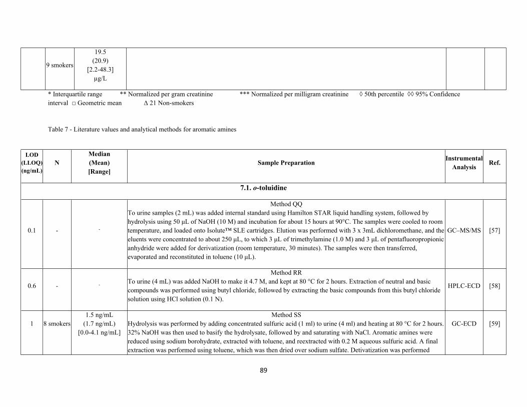
89
9 smokers
19.5(20.9)
[2.2-48.3]µg/L
* Interquartile range ** Normalized per gram creatinine *** Normalized per milligram creatinine ◊ 50th percentile ◊◊ 95% Confidence interval □ Geometric mean Δ 21 Non-smokers
Table 7 - Literature values and analytical methods for aromatic amines
LOD(LLOQ) (ng/mL)
NMedian(Mean)[Range]
Sample Preparation InstrumentalAnalysis Ref.
7.1. o-toluidine
0.1 - -
Method QQTo urine samples (2 mL) was added internal standard using Hamilton STAR liquid handling system, followed by hydrolysis using 50 μL of NaOH (10 M) and incubation for about 15 hours at 90°C. The samples were cooled to room temperature, and loaded onto Isolute™ SLE cartridges. Elution was performed with 3 x 3mL dichloromethane, and the eluents were concentrated to about 250 μL, to which 3 μL of trimethylamine (1.0 M) and 3 μL of pentafluoropropionic anhydride were added for derivatization (room temperature, 30 minutes). The samples were then transferred, evaporated and reconstituted in toluene (10 μL).
GC–MS/MS [57]
0.6 - -
Method RRTo urine (4 mL) was added NaOH to make it 4.7 M, and kept at 80 °C for 2 hours. Extraction of neutral and basic compounds was performed using butyl chloride, followed by extracting the basic compounds from this butyl chloride solution using HCl solution (0.1 N).
HPLC-ECD [58]
1 8 smokers1.5 ng/mL
(1.7 ng/mL)[0.0-4.1 ng/mL]
Method SSHydrolysis was performed by adding concentrated sulfuric acid (1 ml) to urine (4 ml) and heating at 80 °C for 2 hours. 32% NaOH was then used to basify the hydrolysate, followed by and saturating with NaCl. Aromatic amines were reduced using sodium borohydrate, extracted with toluene, and reextracted with 0.2 M aqueous sulfuric acid. A final extraction was performed using toluene, which was then dried over sodium sulfate. Detivatization was performed
GC-ECD [59]
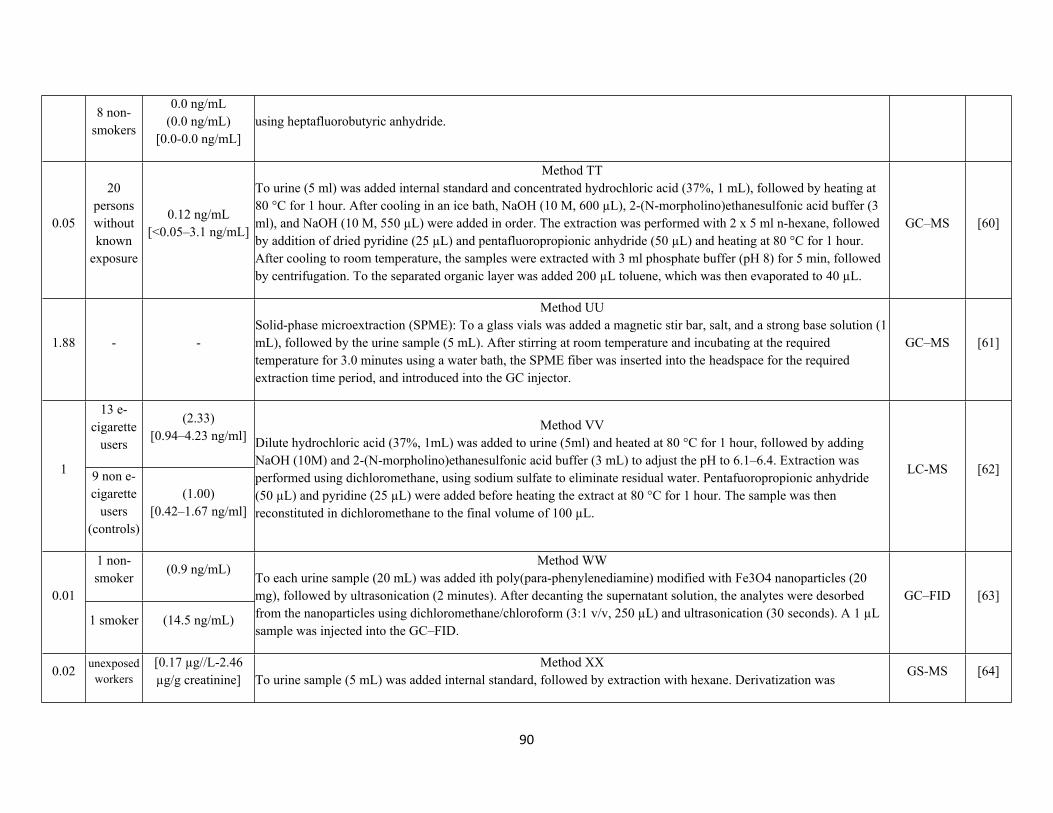
90
8 non-smokers
0.0 ng/mL(0.0 ng/mL)
[0.0-0.0 ng/mL]using heptafluorobutyric anhydride.
0.05
20 persons without known
exposure
0.12 ng/mL[<0.05–3.1 ng/mL]
Method TTTo urine (5 ml) was added internal standard and concentrated hydrochloric acid (37%, 1 mL), followed by heating at 80 °C for 1 hour. After cooling in an ice bath, NaOH (10 M, 600 µL), 2-(N-morpholino)ethanesulfonic acid buffer (3 ml), and NaOH (10 M, 550 µL) were added in order. The extraction was performed with 2 x 5 ml n-hexane, followed by addition of dried pyridine (25 µL) and pentafluoropropionic anhydride (50 µL) and heating at 80 °C for 1 hour. After cooling to room temperature, the samples were extracted with 3 ml phosphate buffer (pH 8) for 5 min, followed by centrifugation. To the separated organic layer was added 200 µL toluene, which was then evaporated to 40 µL.
GC–MS [60]
1.88 - -
Method UUSolid-phase microextraction (SPME): To a glass vials was added a magnetic stir bar, salt, and a strong base solution (1 mL), followed by the urine sample (5 mL). After stirring at room temperature and incubating at the required temperature for 3.0 minutes using a water bath, the SPME fiber was inserted into the headspace for the required extraction time period, and introduced into the GC injector.
GC–MS [61]
13 e-cigarette
users
(2.33)[0.94–4.23 ng/ml]
1 9 non e-cigarette
users (controls)
(1.00)[0.42–1.67 ng/ml]
Method VVDilute hydrochloric acid (37%, 1mL) was added to urine (5ml) and heated at 80 °C for 1 hour, followed by adding NaOH (10M) and 2-(N-morpholino)ethanesulfonic acid buffer (3 mL) to adjust the pH to 6.1–6.4. Extraction was performed using dichloromethane, using sodium sulfate to eliminate residual water. Pentafuoropropionic anhydride (50 µL) and pyridine (25 µL) were added before heating the extract at 80 °C for 1 hour. The sample was then reconstituted in dichloromethane to the final volume of 100 µL.
LC-MS [62]
1 non-smoker
(0.9 ng/mL)
0.01
1 smoker (14.5 ng/mL)
Method WWTo each urine sample (20 mL) was added ith poly(para-phenylenediamine) modified with Fe3O4 nanoparticles (20 mg), followed by ultrasonication (2 minutes). After decanting the supernatant solution, the analytes were desorbed from the nanoparticles using dichloromethane/chloroform (3:1 v/v, 250 µL) and ultrasonication (30 seconds). A 1 µL sample was injected into the GC–FID.
GC–FID [63]
0.02 unexposed workers
[0.17 µg//L-2.46 µg/g creatinine]
Method XXTo urine sample (5 mL) was added internal standard, followed by extraction with hexane. Derivatization was
GS-MS [64]
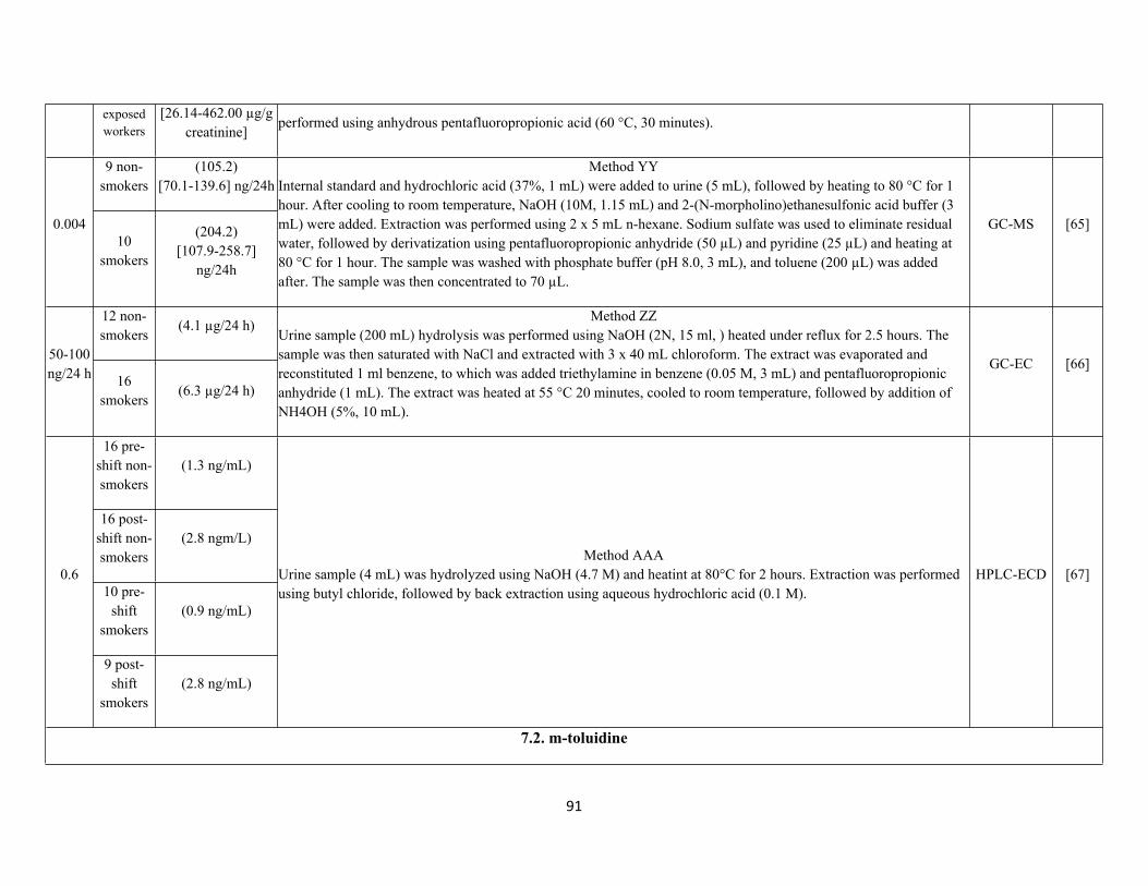
91
exposed workers
[26.14-462.00 µg/g creatinine]
performed using anhydrous pentafluoropropionic acid (60 °C, 30 minutes).
9 non-smokers
(105.2)[70.1-139.6] ng/24h
0.00410
smokers
(204.2)[107.9-258.7]
ng/24h
Method YYInternal standard and hydrochloric acid (37%, 1 mL) were added to urine (5 mL), followed by heating to 80 °C for 1 hour. After cooling to room temperature, NaOH (10M, 1.15 mL) and 2-(N-morpholino)ethanesulfonic acid buffer (3 mL) were added. Extraction was performed using 2 x 5 mL n-hexane. Sodium sulfate was used to eliminate residual water, followed by derivatization using pentafluoropropionic anhydride (50 µL) and pyridine (25 µL) and heating at 80 °C for 1 hour. The sample was washed with phosphate buffer (pH 8.0, 3 mL), and toluene (200 µL) was added after. The sample was then concentrated to 70 µL.
GC-MS [65]
12 non-smokers
(4.1 µg/24 h)
50-100 ng/24 h 16
smokers(6.3 µg/24 h)
Method ZZUrine sample (200 mL) hydrolysis was performed using NaOH (2N, 15 ml, ) heated under reflux for 2.5 hours. The sample was then saturated with NaCl and extracted with 3 x 40 mL chloroform. The extract was evaporated and reconstituted 1 ml benzene, to which was added triethylamine in benzene (0.05 M, 3 mL) and pentafluoropropionic anhydride (1 mL). The extract was heated at 55 °C 20 minutes, cooled to room temperature, followed by addition of NH4OH (5%, 10 mL).
GC-EC [66]
16 pre-shift non-smokers
(1.3 ng/mL)
16 post-shift non-smokers
(2.8 ngm/L)
10 pre-shift
smokers(0.9 ng/mL)
0.6
9 post-shift
smokers(2.8 ng/mL)
Method AAAUrine sample (4 mL) was hydrolyzed using NaOH (4.7 M) and heatint at 80°C for 2 hours. Extraction was performed using butyl chloride, followed by back extraction using aqueous hydrochloric acid (0.1 M).
HPLC-ECD [67]
7.2. m-toluidine
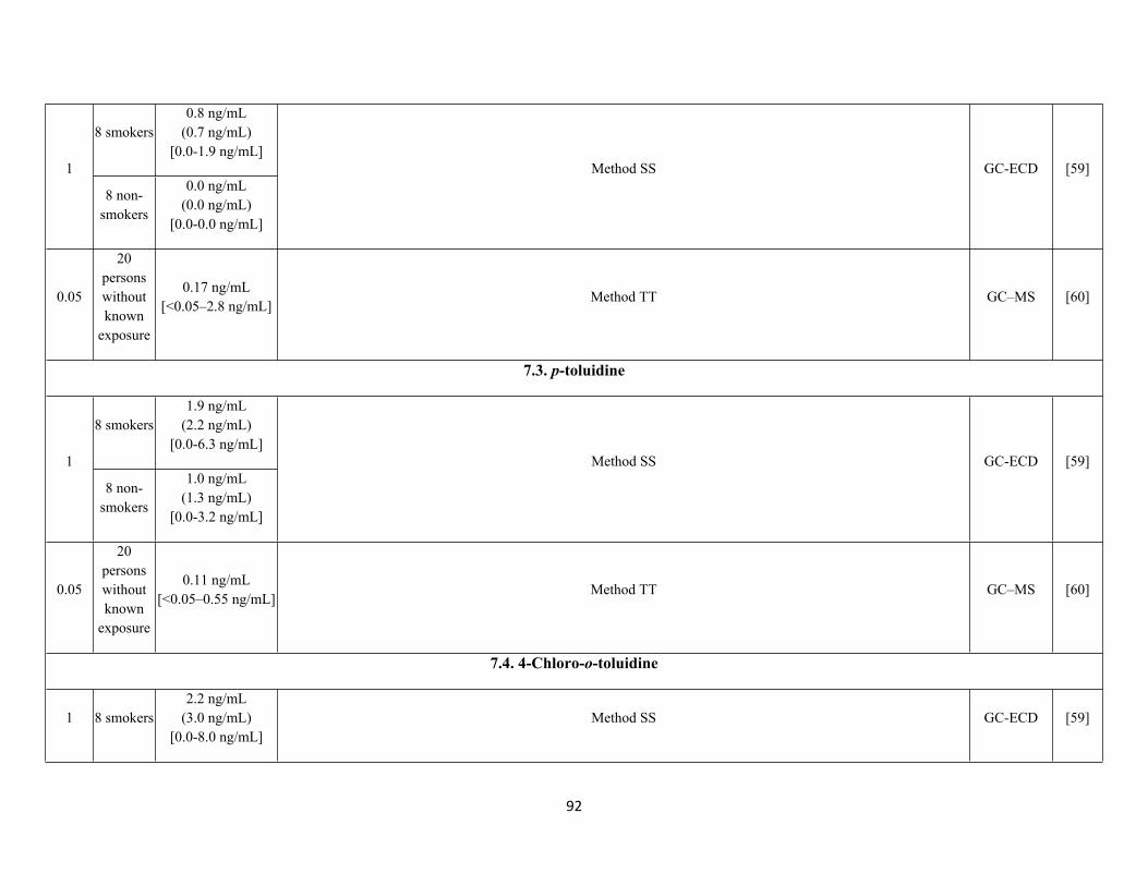
92
8 smokers0.8 ng/mL
(0.7 ng/mL)[0.0-1.9 ng/mL]
1
8 non-smokers
0.0 ng/mL(0.0 ng/mL)
[0.0-0.0 ng/mL]
Method SS GC-ECD [59]
0.05
20 persons without known
exposure
0.17 ng/mL[<0.05–2.8 ng/mL]
Method TT GC–MS [60]
7.3. p-toluidine
8 smokers1.9 ng/mL
(2.2 ng/mL)[0.0-6.3 ng/mL]
1
8 non-smokers
1.0 ng/mL(1.3 ng/mL)
[0.0-3.2 ng/mL]
Method SS GC-ECD [59]
0.05
20 persons without known
exposure
0.11 ng/mL[<0.05–0.55 ng/mL]
Method TT GC–MS [60]
7.4. 4-Chloro-o-toluidine
1 8 smokers2.2 ng/mL
(3.0 ng/mL)[0.0-8.0 ng/mL]
Method SS GC-ECD [59]
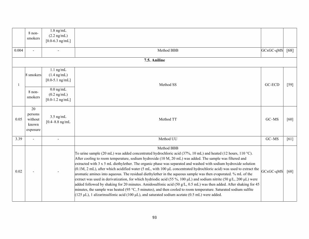
93
8 non-smokers
1.8 ng/mL(2.2 ng/mL)
[0.0-6.3 ng/mL]
0.004 - - Method BBB GCxGC-qMS [68]
7.5. Aniline
8 smokers1.1 ng/mL
(1.4 ng/mL)[0.0-5.1 ng/mL]
1
8 non-smokers
0.0 ng/mL(0.2 ng/mL)
[0.0-1.2 ng/mL]
Method SS GC-ECD [59]
0.05
20 persons without known
exposure
3.5 ng/mL[0.4–8.8 ng/mL
Method TT GC–MS [60]
3.39 - - Method UU GC–MS [61]
0.02 - -
Method BBBTo urine sample (20 mL) was added concentrated hydrochloric acid (37%, 10 mL) and heated (12 hours, 110 °C). After cooling to room temperature, sodium hydroxide (10 M, 20 mL) was added. The sample was filtered and extracted with 3 x 5 mL diethylether. The organic phase was separated and washed with sodium hydroxide solution (0.1M, 2 mL), after which acidified water (5 mL, with 100 μL concentrated hydrochloric acid) was used to extract the aromatic amines into aqueous. The residual diethylether in the aqueous sample was then evaporated. % mL of the extract was used in derivatization, for which hydriodic acid (55 %, 100 μL) and sodium nitrite (50 g/L, 200 μL) were added followed by shaking for 20 minutes. Amidosulfonic acid (50 g/L, 0.5 mL) was then added. After shaking for 45 minutes, the sample was heated (95 °C, 5 minutes), and then cooled to room temperature. Saturated sodium sulfite (125 μL), 1 alizarinsulfonic acid (100 μL), and saturated sodium acetate (0.5 mL) were added.
GCxGC-qMS [68]
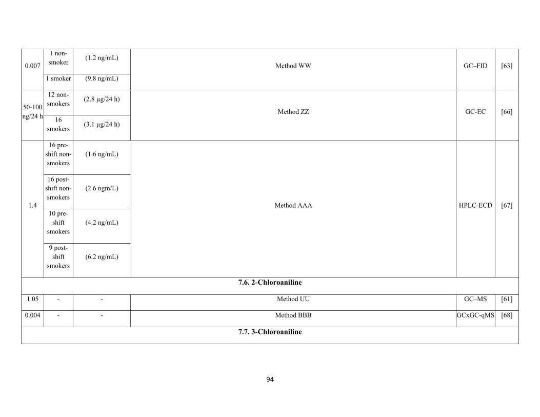
94
1 non-smoker
(1.2 ng/mL)0.007
1 smoker (9.8 ng/mL)
Method WW GC–FID [63]
12 non-smokers
(2.8 µg/24 h)50-100 ng/24 h 16
smokers(3.1 µg/24 h)
Method ZZ GC-EC [66]
16 pre-shift non-smokers
(1.6 ng/mL)
16 post-shift non-smokers
(2.6 ngm/L)
10 pre-shift
smokers(4.2 ng/mL)
1.4
9 post-shift
smokers(6.2 ng/mL)
Method AAA HPLC-ECD [67]
7.6. 2-Chloroaniline
1.05 - - Method UU GC–MS [61]
0.004 - - Method BBB GCxGC-qMS [68]
7.7. 3-Chloroaniline
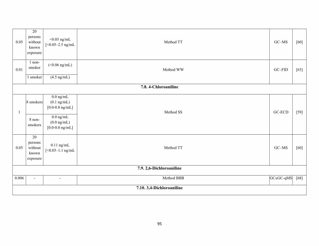
95
0.05
20 persons without known
exposure
<0.05 ng/mL[<0.05–2.5 ng/mL
Method TT GC–MS [60]
1 non-smoker
(<0.06 ng/mL)0.01
1 smoker (4.5 ng/mL)
Method WW GC–FID [63]
7.8. 4-Chloroaniline
8 smokers0.0 ng/mL
(0.1 ng/mL)[0.0-0.8 ng/mL]
1
8 non-smokers
0.0 ng/mL(0.0 ng/mL)
[0.0-0.0 ng/mL]
Method SS GC-ECD [59]
0.05
20 persons without known
exposure
0.11 ng/mL[<0.05–1.1 ng/mL
Method TT GC–MS [60]
7.9. 2,6-Dichloroaniline
0.006 - - Method BBB GCxGC-qMS [68]
7.10. 3,4-Dichloroaniline
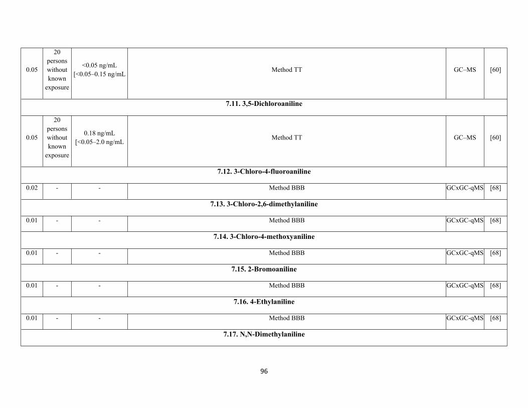
96
0.05
20 persons without known
exposure
<0.05 ng/mL[<0.05–0.15 ng/mL
Method TT GC–MS [60]
7.11. 3,5-Dichloroaniline
0.05
20 persons without known
exposure
0.18 ng/mL[<0.05–2.0 ng/mL
Method TT GC–MS [60]
7.12. 3-Chloro-4-fluoroaniline
0.02 - - Method BBB GCxGC-qMS [68]
7.13. 3-Chloro-2,6-dimethylaniline
0.01 - - Method BBB GCxGC-qMS [68]
7.14. 3-Chloro-4-methoxyaniline
0.01 - - Method BBB GCxGC-qMS [68]
7.15. 2-Bromoaniline
0.01 - - Method BBB GCxGC-qMS [68]
7.16. 4-Ethylaniline
0.01 - - Method BBB GCxGC-qMS [68]
7.17. N,N-Dimethylaniline
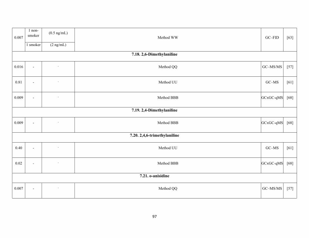
97
1 non-smoker
(0.5 ng/mL)0.007
1 smoker (2 ng/mL)
Method WW GC–FID [63]
7.18. 2,6-Dimethylaniline
0.016 - - Method QQ GC–MS/MS [57]
0.81 - - Method UU GC–MS [61]
0.009 - - Method BBB GCxGC-qMS [68]
7.19. 2,4-Dimethylaniline
0.009 - - Method BBB GCxGC-qMS [68]
7.20. 2,4,6-trimethylaniline
0.40 - - Method UU GC–MS [61]
0.02 - - Method BBB GCxGC-qMS [68]
7.21. o-anisidine
0.007 - - Method QQ GC–MS/MS [57]
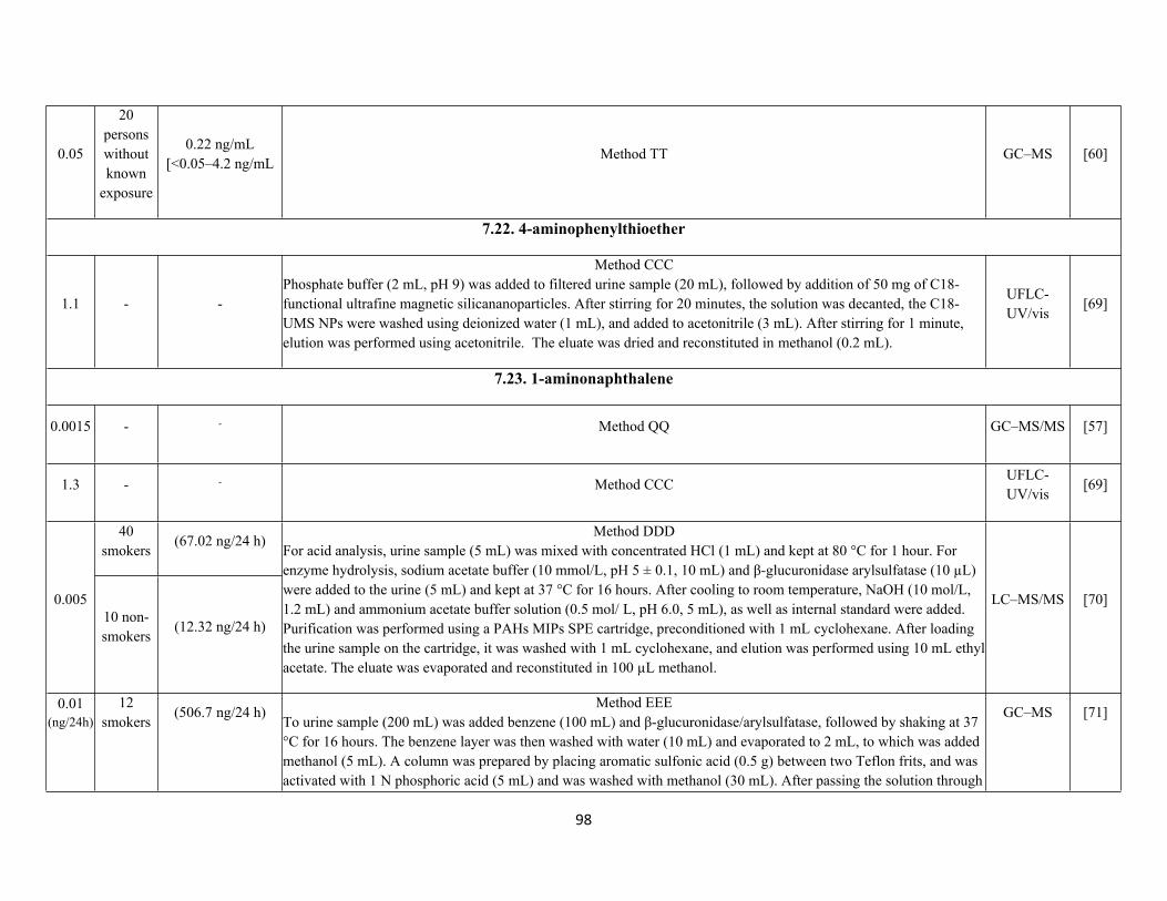
98
0.05
20 persons without known
exposure
0.22 ng/mL[<0.05–4.2 ng/mL
Method TT GC–MS [60]
7.22. 4-aminophenylthioether
1.1 - -
Method CCCPhosphate buffer (2 mL, pH 9) was added to filtered urine sample (20 mL), followed by addition of 50 mg of C18-functional ultrafine magnetic silicananoparticles. After stirring for 20 minutes, the solution was decanted, the C18-UMS NPs were washed using deionized water (1 mL), and added to acetonitrile (3 mL). After stirring for 1 minute, elution was performed using acetonitrile. The eluate was dried and reconstituted in methanol (0.2 mL).
UFLC-UV/vis
[69]
7.23. 1-aminonaphthalene
0.0015 - - Method QQ GC–MS/MS [57]
1.3 - - Method CCCUFLC-UV/vis
[69]
40 smokers
(67.02 ng/24 h)
0.00510 non-smokers
(12.32 ng/24 h)
Method DDDFor acid analysis, urine sample (5 mL) was mixed with concentrated HCl (1 mL) and kept at 80 °C for 1 hour. For enzyme hydrolysis, sodium acetate buffer (10 mmol/L, pH 5 ± 0.1, 10 mL) and β-glucuronidase arylsulfatase (10 µL) were added to the urine (5 mL) and kept at 37 °C for 16 hours. After cooling to room temperature, NaOH (10 mol/L, 1.2 mL) and ammonium acetate buffer solution (0.5 mol/ L, pH 6.0, 5 mL), as well as internal standard were added. Purification was performed using a PAHs MIPs SPE cartridge, preconditioned with 1 mL cyclohexane. After loading the urine sample on the cartridge, it was washed with 1 mL cyclohexane, and elution was performed using 10 mL ethyl acetate. The eluate was evaporated and reconstituted in 100 µL methanol.
LC–MS/MS [70]
0.01(ng/24h)
12 smokers
(506.7 ng/24 h)Method EEE
To urine sample (200 mL) was added benzene (100 mL) and β-glucuronidase/arylsulfatase, followed by shaking at 37 °C for 16 hours. The benzene layer was then washed with water (10 mL) and evaporated to 2 mL, to which was added methanol (5 mL). A column was prepared by placing aromatic sulfonic acid (0.5 g) between two Teflon frits, and was activated with 1 N phosphoric acid (5 mL) and was washed with methanol (30 mL). After passing the solution through
GC–MS [71]
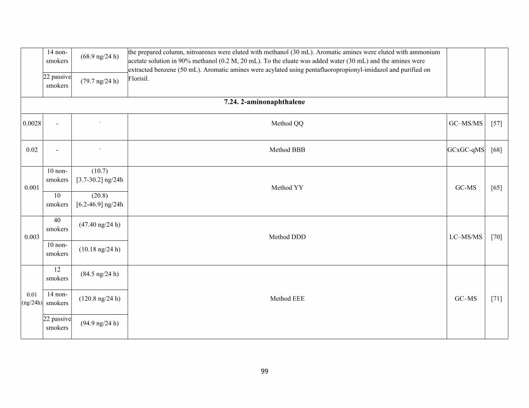
99
14 non-smokers
(68.9 ng/24 h)
22 passivesmokers
(79.7 ng/24 h)
the prepared column, nitroarenes were eluted with methanol (30 mL). Aromatic amines were eluted with ammoniumacetate solution in 90% methanol (0.2 M, 20 mL). To the eluate was added water (30 mL) and the amines were extracted benzene (50 mL). Aromatic amines were acylated using pentafluoropropionyl-imidazol and purified on Florisil.
7.24. 2-aminonaphthalene
0.0028 - - Method QQ GC–MS/MS [57]
0.02 - - Method BBB GCxGC-qMS [68]
10 non-smokers
(10.7)[3.7-30.2] ng/24h
0.00110
smokers(20.8)
[6.2-46.9] ng/24h
Method YY GC-MS [65]
40 smokers
(47.40 ng/24 h)
0.00310 non-smokers
(10.18 ng/24 h)
Method DDD LC–MS/MS [70]
12 smokers
(84.5 ng/24 h)
14 non-smokers
(120.8 ng/24 h)0.01(ng/24h)
22 passivesmokers
(94.9 ng/24 h)
Method EEE GC–MS [71]

100
8 smokers3.1 ng/mL
(3.1 ng/mL)[0.0-7.4 ng/mL]
1
8 non-smokers
0.0 ng/mL(0.5 ng/mL)
[0.0-1.6 ng/mL]
Method SS GC-ECD [59]
7.25. 2-aminobiphenyl
12 smokers
(66.5 ng/24 h)
14 non-smokers
(45.5 ng/24 h)0.01(ng/24h)
22 passivesmokers
(48.1 ng/24 h)
Method EEE GC–MS [71]
7.26. 3-aminobiphenyl
40 smokers
(5.39 ng/24 h)
0.00310 non-smokers
(1.11 ng/24 h)
Method DDD LC–MS/MS [70]
7.27. 4-aminobiphenyl
0.0018 - - Method QQ GC–MS/MS [57]
0.88 - - Method CCCUFLC-UV/vis
[69]
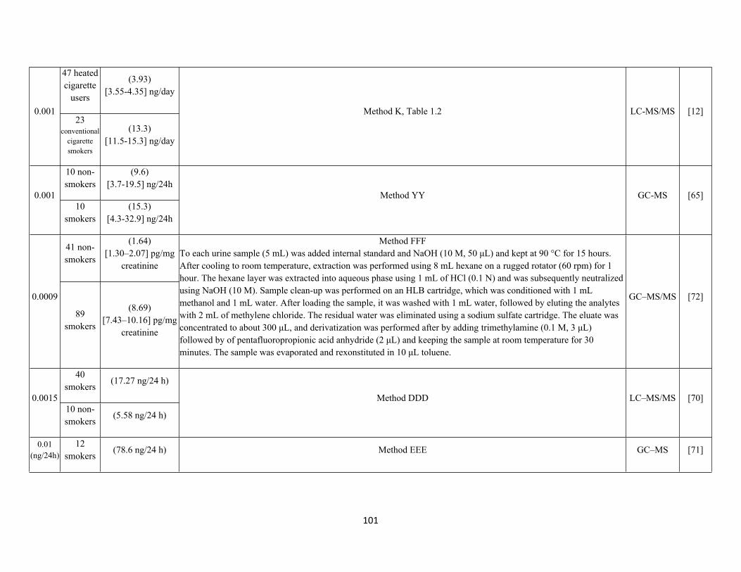
101
47 heated cigarette
users
(3.93)[3.55-4.35] ng/day
0.00123
conventional cigarette smokers
(13.3)[11.5-15.3] ng/day
Method K, Table 1.2 LC-MS/MS [12]
10 non-smokers
(9.6)[3.7-19.5] ng/24h
0.00110
smokers(15.3)
[4.3-32.9] ng/24h
Method YY GC-MS [65]
41 non-smokers
(1.64)[1.30–2.07] pg/mg
creatinine
0.0009
89 smokers
(8.69)[7.43–10.16] pg/mg
creatinine
Method FFFTo each urine sample (5 mL) was added internal standard and NaOH (10 M, 50 μL) and kept at 90 °C for 15 hours. After cooling to room temperature, extraction was performed using 8 mL hexane on a rugged rotator (60 rpm) for 1 hour. The hexane layer was extracted into aqueous phase using 1 mL of HCl (0.1 N) and was subsequently neutralized using NaOH (10 M). Sample clean-up was performed on an HLB cartridge, which was conditioned with 1 mL methanol and 1 mL water. After loading the sample, it was washed with 1 mL water, followed by eluting the analytes with 2 mL of methylene chloride. The residual water was eliminated using a sodium sulfate cartridge. The eluate was concentrated to about 300 μL, and derivatization was performed after by adding trimethylamine (0.1 M, 3 μL) followed by of pentafluoropropionic acid anhydride (2 μL) and keeping the sample at room temperature for 30 minutes. The sample was evaporated and rexonstituted in 10 μL toluene.
GC–MS/MS [72]
40 smokers
(17.27 ng/24 h)
0.001510 non-smokers
(5.58 ng/24 h)
Method DDD LC–MS/MS [70]
0.01(ng/24h)
12 smokers
(78.6 ng/24 h) Method EEE GC–MS [71]
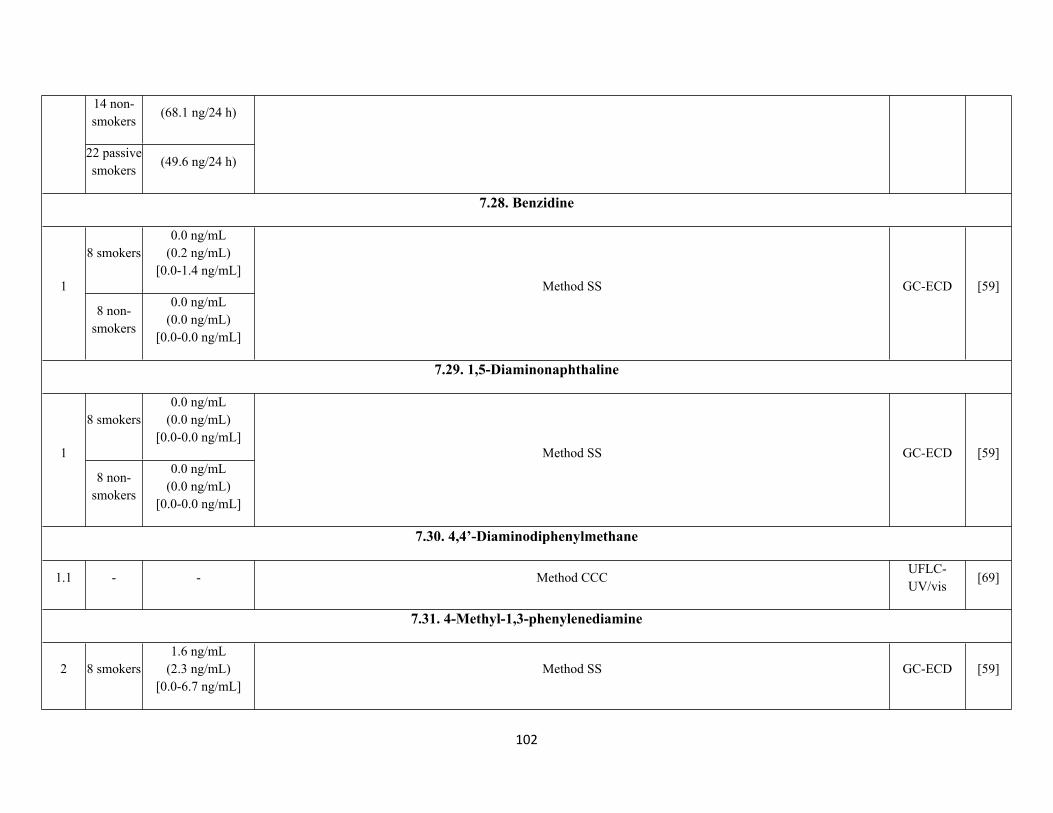
102
14 non-smokers
(68.1 ng/24 h)
22 passivesmokers
(49.6 ng/24 h)
7.28. Benzidine
8 smokers0.0 ng/mL
(0.2 ng/mL)[0.0-1.4 ng/mL]
1
8 non-smokers
0.0 ng/mL(0.0 ng/mL)
[0.0-0.0 ng/mL]
Method SS GC-ECD [59]
7.29. 1,5-Diaminonaphthaline
8 smokers0.0 ng/mL
(0.0 ng/mL)[0.0-0.0 ng/mL]
1
8 non-smokers
0.0 ng/mL(0.0 ng/mL)
[0.0-0.0 ng/mL]
Method SS GC-ECD [59]
7.30. 4,4’-Diaminodiphenylmethane
1.1 - - Method CCCUFLC-UV/vis
[69]
7.31. 4-Methyl-1,3-phenylenediamine
2 8 smokers1.6 ng/mL
(2.3 ng/mL)[0.0-6.7 ng/mL]
Method SS GC-ECD [59]
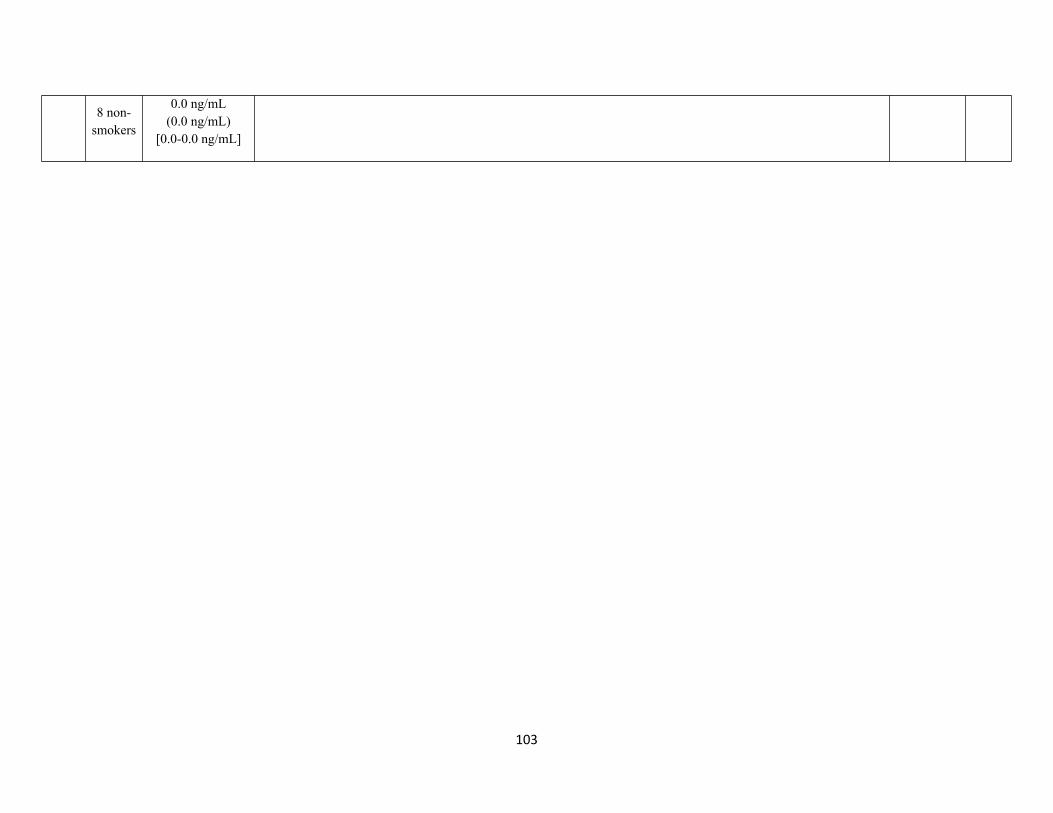
103
8 non-smokers
0.0 ng/mL(0.0 ng/mL)
[0.0-0.0 ng/mL]
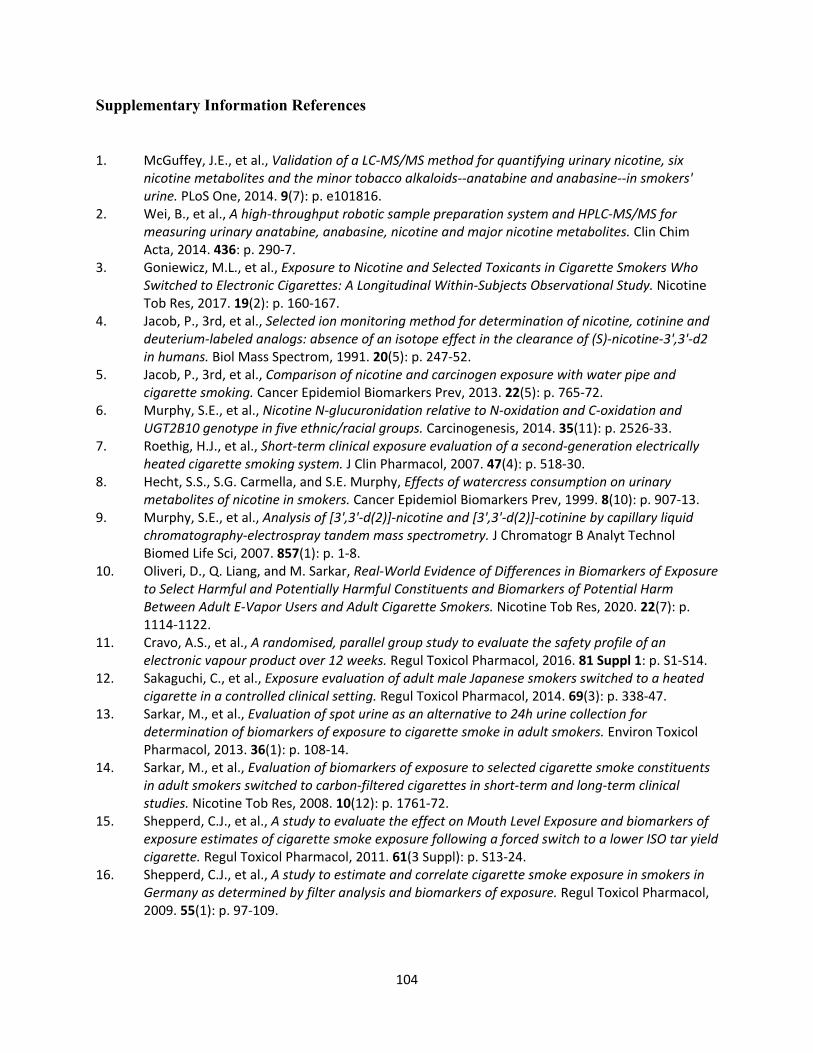
104
Supplementary Information References
1. McGuffey, J.E., et al., Validation of a LC-MS/MS method for quantifying urinary nicotine, six nicotine metabolites and the minor tobacco alkaloids--anatabine and anabasine--in smokers' urine. PLoS One, 2014. 9(7): p. e101816.
2. Wei, B., et al., A high-throughput robotic sample preparation system and HPLC-MS/MS for measuring urinary anatabine, anabasine, nicotine and major nicotine metabolites. Clin Chim Acta, 2014. 436: p. 290-7.
3. Goniewicz, M.L., et al., Exposure to Nicotine and Selected Toxicants in Cigarette Smokers Who Switched to Electronic Cigarettes: A Longitudinal Within-Subjects Observational Study. Nicotine Tob Res, 2017. 19(2): p. 160-167.
4. Jacob, P., 3rd, et al., Selected ion monitoring method for determination of nicotine, cotinine and deuterium-labeled analogs: absence of an isotope effect in the clearance of (S)-nicotine-3',3'-d2 in humans. Biol Mass Spectrom, 1991. 20(5): p. 247-52.
5. Jacob, P., 3rd, et al., Comparison of nicotine and carcinogen exposure with water pipe and cigarette smoking. Cancer Epidemiol Biomarkers Prev, 2013. 22(5): p. 765-72.
6. Murphy, S.E., et al., Nicotine N-glucuronidation relative to N-oxidation and C-oxidation and UGT2B10 genotype in five ethnic/racial groups. Carcinogenesis, 2014. 35(11): p. 2526-33.
7. Roethig, H.J., et al., Short-term clinical exposure evaluation of a second-generation electrically heated cigarette smoking system. J Clin Pharmacol, 2007. 47(4): p. 518-30.
8. Hecht, S.S., S.G. Carmella, and S.E. Murphy, Effects of watercress consumption on urinary metabolites of nicotine in smokers. Cancer Epidemiol Biomarkers Prev, 1999. 8(10): p. 907-13.
9. Murphy, S.E., et al., Analysis of [3',3'-d(2)]-nicotine and [3',3'-d(2)]-cotinine by capillary liquid chromatography-electrospray tandem mass spectrometry. J Chromatogr B Analyt Technol Biomed Life Sci, 2007. 857(1): p. 1-8.
10. Oliveri, D., Q. Liang, and M. Sarkar, Real-World Evidence of Differences in Biomarkers of Exposure to Select Harmful and Potentially Harmful Constituents and Biomarkers of Potential Harm Between Adult E-Vapor Users and Adult Cigarette Smokers. Nicotine Tob Res, 2020. 22(7): p. 1114-1122.
11. Cravo, A.S., et al., A randomised, parallel group study to evaluate the safety profile of an electronic vapour product over 12 weeks. Regul Toxicol Pharmacol, 2016. 81 Suppl 1: p. S1-S14.
12. Sakaguchi, C., et al., Exposure evaluation of adult male Japanese smokers switched to a heated cigarette in a controlled clinical setting. Regul Toxicol Pharmacol, 2014. 69(3): p. 338-47.
13. Sarkar, M., et al., Evaluation of spot urine as an alternative to 24h urine collection for determination of biomarkers of exposure to cigarette smoke in adult smokers. Environ Toxicol Pharmacol, 2013. 36(1): p. 108-14.
14. Sarkar, M., et al., Evaluation of biomarkers of exposure to selected cigarette smoke constituents in adult smokers switched to carbon-filtered cigarettes in short-term and long-term clinical studies. Nicotine Tob Res, 2008. 10(12): p. 1761-72.
15. Shepperd, C.J., et al., A study to evaluate the effect on Mouth Level Exposure and biomarkers of exposure estimates of cigarette smoke exposure following a forced switch to a lower ISO tar yield cigarette. Regul Toxicol Pharmacol, 2011. 61(3 Suppl): p. S13-24.
16. Shepperd, C.J., et al., A study to estimate and correlate cigarette smoke exposure in smokers in Germany as determined by filter analysis and biomarkers of exposure. Regul Toxicol Pharmacol, 2009. 55(1): p. 97-109.
105
17. Morin, A., et al., Estimation and correlation of cigarette smoke exposure in Canadian smokers as determined by filter analysis and biomarkers of exposure. Regul Toxicol Pharmacol, 2011. 61(3 Suppl): p. S3-12.
18. Hecht, S.S., et al., Quantitation of urinary metabolites of a tobacco-specific lung carcinogen after smoking cessation. Cancer Res, 1999. 59(3): p. 590-6.
19. Dempsey, D., et al., Nicotine metabolite ratio as an index of cytochrome P450 2A6 metabolic activity. Clin Pharmacol Ther, 2004. 76(1): p. 64-72.
20. Bernert, J.T., Jr., et al., Development and validation of sensitive method for determination of serum cotinine in smokers and nonsmokers by liquid chromatography/atmospheric pressure ionization tandem mass spectrometry. Clin Chem, 1997. 43(12): p. 2281-91.
21. Jacob, P., 3rd, et al., Determination of the nicotine metabolites cotinine and trans-3'-hydroxycotinine in biologic fluids of smokers and non-smokers using liquid chromatography-tandem mass spectrometry: biomarkers for tobacco smoke exposure and for phenotyping cytochrome P450 2A6 activity. J Chromatogr B Analyt Technol Biomed Life Sci, 2011. 879(3-4): p. 267-76.
22. McRobbie, H., et al., Effects of Switching to Electronic Cigarettes with and without Concurrent Smoking on Exposure to Nicotine, Carbon Monoxide, and Acrolein. Cancer Prev Res (Phila), 2015. 8(9): p. 873-8.
23. Kassem, N.O., et al., Children's exposure to secondhand and thirdhand smoke carcinogens and toxicants in homes of hookah smokers. Nicotine Tob Res, 2014. 16(7): p. 961-75.
24. Yuan, J.M., et al., Urinary levels of volatile organic carcinogen and toxicant biomarkers in relation to lung cancer development in smokers. Carcinogenesis, 2012. 33(4): p. 804-9.
25. Yuan, J.M., et al., Urinary levels of tobacco-specific nitrosamine metabolites in relation to lung cancer development in two prospective cohorts of cigarette smokers. Cancer Res, 2009. 69(7): p. 2990-5.
26. Parsons, W.D., et al., A metabolite of the tobacco-specific lung carcinogen 4-(methylnitrosamino)-1-(3-pyridyl)-1-butanone in the urine of hospital workers exposed to environmental tobacco smoke. Cancer Epidemiol Biomarkers Prev, 1998. 7(3): p. 257-60.
27. Yuan, J.M., et al., Urinary levels of cigarette smoke constituent metabolites are prospectively associated with lung cancer development in smokers. Cancer Res, 2011. 71(21): p. 6749-57.
28. Church, T.R., et al., A prospectively measured serum biomarker for a tobacco-specific carcinogen and lung cancer in smokers. Cancer Epidemiol Biomarkers Prev, 2009. 18(1): p. 260-6.
29. Frigerio, G., et al., Urinary biomonitoring of subjects with different smoking habits. Part I: Profiling mercapturic acids. Toxicol Lett, 2020. 327: p. 48-57.
30. Jacob, P., 3rd, et al., Determination of the nicotine metabolite trans-3'-hydroxycotinine in urine of smokers using gas chromatography with nitrogen-selective detection or selected ion monitoring. J Chromatogr, 1992. 583(2): p. 145-54.
31. Xia, B., et al., Quantitative analysis of five tobacco-specific N-nitrosamines in urine by liquid chromatography-atmospheric pressure ionization tandem mass spectrometry. Biomed Chromatogr, 2014. 28(3): p. 375-84.
32. Jacob, P., 3rd, et al., Subpicogram per milliliter determination of the tobacco-specific carcinogen metabolite 4-(methylnitrosamino)-1-(3-pyridyl)-1-butanol in human urine using liquid chromatography-tandem mass spectrometry. Anal Chem, 2008. 80(21): p. 8115-21.
33. Carmella, S.G., et al., High throughput liquid and gas chromatography-tandem mass spectrometry assays for tobacco-specific nitrosamine and polycyclic aromatic hydrocarbon metabolites associated with lung cancer in smokers. Chem Res Toxicol, 2013. 26(8): p. 1209-17.
34. Carmella, S.G., et al., Effects of smoking cessation on eight urinary tobacco carcinogen and toxicant biomarkers. Chem Res Toxicol, 2009. 22(4): p. 734-41.
106
35. Carmella, S.G., et al., Analysis of total 4-(methylnitrosamino)-1-(3-pyridyl)-1-butanol (NNAL) in human urine. Cancer Epidemiol Biomarkers Prev, 2003. 12(11 Pt 1): p. 1257-61.
36. Carmella, S.G., et al., Intraindividual and interindividual differences in metabolites of the tobacco-specific lung carcinogen 4-(methylnitrosamino)-1-(3-pyridyl)-1-butanone (NNK) in smokers' urine. Cancer Epidemiol Biomarkers Prev, 1995. 4(6): p. 635-42.
37. Carmella, S.G., S. Akerkar, and S.S. Hecht, Metabolites of the tobacco-specific nitrosamine 4-(methylnitrosamino)-1-(3-pyridyl)-1-butanone in smokers' urine. Cancer Res, 1993. 53(4): p. 721-4.
38. Hecht, S.S., et al., A tobacco-specific lung carcinogen in the urine of men exposed to cigarette smoke. N Engl J Med, 1993. 329(21): p. 1543-6.
39. Carmella, S.G., A. Yoder, and S.S. Hecht, Combined analysis of r-1,t-2,3,c-4-tetrahydroxy-1,2,3,4-tetrahydrophenanthrene and 4-(methylnitrosamino)-1-(3-pyridyl)-1-butanol in smokers' plasma. Cancer Epidemiol Biomarkers Prev, 2006. 15(8): p. 1490-4.
40. Alwis, K.U., et al., Simultaneous analysis of 28 urinary VOC metabolites using ultra high performance liquid chromatography coupled with electrospray ionization tandem mass spectrometry (UPLC-ESI/MSMS). Anal Chim Acta, 2012. 750: p. 152-60.
41. Boyle, E.B., et al., Assessment of Exposure to VOCs among Pregnant Women in the National Children's Study. Int J Environ Res Public Health, 2016. 13(4): p. 376.
42. Pluym, N., et al., Analysis of 18 urinary mercapturic acids by two high-throughput multiplex-LC-MS/MS methods. Anal Bioanal Chem, 2015. 407(18): p. 5463-76.
43. Yuan, J.M., et al., 2-Phenethyl Isothiocyanate, Glutathione S-transferase M1 and T1 Polymorphisms, and Detoxification of Volatile Organic Carcinogens and Toxicants in Tobacco Smoke. Cancer Prev Res (Phila), 2016. 9(7): p. 598-606.
44. Carmella, S.G., et al., High throughput liquid chromatography-tandem mass spectrometry assay for mercapturic acids of acrolein and crotonaldehyde in cigarette smokers' urine. J Chromatogr B Analyt Technol Biomed Life Sci, 2013. 935: p. 36-40.
45. Alwis, K.U., et al., Acrolein Exposure in U.S. Tobacco Smokers and Non-Tobacco Users: NHANES 2005-2006. Environ Health Perspect, 2015. 123(12): p. 1302-8.
46. Zhang, X., et al., Simultaneous determination of five mercapturic acid derived from volatile organic compounds in human urine by LC-MS/MS and its application to relationship study. J Chromatogr B Analyt Technol Biomed Life Sci, 2014. 967: p. 102-9.
47. Carmella, S.G., et al., Quantitation of acrolein-derived (3-hydroxypropyl)mercapturic acid in human urine by liquid chromatography-atmospheric pressure chemical ionization tandem mass spectrometry: effects of cigarette smoking. Chem Res Toxicol, 2007. 20(7): p. 986-90.
48. Mascher, D.G., et al., High-performance liquid chromatographic-tandem mass spectrometric determination of 3-hydroxypropylmercapturic acid in human urine. J Chromatogr B Biomed Sci Appl, 2001. 750(1): p. 163-9.
49. Minet, E., et al., An inter-laboratory comparison of urinary 3-hydroxypropylmercapturic acid measurement demonstrates good reproducibility between laboratories. BMC Res Notes, 2011. 4: p. 391.
50. Haiman, C.A., et al., Benzene Uptake and Glutathione S-transferase T1 Status as Determinants of S-Phenylmercapturic Acid in Cigarette Smokers in the Multiethnic Cohort. PLoS One, 2016. 11(3): p. e0150641.
51. Valentin-Blasini, L., B.C. Blount, and A. Delinsky, Quantification of iodide and sodium-iodide symporter inhibitors in human urine using ion chromatography tandem mass spectrometry. J Chromatogr A, 2007. 1155(1): p. 40-6.
107
52. Jacob, P., 3rd, M. Wilson, and N.L. Benowitz, Determination of phenolic metabolites of polycyclic aromatic hydrocarbons in human urine as their pentafluorobenzyl ether derivatives using liquid chromatography-tandem mass spectrometry. Anal Chem, 2007. 79(2): p. 587-98.
53. Hecht, S.S., et al., r-1,t-2,3,c-4-Tetrahydroxy-1,2,3,4-tetrahydrophenanthrene in human urine: a potential biomarker for assessing polycyclic aromatic hydrocarbon metabolic activation. Cancer Epidemiol Biomarkers Prev, 2003. 12(12): p. 1501-8.
54. Carmella, S.G., K.A. Le, and S.S. Hecht, Improved method for determination of 1-hydroxypyrene in human urine. Cancer Epidemiol Biomarkers Prev, 2004. 13(7): p. 1261-4.
55. Ichiba, M., et al., Decreasing urinary PAH metabolites and 7-methylguanine after smoking cessation. Int Arch Occup Environ Health, 2006. 79(7): p. 545-9.
56. Preuss, R., et al., Pilot study on the naphthalene exposure of German adults and children by means of urinary 1- and 2-naphthol levels. Int J Hyg Environ Health, 2004. 207(5): p. 441-5.
57. Mazumder, S., et al., A New Automated Method for the Analysis of Aromatic Amines in Human Urine by GC-MS/MS. J Anal Toxicol, 2019. 43(1): p. 25-35.
58. Teass, A.W., et al., Biological monitoring for occupational exposures to o-toluidine and aniline. Int Arch Occup Environ Health, 1993. 65(1 Suppl): p. S115-8.
59. Riffelmann, M., et al., Biomonitoring of urinary aromatic amines and arylamine hemoglobin adducts in exposed workers and nonexposed control persons. Int Arch Occup Environ Health, 1995. 68(1): p. 36-43.
60. Weiss, T. and J. Angerer, Simultaneous determination of various aromatic amines and metabolites of aromatic nitro compounds in urine for low level exposure using gas chromatography-mass spectrometry. J Chromatogr B Analyt Technol Biomed Life Sci, 2002. 778(1-2): p. 179-92.
61. DeBruin, L.S., P.D. Josephy, and J.B. Pawliszyn, Solid-phase microextraction of monocyclic aromatic amines from biological fluids. Anal Chem, 1998. 70(9): p. 1986-92.
62. Fuller, T.W., et al., Comparison of Bladder Carcinogens in the Urine of E-cigarette Users Versus Non E-cigarette Using Controls. Sci Rep, 2018. 8(1): p. 507.
63. Amiri, A., M. Baghayeri, and S. Nori, Magnetic solid-phase extraction using poly(para-phenylenediamine) modified with magnetic nanoparticles as adsorbent for analysis of monocyclic aromatic amines in water and urine samples. J Chromatogr A, 2015. 1415: p. 20-6.
64. Labat, L., et al., [Assessment of occupational exposure to ortho-toluidine using gas chromatography-mass spectrometry]. Acta Clin Belg, 2006. 61 Suppl 1: p. 63-7.
65. Riedel, K., et al., Determination of three carcinogenic aromatic amines in urine of smokers and nonsmokers. J Anal Toxicol, 2006. 30(3): p. 187-95.
66. el-Bayoumy, K., et al., Identification and quantitative determination of aniline and toluidines in human urine. Cancer Res, 1986. 46(12 Pt 1): p. 6064-7.
67. Ward, E.M., et al., Monitoring of aromatic amine exposures in workers at a chemical plant with a known bladder cancer excess. J Natl Cancer Inst, 1996. 88(15): p. 1046-52.
68. Lamani, X., et al., Determination of aromatic amines in human urine using comprehensive multi-dimensional gas chromatography mass spectrometry (GCxGC-qMS). Anal Bioanal Chem, 2015. 407(1): p. 241-52.
69. Jiang, C., et al., Application of C18-functional magnetic nanoparticles for extraction of aromatic amines from human urine. J Chromatogr B Analyt Technol Biomed Life Sci, 2014. 947-948: p. 49-56.
70. Yu, J., et al., Determination of urinary aromatic amines in smokers and nonsmokers using a MIPs-SPE coupled with LC-MS/MS method. J Chromatogr B Analyt Technol Biomed Life Sci, 2014. 958: p. 130-5.
108
71. Grimmer, G., et al., Detection of carcinogenic aromatic amines in the urine of non-smokers. Sci Total Environ, 2000. 247(1): p. 81-90.
72. Seyler, T.H. and J.T. Bernert, Analysis of 4-aminobiphenyl in smoker's and nonsmoker's urine by tandem mass spectrometry. Biomarkers, 2011. 16(3): p. 212-21.