Embed Size (px)
Citation preview
Aquat. Living Resour. 17, 499–517 (2004)c© EDP Sciences, IFREMER, IRD 2004DOI: 10.1051/alr:2004056www.edpsciences.org/alr
AquaticLivingResources
A review of recent information on the Haplosporidia, with specialreference to Haplosporidium nelsoni (MSX disease)
Eugene M. Burresona,1 and Susan E. Ford2
1 Virginia Institute of Marine Science, College of William and Mary, Gloucester Point, Virginia 23062, USA2 Haskin Shellfish Research Laboratory, Rutgers University, Port Norris, New Jersey 08349, USA
Abstract – The current status of the Haplosporidia is reviewed as well as recent information on Haplosporidium nel-soni, the causative agent of MSX disease in oysters. Recent molecular phylogenetic analyses with greatly increasedtaxon sampling support monophyly of the Haplosporidia and hypothesize placement of the group as sister taxon to thephylum Cercozoa. Oyster pathogens in the genus Bonamia should be considered haplosporidians based on molecularsequence data. Thus, the group contains 4 genera: Uropsoridium, Haplosporidium, Bonamia and Minchinia. Molecularphylogenetic analyses support monophyly of Urosporidium, Bonamia and Minchinia, but Haplosporidium forms a pa-raphyletic clade. Reports of haplosporidia worldwide are reviewed. Molecular detection assays have greatly increasedour ability to rapidly and specifically diagnose important pathogens in the phylum and have also improved our under-standing of the distribution and biology of H. nelsoni and H. costale. Much of the data available for H. nelsoni has beenintegrated into a mathematical model of host/parasite/environment interactions. Model simulations support hypothesesthat recent H. nelsoni outbreaks in the NE United States are related to increased winter temperatures, and that a hostother than oysters is involved in the life cycle. Evidence is presented that natural resistance to H. nelsoni has developedin oysters in Delaware Bay, USA. However, in Chesapeake Bay, USA H. nelsoni has intensified in historically lowsalinity areas where salinities have increased because of recent drought conditions. Efforts to mitigate the impact ofH. nelsoni involve selective breeding programs for disease resistance and the evaluation of disease resistant non-nativeoysters.
Key words: Phylogeny / Diagnostics / Numerical model / Haplosporidia / Haplosporidium nelsoni
1 Introduction
The Haplosporidia constitute a small group of endopara-sites, mostly of marine invertebrates (Perkins 2000), althoughone species is known from freshwater invertebrates. At presentthere are 36 recognized species in the phylum; however, nu-merous others have been reported, but not specifically identi-fied, from many different invertebrate hosts. Several specieshave been associated with epizootic mortalities of commer-cially important molluscs. The most well-studied member ofthe group is Haplosporidium nelsoni, which causes MSX dis-ease in the eastern oyster, Crassostrea virginica, on the eastcoast of North America. This parasite, along with a closelyrelated species, H. costale, which causes SSO disease, alsoin the eastern oyster, were covered in a 1996 review (Fordand Tripp 1996) that considered history and distribution, lifestages, infection and disease processes; epizootiology and en-vironmental influences; and control/management measures,including selective breeding for disease resistance. General re-views of the phylum Haplosporidia include contributions by
a Corresponding author: [email protected]
Perkins (1990, 1991, 2000) as well as an earlier review bySprague (1979).
The present review will emphasize recent developments,which include research on H. nelsoni that has occurred sincethe 1996 publication. They include: 1) progress in charac-terizing, phylogenetically, the Haplosporidia; 2) reports ofnew species of Haplosporidia and new hosts; 3) developmentand implementation of molecular detection assays; 4) numer-ical modeling of H. nelsoni; and 5) changes in the distribu-tion of H. nelsoni epizootics, including prevalence decline inDelaware Bay, and intensification of disease-caused oystermortalities in Chesapeake Bay that have led to the testing ofnon native oysters and selectively-bred native oysters.
2 Phylogenetic position of the Haplosporidia
2.1 Historical perspective
Since the discovery of the first species in the late1800s, the Haplosporidia have been a troublesome group for
500 E.M. Burreson and S.E. Ford: Aquat. Living Resour. 17, 499–517 (2004)
taxonomists and phylogeneticists, and there have been nu-merous classification schemes proposed for placement ofthe group within the protists. Early workers placed speciesin the order Haplosporida, class Sporozoa of the phylumProtozoa. With the advent of electron microscopy in the1950s, the tremendous morphological diversity of single-celled organisms became apparent and many groups of pro-tists were elevated to phylum rank. Sprague (1979) sepa-rated the Haplosporida and Paramyxea from other Sporozoaby including both groups in the new phylum Ascetospora.The phylum Ascetospora was subsequently abandoned and theHaplosporidia and Paramyxea were each elevated to phylumrank (Desportes and Perkins 1990; Perkins 1990, 1991, 2000).However, recently Cavalier-Smith and Chao (2003b) resur-rected Ascetospora as a class in the phylum Cercozoa, subphy-lum Endomyxa. In their scheme class Ascetospora includesthree orders – Haplosporida, Paramyxida and Claustrosporida(but see Sect. 2.2). The Haplosporidia were most recently char-acterized morphologically as a group of parasitic protists hav-ing multinucleate plasmodia and ovoid, walled spores lackingpolar filaments or polar tubes, and with an orifice at one pole.The orifice is covered either externally by a hinged lid or in-ternally by a flap of wall material (Perkins 2000). The place-ment of the genus Bonamia in the Haplosporidia (see Sect. 3.2)muddles this definition of the group because no spore stage hasbeen observed in Bonamia. If a spore stage is truly lacking inBonamia spp. it is unclear at present what morphological char-acters define Haplosporidia.
2.2 Molecular phylogenetic analyses
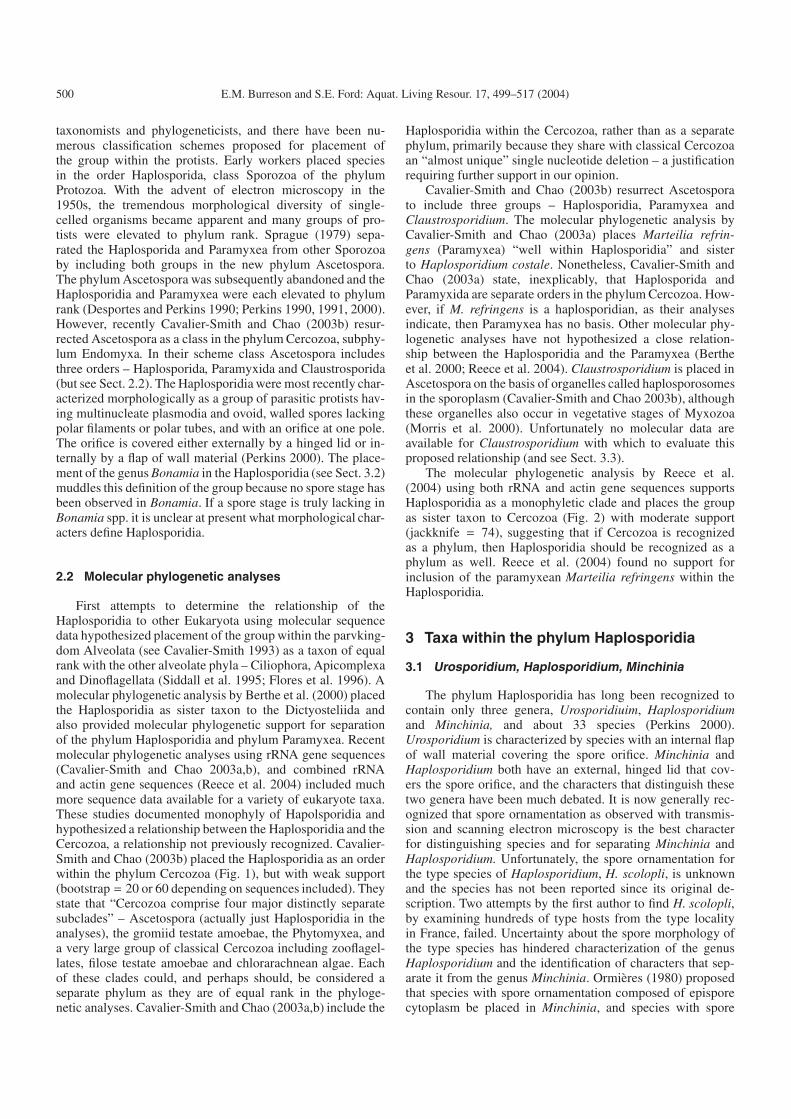
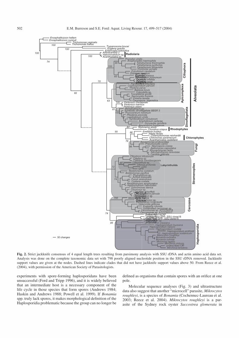
First attempts to determine the relationship of theHaplosporidia to other Eukaryota using molecular sequencedata hypothesized placement of the group within the parvking-dom Alveolata (see Cavalier-Smith 1993) as a taxon of equalrank with the other alveolate phyla – Ciliophora, Apicomplexaand Dinoflagellata (Siddall et al. 1995; Flores et al. 1996). Amolecular phylogenetic analysis by Berthe et al. (2000) placedthe Haplosporidia as sister taxon to the Dictyosteliida andalso provided molecular phylogenetic support for separationof the phylum Haplosporidia and phylum Paramyxea. Recentmolecular phylogenetic analyses using rRNA gene sequences(Cavalier-Smith and Chao 2003a,b), and combined rRNAand actin gene sequences (Reece et al. 2004) included muchmore sequence data available for a variety of eukaryote taxa.These studies documented monophyly of Hapolsporidia andhypothesized a relationship between the Haplosporidia and theCercozoa, a relationship not previously recognized. Cavalier-Smith and Chao (2003b) placed the Haplosporidia as an orderwithin the phylum Cercozoa (Fig. 1), but with weak support(bootstrap = 20 or 60 depending on sequences included). Theystate that “Cercozoa comprise four major distinctly separatesubclades” – Ascetospora (actually just Haplosporidia in theanalyses), the gromiid testate amoebae, the Phytomyxea, anda very large group of classical Cercozoa including zooflagel-lates, filose testate amoebae and chlorarachnean algae. Eachof these clades could, and perhaps should, be considered aseparate phylum as they are of equal rank in the phyloge-netic analyses. Cavalier-Smith and Chao (2003a,b) include the
Haplosporidia within the Cercozoa, rather than as a separatephylum, primarily because they share with classical Cercozoaan “almost unique” single nucleotide deletion – a justificationrequiring further support in our opinion.
Cavalier-Smith and Chao (2003b) resurrect Ascetosporato include three groups – Haplosporidia, Paramyxea andClaustrosporidium. The molecular phylogenetic analysis byCavalier-Smith and Chao (2003a) places Marteilia refrin-gens (Paramyxea) “well within Haplosporidia” and sisterto Haplosporidium costale. Nonetheless, Cavalier-Smith andChao (2003a) state, inexplicably, that Haplosporida andParamyxida are separate orders in the phylum Cercozoa. How-ever, if M. refringens is a haplosporidian, as their analysesindicate, then Paramyxea has no basis. Other molecular phy-logenetic analyses have not hypothesized a close relation-ship between the Haplosporidia and the Paramyxea (Bertheet al. 2000; Reece et al. 2004). Claustrosporidium is placed inAscetospora on the basis of organelles called haplosporosomesin the sporoplasm (Cavalier-Smith and Chao 2003b), althoughthese organelles also occur in vegetative stages of Myxozoa(Morris et al. 2000). Unfortunately no molecular data areavailable for Claustrosporidium with which to evaluate thisproposed relationship (and see Sect. 3.3).
The molecular phylogenetic analysis by Reece et al.(2004) using both rRNA and actin gene sequences supportsHaplosporidia as a monophyletic clade and places the groupas sister taxon to Cercozoa (Fig. 2) with moderate support(jackknife = 74), suggesting that if Cercozoa is recognizedas a phylum, then Haplosporidia should be recognized as aphylum as well. Reece et al. (2004) found no support forinclusion of the paramyxean Marteilia refringens within theHaplosporidia.
3 Taxa within the phylum Haplosporidia
3.1 Urosporidium, Haplosporidium, Minchinia
The phylum Haplosporidia has long been recognized tocontain only three genera, Urosporidiuim, Haplosporidiumand Minchinia, and about 33 species (Perkins 2000).Urosporidium is characterized by species with an internal flapof wall material covering the spore orifice. Minchinia andHaplosporidium both have an external, hinged lid that cov-ers the spore orifice, and the characters that distinguish thesetwo genera have been much debated. It is now generally rec-ognized that spore ornamentation as observed with transmis-sion and scanning electron microscopy is the best characterfor distinguishing species and for separating Minchinia andHaplosporidium. Unfortunately, the spore ornamentation forthe type species of Haplosporidium, H. scolopli, is unknownand the species has not been reported since its original de-scription. Two attempts by the first author to find H. scolopli,by examining hundreds of type hosts from the type localityin France, failed. Uncertainty about the spore morphology ofthe type species has hindered characterization of the genusHaplosporidium and the identification of characters that sep-arate it from the genus Minchinia. Ormières (1980) proposedthat species with spore ornamentation composed of episporecytoplasm be placed in Minchinia, and species with spore
E.M. Burreson and S.E. Ford: Aquat. Living Resour. 17, 499–517 (2004) 501
Fig. 1. Maximum likelihood tree of 50 rhizarian 18S rRNAs using 1638 positions (Γ + I model: α = 0.55084; i = 0.26839). This tree hadthe highest log likelihood (−25 487.62) of those yielded by 11 independent random additions of taxa. New sequences in bold. The figuresare bootstrap percentages (bold if 80% or more) using the same maximum likelihood model. From Cavalier-Smith and Chao (2003b), withpermission of Urban & Fischer Verlag.
ornamentation composed of spore wall material be placedin Haplosporidium. Most recent workers have accepted thisconvention (McGovern and Burreson 1990; Hine and Thorne1998, 2002; Azevedo et al. 1999; Azevedo 2001; Burreson2001). However, Perkins (2000) based generic assignmentsolely on whether spore ornamentation is visible with a lightmicroscope, without regard for ontogenetic origin of the or-namentation. Thus, Perkins (2000) proposed that Minchiniaincludes species in which the ornamentation is visible witha light microscope and Haplosporidium includes species inwhich ornamentation is not visible with a light microscope.
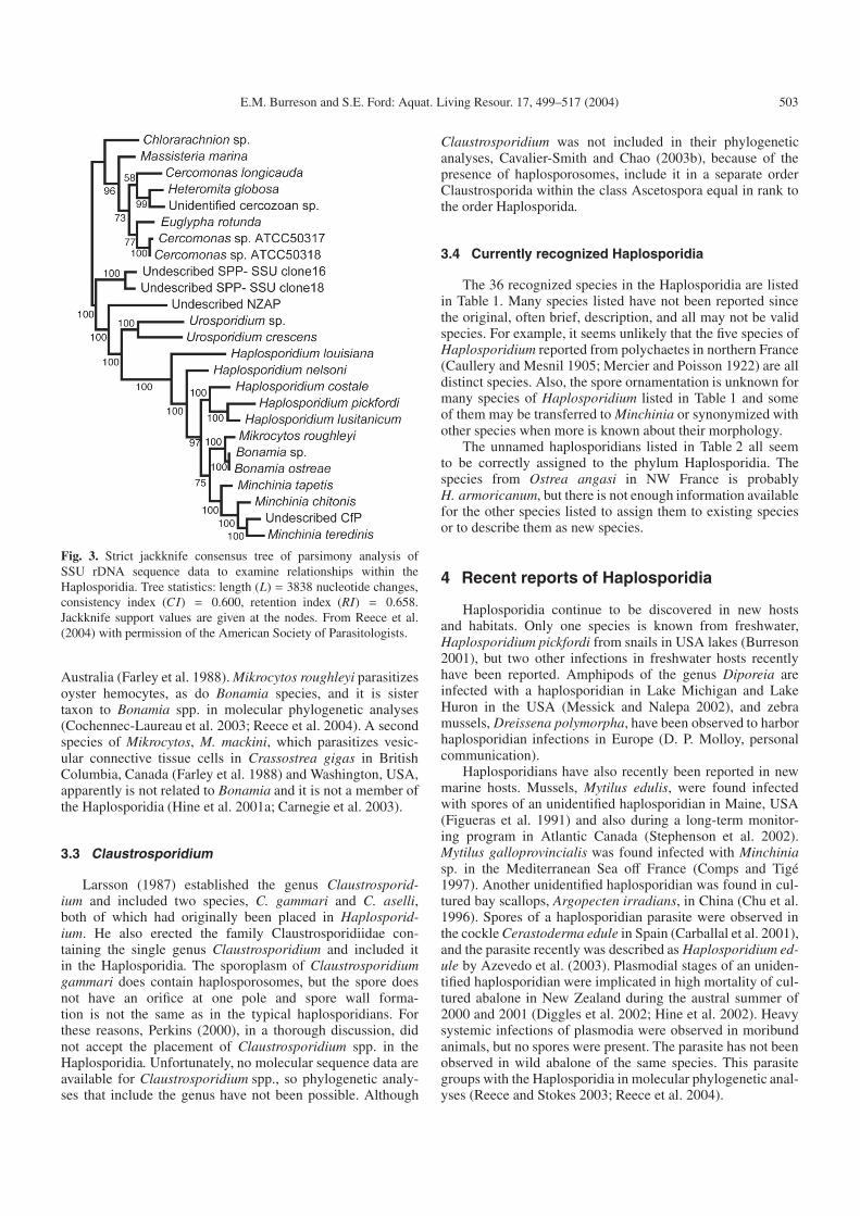
The recent molecular phylogenetic analysis by Reece et al.(2004) supports the importance of ontogenetic origin of sporeornamentation. In their analysis (Fig. 3) the genus Minchiniaformed a monophyletic clade, and all species of Minchiniahave ornamentation composed of epispore cytoplasm. Thegenus Haplosporidium, however, formed a paraphyletic clade(Fig. 3), suggesting that more genera are necessary to encom-pass the morphological diversity of species with ornamenta-tion derived from the spore wall. Unfortunately, new genericassignments cannot be made at the present time because ofthe lack of knowledge on ornamentation of the type speciesof Haplosporidium, H. scolopli, and of many other speciespresently assigned to Haplosporidium.
3.2 Bonamia
Perhaps the most interesting new finding is molecular phy-logenetic support for inclusion of the genus Bonamia in thephylum Haplosporidia (Carnegie et al. 2000; Reece and Stokes2003; Reece et al. 2004). Bonamia has long been suspectedto be a haplosporidian because of the presence of organellescalled haplosporosomes (Perkins 2000), but no spore stagehas been observed, so the genus had previously not been as-signed with certainty to any group. In a recent molecular phy-logenetic analysis (Reece et al. 2004), species of Bonamiaformed a monophyletic clade nested within the traditional hap-losporidian taxa, as sister taxa to Minchinia spp., not as abasal clade (Fig. 3). This alignment as sister taxon to a spore-forming genus suggests that Bonamia does form spores, soperhaps the stages observed to date are intermediate life cy-cle stages and spores are formed in some other, as yet uniden-tified, organism. Alternatively, it is possible that spores havebeen lost in the Bonamia lineage. Loss of spores is sup-ported by the observation that Bonamia ostreae can be trans-mitted directly between oyster hosts in the laboratory via co-habitation (Elston et al. 1986) or by inoculation of purifiedintrahemocyte stages (Hervio et al. 1995). With the possibleexception of H. pickfordi (Barrow 1961), direct transmission
502 E.M. Burreson and S.E. Ford: Aquat. Living Resour. 17, 499–517 (2004)
Fig. 2. Strict jackknife consensus of 4 equal length trees resulting from parsimony analysis with SSU rDNA and actin amino acid data set.Analysis was done on the complete taxonomic data set with 798 poorly aligned nucleotide position in the SSU rDNA removed. Jackknifesupport values are given at the nodes. Dashed lines indicate clades that did not have jackknife support values above 50. From Reece et al.(2004), with permission of the American Society of Parasitologists.
experiments with spore-forming haplosporidans have beenunsuccessful (Ford and Tripp 1996), and it is widely believedthat an intermediate host is a necessary component of thelife cycle in those species that form spores (Andrews 1984;Haskin and Andrews 1988; Powell et al. 1999). If Bonamiaspp. truly lack spores, it makes morphological definition of theHaplosporidia problematic because the group can no longer be
defined as organisms that contain spores with an orifice at onepole.
Molecular sequence analyses (Fig. 3) and ultrastructuredata also suggest that another “microcell” parasite, Mikrocytosroughleyi, is a species of Bonamia (Cochennec-Laureau et al.2003; Reece et al. 2004). Mikrocytos roughleyi is a par-asite of the Sydney rock oyster Saccostrea glomerata in
E.M. Burreson and S.E. Ford: Aquat. Living Resour. 17, 499–517 (2004) 503
Fig. 3. Strict jackknife consensus tree of parsimony analysis ofSSU rDNA sequence data to examine relationships within theHaplosporidia. Tree statistics: length (L) = 3838 nucleotide changes,consistency index (CI) = 0.600, retention index (RI) = 0.658.Jackknife support values are given at the nodes. From Reece et al.(2004) with permission of the American Society of Parasitologists.
Australia (Farley et al. 1988). Mikrocytos roughleyi parasitizesoyster hemocytes, as do Bonamia species, and it is sistertaxon to Bonamia spp. in molecular phylogenetic analyses(Cochennec-Laureau et al. 2003; Reece et al. 2004). A secondspecies of Mikrocytos, M. mackini, which parasitizes vesic-ular connective tissue cells in Crassostrea gigas in BritishColumbia, Canada (Farley et al. 1988) and Washington, USA,apparently is not related to Bonamia and it is not a member ofthe Haplosporidia (Hine et al. 2001a; Carnegie et al. 2003).
3.3 Claustrosporidium
Larsson (1987) established the genus Claustrosporid-ium and included two species, C. gammari and C. aselli,both of which had originally been placed in Haplosporid-ium. He also erected the family Claustrosporidiidae con-taining the single genus Claustrosporidium and included itin the Haplosporidia. The sporoplasm of Claustrosporidiumgammari does contain haplosporosomes, but the spore doesnot have an orifice at one pole and spore wall forma-tion is not the same as in the typical haplosporidians. Forthese reasons, Perkins (2000), in a thorough discussion, didnot accept the placement of Claustrosporidium spp. in theHaplosporidia. Unfortunately, no molecular sequence data areavailable for Claustrosporidium spp., so phylogenetic analy-ses that include the genus have not been possible. Although
Claustrosporidium was not included in their phylogeneticanalyses, Cavalier-Smith and Chao (2003b), because of thepresence of haplosporosomes, include it in a separate orderClaustrosporida within the class Ascetospora equal in rank tothe order Haplosporida.
3.4 Currently recognized Haplosporidia
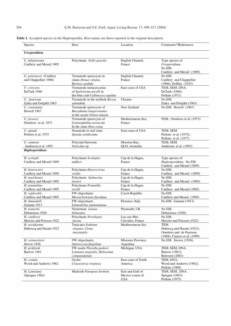
The 36 recognized species in the Haplosporidia are listedin Table 1. Many species listed have not been reported sincethe original, often brief, description, and all may not be validspecies. For example, it seems unlikely that the five species ofHaplosporidium reported from polychaetes in northern France(Caullery and Mesnil 1905; Mercier and Poisson 1922) are alldistinct species. Also, the spore ornamentation is unknown formany species of Haplosporidium listed in Table 1 and someof them may be transferred to Minchinia or synonymized withother species when more is known about their morphology.
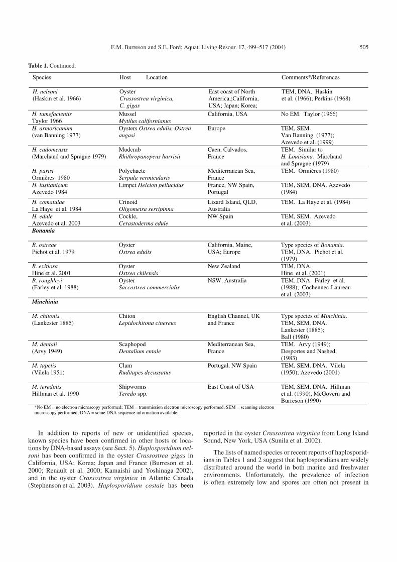
The unnamed haplosporidians listed in Table 2 all seemto be correctly assigned to the phylum Haplosporidia. Thespecies from Ostrea angasi in NW France is probablyH. armoricanum, but there is not enough information availablefor the other species listed to assign them to existing speciesor to describe them as new species.
4 Recent reports of Haplosporidia
Haplosporidia continue to be discovered in new hostsand habitats. Only one species is known from freshwater,Haplosporidium pickfordi from snails in USA lakes (Burreson2001), but two other infections in freshwater hosts recentlyhave been reported. Amphipods of the genus Diporeia areinfected with a haplosporidian in Lake Michigan and LakeHuron in the USA (Messick and Nalepa 2002), and zebramussels, Dreissena polymorpha, have been observed to harborhaplosporidian infections in Europe (D. P. Molloy, personalcommunication).
Haplosporidians have also recently been reported in newmarine hosts. Mussels, Mytilus edulis, were found infectedwith spores of an unidentified haplosporidian in Maine, USA(Figueras et al. 1991) and also during a long-term monitor-ing program in Atlantic Canada (Stephenson et al. 2002).Mytilus galloprovincialis was found infected with Minchiniasp. in the Mediterranean Sea off France (Comps and Tigé1997). Another unidentified haplosporidian was found in cul-tured bay scallops, Argopecten irradians, in China (Chu et al.1996). Spores of a haplosporidian parasite were observed inthe cockle Cerastoderma edule in Spain (Carballal et al. 2001),and the parasite recently was described as Haplosporidium ed-ule by Azevedo et al. (2003). Plasmodial stages of an uniden-tified haplosporidian were implicated in high mortality of cul-tured abalone in New Zealand during the austral summer of2000 and 2001 (Diggles et al. 2002; Hine et al. 2002). Heavysystemic infections of plasmodia were observed in moribundanimals, but no spores were present. The parasite has not beenobserved in wild abalone of the same species. This parasitegroups with the Haplosporidia in molecular phylogenetic anal-yses (Reece and Stokes 2003; Reece et al. 2004).
504 E.M. Burreson and S.E. Ford: Aquat. Living Resour. 17, 499–517 (2004)
Table 1. Accepted species in the Haplosporidia. Host names are those reported in the original description.
Species
Host Location Comments*/References
Urosporidium
U. fuliginosum Caullery and Mesnil 1905
Polychaete Syllis gracilis English Channel, France
Type species of Urosporidium. No EM. Caullery and Mesnil (1905)
U. pelseneeri (Caullery and Chappellier 1906)
Trematode sporocysts in clams Donax vittatus, Barnea candida
English Channel, France
No EM. Caullery and Chappellier (1906); Dollfus (1925)
U. crescens DeTurk 1940
Trematode metacercariae of Spelotrema nicolli in the blue crab Callinectes sapidus
East coast of USA TEM, SEM, DNA. DeTurk (1940); Perkins (1971)
U. fauricum Zaika and Dolgikh 1963
Trematode in the mollusk Rissoa splandida
Ukraine No EM. Zaika and Dolgikh (1963)
U. constantae Howell 1967
Trematode sporocysts of Bucephalus longicornutus in the oyster Ostrea lutaria
New Zealand No EM. Howell (1967)
U. jiroveci Ormières et al. 1973
Trematode sporocysts of Gymnophallus nereicola In the clam Abra ovata
Mediterranean Sea, France
TEM. Ormières et al. (1973)
U. spisuli Perkins et al. 1975
Nematode in surf clam Spisula solidissima
East coast of USA TEM, SEM. Perkins et al. (1975); Perkins et al. (1977)
U. cannoni Anderson et al. 1993
Polyclad flatworm Stylochus sp.
Moreton Bay, QLD, Australia
TEM, SEM. Anderson et al. (1993)
Haplosporidium
H. scolopli Caullery and Mesnil 1899
Polychaete Scoloplos mülleri.
Cap de la Hague, France
Type species of Haplosporidium. No EM. Caullery and Mesnil (1899)
H. heterocirri Caullery and Mesnil 1899
Polychaete Heterocirrus viridis
Cap de la Hague, France
No EM. Caullery and Mesnil (1899)
H. marchouxi Caullery and Mesnil 1905
Polychaete Salmacina dysteri
Cap de la Hague, France
No EM. Caullery and Mesnil (1905)
H. potamillae Caullery and Mesnil 1905
Polychaete Potamilla torelli
Cap de la Hague, France
No EM. Caullery and Mesnil (1905)
H. vejdovskii Caullery and Mesnil 1905
FW oligochaete Mesenchytraeus flaviduus
Czech Republic No EM. Caullery and Mesnil (1905)
H. limnodrili Granata 1913
FW oligochaete Limnodrilus udekemianus
Florence, Italy No EM. Granata (1913)
H. nemertis Debaisieux 1920
Nemertean Lineus bilineatus
Plymouth, UK No EM. Debaisieux (1920)
H. caulleryi Mercier and Poisson 1922
Polychaete Nereilepas fucata
Luc-sur-Mer, Calvados, France
No EM. Mercier and Poisson (1922)
H. ascidiarum Duboscq and Harant 1923
Tunicates Sydnium elegans, Ciona intestinalis
Mediterranean Sea TEM. Duboscq and Harant (1923); Ormières and de Puytorac (1968); Ciancio et al. (1999)
H. cernosvitovi Jírovic 1936
FW oligochaete, Opistocysta flagellum
Misiones Province, Argentina
No EM. Jírovec (1936)
H. pickfordi Barrow 1961
FW snails Physella parkeri, Lymnaea stagnalis, Heliosoma companulatum
Michigan, USA TEM, SEM, DNA. Barrow (1961); Burreson (2001)
H. costale Wood and Andrews 1962
Oyster Crassostrea virginica
East coast of North America
TEM, DNA. Wood and Andrews (1962); Perkins (1969)
H. Louisiana (Sprague 1963)
Mudcrab Panopeus berbstii East and Gulf of Mexico coasts of USA
TEM, SEM., DNA. Sprague (1963); Perkins (1975)
E.M. Burreson and S.E. Ford: Aquat. Living Resour. 17, 499–517 (2004) 505
Table 1. Continued.
H. tumefacientis Taylor 1966
Mussel Mytilus californianus
California, USA No EM. Taylor (1966)
H. armoricanum (van Banning 1977)
Oysters Ostrea edulis, Ostrea angasi
Europe TEM, SEM. Van Banning (1977); Azevedo et al. (1999)
H. cadomensis (Marchand and Sprague 1979)
Mudcrab Rhithropanopeus harrisii
Caen, Calvados, France
TEM. Similar to H. Louisiana. Marchand and Sprague (1979)
H. parisi Ormières 1980
Polychaete Serpula vermicularis
Mediterranean Sea, France
TEM. Ormières (1980)
H. lusitanicum Azevedo 1984
Limpet Helcion pellucidus France, NW Spain, Portugal
TEM, SEM, DNA. Azevedo (1984)
H. comatulae La Haye et al. 1984
Crinoid Oligometra serripinna
Lizard Island, QLD, Australia
TEM. La Haye et al. (1984)
H. edule Azevedo et al. 2003
Cockle, Cerastoderma edule
NW Spain TEM, SEM. Azevedo et al. (2003)
Bonamia
B. ostreae Pichot et al. 1979
Oyster Ostrea edulis
California, Maine, USA; Europe
Type species of Bonamia. TEM, DNA. Pichot et al. (1979)
B. exitiosa Hine et al. 2001
Oyster Ostrea chilensis
New Zealand TEM, DNA. Hine et al. (2001)
B. roughleyi (Farley et al. 1988)
Oyster Saccostrea commercialis
NSW, Australia TEM, DNA. Farley et al. (1988); Cochennec-Laureau et al. (2003)
Minchinia
M. chitonis (Lankester 1885)
Chiton Lepidochitona cinereus
English Channel, UK and France
Type species of Minchinia. TEM, SEM, DNA. Lankester (1885); Ball (1980)
M. dentali (Arvy 1949)
Scaphopod Dentalium entale
Mediterranean Sea, France
TEM. Arvy (1949); Desportes and Nashed, (1983)
M. tapetis (Vilela 1951)
Clam Ruditapes decussatus
Portugal, NW Spain TEM, SEM, DNA. Vilela (1950); Azevedo (2001)
M. teredinis Hillman et al. 1990
Shipworms Teredo spp.
East Coast of USA TEM, SEM, DNA. Hillman et al. (1990), McGovern and Burreson (1990)
*No EM = no electron microscopy performed; TEM = transmission electron microscopy performed, SEM = scanning electron microscopy performed; DNA = some DNA sequence information available.
Species Host Location Comments*/References
H. nelsoni (Haskin et al. 1966)
Oyster Crassostrea virginica, C. gigas
East coast of North America,;California, USA; Japan; Korea;
TEM, DNA. Haskin et al. (1966); Perkins (1968)
In addition to reports of new or unidentified species,known species have been confirmed in other hosts or loca-tions by DNA-based assays (see Sect. 5). Haplosporidium nel-soni has been confirmed in the oyster Crassostrea gigas inCalifornia, USA; Korea; Japan and France (Burreson et al.2000; Renault et al. 2000; Kamaishi and Yoshinaga 2002),and in the oyster Crassostrea virginica in Atlantic Canada(Stephenson et al. 2003). Haplosporidium costale has been
reported in the oyster Crassostrea virginica from Long IslandSound, New York, USA (Sunila et al. 2002).
The lists of named species or recent reports of haplosporid-ians in Tables 1 and 2 suggest that haplosporidians are widelydistributed around the world in both marine and freshwaterenvironments. Unfortunately, the prevalence of infectionis often extremely low and spores are often not present in
506 E.M. Burreson and S.E. Ford: Aquat. Living Resour. 17, 499–517 (2004)
Table 2. Reports of unnamed haplosporidians.
infected hosts, making it difficult to obtain sufficient materialfor species descriptions.
5 Development and implementationof molecular detection assays
5.1 General considerations
Molecular detection assays for aquatic pathogens are be-ing developed at an increasingly rapid rate. Unfortunately,the assays often have not been validated against traditionaltechniques, and most of these assays have not been thor-oughly tested for inclusivity (Do they detect all strains of thepathogen?) or specificity (Do they cross react with any other
organism?). The basic problem is that molecular detection as-says too often are developed from a few sequences from a lim-ited geographic range of the pathogen without a good under-standing of the overall sequence variability within the species,and they are often not sufficiently tested for specificity. Thus,assays may not detect all genetic strains of the species through-out its range or they may cross react with other species. In ad-dition, it is important to realize that the polymerase chain reac-tion (PCR) detects DNA and not necessarily a viable pathogen.To confirm the presence of a viable pathogen, PCR shouldbe used in conjunction with other methods that allow visual-ization of the pathogen in tissue, such as histology or in situhybridization with DNA probes.
Nonetheless, the development of sensitive and specificmolecular detection assays has greatly increased our ability torapidly and specifically diagnose important pathogens in the
E.M. Burreson and S.E. Ford: Aquat. Living Resour. 17, 499–517 (2004) 507
phylum Haplosporidia. The use of the assays has significantlyimproved our understanding of the distribution and biology ofpathogenic members of the phylum.
5.2 Specific assays
As might be expected, the first molecular assays were de-veloped for Haplosporidium nelsoni, the causative agent ofMSX disease in oysters along the east coast of North America.The assays target variable regions of the small subunit rRNAgene. A DNA probe sequence for H. nelsoni was identifiedby Fong et al. (1993), and it was tested on H. nelsoni cells inhemolymph smears. PCR primers (Stokes et al. 1995a) and aDNA probe (Stokes and Burreson 1995) for H. nelsoni weretested for sensitivity and specificity and have been used byvarious researchers to identify H. nelsoni in oysters. The pres-ence of H. nelsoni in Crassostrea gigas was verified usingthese molecular detection assays (see Sect. 4). These molec-ular diagnostic tools have more recently been used to ver-ify H. nelsoni as the cause of epizootic oyster mortality inNova Scotia, Canada (Stephenson et al. 2003). In addition, theprimer sequences identified by Stokes et al. (1995a) have beenused by others to develop a competitive, quantitative PCR as-say for H. nelsoni (Day et al. 2000) and a multiplex PCR assay(Penna et al. 2001; Russell et al. 2004) that detects H. nelsoni,H. costale and Perkinsus marinus.
DNA-based diagnostic assays have also been developedfor other haplosporidians. Specific PCR primers and a DNAprobe have been developed for Minchinia teredinis, a para-site of shipworms, Teredo spp. along the east coast of NorthAmerica (Stokes et al. 1995b). Molecular diagnostic assayshave also been developed for Bonamia spp. These are dis-cussed in detail in the paper by Carnegie and Cochennec-Laureau in this issue of Aquatic Living Resources entitled:Microcell parasites of oysters: recent insights and futuretrends.
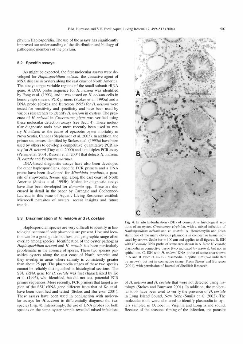
5.3 Discrimination of H. nelsoni and H. costale
Haplosporidian species are very difficult to identify in his-tological sections if only plasmodia are present. Host and loca-tion can be a good guide, but host and geographic range oftenoverlap among species. Identification of the oyster pathogensHaplosporidium nelsoni and H. costale has been particularlyproblematic in the absence of spores. These two species par-asitize oysters along the east coast of North America andthey overlap in areas where salinity is consistently greaterthan about 25 ppt. The plasmodia stages of these two speciescannot be reliably distinguished in histological sections. TheSSU rRNA gene for H. costale was first characterized by Koet al. (1995), who identified, but did not test, potential PCRprimer sequences. More recently, PCR primers that target a re-gion of the SSU rRNA gene different from that of Ko et al.have been identified and tested (Stokes and Burreson 2001).These assays have been used in conjunction with molecu-lar assays for H. nelsoni to differentially diagnose the twospecies (Fig. 4). Interestingly, the use of DNA probes for bothspecies on the same oyster sample revealed mixed infections
Fig. 4. In situ hybridization (ISH) of consecutive histological sec-tions of an oyster, Crassostrea virginica, with a mixed infection ofHaplosporidian nelsoni and H. costale. A. Hematoxylin and eosinstain; two of the many obvious plasmodia in connective tissue indi-cated by arrows. Scale bar = 100 µm and applies to all figures. B. ISHwith H. costale DNA probe of same area shown in A. Note H. costaleplasmodia in connective tissue (two indicated by arrows), but not inepithelium. C. ISH with H. nelsoni DNA probe of same area shownin A and B. Note H. nelsoni plasmodia in epithelium (two indicatedby arrows), but not in connective tissue. From Stokes and Burreson(2001), with permission of Journal of Shellfish Research.
of H. nelsoni and H. costale that were not detected using his-tology (Stokes and Burreson 2001). In addition, the molecu-lar tools have been used to verify the presence of H. costalein Long Island Sound, New York (Sunila et al. 2002). Themolecular tools were also used to identify plasmodia in oys-ters sampled in October in Virginia and Long Island sound.Because of the seasonal timing of the infection, the parasite
508 E.M. Burreson and S.E. Ford: Aquat. Living Resour. 17, 499–517 (2004)
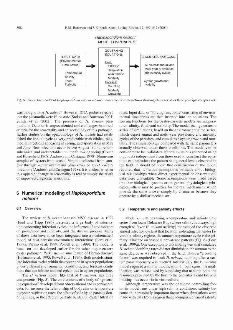
Fig. 5. Conceptual model of Haplosporidium nelsoni – Crassostrea virginica interactions showing elements of its three principal components.
was thought to be H. nelsoni. However, DNA probes revealedthat the plasmodia were H. costale (Stokes and Burreson 2001;Sunila et al. 2002). The presence of H. costale plas-modia in October is unprecedented and challenges historicalcriteria for the seasonality and epizootiology of this pathogen.Earlier studies on the epizootiology of H. costale had estab-lished the annual cycle as very predictable with clinical plas-modial infections appearing in spring, and sporulation in Mayand June. New infections occur before August 1st, but remainsubclinical and undetectable until the following spring (Couchand Rosenfield 1968; Andrews and Castagna 1978). Numeroussamples of oysters from coastal Virginia collected from sum-mer through winter over many years revealed no H. costaleinfections (Andrews and Castagna 1978). It is unclear whetherthis apparent change in seasonality is real or simply the resultof improved diagnostic sensitivity.
6 Numerical modeling of Haplosporidiumnelsoni
6.1 Overview
The review of H. nelsoni-caused MSX disease in 1996(Ford and Tripp 1996) presented a large body of informa-tion concerning infection cycles, the influence of environmenton prevalence and intensity, and the disease process. Manyof these data have since been integrated into a mathematicalmodel of host-parasite-environment interactions (Ford et al.1999a; Paraso et al. 1999; Powell et al. 1999). The model isbased on one developed earlier for the other major easternoyster pathogen, Perkinsus marinus (cause of Dermo disease)(Hofmann et al. 1995; Powell et al. 1996). Both models simu-late infection cycles within the oyster and in oyster populationsunder different environmental conditions, and forecast condi-tions that can initiate and end epizootics in oyster populations.
The H. nelsoni model, like that of P. marinus, has threecomponents (Fig. 5). The core consists of a body of “govern-ing equations” developed from observational and experimentaldata: for instance the relationship of body size or temperatureto oyster respiration rates, the effect of salinity on parasite dou-bling times, or the effect of parasite burden on oyster filtration
rates. Input data, or “forcing functions,” consisting of environ-mental time series are then inserted into the equations. Theforcing functions for the oyster-parasite models are tempera-ture, salinity, food, and turbidity. The model then generates aseries of simulations, based on the environmental time series,which depict annual and multi-year prevalence and intensitycycles of the parasites, and cumulative oyster growth and mor-tality. The simulations are compared with the same parametersactually observed under those conditions. The model can beconsidered to be “validated” if the simulations generated usinginput data independent from those used to construct the equa-tions can reproduce the pattern and general levels observed inthe field. It should be noted that construction of the modelrequired that numerous assumptions be made about biolog-ical relationships when direct experimental or observationaldata were unavailable. Some assumptions were made basedon other biological systems or on general physiological prin-ciples; others may be proxies for the real mechanism, whichprovide the same answer simply by chance or because theyoperate by a similar mechanism.
6.2 Temperature and salinity effects
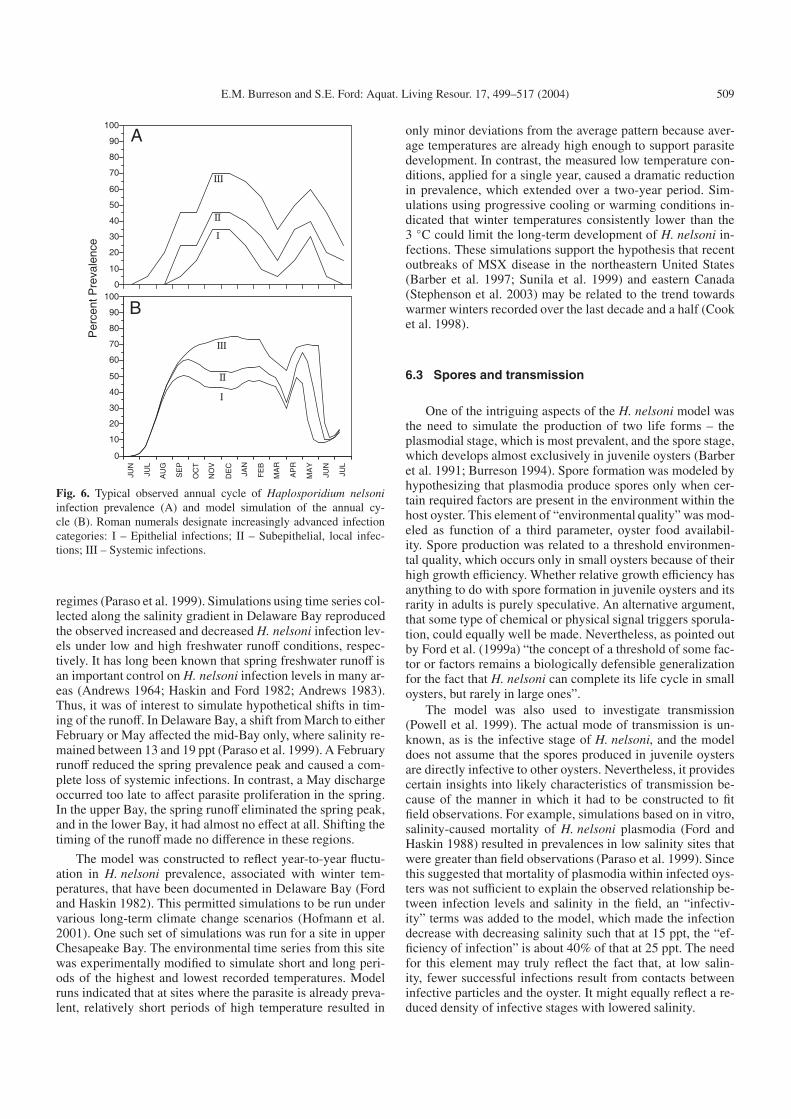
Model simulations using a temperature and salinity timeseries from lower Delaware Bay (where salinity is always highenough to favor H. nelsoni activity) reproduced the observedannual infection cycle at that location, indicating that under fa-vorable salinity regime, the annual temperature cycle is the pri-mary influence on seasonal prevalence patterns (Fig. 6) (Fordet al. 1999a). One exception to this finding was that simulatedH. nelsoni doubling rates did not diminish in the autumn to thesame degree as was observed in the field. Thus, a “crowdingfactor” was required to limit H. nelsoni doubling after a cer-tain parasite density was reached. Interestingly, the P. marinusmodel required a similar modification. In both cases, the mod-ification was rationalized by supposing that at some point theresources provided by the host to the parasites would becomelimiting – as occurs in in vitro culture.
Although temperature was the dominate controlling fac-tor in model runs under high salinity conditions, salinity be-came an increasingly important factor when simulations weremade with data from a region that encompassed varied salinity
E.M. Burreson and S.E. Ford: Aquat. Living Resour. 17, 499–517 (2004) 509
Fig. 6. Typical observed annual cycle of Haplosporidium nelsoniinfection prevalence (A) and model simulation of the annual cy-cle (B). Roman numerals designate increasingly advanced infectioncategories: I – Epithelial infections; II – Subepithelial, local infec-tions; III – Systemic infections.
regimes (Paraso et al. 1999). Simulations using time series col-lected along the salinity gradient in Delaware Bay reproducedthe observed increased and decreased H. nelsoni infection lev-els under low and high freshwater runoff conditions, respec-tively. It has long been known that spring freshwater runoff isan important control on H. nelsoni infection levels in many ar-eas (Andrews 1964; Haskin and Ford 1982; Andrews 1983).Thus, it was of interest to simulate hypothetical shifts in tim-ing of the runoff. In Delaware Bay, a shift from March to eitherFebruary or May affected the mid-Bay only, where salinity re-mained between 13 and 19 ppt (Paraso et al. 1999). A Februaryrunoff reduced the spring prevalence peak and caused a com-plete loss of systemic infections. In contrast, a May dischargeoccurred too late to affect parasite proliferation in the spring.In the upper Bay, the spring runoff eliminated the spring peak,and in the lower Bay, it had almost no effect at all. Shifting thetiming of the runoffmade no difference in these regions.
The model was constructed to reflect year-to-year fluctu-ation in H. nelsoni prevalence, associated with winter tem-peratures, that have been documented in Delaware Bay (Fordand Haskin 1982). This permitted simulations to be run undervarious long-term climate change scenarios (Hofmann et al.2001). One such set of simulations was run for a site in upperChesapeake Bay. The environmental time series from this sitewas experimentally modified to simulate short and long peri-ods of the highest and lowest recorded temperatures. Modelruns indicated that at sites where the parasite is already preva-lent, relatively short periods of high temperature resulted in
only minor deviations from the average pattern because aver-age temperatures are already high enough to support parasitedevelopment. In contrast, the measured low temperature con-ditions, applied for a single year, caused a dramatic reductionin prevalence, which extended over a two-year period. Sim-ulations using progressive cooling or warming conditions in-dicated that winter temperatures consistently lower than the3 ◦C could limit the long-term development of H. nelsoni in-fections. These simulations support the hypothesis that recentoutbreaks of MSX disease in the northeastern United States(Barber et al. 1997; Sunila et al. 1999) and eastern Canada(Stephenson et al. 2003) may be related to the trend towardswarmer winters recorded over the last decade and a half (Cooket al. 1998).
6.3 Spores and transmission
One of the intriguing aspects of the H. nelsoni model wasthe need to simulate the production of two life forms – theplasmodial stage, which is most prevalent, and the spore stage,which develops almost exclusively in juvenile oysters (Barberet al. 1991; Burreson 1994). Spore formation was modeled byhypothesizing that plasmodia produce spores only when cer-tain required factors are present in the environment within thehost oyster. This element of “environmental quality” was mod-eled as function of a third parameter, oyster food availabil-ity. Spore production was related to a threshold environmen-tal quality, which occurs only in small oysters because of theirhigh growth efficiency. Whether relative growth efficiency hasanything to do with spore formation in juvenile oysters and itsrarity in adults is purely speculative. An alternative argument,that some type of chemical or physical signal triggers sporula-tion, could equally well be made. Nevertheless, as pointed outby Ford et al. (1999a) “the concept of a threshold of some fac-tor or factors remains a biologically defensible generalizationfor the fact that H. nelsoni can complete its life cycle in smalloysters, but rarely in large ones”.
The model was also used to investigate transmission(Powell et al. 1999). The actual mode of transmission is un-known, as is the infective stage of H. nelsoni, and the modeldoes not assume that the spores produced in juvenile oystersare directly infective to other oysters. Nevertheless, it providescertain insights into likely characteristics of transmission be-cause of the manner in which it had to be constructed to fitfield observations. For example, simulations based on in vitro,salinity-caused mortality of H. nelsoni plasmodia (Ford andHaskin 1988) resulted in prevalences in low salinity sites thatwere greater than field observations (Paraso et al. 1999). Sincethis suggested that mortality of plasmodia within infected oys-ters was not sufficient to explain the observed relationship be-tween infection levels and salinity in the field, an “infectiv-ity” terms was added to the model, which made the infectiondecrease with decreasing salinity such that at 15 ppt, the “ef-ficiency of infection” is about 40% of that at 25 ppt. The needfor this element may truly reflect the fact that, at low salin-ity, fewer successful infections result from contacts betweeninfective particles and the oyster. It might equally reflect a re-duced density of infective stages with lowered salinity.
510 E.M. Burreson and S.E. Ford: Aquat. Living Resour. 17, 499–517 (2004)
Simulations also needed to replicate the observation thatchanges in H. nelsoni prevalence occur rapidly and over largeareas of estuaries and that these changes occur independentlyof local salinity. To reproduce this observation, the model em-ploys bay-wide oscillations in infective particle availabilitythat are tied to multi-year salinity fluctuations. Simulationsmirrored long-term prevalence time series in both Delawareand Chesapeake Bays (Powell et al. 1999). Since the modeldoes not connect oyster infection levels with subsequent trans-mission, the linking of infective particle availability to long-term salinity change suggests that a non oyster reservoir forinfective stages itself is influenced by salinity, or that salin-ity is a surrogate for some other parameter such as river flow,water residence time, or dilution.
Because attempts to demonstrate direct transmission ofH. nelsoni between oysters have consistently failed, specula-tion has persisted that another host exists, acting either as areservoir for infective stages or as an intermediate host fortransmission (Burreson 1988; Haskin and Andrews 1988; Fordand Tripp 1996). The modeling exercise highlighted the char-acteristics of a potential host: 1) it must be capable of releasinglarge number of infective particles rapidly and continuouslyduring the warm months; 2) normal temperature and salinityvariation cannot affect it; 3) it must be affected by cold win-ters, but capable of recovery within a year or two; 4) it mustproduce infective particles independently of H. nelsoni levelsin the oyster population; and 5) it must exist at relatively highsalinity (Powell et al. 1999). These characteristics are similarto those proposed by Haskin and Andrews (1988) based onfield data.
6.4 Comparisons between H. nelsoni and P. marinus
The data used to construct the P. marinus and H. nelsonimodels, as well as the models themselves, provide interestingcomparisons between the two parasites. Both models operateby causing parasites to multiply or to die in vivo and thus re-quire quantitative data on parasite abundance rather than thesemiquantitative staging systems routinely used to assess in-fection intensity of both parasites. A conversion between theP. marinus infection stages and parasite abundance was devel-oped using a process that frees the parasites from host tissuesso their densities can be determined (Choi et al. 1989). Be-cause H. nelsoni plasmodia are more fragile and would notsurvive a similar treatment, densities of this parasite were esti-mated by counting parasites in a known volume (area countedx section thickness) of representative tissue sections and ex-trapolating those concentrations to the density of plasmodiaper unit weight (Ford et al. 1999a). On the other hand, bothparasites can be obtained in hemolymph samples and theirconcentrations determined directly (Burreson et al. 1988; Fordand Kanaley 1988). For both parasites, average maximumdensities in the hemolymph are in the range of 5 × 10⁵ to106 ml−1 and those estimated for the soft tissue are on theorder of 106 parasites g−1 wet weight, which also seems to bethe lethal level as higher densities are rarely found in livingoysters. As mentioned earlier, models for both parasites re-quire a “crowding factor”, which slows the replication ratewhen parasite densities become high. The parasite density at
which crowding begins to influence P. marinus growth, ob-tained from field and experimental data (Saunders et al. 1993;Ford et al. 1999b), is similar to that estimated for H. nelsoni byfitting model simulation to disease prevalence and intensity: 1to 7×10⁵ parasites g−1 wet weight. The resemblance of thresh-old values suggests fundamental similarities in the per-parasiteuse of nutrients from, and the damage caused to, the host oys-ter by each parasite. Interestingly, the limit of consistently re-liable detection for P. marinus, using the standard Ray/Mackinmethod of incubating tissues in Fluid Thioglycollate Medium,is estimated to be 103 to 104 parasites g−1 wet weight (Choiet al. 1989; Bushek et al. 1994), which is similar to that calcu-lated for H. nelsoni, using tissue section histology (Ford et al.1999a).
In the model itself, the in vivo proliferation rate ofH. nelsoni is based on a Q10 of 3.2. This value was requiredto match proliferation rates at elevated temperature, inferredfrom prevalence increases. It is unusually high and impliesthat H. nelsoni is very sensitive to temperature change. Bycomparison, a more typical Q10 of 2 provided adequate dou-bling in P. marinus simulations (Hofmann et al. 1995). Thus,under condition of rising temperature, H. nelsoni proliferationrates should increase faster than those of P. marinus and underfalling temperatures, they should decrease faster. When super-imposed, however, the modeled doubling times for the two par-asites indicate that H. nelsoni has the higher proliferation rateacross the entire temperature range over which both co-exist,approximately 0 to 35 ◦C. These comparisons are consistentwith field observations showing that when oysters are exposedto both parasites in the field, H. nelsoni typically begins killingbefore P. marinus does (Andrews 1967; Chintala et al. 1994).A similar observation would result from a relatively higherdose of H. nelsoni, and although densities of P. marinus havebeen measured in the water and dose-response curves gener-ated (Ragone Calvo et al. 2003), comparable information isunavailable for H. nelsoni.
7 Recent changes in the distributionand intensity of MSX disease outbreaks
7.1 History of MSX disease outbreaks
The first recorded disease outbreak caused by H. nelsoni ineastern oysters occurred in the spring of 1957 in Delaware Bay,New Jersey, USA (Haskin et al. 1966). In 1959, H. nelsoni be-gan causing mortalities in Mobjack Bay, a subestuary of lowerChesapeake Bay, and the parasite subsequently spread upes-tuary during a drought in the mid 1960s (Andrews and Wood1967; Farley 1975). The parasite was found in oysters alongthe Atlantic coasts of New Jersey, Maryland, and Virginia in1958 and 1959, and in 1960 it was reported on the Connecticutshore of Long Island Sound (Haskin and Andrews 1988). In1965, it was found in Great South Bay on the south shore ofLong Island, New York (Andrews and Wood 1967; Haskinand Andrews 1988) and in 1967 in Wellfleet Harbor, on thenorth side of Cape Cod, Massachusetts (Krantz et al. 1972).In the 1980s, the reported range of the parasite was extendedalong the entire east coast of the United States, from Maine to
E.M. Burreson and S.E. Ford: Aquat. Living Resour. 17, 499–517 (2004) 511
Florida (Haskin and Andrews 1988; Hillman et al. 1988; Kern1988; Lewis et al. 1992). More importantly, epizootics withsevere mortality occurred in Oyster Bay on the north shoreof Long Island, New York and in Southern Massachusettsduring this decade (Haskin and Andrews 1988; Matthiessenet al. 1990). Between 1984 and 1987, oyster production fromthe Connecticut shore of Long Island Sound dropped from244 000 bushels to 70 000, suggesting that the Long Island areaepizootic may not have been localized to Oyster Bay (Sunilaet al. 1999). At the same time, H. nelsoni infections spreadand intensified in Chesapeake and Delaware Bays (Haskin andFord 1986; Burreson and Andrews 1988). In the 1990s, furtherepizootics with heavy mortalities occurred in southern Maine(Barber et al. 1997) and Long Island Sound (Sunila et al.1999), and in 2002 H. nelsoni caused localized heavy mortal-ities in the Bras d’Or Lakes region of Nova Scotia, Canada(Stephenson et al. 2003). In Chesapeake Bay, the decade ofthe 1990s has seen continued spread of both H. nelsoni andP. marinus into regions of the upper Bay and tributaries wherethey have infected susceptible oysters and caused heavy mor-talities (Tarnowski 2002; Ragone Calvo and Burreson 2003)
The demonstration by molecular detection that H. nelsoniis present in the Pacific oyster, C. gigas in Asia and thewestern United States (Burreson et al. 2000; Kamaishi andYoshinaga 2002) indicates that H. nelsoni was introduced fromthe Pacific; however, neither the mechanism nor the timingis known. It is usually inferred that the parasite entered theUnited States in shipments of infected C. gigas made by oys-ter growers or scientists. Deliberate introductions might wellhave been the source, but other possibilities must be consid-ered. Particularly noteworthy is the great increase in ship tran-sit between Pacific and Atlantic ports that occurred during andafter World War II. Shipping could have introduced H. nelsonivia infected C. gigas attached to ship’s hulls or via release ofH. nelsoni spores in the discharge of ballast water. The sporeis a thick-walled stage in the life cycle of H. nelsoni. Its role intransmission is not known, but the spore in other species is typ-ically a transmission stage that can remain “dormant” for longperiods and that is highly tolerant of environmental extremes.Further, it is often concluded that H. nelsoni was introducedinto Delaware Bay and then “spread” to Chesapeake Bay andother areas. However, the time required for an epizootic to oc-cur after an introduction has taken place is unknown and thefinding of H. nelsoni from Long Island Sound to ChesapeakeBay within the space of 3 years, makes it difficult to ascertainwhere the “first” introduction occurred, or even if there wasa single introduction only. Certainly, the parasite must havebeen present for some time before it caused epizootics. In fact,it was not until the mid 1980s, more than 20 years after it wasfirst detected in Long Island Sound, that epizootic mortalitieswere recorded in the region.
7.2 Climate-related intensification and spread of MSXdisease outbreaks
Changes in climate are sometimes linked to disease out-breaks (Harvell et al. 1999; Harvell et al. 2002), including therange extension of Dermo disease epizootics into the north-eastern United States (Ford 1996). Given the known sensitivity
of H. nelsoni to salinity and temperature, it is reasonable to ex-amine the role of these parameters in the apparent northward“spread” of MSX disease outbreaks of the 1980s and 1990s,as well as the intensification of the disease in Chesapeake Bay.In the Chesapeake Bay and its tributaries, salinity gradientsare strong and large areas were formerly protected from highH. nelsoni infection levels by freshwater runoff that kept salin-ities low (Andrews 1968). Since the early 1980s, however, aseries of extreme, multi-year droughts has increased salinitiesand permitted the spread of H. nelsoni, as well as P. marinus,into new areas of the estuary (Burreson and Andrews 1988;Smith and Jordan 1993; Burreson and Ragone Calvo 1996;Tarnowski 2002; Ragone Calvo and Burreson 2003). The re-sult has been widespread and heavy oyster mortalities, anda severe loss of production of this commercially importantspecies. In Delaware Bay, too, H. nelsoni also spread upbayduring a severe drought in the mid-1980s (Haskin and Ford1986), but with apparently different consequences (see below).In most of the other oyster-growing waters of the northeast,salinities are at least 20 ppt, so that low salinity should nothave been a factor limiting H. nelsoni proliferation, althoughdrought-associated lack of flushing during recent periods oflow river flow might allow concentration of infective stages.
Alternatively, a change in temperature regime might ex-plain the northern MSX disease outbreaks, as suggested by themathematical modeling exercise described above (Hofmannet al. 2001). Clearly, temperatures have been increasing in thisarea over the past two decades and it is particularly notice-able in higher winter temperatures (Karl et al. 1996; IPCC2001), which would relax the control that cold winters appearto have on H. nelsoni. Hofmann et al. (2001), however, pointedout an inconsistency in the argument that low temperature hadbeen the mechanism preventing MSX disease outbreaks in thenorth. If this were true, why have there been no outbreaks inthe southeastern United States, where the parasite is present,but at relatively low prevalence and not associated with large-scale mortalities (Lewis et al. 1992; Bobo et al. 1996). Per-haps prolonged high temperatures play a role (Ford and Haskin1982), but there is no evidence that elevated temperature in-hibits H. nelsoni. Alternatively, some condition other than adirect temperature effect is unfavorable or perhaps a secondhost is scarce in this region.
7.3 Decline in MSX disease prevalence in DelawareBay associated with natural resistance
The epizootic of 1957-1959 killed about 90−95% of alloysters in lower Delaware Bay, where salinities are nearlyalways favorable for H. nelsoni, and mortalities were esti-mated to be 50−60% in the lower-salinity beds (Haskin et al.1966). This tremendous selective mortality resulted in mea-surably increased survival of the native Delaware Bay oysterpopulation, which was comparable to that after one generationof selective breeding (Haskin and Ford 1979). After the initialimprovement, however, no further change was documented fornearly 30 years because little or no additional selective mortal-ity occurred on the upbay beds where most of the oysters werelocated. In the mid 1980s, drought allowed H. nelsoni to pene-trate far upbay. Prevalences reached up to 80%, the highest on
512 E.M. Burreson and S.E. Ford: Aquat. Living Resour. 17, 499–517 (2004)
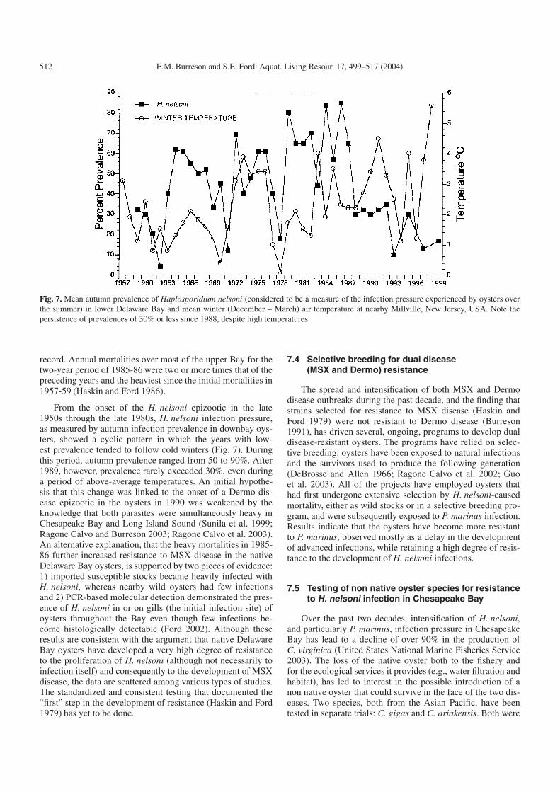
Fig. 7. Mean autumn prevalence of Haplosporidium nelsoni (considered to be a measure of the infection pressure experienced by oysters overthe summer) in lower Delaware Bay and mean winter (December – March) air temperature at nearby Millville, New Jersey, USA. Note thepersistence of prevalences of 30% or less since 1988, despite high temperatures.
record. Annual mortalities over most of the upper Bay for thetwo-year period of 1985-86 were two or more times that of thepreceding years and the heaviest since the initial mortalities in1957-59 (Haskin and Ford 1986).
From the onset of the H. nelsoni epizootic in the late1950s through the late 1980s, H. nelsoni infection pressure,as measured by autumn infection prevalence in downbay oys-ters, showed a cyclic pattern in which the years with low-est prevalence tended to follow cold winters (Fig. 7). Duringthis period, autumn prevalence ranged from 50 to 90%. After1989, however, prevalence rarely exceeded 30%, even duringa period of above-average temperatures. An initial hypothe-sis that this change was linked to the onset of a Dermo dis-ease epizootic in the oysters in 1990 was weakened by theknowledge that both parasites were simultaneously heavy inChesapeake Bay and Long Island Sound (Sunila et al. 1999;Ragone Calvo and Burreson 2003; Ragone Calvo et al. 2003).An alternative explanation, that the heavy mortalities in 1985-86 further increased resistance to MSX disease in the nativeDelaware Bay oysters, is supported by two pieces of evidence:1) imported susceptible stocks became heavily infected withH. nelsoni, whereas nearby wild oysters had few infectionsand 2) PCR-based molecular detection demonstrated the pres-ence of H. nelsoni in or on gills (the initial infection site) ofoysters throughout the Bay even though few infections be-come histologically detectable (Ford 2002). Although theseresults are consistent with the argument that native DelawareBay oysters have developed a very high degree of resistanceto the proliferation of H. nelsoni (although not necessarily toinfection itself) and consequently to the development of MSXdisease, the data are scattered among various types of studies.The standardized and consistent testing that documented the“first” step in the development of resistance (Haskin and Ford1979) has yet to be done.
7.4 Selective breeding for dual disease(MSX and Dermo) resistance
The spread and intensification of both MSX and Dermodisease outbreaks during the past decade, and the finding thatstrains selected for resistance to MSX disease (Haskin andFord 1979) were not resistant to Dermo disease (Burreson1991), has driven several, ongoing, programs to develop dualdisease-resistant oysters. The programs have relied on selec-tive breeding: oysters have been exposed to natural infectionsand the survivors used to produce the following generation(DeBrosse and Allen 1966; Ragone Calvo et al. 2002; Guoet al. 2003). All of the projects have employed oysters thathad first undergone extensive selection by H. nelsoni-causedmortality, either as wild stocks or in a selective breeding pro-gram, and were subsequently exposed to P. marinus infection.Results indicate that the oysters have become more resistantto P. marinus, observed mostly as a delay in the developmentof advanced infections, while retaining a high degree of resis-tance to the development of H. nelsoni infections.
7.5 Testing of non native oyster species for resistanceto H. nelsoni infection in Chesapeake Bay
Over the past two decades, intensification of H. nelsoni,and particularly P. marinus, infection pressure in ChesapeakeBay has lead to a decline of over 90% in the production ofC. virginica (United States National Marine Fisheries Service2003). The loss of the native oyster both to the fishery andfor the ecological services it provides (e.g., water filtration andhabitat), has led to interest in the possible introduction of anon native oyster that could survive in the face of the two dis-eases. Two species, both from the Asian Pacific, have beentested in separate trials: C. gigas and C. ariakensis. Both were
E.M. Burreson and S.E. Ford: Aquat. Living Resour. 17, 499–517 (2004) 513
deployed at duplicate low (<15 ppt), medium (15−25 ppt),and high (>25 ppt) salinity sites in lower Chesapeake Bay andalong the Atlantic coast of Virginia. Growth, survival, and in-fection levels were compared with those of C. virginica de-ployed at the same sites (Calvo et al. 1999; Calvo et al. 2001).To minimize the potential for unintended reproduction, onlytriploid non natives, which are largely sterile, were used in thetests. Crassostrea gigas grew faster and survived better thanC. virginica at the high salinity sites, performed similarly atthe medium salinity sites, and did less well at the low salinitylocations (Calvo et al. 1999). Crassostrea ariakensis outper-formed the C. virginica at all locations (Calvo et al. 2001).At high salinity sites in both trials, C. virginica became heav-ily infected with P. marinus (up to 100%) and to a consider-ably lesser degree (maximum of 16 to 25%) with H. nelsoni.Both C. ariakensis and C. gigas also acquired P. marinus in-fections (up to 60−67%, respectively), but the infections re-mained mostly light and non lethal. No H. nelsoni infectionswere detected in either of the non native oysters. It should berecalled that H. nelsoni has been detected in C. gigas in thePacific region, but always with very large sample sizes to de-tect prevalences that averaged <1% (Kern 1976; Kang 1980;Burreson et al. 2000).
Acknowledgements. We thank Kimberly Reece and Ryan Carnegie,Virginia Institute of Marine Science, for helpful comments on themolecular phylogenetics section.
References
Anderson T.J., Newman L.J., Lester R.J.G., 1993, Light and elec-tron microscope study of Urosporidium cannoni n. sp., a hap-losporidian parasite of the polyclad turbellarian Stylochus sp. J.Euk. Microbiol. 40, 162-168.
Andrews J.D., 1964, Oyster mortality studies in Virginia. IV. MSXin James River public seed beds. Proc. Nat. Shellfish Assoc. 53,65-84.
Andrews J.D., 1967, Interaction of two diseases of oysters in naturalwaters. Proc. Nat. Shellfish Assoc. 57, 38-48.
Andrews J.D., 1968, Oyster mortality studies in Virginia. VII. Reviewof epizootiology and origin of Minchinia nelsoni. Proc. Nat.Shellfish Assoc. 58, 23-36.
Andrews J.D., 1983, Minchinia nelsoni (MSX) infections in theJames River seed-oyster area and their expulsion in spring.Estuar. Coast. Shelf Sci. 16, 255-269.
Andrews J.D., 1984, Epizootiology of diseases of oysters(Crassostrea virginica), and parasites of associated organ-isms in eastern North America. Helgol. Meer. 37, 149-166.
Andrews J.D., Castagna M., 1978, Epizootiology of Minchiniacostalis in susceptible oysters in seaside bays of Virginia’s east-ern shore, 1959-1976. J. Invertebr. Pathol. 32, 124-138.
Andrews J.D., Wood J.L., 1967, Oyster mortality studies in Virginia.VI. History and distribution of Minchinia nelsoni, a pathogen ofoysters, in Virginia. Ches. Sci. 8, 1-13.
Armstrong D.A., Armstrong J.L., 1974, A haplosporidan infection ingaper clams, Tresus capax (Gould), from Yaquina Bay, Oregon.Proc. Natl Shellfish Assoc. 64, 68-72.
Arvy, 1949, Présentation de documents relatifs à l’ovogenèse chez leDentale et à deux parasites de ce Scaphopode: Cercaria prenantin. sp. et Haplosporidium dentali. Bull. Soc. Zool. France 74,292-294.
Azevedo C., 1984, Ultrastructure of the spore of Haplosporidiumlusitanicum sp. n. (Haplosporida, Haplosporidiidae), parasite ofa marine mollusc. J. Parasitol. 70, 358-371.
Azevedo C., 2001, Ultrastructural description of the spore matura-tion stages of the clam parasite Minchinia tapetis (Vilela 1951)(Haplosporida: Haplosporidiidae). Syst. Parasitol. 49, 189-194.
Azevedo C., Conchas R.F., Montes C., 2003, Description ofHaplosporidium edule n. sp. (Phylum Haplosporidia), a parasiteof Cerastoderma edule (Mollulsca, Bivalvia) with complex sporeornamentation. Eur. J. Protistol. 39, 161-167.
Azevedo C., Montes J., Corral L., 1999, A revised description ofHaplosporidium armoricanum, parasite of Ostrea edulis L. fromGalicia, northwestern Spain, with special reference to the spore-wall filaments. Parasitol. Res. 85, 977-983.
Bachere E., Chagot D., Tigé G., Grizel H., 1987, Study of a hap-losporidian (Ascetospora), parasitizing the Australian flat oysterOstrea angasi. Aquaculture 67, 266-268.
Ball S.J., 1980, Fine structure of the spores of Minchinia chito-nis (Lankester 1885) Labbé, 1896 (Sporozoa: Haplosporida), aparasite of the chiton, Lepidochitona cinereus. Parasitology 81,169-176.
Barber B.J., Langan R., Howell T.L., 1997, Haplosporidium nelsoni(MSX) epizootic in the Piscataqua river estuary (Maine NewHampshire, USA). J. Parasitol. 83, 148-150.
Barber R.D., Kanaley S.A., Ford S.E., 1991, Evidence for regu-lar sporulation by Haplosporidium nelsoni (MSX) (Ascetospora:Haplosporidiidae) in spat of the American oyster, Crassostreavirginica. J. Protozool. 38, 305-306.
Barrow J.H. Jr., 1961, Observations of a haplosporidian,Haplosporidium pickfordi sp. nov. in fresh water snails.Trans. Am. Microsc. Soc. 80, 319-329.
Berthe F.C.J., Le Roux F., Peyretaillade E., Peyret P., Rodriguez D.,Gouy M., Vivarés C.P., 2000, Phylogenetic analysis of the smallsubunit ribosomal RNA of Marteilia refringens validates the ex-istence of phylum Paramyxea (Desportes and Perkins, 1990). J.Euk. Microbiol. 47, 288-293.
Bobo M.Y., Richardson D., Cheng T.C., McGovern E., Coen L., 1996,Seasonal cycle of Haplosporidium nelsoni (MSX) in intertidaloysters, Crassostrea virginica, in South Carolina. J. Shellfish Res.15, 525.
Burreson E.M., 1988, Use of immunoassays in haplosporidan lifecycle studies. In: Fisher W.S. (Eds.). Disease Processes inMarine Bivalve Molluscs. Am. Fish. Soc., Bethesda, Maryland,pp. 298-303.
Burreson E.M., 1991, Effects of Perkinsus marinus infection inthe eastern oyster I: Susceptibility of native and MSX-resistantstocks. J. Shellfish Res. 10, 417-424.
Burreson E.M., 1994, Further evidence of regular sporulation byHaplosporidium nelsoni in small oysters, Crassostrea virginica.J. Parasitol. 80, 1036-1038.
Burreson E.M., 2001, Spore ornamentation of Haplosporidium pick-fordi Barrow, 1961 (Haplosporidia), a parasite of freshwatersnails in Michigan, USA. J. Euk. Microbiol. 48, 622-626.
Burreson E.M., Andrews J.D., 1988, Unusual intensification ofChesapeake Bay oyster diseases during recent drought condi-tions. Oceans ’88 Proceedings, pp. 799-802.
Burreson E.M., Ragone Calvo L.M., 1996, Epizootiology ofPerkinsus marinus disease of oysters in Chesapeake Bay, withemphasis on data since 1985. J. Shellfish Res. 15, 17-34.
Burreson E.M., Robinson M.E., Villalba A., 1988, A comparisonof paraffin histology and hemolymph analysis for the diagno-sis of Haplosporidium nelsoni (MSX) in Crassostrea virginica(Gmelin). J. Shellfish Res. 7, 19-24.
514 E.M. Burreson and S.E. Ford: Aquat. Living Resour. 17, 499–517 (2004)
Burreson E.M., Stokes N.A., Friedman C.S., 2000, Increased viru-lence in an introduced pathogen: Haplosporidium nelsoni (MSX)in the eastern oyster Crassostrea virginica. J. Aquat. Anim.Health 12, 1-8.
Bushek D., Ford S.E., Allen J.S.K., 1994, Evaluation of methods us-ing Ray’s fluid thioglycollate medium for diagnosis of Perkinsusmarinus infection in the eastern oyster, Crassostrea virginica.Ann. Rev. Fish Dis. 4, 201-217.
Calvo G.W., Luckenbach M.W., Allen S.K., Burreson E.M., 1999,Comparative field study of Crassostrea gigas (Thunberg, 1793)and Crassostrea virginica (Gmelin, 1791) in relation to salinity inVirginia. J. Shellfish Res. 18, 465-473.
Calvo G.W., Luckenbach M.W., Allen S.K., Burreson E.M., 2001, Acomparative field study of Crassostrea ariakensis (Fujita 1913)and Crassostrea virginica (Gmelin 1791) in relation to salinity inVirginia. J. Shellfish Res. 20, 221-229.
Carballal M.J., Iglesias D., Santamarina J., Ferro-Soto B., VillalbaA., 2001, Parasites and pathologic conditions of the cockleCerastoderma edule populations of the coast of Galicia (NWSpain). J. Invertebr. Pathol. 78, 87-97.
Carnegie R.G., Barber B.J., Culloty S.C., Figueras A.J., Distel D.L.,2000, Development of a PCR assay for detection of the oysterpathogen Bonamia ostreae and support for its inclusion in theHaplosporidia. Dis. Aquat. Org. 42, 199-206.
Carnegie R.G., Meyer G.R., Blackbourn J., Cochennec-Laureau N.,Berthe F.C.J., Bower S.M., 2003, Detection of the oyster para-site Mikrocytos mackini by PCR and fluorescent in situ hybridiza-tion, and a preliminary phylogenetic analysis. Dis. Aquat. Org.54, 219-227.
Caullery M., Chappellier A., 1906, Anurosporidium pelseneeri n.g.,n. sp., Haplosporidie infectant les sporocysts d’un trematode par-asite de Donax trunculus L. C. R. Soc. Biol. 60, 325-328.
Caullery M., Mesnil F., 1899, Sur le genre Aplosporidium (nov) etl’ordre nouveau des Aplosporidies. C. R. Soc. Biol. (Paris) 51,789-791.
Caullery M., Mesnil F., 1905, Sur quelques nouvelles haplosporidiesd’Annélide. C. R. Soc. Biol. (Paris) 58, 580-583.
Cavalier-Smith T., 1993, Kingdom Protozoa and its 18 phyla.Microbiol. Rev. 57, 953-994.
Cavalier-Smith T., Chao E.E.-Y., 2003a, Phylogeny of Coanozoa,Apusozoa, and other Protozoa and early eukaryote megaevolu-tion. J. Mol. Evol. 56, 540-563.
Cavalier-Smith T., Chao E.E.-Y., 2003b, Phylogeny and classificationof phylum Cercozoa (Protozoa). Protist 154, 341-358.
Chintala M.M., Ford S.E., Fisher W.S., Ashton-Alcox K.A., 1994,Oyster serum agglutinins and resistance to protozoan parasites. J.Shellfish Res. 13, 115-121.
Choi K.-S., Wilson E.A., Lewis D.H., Powell E.N., Ray S.M., 1989,The energetic cost of Perkinsus marinus parasitism in oysters.Quantification of the thioglycollate method. J. Shellfish Res. 8,117-125.
Chu F.-L.E., Burreson E.M., Zhang F.S., Chew K.K., 1996, Anunidentified haplosporidian parasite of bay scallop Argopectenirradians cultured in the Shandong and Liaoning provinces ofChina. Dis. Aquat. Org. 42, 207-214.
Ciancio A., Scippa S., Izzo C., 1999, Ultrastructure of vegetative andsporulation stages of Haplosporidium ascidiarum from the ascid-ian Ciona intestinalis L. Eur. J. Protistol. 35, 175-182.
Cochennec-Laureau N., Reece K.S., Berthe F.C.J., Hine P.M., 2003,Mikrocytos roughleyi taxonomic affiliation leads to the genusBonamia (Haplosporidia). Dis. Aquat. Org. 54, 209-217.
Comps M., Pichot Y., 1991, Fine spore structure of a haplospori-dan parasitizing Crassostrea gigas: taxonomic implications. Dis.Aquat. Org. 11, 73-77.
Comps M., Tigé G., 1997, Fine structure of Minchinia sp., a hap-losporidan infecting the mussel Mytilus galloprovincialis L. Syst.Parasitol. 38, 45-50.
Cook T., Folli M., Klinck J., Ford S., Miller J., 1998, The relation-ship between increasing sea surface temperature and the north-ward spread of Perkinsus marinus (Dermo) disease epizootics inoysters. Estuar. Coast. Shelf Sci. 40, 587-597.
Couch J.A., Rosenfield A., 1968, Epizootiology of Minchinia costalisand Minchina nelsoni in oysters introducted into ChincotaeagueBay. Proc. Natl. Shellfish Assoc. 58, 51-59.
Day J.M., Franklin M.E., Brown B.L., 2000, Use of competitivePCR to detect and quantify Haplosporidium nelsoni infection(MSX disease) in the eastern oyster (Crassostrea virginica). Mar.Biotechnol. 1, 147-154.
Debaisieux P., 1920, Haplosporidium (Minchinia) chitonis Lank.,Haplosporidium nemertis, Nov. sp. La Cellule 30, 293-309.
DeBrosse G.A., Allen S.K., 1966, Cooperative regional oyster se-lective breeding (CROSBREED) project. J. Shellfish Res. 15,514-515.
Desportes I., Nashed N.N., 1983, Ultrastructure of sporulationin Minchinia dentali (Arvy), an haplosporean parasite ofDentalium entale (Scaphopoda, Mollusca); taxonomic implica-tions. Protistologica 19, 435-460.
Desportes I., Perkins F.O., 1990, Phylum Paramyxea. In: MargulisL., Corliss J.O., Melkonian M., Chapman D.J. (Eds.), Handbookof Protoctista, Jones and Bartlett Publishing, Boston, MA,pp. 30-35.
DeTurk W.E., 1940, The occurrence and development of a hypera-site, Urosporidium crescens sp. nov. (Sporozoa, Haplosporidia),which infecsts the metacercariae of Spelotrema nicolli, parasiticin Callinectes sapidus. J. Elisha Mitchell Sci. Soc. 56, 231-232.
Diggles B.K., Nichol J., Hine P.M., Wakefield S., Cochennec-LaureauN., Roberts R.D., Friedman C.S., 2002, Pathology of culturedpaua Haliotis iris infected with a novel haplosporidian parasite,with some observations on the course of disease. Dis. Aquat. Org.50, 219-231.
Dollfus R.Ph., 1925, Liste critique des cercaires marines à queuesétigère signalées jusqu’à présent. Trav. Station Zool. Wimereux9, 3-65.
Duboscq O., Harant H., 1923, Sur les Sporozoaires des Tuniciers. C.R. Acad. Sci. Paris 177, 432-433.
Elston R.A., Farley C.A., Kent M.L., 1986, Occurrence and signif-icance of bonamiasis in European flat oysters Ostrea edulis inNorth America. Dis. Aquat. Org. 2, 49-54.
Farley C.A., 1975, Epizootic and enzootic aspects of Minchinianelsoni (Haplosporida) disease in Maryland oysters. J. Protozool.22, 418-427.
Farley C.A., Wolf P.H., Elston R.A., 1988, A long-term study of “mi-crocell” disease in oysters with a description of a new genus,Mikrocytos (g. n.), and two new species, Mikrocytos mackini (sp.n.) and Mikrocytos roughleyi (sp. n.). Fish. Bull. 86, 581-593.
Figureras A.J., Jardon C.F., Caldas J.R., 1991, Diseases and parsitesof mussels (Mytilus edulis Linneaus, 1758) from two sites on theeast coast of the United States. J. Shellfish Res. 10, 89-94.
E.M. Burreson and S.E. Ford: Aquat. Living Resour. 17, 499–517 (2004) 515
Flores B.S., Siddall M.E., Burreson E.M., 1996, Phylogeny of theHaplosporidia (Eukaryota: Alveolata) based on small subunit ri-bosomal RNA gene sequence. J. Parasitol. 82, 616-623.
Fong D., Chan M.M.-Y., Rodriguez R., Chen C.-C., Liang Y.,Littlewood, D.T.J., Ford S.E., 1993, Small subunit ribosomalRNA gene sequence of the parasitic protozoan Haplosporidiumnelsoni provides a molecular probe for the oyster MSX disease.Mol. Biochem. Parasitol. 62, 139-143.
Ford S.E., 1996, Range extension by the oyster parasite Perkinsusmarinus into the northeastern US: Response to climate change?J. Shellfish Res. 15, 45-56.
Ford S.E., 2002, Development of high disease resistance in a wildoyster population. J. Shellfish Res. 21, 387.
Ford S.E., Haskin H.H., 1982, History and epizootiology ofHaplosporidium nelsoni (MSX), an oyster pathogen, in DelawareBay, 1957-1980. J. Invertebr. Pathol. 40, 118-141.
Ford S.E., Haskin H.H., 1988, Comparison of in vitro salinity tol-erance of the oyster parasite Haplosporidium nelsoni (MSX) andhemocytes from the host, Crassostrea virginica. Comp. Biochem.Physiol. 90A, 183-187.
Ford S.E., Kanaley S.A., 1988, An evaluation of hemolymph diag-nosis for detection of the oyster parasite Haplosporidium nelsoni(MSX). J. Shellfish Res. 7, 11-18.
Ford S., Powell E., Klinck J., Hofmann E., 1999a, Modeling theMSX parasite in eastern oyster (Crassostrea virginica) popula-tions. I. Model development, implementation, and verification. J.Shellfish Res. 18, 475-500.
Ford S.E., Schotthoefer A., Spruck C., 1999b, In vivo dynamics of themicroparasite Perkinsus marinus during progression and regres-sion of infections in eastern oysters. J. Parasitol. 85, 273-282.
Ford S.E., Tripp M.R., 1996, Diseases and defense mechanisms. In:Kennedy V.S., Newell R.I.E., Eble A.F. (Eds.), The eastern oysterCrassostrea virginica, Maryland Sea Grant, College Park, MD,pp. 581-660.
Granata L., 1913, Ciclo de sviluppo di Haplosporidium limnodrili n.sp. Rend. R. Accad. Lincei 22, 734.
Guo X., Ford S., DeBrosse G., 2003, Breeding and evaluation of east-ern oyster strains selected for MSX, Dermo and JOD resistance.J. Shellfish Res. 22, 333.
Harvell C.D.K.K., Burkholder J.M., Colwell R.R., Epstein P.R.,Grimes D.J., Hofmann E.E., Lipp E.K., Osterhaus A.D.M.E.,Overstreet R.M., Porter J.W., Smith G.W., Vasta G.R., 1999,Emerging marine disease - climate links and anthropogenic fac-tors. Science 285, 1505-1510.
Harvell C.D., Mitchell C.E., Ward J.R., Altizer S., Dobson A.P.,Ostfeld R.S., Samuel M.D., 2002, Climate warming and diseaserisks for terrestrial and marine biota. Science 296, 2158-2162.
Haskin H.H., Andrews J.D., 1988, Uncertainties and specula-tions about the life cycle of the eastern oyster pathogenHaplosporidium nelsoni (MSX). In: Fisher W.S. (Ed.), DiseaseProcesses in Marine Bivalve Molluscs. Am. Fish. Soc. Spec.Publ. 18, 5-22.
Hervio D., Bachère E., Boulo V., Cochennec N., Vuillemin V.,Le Coguic Y., Cailletaux G., Mazurié J., Mialhe E., 1995,Establishment of an experimental infection protocol for the flatoyster, Ostrea edulis, with the intrahaemocytic protozoan para-site, Bonamia ostreae: application in the selection of parasite-resistant oysters. Aquaculture 132, 183-194.
Haskin H.H., Ford S.E., 1979, Development of resistance toMinchinia nelsoni (MSX) mortality in laboratory-reared and na-tive oyster stocks in Delaware Bay. Mar. Fish. Rev. 41, 54-63.
Haskin H.H., Ford S.E., 1982, Haplosporidium nelsoni (MSX) onDelaware Bay seed oyster beds: a host-parasite relationship alonga salinity gradient. J. Invertebr. Pathol. 40, 388-405.
Haskin H.H., Ford S.E., 1986, Report to the Bureau of Shellfisheries,NJ Department of Environmental Protection on the Delaware BayOyster Research Program 1984-1986. Rutgers University, PortNorris, New Jersey.
Haskin H.H., Stauber L.A., Mackin J.A., 1966, Minchinia nelsonin. sp. (Haplosporida, Haplosporidiidae): causative agent of theDelaware Bay oyster epizootic. Science 153, 1414-1416.
Hillman R.E., Boehm P.D., Freitas S.Y., 1988, A pathology potpourrifrom the NOAA mussel watch program. J. Shellfish Res. 7, 216.
Hillman R.E., Ford S.E., Haskin H.H., 1990, Minchinia teredinisn. sp. (Balanosporida, Haplosporidiidae), a parasite of teredinidshipworms. J. Protozool. 37, 364-368.
Hine P.M., Thorne T., 1998, Haplosporidium sp. (Haplosporidia) inhatchery-reared pearl oysters, Pinctada maxima (Jameson, 1901),in north western Australia. J. Invertebr. Pathol. 71, 48-52.
Hine P.M., Thorne T., 2002, Haplosporidium sp. (Alveolata:Haplosporidia) associated with mortalities among rock oystersSaccostrea cuccullata in north Western Australia. Dis. Aquat.Org. 51, 123-133.
Hine P.M., Bower S.M., Meyer G.R., Cochennec-Laureau N., BertheF.C.J., 2001a, Ultrastructure of Mikrocytos mackini, the cause ofDenman Island disease in oysters Crassostrea spp. and Ostreaspp. in British Columbia, Canada. Dis. Aquat. Org. 45, 215-227.
Hine P.M., Cochennec-Laureau N., Berthe F.C.J., 2001b, Bonamiaexitiosus n. sp. (Haplosporidia) infecting flat oysters Ostreachilensis in New Zealand. Dis. Aquat. Org. 47, 63-72.
Hine P.M., Wakefield S., Diggles B.K., Webb V.L., Maas E.W., 2002,The ultrastructure of a haplosporidian containing Rickettsiae, as-sociated with mortalities among cultured paua Haliotis iris. Dis.Aquat. Org. 49, 207-219.
Hofmann E., Ford S., Powell E., Klinck J., 2001, Modeling studiesof the effect of climate variability on MSX disease in easternoyster (Crassostrea virginica) populations. Hydrobiologia 460,195-212.
Hofmann E.E., Powell E.N., Klinck J.M., Saunders G., 1995,Modelling diseased oyster populations I. Modelling Perkinsusmarinus infections in oysters. J. Shellfish Res. 14, 121-151.
Howell M., 1967, The trematode Bucephalus longicornutus (Manter,1954) in the New Zealand Mud-oyster, Ostrea lutdaria. Trans. R.Soc. New Zealand 8, 221-237.
IPCC, 2001, Summary for Policy Makers. Report of WorkingGroup I. Intergovernmental Panel on Climate Change.http://www.ipcc.ch/pub/pub.htm.
Jírovec O., 1936, Haplosporidium cernosvitovi n. sp. eine neueHaplosporidienart aus Opistocysta (Pristina) flagellum Leidy.Arch. Protistenk. 86, 500-508.
Kamaishi T., Yoshinaga T., 2002, Detection of Haplosporidium nel-soni in Pacific oyster Crassostrea gigas in Japan. Fish Pathol. 37,193-195.
Kang P.A., 1980, On the Minchinia sp. infection in the oysters fromChungmu area. Bull. Fish. Resour. Develop. Agency (Republicof Korea) 25, 25-28.
Karl T.R., Knight R.W., Easterling D.R., Quayle R.G., 1996, Indicesof climate change for the United States. Bull. Am. Meteorol. Soc.77, 279-291.
Katkansky S.C., Warner R.W., 1970, Sporulation of a haplosporid-ian in a Pacific oyster (Crassostrea gigas) in Humboldt Bay,California. J. Fish. Res. Board Can. 27, 1320-1321.
Kern F.G., 1976, Sporulation of Minchinia sp. (Haplosporida,Haplosporidiidae) in the Pacific oyster Crassostrea gigas(Thunberg) from the republic of Korea. J. Protozool. 23, 498-500.
Kern F.G., 1988, Recent changes in the range of “MSX”Haplosporidium nelsoni. J. Shellfish Res. 7, 543.
516 E.M. Burreson and S.E. Ford: Aquat. Living Resour. 17, 499–517 (2004)
Ko Y.-T., Ford S.E., Fong D., 1995, Characterization of the small sub-unit RNA gene of the oyster parasite Haplosporidium costale.Mol. Mar. Biol. Biotechnol. 4, 236-240.
Krantz E.L., Buchanan L.R., Farley C.A., Carr A.H., 1972, Minchinianelsoni in oysters from Massachusetts waters. Proc. Nat. ShellfishAssoc. 62, 83-88.
La Haye C.A., Holland N.D., McLean N., 1984, Electron mi-croscopic study of Haplosporidium comatulae n. sp. (PhylumAscetospora: Class Stellatosporea), a haplosporidian endopara-site of an Australian crinoid, Oligometra serripinna (PhylumEchinodermata). Protistologica 20, 507-515.
Lankester E.R., 1885, Protozoa. In: Encyclopaedia Britannica, 9thedn. 19, 830-866.
Larsson R., 1987, On Haplosporidium gammari, a parasite of the am-phipod Rivulogamarus pulex, and it relationships with the phy-lum Ascetospora. J. Invertebr. Pathol. 49, 159-169.
Lewis E.J., Kern F.G., Rosenfield A., Stevens S.A., Walker R.L.,Heffernan P.B., 1992, Lethal parasites in oysters from coastalGeorgia, with discussion of disease and management implica-tions. Mar. Fish. Rev. 52, 1-6.
Marchand J., Sprague V., 1979, Ultrastructure de Minchiniacadomensis sp. n. (Haplosporida) parasite du DécapodeRhithropanopeus harrisii tridentatus Matiland dans le Canal deCaen à la Mer (Calvados, France). J. Protozool. 26, 179-185.
Matthiessen G.C., Feng S.Y., Leibovitz L., 1990, Patterns of MSX(Haplosporidium nelsoni) infection and subsequent mortality inresistant and susceptible strains of the eastern oyster Crassostreavirginica (Gmelin, 1791) in New England. J. Shellfish Res. 9,359-366.
McGovern E.R., Burreson E.M., 1990, Ultrastructure of Minchiniasp. spores from shipworms (Teredo spp.) in the western NorthAtlantic, with a discussion of taxonomy of the Haplosporidiidae.J. Protozool. 37, 212-218.
Mercier L., Poisson R., 1922, Une haplosporidie, Haplosporidiumcaulleryi nov. sp., parasite de Nereilepas fucata Sav. C.R. Acad.Sci. Paris 174, 1205-1207.
Messick G.A., Nalepa T.F., 2002, Parasites in Diporeia spp. am-phipods from Lakes Michigan and Huron. J. Shellfish Res. 21,391.
Mix M.C., Sprague V., 1974, Occurrence of a haplosporidian in na-tive oysters (Ostrea lurida) from Yaquina Bay and Alsea Bay,Oregon. J. Invertebr. Pathol. 23, 2552-254.
Morris D.J., Adams A., Richards R.H., 2000, Observations on theelectron-dense bodies of the PKX parasite, agent of proliferativekidney disease in salmonids. Dis. Aquat. Org. 39, 201-209.
Newman M.W., Johnson C.A. III, Pauley G.B., 1976, A Minchinia-like haplosporidan parasitizing blue crabs, Callinectes sapidus. J.Invertebr. Pathol. 27, 311-315.
Ormières R., 1980, Haplosporidium parisi n. sp. Haplosporidie para-site de Serpula vermicularis L. étude ultrastructurale de la spore.Protistologica 16, 467-474.
Ormières R., de Puytorac P., 1968, Ultrastructure des spores del’haplosporidie Haplosporidium ascidiarum endoparasite du tu-nicier Sydniuim elegans Giard. C. R. Acad. Sci. Paris 266,1134-1136.
Ormières R., Sprague V., Bartoli P., 1973, Light and electron micro-scope study of a new species of Urosporidium (Haplosporida),hyperparasite of trematode sporocysts in the clam Abra ovata. J.Invertebr. Pathol. 21, 71-86.
Paraso M.C., Ford S.E., Powell E.N., Hofmann E.E., Klinck J.M.,1999, Modeling the MSX parasite in eastern oyster (Crassostreavirginica) populations. II. Salinity effects. J. Shellfish Res. 18,501-516.
Penna M.S., Khan M., French R.A., 2001, Development of amultiplex PCR for the detection of Haplosporidium nelsoni,Haplosporidium costale and Perkinsus marinus in the easternoyster (Crassostrea virginica Gmelin, 1791). Mol. Cell. Probes15, 385-390.
Perkins F.O., 1968, Fine structure of the oyster pathogen Minchinianelsoni (Haplosporida, Haplosporidiidae). J. Invertebr. Pathol.10, 287-305.
Perkins F.O., 1969, Electron microscope studies of sporulation in theoyster pathogen, Minchinia costalis (Sporozoa: Haplosporida). J.Parasitol. 55, 897-920.
Perkins F.O., 1971, Sporulation in the trematode hyperpar-asite Urosporidium crescens DeTurk, 1940 (Haplosporida:Haplosporidiidae) – an electron microscope study. J. Parasitol.57, 9-23.
Perkins F.O., 1975, Fine structure of Minchinia sp. (Haplosporida)sporulation in the mud crab, Panopeus herbstii. Mar. Fish. Rev.37, 46-60.
Perkins F.O., 1990, Phylum Haplosporidia. In: Margulis L.,Corliss J.O., Melkonian M., Chapman D.J. (Eds.), Handbookof Protoctista, Jones and Bartlett Publishing, Boston, MA,pp. 19-29.
Perkins F.O., 1991, “Sporozoa”: Apicomplexa, Microsporidia,Haplosporidia, Paramyxea, Myxosporidia, and Actinosporidia.In: Harrison F.W., Corliss J.O. (Eds.), Microscopic anatomy ofInvertebrates, Vol. 1, Wiley-Liss, NY, pp. 261-331.
Perkins F.O., 2000, Phylum Haplosporidia Caullery & Mesnil, 1899,In: Lee J.J., Leedale G.F., Bradbury P. (Eds.), An illustrated guideto the protozoa, 2nd edition, Vol. 2, Society of Protozoologists,Lawrence, KS, pp. 1328-1341.
Perkins F.O., Madden P.A., Sawyer T.K., 1977, Ultrastructural studyof the spore surface of the haplosporidian Urosporidium spisuli.Trans. Am. Micros. Soc. 96, 376-382.
Perkins F.O., Zwerner D.E., Dias R.K., 1975, The hyperparasite,Urosporidium spisuli sp. n. (Haplosporea), and its effects on thesurf clam industry. J. Parasitol. 61, 944-949.
Pichot Y., Comps M., Tigé G., Grizel H., Rabouin M.A., 1979,Recherches sur Bonamia ostreae gen. n., sp. n., parasite nouveaude l’huître plate Ostrea edulis L. Trav. Inst. Pêches Marit. 443,131-140.
Powell E.N., Klinck J.M., Ford S.E., Hofmann E.E., Jordan S.J.,1999, Modeling the MSX parasite in eastern oyster (Crassostreavirginica) populations. III. Regional application and the problemof transmission. J. Shellfish Res. 18, 517-537.
Ragone Calvo L.M., Burreson E.M., 2003, Status of the Major OysterDiseases in Virginia 2002, A Summary of the Annual MonitoringProgram. Marine Resource Report No. 03-02. Virginia Instituteof Marine Science, The College of William and Mary, GloucesterPoint, Virginia. March 2003.
Ragone Calvo L.M., Calvo G.W., Burreson E.M., 2002, Dual diseaseresistance in a selectively bred eastern oyster, Crassostrea vir-ginica, strain tested in Chesapeake Bay. Aquaculture 220, 69-87.
Ragone Calvo L.M., Dungan C.F., Roberson B.S., Burreson E.M.,2003, Systematic evaluation of factors controlling Perkinsusmarinus transmission dynamics in lower Chesapeake Bay. Dis.Aquat. Org. 56, 75-86.
Reece K.S., Siddall M.E., Stokes N.A., Burreson E.M., 2004,Molecular phylogeny of the Haplosporidia based on two inde-pendent gene sequences. J. Parsitol. 90, 1111-1122.
Reece K.S., Stokes N.A., 2003, Molecular analysis of a haplosporid-ian parasite from cultured New Zealand abalone Haliotis iris. Dis.Aquat. Org. 53, 61-66.
E.M. Burreson and S.E. Ford: Aquat. Living Resour. 17, 499–517 (2004) 517
Renault T., Stokes N.A., Chollet B., Cochennec N., Berthe F.,Burreson E.M., 2000, Haplosporidiosis in the Pacific oyster,Crassostrea gigas, from the French Atlantic coast. Dis. Aquat.Org. 42, 207-214.
Russell S., Frasca Jr. S., Sunila I., French R.A., 2004, Application ofa multiplex PCR for the detection of protozoan parasites of theeastern oyster Crassostrea virginica in field samples. Dis. Aquat.Org. 59, 85-91
Saunders G.L., Powell E.N., Lewis D.H., 1993, A determinationof in vivo growth rates for Perkinsus marinus, a parasite ofCrassostrea virginica. J. Shellfish Res. 12, 229-240.
Siddall M.E., Stokes N.A., Burreson E.M., 1995, Molecular phylo-genetic evidence that the phylum Haplosporidia has an alveolateancestry. Mol. Biol. Evol. 12, 573-581.
Smith G., Jordan S., 1992, Monitoring Maryland’s Chesapeake BayOysters. A comprehensive characterization of Modified FallSurvey Results 1990-1991. Maryland Department of NaturalResources, Chesapeake Bay Research and Monitoring DivisionReport No. CBRM-OX-93-3, Oxford, Maryland. September1992.
Sprague V., 1963, Minchinia louisiana n. sp. (Haplosporidia,hdaplosporidiidae), a parasite of Panopeus herbstii. J. Protozool.10, 267-274.
Sprague V., 1979, Classification of the Haplosporidia. Mar. Fish. Rev.41, 40-44.
Stephenson M.F., McGladdery S.E., Stokes N.A., 2002, Detectionof a previously undescribed haplosporidian-like infection of bluemussel (Mytilus edulis) in Atlantic Canada. J. Shellfish Res. 21,389.
Stephenson M.F., McGladdery S.E., Maillet M., Veniot A., Meyer G.,2003, First reported occurrence of MSX in Canada. J. ShellfishRes. 22, 355.
Stokes N.A., Burreson E.M., 1995, A sensitive and specific DNAprobe for the oyster pathogen Haplosporidium nelsoni. J. Euk.Microbiol. 42, 350-357.
Stokes N.A., Burreson E.M., 2001, Differential diagnosis of mixedHaplosporidium costale and Haplosporidium nelsoni infectionsin the eastern oyster, Crassostrea virginica, using DNA probes.J. Shellfish Res. 20, 207-213.
Stokes N.A., Siddall M.E., Burreson E.M., 1995a, Detection ofHaplosporidium nelsoni (Haplosporidia: Haplosporidiidae) inoysters by PCR amplification. Dis. Aquat. Org. 23, 145-152.
Stokes N.A., Siddall M.E., Burreson E.M., 1995b, Small sub-unit ribosomal RNA gene sequence of Minchinia teredinis(Haplosporidia: Haplosporidiidae) and a specific DNA probe andPCR primers for its detection. J. Invertebr. Pathol. 65, 300-308.
Sunila I., Karolus J., Volk J., 1999, A new epizootic ofHaplosporidium nelsoni (MSX), a haplosporidian oyster parasite,in Long Island Sound, Connecticut. J. Shellfish Res. 18, 169-174.
Sunila I., Stokes N.A., Smolowitz R., Karney R.C., Burreson E.M.,2002, Haplosporidium costale (Seaside Organism), a parasite ofthe eastern oyster, is present in Long Island sound. J. ShellfishRes. 21, 113-118.
Tarnowski M., 2002, Maryland Oyster Population Status Report:2001 Fall Survey. Maryland Department of Natural Resources,Annapolis, Maryland. November, 2002.
Taylor R.L., 1966, Haplosporidium tumefacientis sp. n., the etiologicagent of a disease of the California sea mussel, Mytilus californi-anus Conrad. J. Invertebr. Pathol. 8, 109-121.
United States National Marine Fisheries Service, 2003, AnnualCommercial Landings Statistics:http://www.st.nmfs.gov/st1/commercial/landings/annual_landings.html.
van Banning P., 1977, Minchinia armoricana sp. nov. (Haplosporida),a parasite of the European flat oyster, Ostrea edulis. J. Invertebr.Pathol. 30, 199-206.
Vilela H., 1950, Sporozoaires parasites de la Palourde, Tapes decus-satus (L.). Rev. Fac. Ciências, Lisboa, Sér. C, 1, 379-386.
Wood J.L., Andrews J.D., 1962, Haplosporidium costale (Sporozoa)associated with a disease of Virginia oysters. Science 136,710-711.
Zaika V.E., Dolgikh A.V., 1963, A rare case of haplosporidian hy-perparasitism by Urosporidium tauricum sp. n. in partenites oftrematodes of the family Hemiuridae Lühe from the molluskRissoa splendida Eichw. Zool. Zhur. 42, 1727-1729 (in Russian).