Embed Size (px)
Citation preview
A Review of Alopecia
Saint Joseph Mercy Hospital, Ann Arbor, Michigan
October 18, 2015
Disclosures
No financial relationships exist with
commercial interests
Saint Joseph Mercy Dermatology Residency
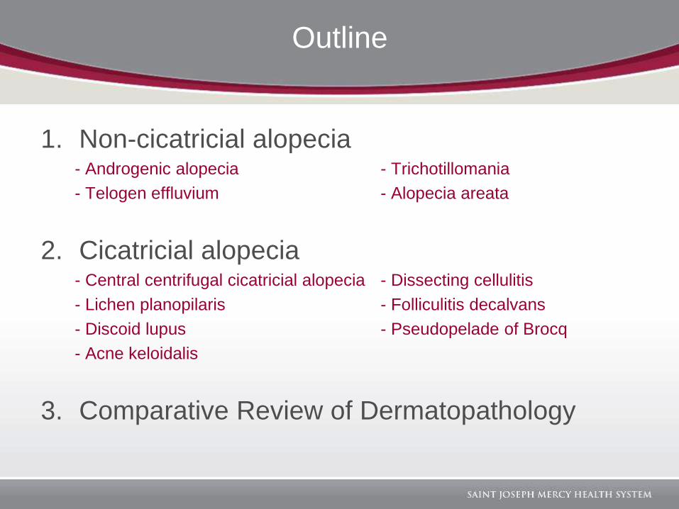
Outline
1. Non-cicatricial alopecia- Androgenic alopecia - Trichotillomania
- Telogen effluvium - Alopecia areata
2. Cicatricial alopecia- Central centrifugal cicatricial alopecia - Dissecting cellulitis
- Lichen planopilaris - Folliculitis decalvans
- Discoid lupus - Pseudopelade of Brocq
- Acne keloidalis
3. Comparative Review of Dermatopathology
NON-CICATRICIAL ALOPECIA
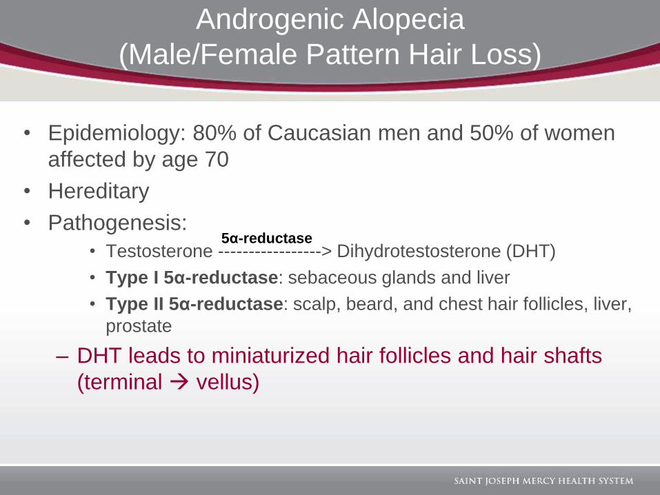
Androgenic Alopecia
(Male/Female Pattern Hair Loss)
• Epidemiology: 80% of Caucasian men and 50% of women
affected by age 70
• Hereditary
• Pathogenesis:
• Testosterone -----------------> Dihydrotestosterone (DHT)
• Type I 5α-reductase: sebaceous glands and liver
• Type II 5α-reductase: scalp, beard, and chest hair follicles, liver,
prostate
– DHT leads to miniaturized hair follicles and hair shafts
(terminal vellus)
5α-reductase
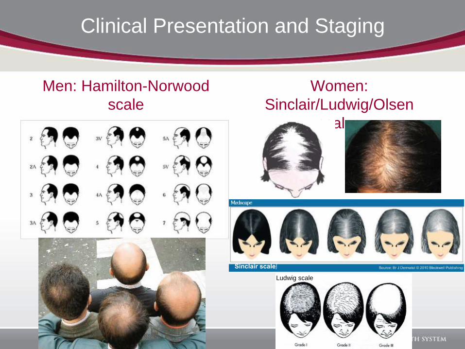
Clinical Presentation and Staging
Men: Hamilton-Norwood
scale
Women:
Sinclair/Ludwig/Olsen
scales
Sinclair scale∫
Ludwig scale
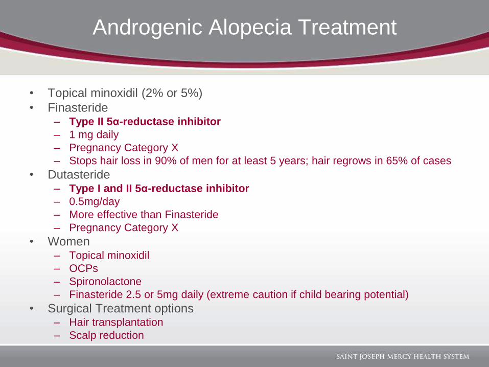
Androgenic Alopecia Treatment
• Topical minoxidil (2% or 5%)
• Finasteride– Type II 5α-reductase inhibitor
– 1 mg daily
– Pregnancy Category X
– Stops hair loss in 90% of men for at least 5 years; hair regrows in 65% of cases
• Dutasteride– Type I and II 5α-reductase inhibitor
– 0.5mg/day
– More effective than Finasteride
– Pregnancy Category X
• Women– Topical minoxidil
– OCPs
– Spironolactone
– Finasteride 2.5 or 5mg daily (extreme caution if child bearing potential)
• Surgical Treatment options– Hair transplantation
– Scalp reduction
• Camouflage techniques– Hair pieces
– Wigs
– Creative styling
– Hair dyes/Spray on hair/Hair fibers
• New and emerging treatments– Platelet rich plasma (PRP)
• More studies needed to evaluate mechanism
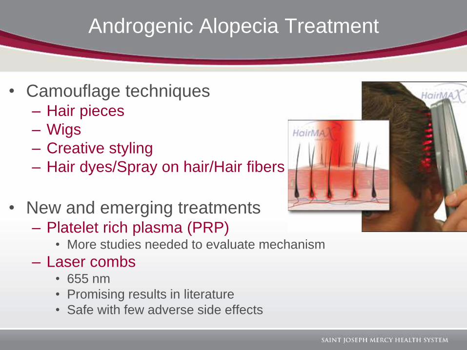
– Laser combs• 655 nm
• Promising results in literature
• Safe with few adverse side effects
Androgenic Alopecia Treatment
Telogen Effluvium
• Pathogenesis:– Premature conversion of anagen hairs to telogen hairs secondary to a
precipitating event/trigger
• Common triggers: – Surgery, fever, medications
– Crash dieting, iron deficiency
– Papulosquamous disease affecting scalp
– Thyroid disease
– Pregnancy (2-3 months after delivery)
– Severe emotional distress
• Chronic form with no precipitating factors
• Clinical Presentation:– Increased shedding of entire scalp (150-400 hairs/day)
– Positive hair pull test (>10% club hairs)
– Physician may not appreciate a decrease in hair density, while patient may complain of noticeably thinner hair
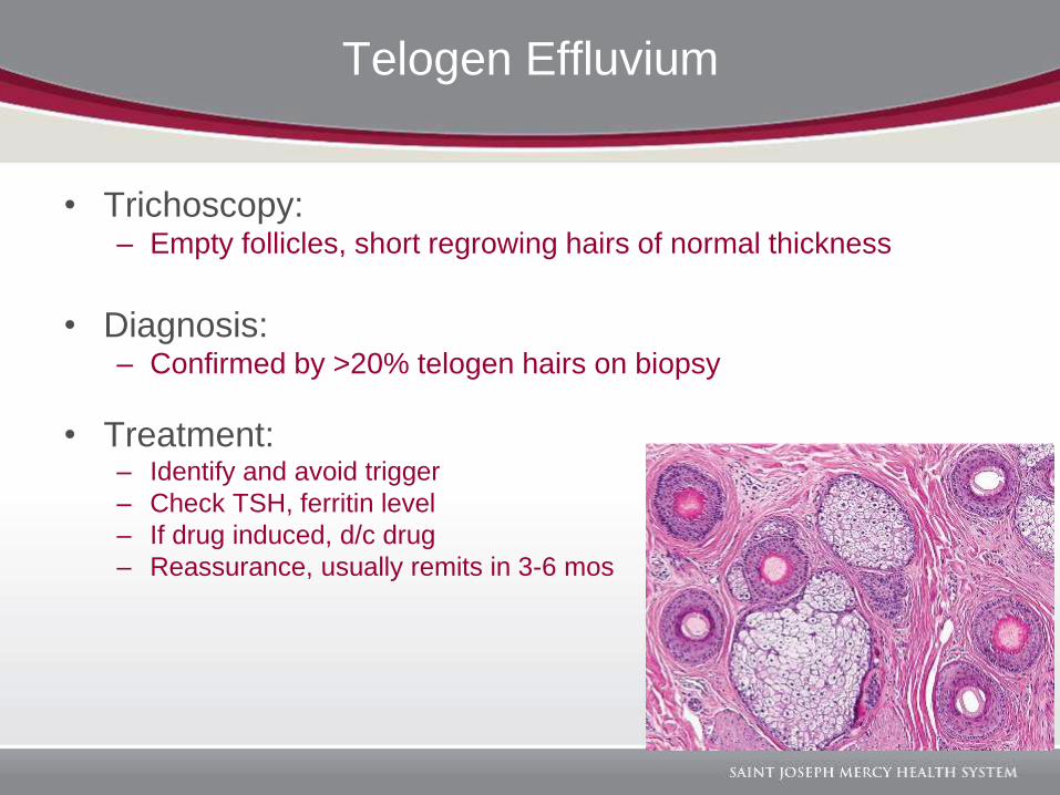
• Trichoscopy:– Empty follicles, short regrowing hairs of normal thickness
• Diagnosis:– Confirmed by >20% telogen hairs on biopsy
• Treatment:– Identify and avoid trigger
– Check TSH, ferritin level
– If drug induced, d/c drug
– Reassurance, usually remits in 3-6 mos
Telogen Effluvium
Trichotillomania
• Classified under American psychiatric association’s DSM-V as an “obsessive-compulsive disorder”
• Epidemiology– MC in girls <10
• Etiology– Habitual hair pulling
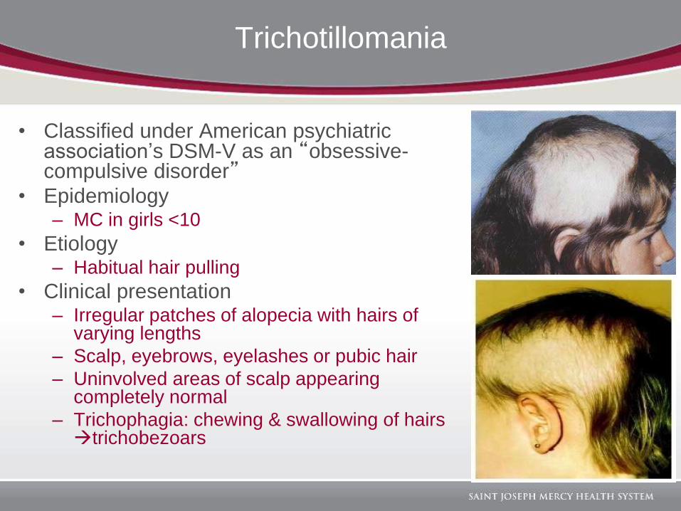
• Clinical presentation– Irregular patches of alopecia with hairs of
varying lengths
– Scalp, eyebrows, eyelashes or pubic hair
– Uninvolved areas of scalp appearing completely normal
– Trichophagia: chewing & swallowing of hairs trichobezoars
Trichotillomania
• Diagnosis made clinically
• Treatment– Behavior modification
– Psychotherapy
– SSRIs
– N-acetylcysteine• Increases glutamate concentration reduced compulsive behavior
Alopecia Areata
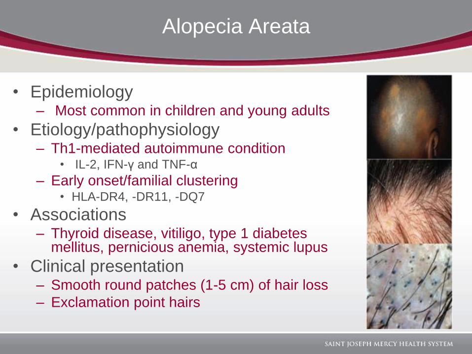
• Epidemiology– Most common in children and young adults
• Etiology/pathophysiology– Th1-mediated autoimmune condition
• IL-2, IFN-γ and TNF-α
– Early onset/familial clustering• HLA-DR4, -DR11, -DQ7
• Associations– Thyroid disease, vitiligo, type 1 diabetes
mellitus, pernicious anemia, systemic lupus
• Clinical presentation– Smooth round patches (1-5 cm) of hair loss
– Exclamation point hairs
Alopecia Areata Subtypes
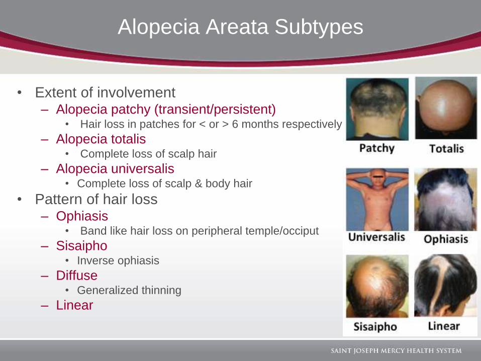
• Extent of involvement– Alopecia patchy (transient/persistent)
• Hair loss in patches for < or > 6 months respectively
– Alopecia totalis• Complete loss of scalp hair
– Alopecia universalis• Complete loss of scalp & body hair
• Pattern of hair loss– Ophiasis
• Band like hair loss on peripheral temple/occiput
– Sisaipho• Inverse ophiasis
– Diffuse• Generalized thinning
– Linear
Alopecia Areata
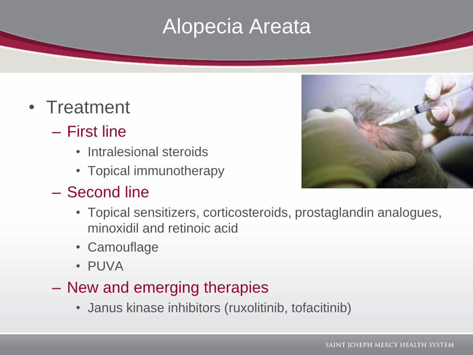
• Treatment
– First line
• Intralesional steroids
• Topical immunotherapy
– Second line
• Topical sensitizers, corticosteroids, prostaglandin analogues,
minoxidil and retinoic acid
• Camouflage
• PUVA
– New and emerging therapies
• Janus kinase inhibitors (ruxolitinib, tofacitinib)
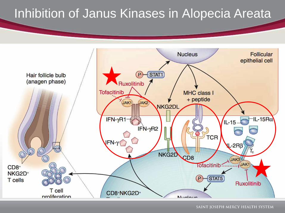
Inhibition of Janus Kinases in Alopecia Areata
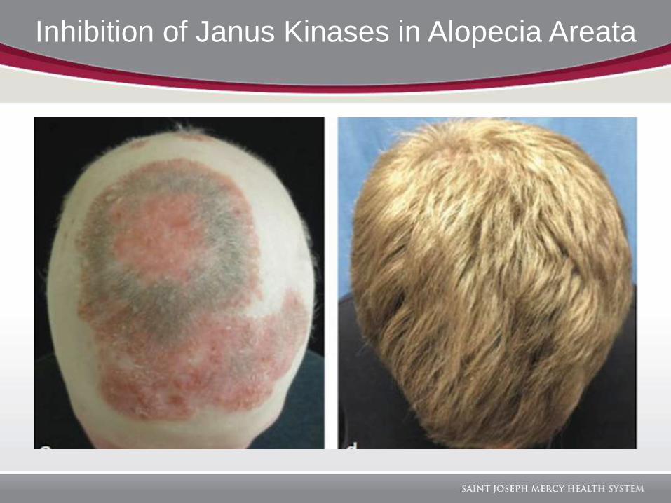
Inhibition of Janus Kinases in Alopecia Areata
CICATRICIAL ALOPECIA
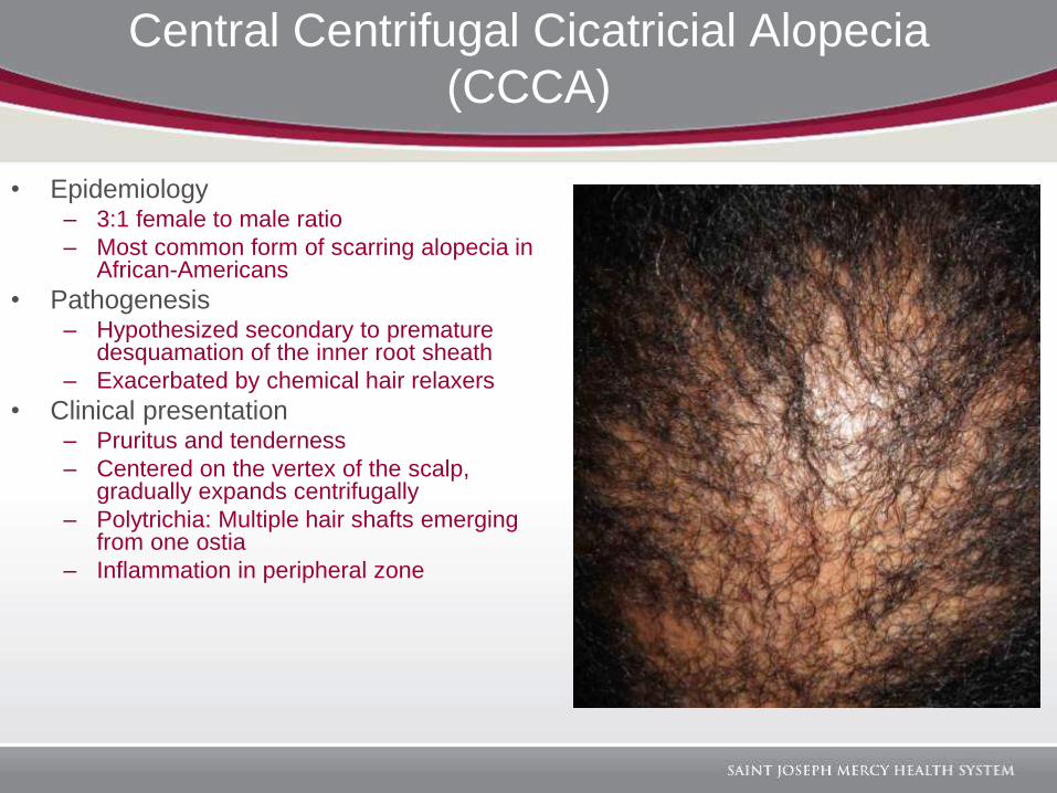
Central Centrifugal Cicatricial Alopecia
(CCCA)
• Epidemiology– 3:1 female to male ratio
– Most common form of scarring alopecia in African-Americans
• Pathogenesis – Hypothesized secondary to premature
desquamation of the inner root sheath
– Exacerbated by chemical hair relaxers
• Clinical presentation– Pruritus and tenderness
– Centered on the vertex of the scalp, gradually expands centrifugally
– Polytrichia: Multiple hair shafts emerging from one ostia
– Inflammation in peripheral zone
Central Centrifugal Cicatricial Alopecia
(CCCA)
• Treatment– Discontinuation of traumatic hairstyling
– Corticosteroids• Topical and intralesional
– Oral antibiotics• Tetracycline family
• New and emerging therapy– Hair transplantation
Lichen Planopilaris
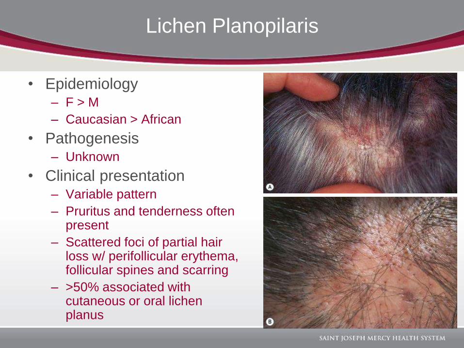
• Epidemiology– F > M
– Caucasian > African
• Pathogenesis– Unknown
• Clinical presentation– Variable pattern
– Pruritus and tenderness often present
– Scattered foci of partial hair loss w/ perifollicular erythema, follicular spines and scarring
– >50% associated with cutaneous or oral lichen planus
Lichen Planopilaris
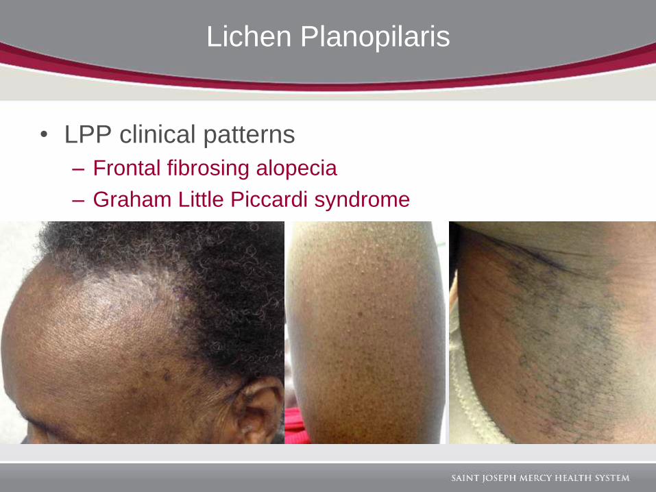
• LPP clinical patterns
– Frontal fibrosing alopecia
– Graham Little Piccardi syndrome
Lichen Planopilaris
• Treatment– Often resistant to therapy
– Corticosteroids• Topical, intralesional and oral
– Antimalarials
– Anecdotal • Cyclosporine, mycophenolate mofetil, systemic retinoids, or
low-dose methotrexate
• New and emerging treatment– Pioglitazone 15mg daily
• PPAR gamma agonist
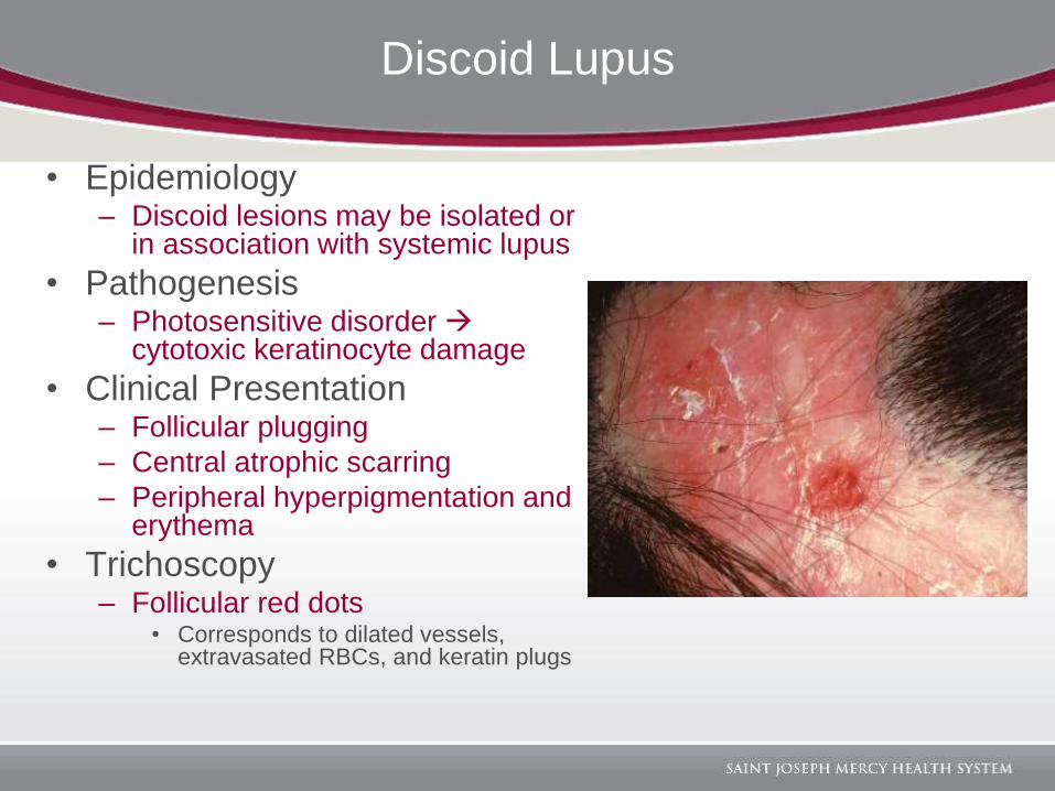
Discoid Lupus
• Epidemiology– Discoid lesions may be isolated or
in association with systemic lupus
• Pathogenesis– Photosensitive disorder
cytotoxic keratinocyte damage
• Clinical Presentation– Follicular plugging
– Central atrophic scarring
– Peripheral hyperpigmentation and erythema
• Trichoscopy– Follicular red dots
• Corresponds to dilated vessels, extravasated RBCs, and keratin plugs
Discoid Lupus
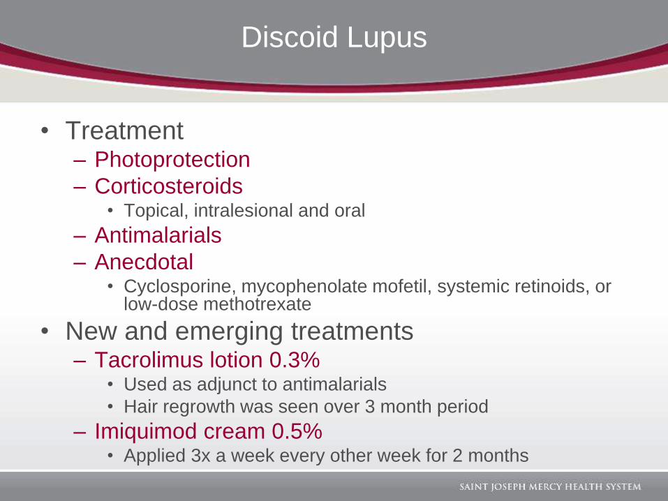
• Treatment– Photoprotection
– Corticosteroids• Topical, intralesional and oral
– Antimalarials
– Anecdotal • Cyclosporine, mycophenolate mofetil, systemic retinoids, or
low-dose methotrexate
• New and emerging treatments– Tacrolimus lotion 0.3%
• Used as adjunct to antimalarials
• Hair regrowth was seen over 3 month period
– Imiquimod cream 0.5% • Applied 3x a week every other week for 2 months
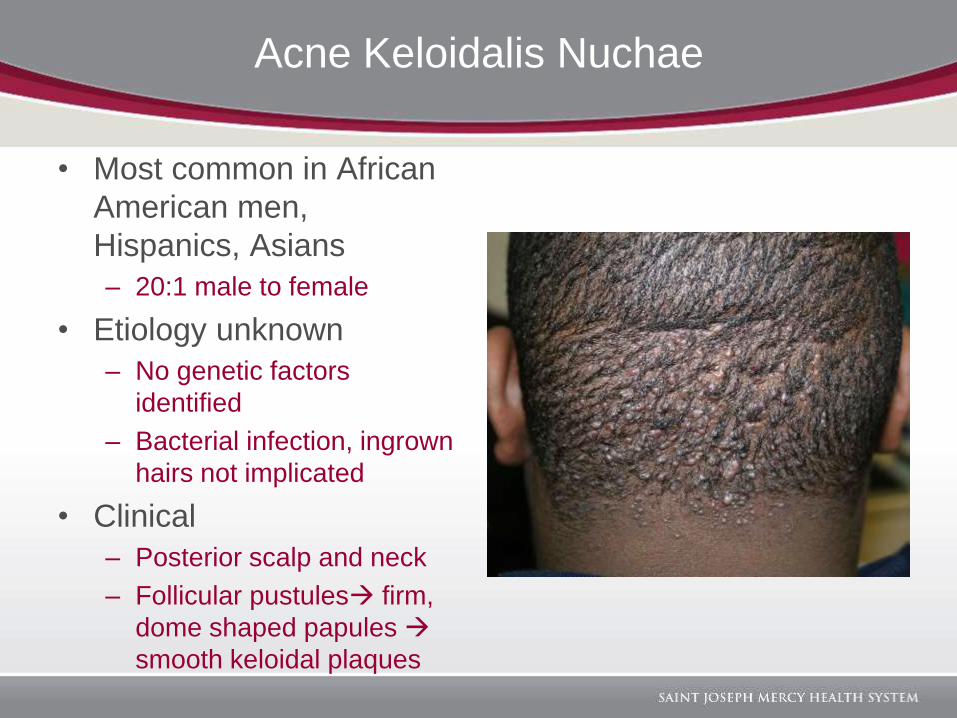
Acne Keloidalis Nuchae
• Most common in African
American men,
Hispanics, Asians
– 20:1 male to female
• Etiology unknown
– No genetic factors
identified
– Bacterial infection, ingrown
hairs not implicated
• Clinical
– Posterior scalp and neck
– Follicular pustules firm,
dome shaped papules
smooth keloidal plaques
Acne Keloidalis Nuchae
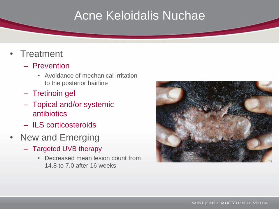
• Treatment
– Prevention
• Avoidance of mechanical irritation
to the posterior hairline
– Tretinoin gel
– Topical and/or systemic
antibiotics
– ILS corticosteroids
• New and Emerging– Targeted UVB therapy
• Decreased mean lesion count from
14.8 to 7.0 after 16 weeks
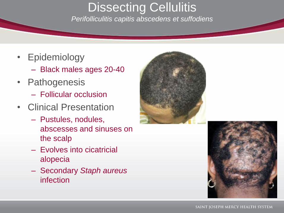
Dissecting CellulitisPerifolliculitis capitis abscedens et suffodiens
• Epidemiology
– Black males ages 20-40
• Pathogenesis
– Follicular occlusion
• Clinical Presentation
– Pustules, nodules,
abscesses and sinuses on
the scalp
– Evolves into cicatricial
alopecia
– Secondary Staph aureus
infection
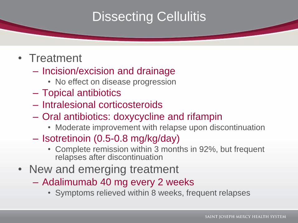
Dissecting Cellulitis
• Treatment– Incision/excision and drainage
• No effect on disease progression
– Topical antibiotics
– Intralesional corticosteroids
– Oral antibiotics: doxycycline and rifampin• Moderate improvement with relapse upon discontinuation
– Isotretinoin (0.5-0.8 mg/kg/day)• Complete remission within 3 months in 92%, but frequent
relapses after discontinuation
• New and emerging treatment– Adalimumab 40 mg every 2 weeks
• Symptoms relieved within 8 weeks, frequent relapses
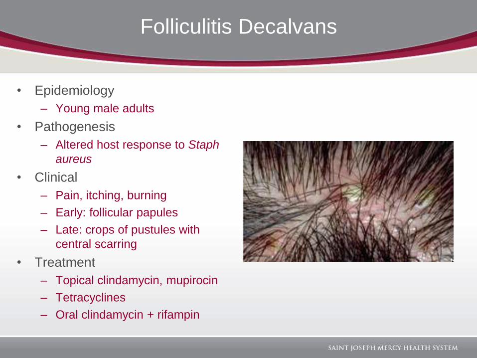
Folliculitis Decalvans
• Epidemiology
– Young male adults
• Pathogenesis
– Altered host response to Staph
aureus
• Clinical
– Pain, itching, burning
– Early: follicular papules
– Late: crops of pustules with
central scarring
• Treatment
– Topical clindamycin, mupirocin
– Tetracyclines
– Oral clindamycin + rifampin
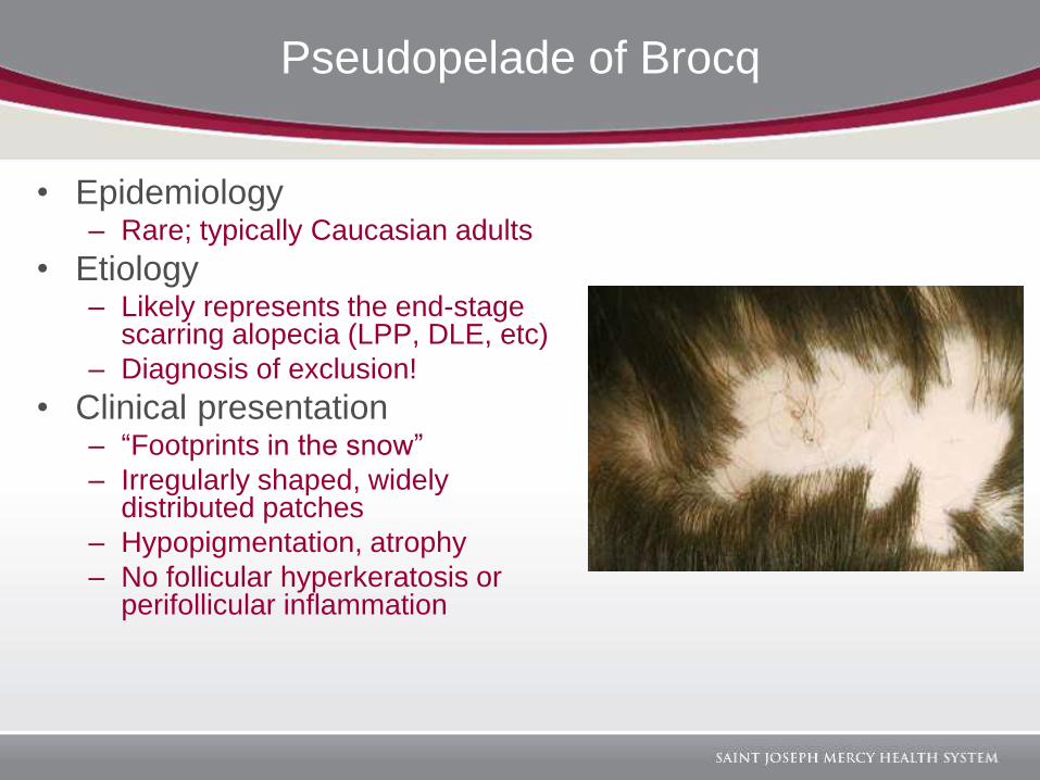
Pseudopelade of Brocq
• Epidemiology– Rare; typically Caucasian adults
• Etiology– Likely represents the end-stage
scarring alopecia (LPP, DLE, etc)
– Diagnosis of exclusion!
• Clinical presentation– “Footprints in the snow”
– Irregularly shaped, widely distributed patches
– Hypopigmentation, atrophy
– No follicular hyperkeratosis or perifollicular inflammation
DERMATOPATHOLOGY
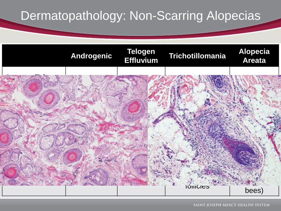
Dermatopathology: Non-Scarring Alopecias
AndrogenicTelogen
EffluviumTrichotillomania
Alopecia
Areata
Anagen:Teloge
n RatioDecreased Decreased Variable Decreased
Catagen Hairs Rare - Common Common
Miniaturization Yes No No Yes
Additional
FindingsAnisotrichosis
>20%
telogen
hairs
Trichomalacia,
pigment casts,
empty anagen
follicles
Peribulbar
lymphocytic
inflammation
(swarm of
bees)
Dermatopathology: Scarring Alopecias
LPP DLE CCCADissecting
Cellulitis
Predominant
Inflammatory
Cell
Lymphocyte Lymphocyte LymphocyteNeutrophil,
then mixed
Level of
Inflammation
Infundibulum,
isthmusIsthmus
Infundibulum,
isthmus
Extensive,
extending into
subQ
Characteristic
Features
Lichenoid
interface,
Civatte
bodies
Vacuolar
interface,
follicular
plugging,
mucin, +DIF
Eccentric
epithelial
atrophy,
naked hair
shafts
Sinus tracts
lined by
squamous
epithelium,
subQ and
dermal
abscesses

Summary
1. Non-cicatricial alopecia- Androgenic alopecia - Trichotillomania
- Telogen effluvium - Alopecia areata
2. Cicatricial alopecia- Central centrifugal cicatricial alopecia - Dissecting cellulitis
- Lichen planopilaris - Folliculitis decalvans
- Discoid lupus - Pseudopelade of Brocq
- Acne keloidalis
3. Comparative Review of Dermatopathology
• Amato L, Mei S, Massi D. Cicatricial alopecia; a dermatopathologic and immunopathologic study of 33 patients (pseudopelade
of brocq is not a specific clinico-pathologic entity). Int J Dermatol. 2002;41:8-15.
• Bolduc, C. Alopecia Areata treatment-Corticosteroid injection. Medscape. 2015. Retrieved from:
http://img.medscapestatic.com/pi/meds/ckb/89/27989tn.jpg.
• Bolognia, J. “Alopecias.” Dermatology. 3rd ed. Vol. 1. Philadelphia: Elsevier Saunders, 2012. 1105-07.
• Blume-Peytavi, U, Hillmann K, Dietz E, et al. A randomized, single-blind trial of 5% minoxidil foam once daily versus 2%
minoxidil solution twice daily in the treatment of androgenic alopecia in women. J Am Acad Dermatol. 2011 Dec;65(6):1126-
1134.e2.
• Callender VD, Lawson CN, Onwudiwe OC. Hair transplantation in the surgical treatment of central centrifugal cicatricial
alopecia. Dermatol Surg. 2014;40(10):1125-1130.
• Divito, S. and T. Kupper. Inhibiting Janus kinases to treat alopecia areata. Nature. 2014;20:9:989-990.
• Elder, D. Lever’s Histopathology of the Skin. 11th ed. Philadelphia: Wolters Kluwer, 2015. 329-33.
• Gkini MA, Kouskoukis AE, Tripsianis G, et al. Study of platelet-rich plasma injections in the treatment of androgenetic alopecia
through an one-year period. J Cutan Aesthet Surg. 2014 Oct-Dec;7(4):213-9
• Gubelin H, Barboza M, Tsai TF, et al. A randomized, active- and placebo-controlled study of the efficacy and safety of different
doses of dutasteride versus placebo and finasteride in the treatment of male subjects with androgenetic alopecia. J Am Acad
Dermatol. 2014 Mar;70(3):489-498.e3
• Ioffreda M. Inflammatory diseases of hair follicles, sweat glands, and cartilage. In: Elder D, Elenitsas R, Johnson B, et al, eds.
Lever’s Histopathology of the Skin. 10th ed. Philadelphia, PA: Lippincott Williams & Wilkins; 2009.
• James, W. Berger, T. and D. Elston. Diseases of the Skin Appendages. Andrews Disease of the Skin Clinical Dermatology.
2011;11:741-743.
• Madu P, Kundu RV. Follicular and Scarring Disorders in Skin of Color: Presentation and Management. Am J Clin Dermatol.
2014;15:307-321.
• Mahadevia, B. Images of Alopecia areata-totalis and universalis. Goodbyehairloss. Retrieved from:
http://goodbyehairloss.blogspot.com/2010/03/alopecia-areata.html.
References
References
• Mesinkovska NA, Tellez A, Dawes D, Piliang M, Bergfeld W. The use of oral pioglitazone in the treatment of lichen planopilaris.
J Am Acad Dermatol. 2015. 72(2):355-6.
• Milam E, Ramachandran S, Franks A. Treatment of Scarring Alopecia in Discoid Variant of Chronic Cutaneous Lupus
Erythematosus with Tacrolimus Lotion, 0.3%. JAMA Dermatol. 2015. Web. 29 July 2015.
• Leavitt M, Charles G, Heyman E, et al. HairMax LaserComb laser phototherapy device in the treatment of male androgenetic
alopecia: A randomized, double-blind, sham device-controlled, multicentre trial. Clin Drug Investig. 2009;29(5):283-92.
• Mirmirani P, Willey A, Headington J. Primary cicatricial alopecia: Histopathologic findings do not distinguish clinical variants. J
Am Acad Dermatol. 2005;52:637-643.
• Rapini, R. Adenexal Inflammatory Diseases. Practical Dermatopathology. 2012;10:157.
• Rodney I, Onwudiwe O, Callendar V, et al. Hair and scalp disorders in ethnic populations. Journal of Drugs and Dermatology.
2013 Apr;12(4):420-7.
• Rodriguez-Garcia C, Gonzalez-Hernandez S, Hernandez-Martin A. Aplasia cutis congenita and other anomalies associated
with methimazole exposure during pregnancy. Pediatr Dermatol. 2011;28(6)743-6.
• Seetharam, K. Alopecia areata: An update. Indian J Dermatol Venereol Leprol. 2013;79:563-575.
• Sperling L, Solomon A, Whiting D. A new look at scarring alopecia. Arch Dermatol. 2000;136:235-242.
• Turan E, Sinem B, Turgut E, et al. “Successful treatment of generalized discoid lupus erythematosus with imiquimod cream
5%: a case report and review of the literature.” Acta Dermatovenerol Croat. (2014);22(2): 150-9.
• Vivehanantha, S. and J. Berth-Jones. Alopecia areata. Lebwohl Treatment of Skin Disease: Comprehensive Therapeutic
Strategies. 2014;10:29.
• Zhao, Y. Zhang, B. Caulloo, S. et. al. Diffuse alopecia areata is associated with intense inflammatory infiltration and CD8+ T
cells in hair loss regions and an increase in serum IgE level. Indian J Dermatol Venereol Leprol. 2012;78:709-714.

Thank You


![Symmetric alopecia in the dog [Read-Only]alaskanmalamute.org/.../uploads/2015/11/Symmetric-alopecia-in-the … · of alopecia in the dog Pathogenesis Clinical appearance of alopecia](https://img.pdfslide.us/doc/110x75/5ebdda54a09b4c70d34c1b77/symmetric-alopecia-in-the-dog-read-only-of-alopecia-in-the-dog-pathogenesis.jpg)

![NIH Public Access novel perspectives and therapeutic ...€¦ · 03/06/2019 · alopecia, effluvium, hirsutism), acne vulgaris and atopic dermatitis [1–7]. In this article, the](https://img.pdfslide.us/doc/110x75/5f9abefff472d223b041b2ea/nih-public-access-novel-perspectives-and-therapeutic-03062019-alopecia.jpg)