Embed Size (px)
Citation preview

469GEODIVERSITAS • 2012 • 34 (3) © Publications Scientifiques du Muséum national d’Histoire naturelle, Paris. www.geodiversitas.com
KEY WORDSClimatiiformes,
Lower Devonian,Lochkovian,
gnathostome,systematics.
Hanke G. F. & Davis S. P. 2012. — A re-examination of Lupopsyrus pygmaeus Bernacsek & Dineley, 1977 (Pisces, Acanthodii). Geodiversitas 34 (3): 469-487. http://dx.doi.org/10.5252/g2012n3a1
ABSTRACTNew anatomical details are described for the acanthodian Lupopsyrus pygmaeus Bernacsek & Dineley, 1977, based on newly prepared, nearly complete body fossils from the MOTH locality, Northwest Territories, Canada. New interpre-tations of previously known structures are provided, while the head, tail, and sensory lines of L. pygmaeus are described for the first time. The pectoral girdle of L. pygmaeus shows no evidence of pinnal and lorical plates as mentioned in the original species description. Instead, the dermal elements of the pectoral region appear to comprise a single pair of prepectoral spines which rest on transversely oriented procoracoids, and large, shallowly inserted, ornamented pectoral fin spines which contact both the procoracoids and scapulocoracoids. The scales of L. pygmaeus lack growth zones and mineralized basal tissue, and superficially resemble scales of thelodonts or monodontode placoid scales of early chondrichthyans, and not the typical scales of acanthodians. However, L. pygmaeus possesses perichondrally-ossified pork-chop shaped scapulocora-coids, a series of hyoidean gill plates, and scale growth that originates near the caudal peduncle; these features suggest a relationship to acanthodians. Prior to this study, both authors conducted separate cladistic analyses which resulted in differing tree positions for L. pygmaeus and its relationships within the Acanthodii. However, both analyses did agree that there is no evidence allying L. pygmaeus to the traditional “climatiid” acanthodians contrary to previous historical classifications.
Gavin F. HANKERoyal British Columbia Museum,
675 Belleville Street, Victoria, British Columbia V8W 9W2 (Canada)[email protected]
Samuel P. DAVIS 505 King Court, Smithfield, Virginia, 23430-6288 (USA)
A re-examination of Lupopsyrus pygmaeus Bernacsek & Dineley, 1977 (Pisces, Acanthodii)

470 GEODIVERSITAS • 2012 • 34 (3)
Hanke G. F. & Davis S. P.
INTRODUCTION
The first few acanthodian Denison (1999) speci-mens collected from the MOTH locality, Mackenzie Mountains, N.W.T., Canada, were either badly weathered prior to collection, or were over-prepared, and therefore exhibited limited anatomical infor-mation. The first specimens of Lupopsyrus pygmaeus Bernacsek & Dineley, 1977 were no exception. Bernacsek & Dineley (1977) described L. pygmaeus and assigned the species to the family Climatiidae Berg, 1940 (Climatiiformes) based on the presence of a series of prepelvic spines, a pair of prepectoral spines, dermal pectoral armour, coupled with the perception that the ornamentation of L. pygmaeus body scales was similar to that of Climatius reticulatus Agassiz, 1845. Specimens of Lupopsyrus pygmaeus which were available to Bernacsek & Dineley (1977) showed details of the pectoral girdle and flank, but none of their specimens had a well-preserved head
or caudal fin. Details from the species diagnosis and the reconstruction presented by Bernacsek & Dineley (1977) were presented in Denison’s (1979) summary of acanthodian fishes, and until now, L. pygmaeus has not been re-evaluated, even though many new and better-preserved specimens exist.
The phylogenetic position of Lupopsyrus Ber-nacsek & Dineley, 1977, has been difficult to determine partly due to the morphological inac-curacies of the original description. Bernacsek & Dineley (1977), and then Denison (1979) placed L. pygmaeus among climatiid fishes, whereas Long’s (1986: fig. 9) character summary placed L. pygmaeus with the diplacanthids. Unfortunately, Long (1986) suggested only that the fin-spine ornamen-tation (i.e. “simple linear type”), and the presence of a “free” pectoral fin spine of L. pygmaeus was primitive relative to that in C. reticulatus, and that the presence of a small lorical plate indicated that Lupopsyrus should be grouped with diplacanthids
RÉSUMÉNouvelle étude de Lupopsyrus pygmaeus Bernacsek & Dineley, 1977 (Pisces, Acanthodii).De nouveaux détails anatomiques de Lupopsyrus pygmaeus Bernacsek & Dineley, 1977 sont décrits à partir d’un matériel de fossiles sub-complets et récemment préparés de la localité MOTH, Territoires du Nord, Canada. De nouvelles inter-prétations des structures anatomiques déjà connues sont présentées, tandis que la tête, la queue et les lignes sensorielles de L. pygmaeus sont décrites pour la première fois. La ceinture pectorale de L. pygmaeus ne présente pas de plaques pinnales ou loricales, comme mentionnées dans la description originale de l’espèce. En revanche, les éléments dermiques de la région pectorale sont constitués d’une paire simple d’épines prépectorales reposant sur des procoracoïdes orientés transversalement et de larges épines pectorales ornementées, superficiellement insérées, en contact avec les procoracoïdes et les scapulocoracoïdes. Les écailles de L. pygmaeus ne présentent pas de zones de croissance ni de tissu basal minéralisés, ressemblant ainsi aux écailles des Thélodontes ou aux écailles placoïdes monodontodes des chondrichtyens basaux, et non aux écailles typiques d’acanthodiens. Cependant, L. pygmaeus possède des scapulocoracoïdes recouverts d’os périchondral et en forme de côtes de porc, des séries de plaques branchiales hyoïdiennes, et un centre de développement de la squamation situé près du pédoncule caudal ; ces caractères suggèrent une relation avec les Acanthodiens. En amont de cette étude, les auteurs ont conduit des analyses cladistiques séparées amenant à différentes positions phylogénétiques au sein des Acanthodii. Néanmoins, aucune de ces deux analyses ne met en évidence l’asso-ciation de L. pygmaeus aux traditionnels acanthodiens climatiides, contrairement à ce qu’affirmaient les précédentes classifications.
MOTS CLÉSClimatiiformes,
Dévonien inférieur,Lochkovien,
gnathostome,systématique.

471
A re-examination of Lupopsyrus pygmaeus
GEODIVERSITAS • 2012 • 34 (3)
drawings were made with a camera lucida attached to the NIKON SMZ 1500 dissecting microscope.
Specimens are deposited in the collections of the Laboratory for Vertebrate Palaeontology (Depart-ment of Biological Sciences), University of Alberta, Edmonton, Alberta, Canada, and carry the prefix UALVP on catalogue numbers. The holotype and several other specimens of Lupopsyrus pygmaeus are housed at the Canadian Museum of Nature and when these specimens were accessioned, the catalogue numbers were given the prefix NMC de-noting the early name for the institution, National Museums of Canada. Geological Survey of Canada localities carry the prefix GSC. At the time of writ-ing, the holotype of L. pygmaeus (NMC 22715) is still on loan in England and was not available for examination.
AbbreviAtionsaf. anal fin web;afs. anal fin spine;art.pfs. pectoral fin spine articulation fossa;d.end.e. ductus endolymphaticus externus;dfa. anterior dorsal fin web;dfa.sp. anterior dorsal fin spine;dfp. posterior dorsal fin web;dfp.sp. posterior dorsal fin spine;dt. dentine tubules;eks. enlarged keeled scutes;hgc. hyoidean gill cover;hl. hypochordal lobe of caudal fin;ins.a. insertion area of spines;lc. main lateral sensory canal;lt. left side;mpl. middle pit line;occ. occipital cross-commissure;ot. otic material;pc. pulp cavity;pcb. basal opening of pulp cavity;pcf. pectoral fin web;pfs. pectoral fin spine;pls. pelvic fin spine;poc. preopercular sensory line;ppl. posterior pit line;p.ps. prepectoral spine;prc. procoracoid;prp. prepelvic spine;pv.f. pelvic fin web;rt. right side;sco. scapulocoracoid;smc. supramaxillary sensory line;soc. supraorbital sensory canal.
(his “diplacanthoids”). Some MORS (Middle Old Red Sandstone) diplacanthids from the Middle Devonian of Scotland have paired ventral dermal plates attached to the procoracoids (SPD pers. obs.; Miles 1973), but these are enlarged, nearly flat, and ornamented, and therefore are very dif-ferent to any of the structures associated with the pectoral girdle of L. pygmaeus (even as described by Bernacsek & Dineley [1977]).
In this paper we present a new interpretation of the pectoral girdle of L. pygmaeus and provide descriptions of several structures which were not available to Bernacsek & Dineley (1977). In addi-tion to the morphological elements of L. pygmaeus which provided data for the cladistical analyses by Davis (2002), we also provide a description of scale microstructure and an account of scale vari-ation across the body. This updated histological and morphological account was used to generate characters in the cladistic analysis by Hanke & Wilson (2004) and will be useful for those wish-ing to include L. pygmaeus in future phylogenetic analyses.
MATERIAL AND METHODS
Recently collected specimens of L. pygmaeus were cleaned using a combination of acetic acid and mechanical preparation. The siliciclastic residues remaining after acetic acid treatment were removed using a soft brush and 00-insect-pins. Fossils were stabilized using a 5% solution of Glyptal™ cement. Ammonium chloride sublimate was used to whiten specimens before photography.
Small groups of scales were embedded in Lumi-nate 83 HA-4 epoxy, and then polished to expose histological structure using 600 and 1000 grit wet-dry sandpaper, with a final polish using moistened alumina powder on a glass plate. Images were taken using a Nikon Coolpix 990 digital camera attached to a Nikon SMZ 1500 dissecting microscope. The external structure of scales was examined using the JEOL JSM 6301 FXV electron microscope in the Department of Earth and Atmospheric Sciences, University of Alberta. Scale specimens were sputter-coated with gold prior to electron microscopy. Line
472 GEODIVERSITAS • 2012 • 34 (3)
Hanke G. F. & Davis S. P.
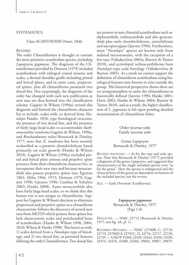
SYSTEMATICS
Class ACANTHODII Owen, 1846
remArks
The order Climatiiformes is thought to contain the most primitive acanthodian species, including Lupopsyrus pygmaeus. The diagnosis of the Cli-matiiformes provided by Denison (1979) included acanthodians with enlarged cranial tesserae and scales, a dermal shoulder girdle including pinnal and lorical plates, and in some cases, prepecto-ral spines, plus all climatiiforms possessed two dorsal fins. Not surprisingly, the diagnosis of the order has changed with each new publication as new taxa are shoe-horned into the classification scheme. Gagnier & Wilson (1996a) revised this diagnosis and limited the climatiiform character list to include: scales with, or derived from Nostolepis Pander, 1856, type histological structure, the presence of two dorsal fins, and the presence of fairly large head scales to accommodate Kathemacanthus rosulentus Gagnier & Wilson, 1996a, and Brochoadmones milesi Bernacsek & Dineley, 1977; note that K. rosulentus has been recently reclassified as a putative chondrichthyan based primarily on scale growth (Hanke & Wilson 2010). Gagnier & Wilson (1996a) excluded pin-nal and lorical plate armour and prepelvic spine presence from their climatiiform character list, to incorporate their new taxa and because mesacan-thids also possess prepelvic spines (see: Egerton 1861; Miles 1966, 1973; Denison 1979; Gag-nier 1996; Upenice 1996; Cumbaa & Schultze 2002; Hanke 2008). Some mesacanthids also have fairly large head scales, so we think that this feature too is not unique to climatiiforms. Sup-port for Gagnier & Wilson’s decision to eliminate prepectoral and prepelvic spines as a climatiiform characteristic follows the discovery of several new taxa from MOTH which possess these spines but lack characteristic scales and perichondral bone of acanthodians (Hanke & Wilson 1998, 2004, 2010; Wilson & Hanke 1998). This leaves us with: 1) scales derived from a Nostolepis-type of histol-ogy; and 2) two dorsal fins, as potential features defining the order Climatiiformes. Two dorsal fins
are present in non climatiid acanthodians such as: diplacanthids, ischnacanthids and also gyracan-thids plus early chondrichthyans, osteichthyans and sarcopterygians (Janvier 1996). Furthermore, most “Nostolepis” species are known only from isolated microremains, with the exception of a few taxa (Valiukevičius 2003a; Burrow & Turner 2010), and acritolepid ischnacanthiforms have Nostolepis-type scale histology (Valiukevičius & Burrow 2005). As a result we cannot support the definition of climatiiform acanthodians using his-tological features now known to exist outside the group. This historical perspective shows there are no synapomorphies to unite the climatiiforms as historically defined (Janvier 1996; Hanke 2001; Davis 2002; Hanke & Wilson 2004; Burrow & Turner 2010), and as a result, the higher classifica-tion of L. pygmaeus is left open pending detailed reexamination of climatiiform fishes.
Order incertae sedis Family incertae sedis
Genus Lupopsyrus Bernacsek & Dineley, 1977
revised diAgnosis. — As for the type and only spe-cies. Note that Bernacsek & Dineley (1977) provided a diagnosis of the genus Lupopsyrus, and suggested that characteristics of the single included species were “as for the genus”. Here the species is rediagnosed and the characteristics of the genus are dependent on features of the included species, not the reverse.
Age. — Early Devonian (Lochkovian).
Lupopsyrus pygmaeus Bernacsek & Dineley, 1977
(Figs 1-6)
Holotype. — NMC 22715 (Bernacsek & Dineley 1977: text-fig. 3A, pl. 1).
referred mAteriAl. — NMC 22700B, C, 22718, 22719, 22700D-F, 22701C, D, 22716, 22717, 22720, 22745. — UALVP 19260, 32420, 32442, 32456, 32458, 32474, 32476, 32480, 32482, 39065, 39067, 39079-

473
A re-examination of Lupopsyrus pygmaeus
GEODIVERSITAS • 2012 • 34 (3)
39082, 39121, 41493, 41629, 41632, 41665, 41931, 41939, 41945, 42000, 42002, 42008, 42012, 42013, 42027, 42046, 42061, 42113, 42142, 42150, 42173, 42208, 42274, 42518, 42524, 42529, 42530, 42533, 42538, 42453-42455, 42544, 42597, 42605, 43064, 43091, 43092, 43094, 43095, 43256, 43409, 43456, 45154, 45155.
LocaLity and age. — Lupopsyrus specimens are recovered from talus below a Lower Devonian (Lochkovian) horizon between 430-435 m in the MOTH locality section (as measured in 1996), central Mackenzie Mountains, N.W.T., Canada (see Hanke et al. 2001b: figs 1, 2); approximately 411 m in the section measured by the Geological Sur-vey of Canada (Gabrielse et al. 1973). The Devonian fish layer in the MOTH locality section is equivalent to GSC locality 69014 in section 43 of Gabrielse et al. (1973) and locality 129 in the UALVP catalogue system. Although previous authors have suggested habitats rang-ing from intertidal lagoons to deep-water shelf settings, recent sedimentological, ichnological and taphonomic
study suggests an oxygen-poor, intra-shelf topographic low below storm wave base (Zorn et al. 2005) on a shelf that fringed western Laurussia (combined Laurentia and Baltica; Li et al. 1993).
revised diAgnosis. — Acanthodians with two longi-tudinal rows of enlarged keeled scutes situated along the posterior half of each side of the body and caudal fin axis; largest keeled scutes located below the second dorsal fin base; three dermal opercular gill plates per side; pectoral, pelvic, anal and dorsal spines with widely separated ribs with fine nodular ornamentation; single prepectoral spine positioned over lateral end of each procoracoid; prepelvic fin spines with unornamented blade-like posterior lamina; circumorbital scales iden-tical to head scales; head and body scales have crown with prominent median keel and one lateral flange per side; central keel and lateral flanges of each scale terminating posteriorly in a single point; scales are monodontode with mesodentine, Stranggewebe-like crown histology.
A
Bdfp.
eks.
dfp.sp.dfa. dfa.sp.
lc.
smc.rt.p.ps
lt.p.psrt.pfs.
rt.2nd.prp.
rt.3rd.prp.rt.4th.prp.rt.pls.rt.pv.f.afs.af.hl.
rt.pcf.(rt.1st.prp.
underneath)
orbit
occ.
hgc.poc.ins.a.
Fig. 1. — Lupopsyrus pygmaeus Bernacsek & Dineley, 1977: A, photograph of UALVP 41493 in left lateral view; B, camera lucida draw-ing of the same specimen with interpretation of structures. Abbreviations: see Material and methods. Scale bars: 1 cm.

474 GEODIVERSITAS • 2012 • 34 (3)
Hanke G. F. & Davis S. P.
description
Much of the dermal covering over the head of Lupopsyrus pygmaeus is preserved on UALVP 41493 (Fig. 1), and other new Lupopsyrus specimens in the University of Alberta collections. The head of L. pygmaeus as preserved is short at 10 % of body length, but the rostrum and jaws either have lost scale cover in all specimens, or lacked scale cover in life and therefore, the full snout length cannot be estimated (Figs 1; 2B); the jaws may also have fallen clear of the carcass in all specimens during decay. The dorsal surface of the head is covered with small scales which are similar in structure to body scales. These head scales differ slightly from those on the body in that the posterior apex of the crown of each scale is short, blunt and does not extend posterior to the basal rim surrounding the pulp ca-nal. The gradual transition between head and body scales occurs over the branchial chamber; enlarged head scales and/or tesserae are absent (Figs 1; 2).
The orbits of L. pygmaeus lack ossified sclerotic plates and are highlighted by an area of silvery-black material within the ring of micromeric cir-cumorbital scales (note that terminology follows Burrow et al. [2011]). The dorsal and posterior margin of the orbit has scales which are identical to normal head scales, and there are no enlarged
circumorbital scales (Figs 1; 2). There are no scales anterior and ventral to the orbit in any of the avail-able specimens, therefore the lachrymal and labial portions of the head are unknown and may have lacked scales in life.
The jaws, branchial arches and endocranium are not mineralized. Teeth are absent and there is no indication of jaw extent or shape. The position of the otic capsule, and therefore, the position near the posterior end of the braincase is indicated by small masses of sandy, otic material, which Sahney & Wilson (2001) interpret as otic statoconia. When scale cover is complete, these masses appear only as paired bulges (Figs 2B; 4B), but where scales are lost, the light coloured sandy material is exposed. Many L. pygmaeus specimens have heads preserved as dorsoventral compressions, and the masses of otic material are well-separated (Figs 2B; 4B). This preservation suggests that the braincase and head of L. pygmaeus was fairly broad rather than laterally compressed; a broad, depressed braincase is characteristic in several clades of gnathostomes (Janvier 1996) and likely is a primitive feature for gnathostomes.
Sensory lines preserved on very few L. pygmaeus specimens run between scales (Figs 1; 2). The su-pramaxillary sensory canal and preopercular sensory
Alt.p.ps.
hgc.
lt.pfs. lt.sco. lc.?
soc.
ot. rt.sco.
lt.pfs.
hgc.
orbit
rt.sco.d.end.eppl.mpl.
soc.
lc.
lt.prc.
B
Fig. 2. — Head of Lupopsyrus pygmaeus Bernacsek & Dineley, 1977: A, pectoral girdle and head of UALVP 39079 in dorsal view; B, head of UALVP 42208 in dorsolateral view. Abbreviations: see Material and methods. Scale bars: A, 1 cm; B, 0.5 cm.

475
A re-examination of Lupopsyrus pygmaeus
GEODIVERSITAS • 2012 • 34 (3)
canals converge posteriorly across the cheek anterior to the hyoidean gill plates (Fig. 1); the latter canal meets the main sensory line dorsally. The supraor-bital sensory canal traces converge posteriorly, but its entire course cannot be determined in the avail-able specimens (Fig. 2A, B). One specimen shows the trace of the infraorbital canal posteroventral to the orbit (Fig. 2B). There also are traces of short, paired, converging, middle pit lines, and posterior pit lines preserved near the level of the otic region of the braincase (Fig. 2A). Behind the posterior pit lines are a pair of gaps in the scale cover which may indicate the position of the external endo-lymphatic duct openings (Fig. 2A). The occipital cross-commissure is seen as a short canal trace leading dorsally from the main lateral canal level with the origin of the pectoral fin spine (Fig. 1B).
The branchial chamber of Lupopsyrus pygmaeus is not well-preserved in any specimens, but ap-
pears compact relative to the orbito-otic region; the extent of the branchial chamber is estimated from the position of the otic material and the position of the pectoral girdle (Fig. 2B). There is no evidence of gill openings, single or multi-ple, but the presence of three dagger-like dermal plates located in an arc over the opercular region indicates that a single opercular flap was present rather than a series of narrow separate opercular flaps (Figs 1; 2A, B). The middle plate of the operculum is larger than the dorsal and ventral plates, and each plate has a single longitudinal keel, surrounded by small tubercles. This keel is serrated, and its summit is near the mid-length of the plate. The underside of each opercular plate possesses a shallow trough which continues along the entire length of the plate; the basal rim is tear-drop shaped, widest anteriorly, and tapers posteriorly.
Adfp. dfp.sp.
af.
hl.
hl.
eks.
afs. rt.pv.f.
C
B
D
Fig. 3. — Lupopsyrus pygmaeus Bernacsek & Dineley, 1977: A, posterior dorsal fin of UALVP 41493; B, the anal fin spine and parts of the anal fin of UALVP 41493; C, the caudal peduncle and caudal fin of UALVP 45154; D, the leading edge of the hypochordal lobe of the caudal fin of UALVP 42208. Abbreviations: see Material and methods. Scale bars: A, B, D, 1 mm; C, 5 mm.

476 GEODIVERSITAS • 2012 • 34 (3)
Hanke G. F. & Davis S. P.
The dorsal fins and spines were described in detail by Bernacsek & Dineley (1977), although they did not mention that the anterior dorsal fin spine had a shallow insertion area (Fig. 1). This basal portion presumably anchored the spine into the epaxial mus-culature. Lupopsyrus pygmaeus lacks enlarged scales around the base of each fin spine.
The anterior and posterior dorsal fin spines sup-port fin webs which possess a convex trailing margin extending posterior to the apex of the fin spine, and also have irregularly-arranged scales (Figs 1; 3A). The distal half of each dorsal fin web appears to be de-tatched from the dorsal fin spine (Fig. 3A), but this may be an artefact of preservation or preparation. Fin-web scales are minute, and apart from size, are identical to typical body scales. There is a gradual size transition between typical body scales and small fin web scales (Fig. 3A, B).
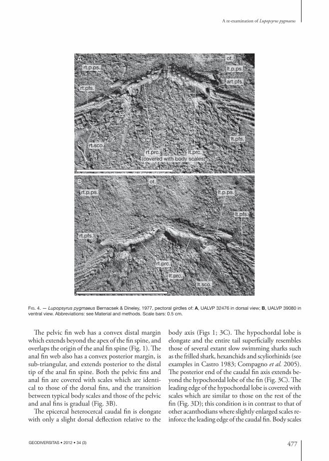
The pectoral fin spine was described in detail by Bernacsek & Dineley (1977) and no new informa-tion can be added in our account. However, details of the pectoral dermal plate armour require further clarification. Bernacsek & Dineley (1977) mentioned that a single lorical plate, with a single, median spine was present on the holotype of Lupopsyrus pygmaeus. They also indicated that the lorical plate was poorly preserved, as is evident in the photographs published with the species description. A median lorical plate is not known in any University of Alberta Lupopsyrus specimens (Figs 2A, B; 4), although in one, a small pile of displaced scales positioned posterior to the pectoral girdle creates the impression of a small, elevated node (Fig. 4B). It may be possible that the “lorical plate” described by Bernacsek & Dineley (1977) was a similar mass of scales. Specimens exam-ined at the Canadian Museum of Nature lack both lorical plates and median “lorical” spines.
The margins of the prepectoral spines cannot be distinguished in the figures presented by Bernacsek & Dineley (1977), and they suggest that the prepectoral spines are completely fused to what they described as compound pinnal plates. The University of Alberta specimens show that the prepectoral spines have a distinct basal rim which denotes the perimeter of each spine (Figs 2A; 4), and these spines sit over the lateral end of procoracoid bones (see below). The prepectoral spines are curved, have a broad
basal cavity, and possess longitudinal ribs which are ornamented with fine nodes (Figs 2A; 4).
Bernacsek & Dineley (1977) wrote that L. pygmaeus had compound pinnal plates, and went so far as to suggest a homology between the first and second pinnal plates of Climatius reticulatus. In fact, the bones that subtend both prepectoral spines and contact the anterior-most base of the pectoral spines are smooth, unornamented, and have a surface texture similar to that of the perichondrally-ossified scapu-locoracoids, not like the dermal tuberculated plates which are characteristic of climatiid pectoral dermal armour (Miles 1973; Watson 1937; Denison 1979). In L. pygmaeus, these bones extend anteromedial to the scapulocoracoid and meet at the ventral midline (Figs 2A, B; 4). We interpret these smoothly ossified structures to be procoracoids, based on structure and similarity of position to procoracoids of other acanthodians. The procoracoids have a small fossa on the lateral margin (Fig. 4) which may have served as the point of articulation for the proximal end of the pectoral fin spine. A revised reconstruction of the pectoral girdle of Lupopsyrus pygmaeus, based on UALVP 32476, 39079 and 39080 (Figs 2; 3) is presented in Figure 5. Any apparent ornamentation on the procoracoids (Fig. 4A) consists of overlying scales which had settled on the bone during decay and preservation of the carcass.
Two specimens (UALVP 39080 and 41493) show that a pectoral fin web was present posterior to the pectoral fin spine. The fin web is covered with minute scales, and the trailing edge of the fin probably was convex (Fig. 1). The fin web is not attached to the fin spine in UALVP 41493; additional specimens will be needed to determine whether the detatch-ment of the pectoral fin web in UALVP 41493 is a taphonomic artefact.
Little can be added to the descriptions of the prepelvic and pelvic fin spines. However, since the material available to Bernacsek & Dineley (1977) was poorly preserved, they could not determine which prepelvic spine had a posterior, flat lamina. Based on the material at the University of Alberta, all prepelvic spines of Lupopsyrus pygmaeus possess this flat, unornamented, trailing lamina, which is most prominent on the posterior-most prepelvic spine pair (Fig. 1).
477
A re-examination of Lupopsyrus pygmaeus
GEODIVERSITAS • 2012 • 34 (3)
The pelvic fin web has a convex distal margin which extends beyond the apex of the fin spine, and overlaps the origin of the anal fin spine (Fig. 1). The anal fin web also has a convex posterior margin, is sub-triangular, and extends posterior to the distal tip of the anal fin spine. Both the pelvic fins and anal fin are covered with scales which are identi-cal to those of the dorsal fins, and the transition between typical body scales and those of the pelvic and anal fins is gradual (Fig. 3B).
The epicercal heterocercal caudal fin is elongate with only a slight dorsal deflection relative to the
body axis (Figs 1; 3C). The hypochordal lobe is elongate and the entire tail superficially resembles those of several extant slow swimming sharks such as the frilled shark, hexanchids and scyliorhinids (see examples in Castro 1983; Compagno et al. 2005). The posterior end of the caudal fin axis extends be-yond the hypochordal lobe of the fin (Fig. 3C). The leading edge of the hypochordal lobe is covered with scales which are similar to those on the rest of the fin (Fig. 3D); this condition is in contrast to that of other acanthodians where slightly enlarged scales re-inforce the leading edge of the caudal fin. Body scales
B
A
(covered with body scales)rt.prc. lt.prc.
rt.sco.lt.pfs.
rt.pfs.
rt.p.ps.
ot.
lt.p.ps.
art.pfs.
rt.p.ps.
ot.
lt.p.ps.
lt.pfs.
lt.sco.
lt.prc.
rt.prc.
rt.pfs.
Fig. 4. — Lupopsyrus pygmaeus Bernacsek & Dineley, 1977, pectoral girdles of: A, UALVP 32476 in dorsal view; B, UALVP 39080 in ventral view. Abbreviations: see Material and methods. Scale bars: 0.5 cm.

478 GEODIVERSITAS • 2012 • 34 (3)
Hanke G. F. & Davis S. P.
on the caudal fin axis grade into typical caudal fin scales with no abrupt change in scale size, and these irregularly-arranged scales decrease in size along the caudal axis toward the posterior tip (Figs 1A; 3C).
The scales of L. pygmaeus change little in size over the body (Fig. 1), although as mentioned above, smaller scales are found on the fin webs, the posterior portions of the caudal fin axis, and on the head (Figs 1A; 2; 3). Bernacsek & Dineley (1977) described the external ornamentation of the scales of L. pygmaeus, but their photographs did not show the fine details of scale structure and pattern. All scales are ornamented with a central crest and a pair of lateral flanges which possess smooth edges (Fig. 6A-D) and merge posteriorly to form the trailing tip of each scale. The pulp cavity extends into the posterior portions of the central crest (Fig. 6C). Lupopsyrus scales have a developed neck, but no neck canals were identi-fied in the scales which were sectioned. Internally, the scales of L. pygmaeus are simple monodontode structures (Fig. 6G). The scale crowns appear to be mesodentinous with parallel cell spaces resembling that of Stranggewebbe; the tissue around the basal rim of the neck also may be cellular (Fig. 6G), but the poor preservation of histological detail prevents detailed comparison. The underside of the scale neck forms a concave, rhombic rim which lacks traces of Sharpey’s fibres or cellular basal tissue and the pulp cavity remains open (Fig. 6A, B). The apparent lack of Sharpey’s fibres is problematic given that MOTH fishes have poor histological preservation. It is also impossible to determine whether the scales of L. pygmaeus were aligned in life, although the rhombic shaped neck and basal rim (Fig. 6B) suggests that scales could have aligned in oblique rows.
Similar monodontode scales lacking basal tissue are known in thelodonts and certain chondrichthyans as well as modern elasmobranchs (Karatajute-Talimaa 1973, 1992, 1998; Turner 1991; Hanke & Wilson 2004; Märss et al. 2002). Bernacsek & Dineley (1977) suggested that the scales of L. pygmaeus were simple as a secondary specialization rather than a primi-tive feature relative to the complex, layered scales of other acanthodians (and compound scales of some putative chondrichthyans).
Little can be added to the description of the flank scutes of Lupopsyrus pygmaeus (Fig. 6E, F). The scutes
are largest at the level of the second dorsal fin and on the caudal peduncle (Figs 1; 3C). This region cor-responds to the region where scales are first added during ontogeny in acanthodid species (Zidek 1985, 1988), and may indicate a similar origin for scale development in L. pygmaeus. There is no evidence that the scutes conducted the main sensory canal as discussed by Bernacsek & Dineley (1977), but the scutes may have functioned to direct water along the caudal peduncle and reduce turbulence as do scales of extant sharks (Reif 1978; Reif & Dinkelacker 1982; Dean & Bhushan 2010).
DISCUSSION
The hyoidean gill plates of Lupopsyrus pygmaeus require additional discussion relative to equivalent features of other acanthodians. Bernacsek & Dineley (1977) mentioned that the enlarged plates antero-dorsal to the pectoral girdle may be branchiostegal plates. Branchiostegal and hyoidean gill covers have been used interchangeably in the literature to describe the plates reinforcing the opercula of acanthodians (see for example Denison 1979: fig. 4). We prefer the term hyoidean gill plates for the gill armour dorsal to the palatoquadrate-Meckel’s cartilage articulation, and branchiostegal plates for armour ventromedial to the lower jaw, below the palato-quadrate-Meckel’s cartilage articulation. The plates in L. pygmaeus are level with the orbit (Fig. 1), and therefore are above the jaw articulation. It should also be noted that there is enough evidence on the holotype to show that the throat (gular) region of L. pygmaeus lacks dermal plates and consists solely of separate scales. The MORS (Middle Old Red Sandstone) diplacanthids also have a gular covering of individual scales rather than plates. For all of the Scottish MORS climatiid acanthodians, the gular region is filled with short to elongate branchiostegal plates (Davis 2002); the dermal covering of the gular region of Brachyacanthus is unknown. Given the variability in distribution and opercular plate number in acanthodians, it is possible that their gill covers are a unique form of gill reinforcement set superficially in the skin. Therefore, we suggest that osteichthyan opercular plates, branchiostegals and

479
A re-examination of Lupopsyrus pygmaeus
GEODIVERSITAS • 2012 • 34 (3)
acanthodian “hyoidean gill covers” be re-evaluated in detail to improve future comparison of opercu-lar plates across gnathostome taxa. Furthermore, each hyoidean gill plate in Lupopsyrus has a single crest with no subdivision of the plate, and there-fore, is different than the polyodontode necklace scale/“artichoke” scale of Kathemacanthus rosulentus (Hanke & Wilson 2010: fig. 7F). The hyoidean gill plates of Lupopsyrus are thin structures, not like the robust prepectoral spines which curve dorsally behind the branchial chamber of Seretolepis elegans and Kathemacanthus rosulentus (Hanke & Wilson 2010: figs 4, 5, 11).
Scale structure and the original description of pectoral dermal armour also has mislead researchers wishing to place Lupopsyrus in acanthodian clas-sifications. Until now, the histological structure of Lupopsyrus pygmaeus scales was unknown and its pectoral armour was thought to be a complex der-mal structure. As a result, Bernacsek & Dineley’s (1977) and Denison’s (1979) alignment of L. pygmaeus with the climatiids has gone unchallenged. Bernacsek & Dineley (1977) originally stated that L. pygmaeus was a derived climatiiform based on its small, ornamented body scales which resembled those of Climatius reticulatus (see: Ørvig 1967:
A
art.pfs. art.pfs.
lt.pfs.
lt.pfs.
lt.pfs.
rt.pfs.
rt.pfs.
rt.sco.
rt.sco.
rt.prc.
rt.prc.
lt.prc.
lt.prc.
lt.prc.
lt.sco.
lt.sco.
lt.sco.
rt.p.sp. lt.p.sp.
B
C
rt.p.sp. lt.p.sp.
lt.p.sp.
Fig. 5. — Reconstruction of the pectoral girdle of Lupopsyrus pygmaeus Bernacsek & Dineley, 1977: A, ventral view; B, anterior view; C, lateral view. Abbreviations: see Material and methods.

480 GEODIVERSITAS • 2012 • 34 (3)
Hanke G. F. & Davis S. P.
pl. 4; Denison 1979). However, highly sculptured scales are common in climatiiform acanthodians (Vieth 1980; Valiukevičius 1998, 2003a, b, c, 2005; Vergoosen 1999, 2000; Burrow 2002) and a wide range of chondrichthyans (Vieth 1980; Reif 1985; Johns et al. 1997; Karatajute-Talimaa 1997; Hanke 2001; Hanke & Wilson 2004, 2010), and prob-ably reflects repeated selection for hydrodynamic efficiency (Reif 1978; Reif & Dinkelacker 1982; Dean & Bhushan 2010) rather than relationship.
The scales of L. pygmaeus lack growth zones and mineralized basal tissue, and resemble scales of many chondrichthyans (see examples in Reif 1985; Johns et al. 1997), and thelodonts (Karata-jute-Talimaa 1978; Vieth 1980; Turner & Dring 1981; Turner 1995; Turner & Van der Brugghen 1995; Märss 1996, 1999; Märss & Ritchie 1998; Blom & Goujet 2002; Vergoossen 2002). In con-trast, typical acanthodian scales show superpositional growth, in that new odontodes are added on top of older ones, forming an “onion-like” cross-section (Denison 1979; Janvier 1996; Karatajute-Talimaa 1998; Valiukevičius 1998, 2003a, b, c, 2004); some acanthodians (assuming they are correctly classi-fied, e.g., Nostolepis robusta (Brotzen, 1934)) have scales that grew areally as well as superpositionally (Valiukevičius & Burrow 2005). Euthacanthus macnicoli scales show superpositional scale growth with few, fairly thick growth zones, cellular basal tissue, with enclosed cell bases (SPD pers. obs.), and a large pulp cavity or vascular canal supplying each odontode (Denison 1979: fig. 10a), and therefore are more complex than scales of L. pygmaeus. The “ganoid” scales of osteichthyans show superposi-tional growth, but these scales are usually larger, more complex rhombic structures with unique peg-in-socket articulation and thus significantly different compared to acanthodian scales. We sug-gest that the presence of simple monodontode body scales in L. pygmaeus represents a primitive feature, with complex polyodontode scales of other taxa representing derived features (Zhu et al. 2009); monodontode scales are found on thelodonts, some basal chondrichthyans and modern elasmobranchs (see examples in Karatajute-Talimaa 1968, 1973, 1992, 1998; Vieth 1980; Turner 1973, 1982, 1991; Turner & Murphy 1988; Turner & Van der Brug-
ghen 1995; Märss et al. 2002, 2006). Scales of E. macnicoli are composed of mesodentine (Denison 1979: fig. 10A) and this tissue should be considered primitive for the Acanthodii given that it also is found in placoderms (Denison 1978; Janvier 1996; Smith & Sansom 1997; Burrow & Turner 1999; Valiukevičius & Burrow 2005).
Denison (1979) placed Lupopsyrus in his version of the family Climatiidae, based on the misconcep-tion that L. pygmaeus had pectoral dermal plate armour with fused prepectoral spines. As men-tioned above, L. pygmaeus lacks this armour, and therefore, lacks the features to support Denison’s classification. Long (1986) placed L. pygmaeus with the diplacanthids (see Long 1986: fig. 9) with the presence of a median lorical aligning L. pygmaeus with the climatiids and diplacanthids, again follow-ing the observations made by Bernacsek & Dineley (1977). Lupopsyrus pygmaeus lacks the enlarged circumorbital plates, prepelvic spine structure and pectoral dermal ornament distribution that are all characteristic of diplacanthid fishes (Janvier 1996; Gagnier 1996; Hanke et al. 2001a; Hanke & Davis 2008), and also lacks the pinnal and lorical plates of LORS (Lower Old Red Sandstone) climatiids (Miles 1973).
Older classification schemes and hand-drawn phylogenetic schemes have portrayed the Climatii-formes as a well-defined, monophyletic species as-semblage (see examples: Novitskaya & Obruchev 1964; Moy-Thomas & Miles 1971; Denison 1979; Long 1986; Maisey 1986; Janvier 1996). While this assumption of climatiiform monophyly has been questioned recently (Gagnier & Wilson 1996a; Janvier 1996; Hanke & Wilson 2004), many au-thors have accepted that the “climatiiforms” are an assemblage of primitive acanthodians (Berg 1940; Novitskaya & Obruchev 1964; Moy-Thomas & Miles 1971; Miles 1973; Denison 1979; Long 1986; Gagnier & Wilson 1996a, b; Janvier 1996; Hanke & Wilson 2004; Nelson 2006).
Several classification schemes have been pub-lished in an attempt to organize the climatiiforms into families (see Miles 1966 and Denison 1979 for a review), but these schemes did not detail the relationships among species within families. Berg (1940) created a family to distinguish Euthacanthus

481
A re-examination of Lupopsyrus pygmaeus
GEODIVERSITAS • 2012 • 34 (3)
macnicoli Powrie, 1864, and Miles (1966) expanded the family to include both Euthacanthus Powrie, 1864 and Brachy acanthus Egerton, 1860, as tooth-
less climatiiforms with an incomplete hyoidean gill cover. Brachyacanthus later was moved to the family Climatiidae by Miles (1973), leaving Euthacanthus
pcb.
pc.
A
D
E F
G
B
C
dt.
pcb.
Fig. 6. — Lupopsyrus pygmaeus Bernacsek & Dineley, 1977: A, patch of typical body scales; B, detail of a scale in basal view; C, detail of body scale with a broken tip showing the pulp cavity; D, single body scale in oblique, crown view; E, detail of a body scute with crown intact; F, detail of a body scute with a broken crown showing the large, central pulp cavity; G, drawing of a body scale in sagittal section. A-C, G, UALVP 43409; D-F, UALVP 42530. Abbreviations: see Material and methods. Scale bars: A, 0.5 mm; B-G, 100 µm.
482 GEODIVERSITAS • 2012 • 34 (3)
Hanke G. F. & Davis S. P.
Powrie, 1864, once again in its own family. Deni-son (1979) thought that Euthacanthus macnicoli was the most primitive acanthodian known at that time, based on the simplicity of its pectoral dermal armour, lack of teeth, and scale microstructure, but despite this apparent simplicity, he reassigned Euthacanthus to the family Climatiidae along with heavily armoured fishes. Denison (1979), Maisey (1986) and GFH (in this paper) suggest that the heavy pectoral armour in some climatiiforms evolved as a specialization from a less complexly armoured ancestor, but until recently, almost any interpreta-tion of acanthodian character polarity was possible due to the limited number of comparable outgroups available for study (Maisey 1986).
Hanke & Wilson (2004), Brazeau (2009) and Burrow & Turner (2010) published the three most recent attempts to examine acanthodian relation-ships with cladistic methods, and the analysis by Hanke & Wilson (2004), using data derived from the present interpretation of Lupopsyrus pygmaeus, placed L. pygmaeus below E. macnicoli in their topology. Brazeau (2009: 307) split acan-thodians such that some climatiids were grouped with chondrichthyans, diplacanthiforms were left unresolved, and ischnacanthiforms, Euthacanthus, Cassidiceps Gagnier & Wilson, 1996, and acanthodiforms were grouped with bony fishes; this arrangement essentially mimics the “odd clad-ogram” produced by Janvier (1996: 331, fig. 9.1). Based on endocranial data, Brazeau claimed that the acanthodian Ptomacanthus anglicus Miles, 1973, is the sister taxon to the gnathostome crown group, however his consensus cladograms neither resolve P. anglicus relative to climatiid acanthodi-ans and chondrichthyans (Brazeau 2009: fig. 3A), nor between osteichthyans, chondrichthyans and placoderms (Brazeau 2009: fig. 3B). Lupopsyrus pygmaeus posesses an interesting combination of chondrichthyan-like scales and a fin spine complement previously thought to be exclusive to acanthodians, was available for study, and yet was not included in Brazeau’s (2009) analysis. Hanke & Wilson’s (2004) analysis will be re-peated in a forthcoming paper with the addition of data from new acanthodians and Seretolepis elegans Karatajute-Talimaa, 1968, Kathemacanthus
rosulentus, Doliodus problematicus (Woodward, 1892), and Gyracanthides murrayi Woodward, 1906 (Gagnier & Wilson 1996a; Warren et al. 2000; Miller et al. 2003; Hanke & Wilson 2010).
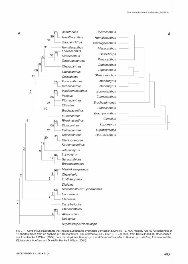
In the study by Davis (2002) (Fig. 7A), Lupopsyrus was positioned as a member of a sister clade to the diplacanthids, and by Hanke & Wilson (2004), as the sister taxon to all other acanthodians (Fig. 7B). Character distribution and the topology of each cladogram are discussed in the original studies and will not be repeated here. This variation in results shows that choice of outgroup bears significantly on the positional outcome for L. pygmaeus but in both analyses (Fig. 7), we agree that L. pygmaeus is not sister to, nor within the climatiid/climatiiform clade as previously thought.
We think the following characters suggest an acanthodian affinity for Lupopsyrus pygmaeus: 1) pork-chop-shaped, perichondrally ossified scapu-locoracoid; 2) presence of multiple hyoidean gill plates; and 3) scale growth that originates just anterior to the caudal peduncle as described by Zidek (1985, 1988). The origin of scale growth in L. pygmaeus was determined from a juvenile in the University of Alberta collection, UALVP 45155; this specimen will be described separately in a treatment of growth in Lupopsyrus. Origin of scale growth during ontogeny will have to be examined in several other Palaeozoic taxa to see whether this character is indeed a feature only of acanthodians. Schultze & Bardack (1987: figs 9, 15, 16) show that scale growth in some palaeoniscids originates anteriorly on the mid-flank just behind the operculum; it remains to be seen where scale growth originates in early chondrichthyans. In this paper we simply show that L. pygmaeus cannot be grouped as a climatiid/climatiiform as previous authors have done. More rigorous analysis awaits the forthcoming publication by SPD and Michael Coates (University of Chicago) with their clad-istic analysis of early gnathostomes; the position of L. pygmaeus will be tested in their publication. Most other features of Lupopsyrus pygmaeus, such as paired fin spines, prepectoral spines, prepelvic spines, and an anal fin spine are now known to be widespread among acanthodians and putative chondrichthyans.
483
A re-examination of Lupopsyrus pygmaeus
GEODIVERSITAS • 2012 • 34 (3)
A 37 Acanthodes
Howittacanthus
Traquairichthys
HomalacanthusLodeacanthus
Mesacanthus
Triazeugacanthus
Cheiracanthus
Latviacanthus
Cassidiceps
Poracanthodes
Ischnacanthus
Vernicomacanthus
Parexus
Ptomacanthus
Climatius
Brachyacanthus
Euthacanthus
Rhadinacanthus
Diplacanthus
Culmacanthus
Uraniacanthus
GladiobranchusKathemacanthus
TetanopsyrusLupopsyrus
GyracanthidesBrochoadmones
Mimia/Howqualepis
Cheirolepis
Eusthenopteron
DialipinaDicksonosteus/Kujanowiaspis
Coccosteus
Ctenurella
Campbellodus
Akmonistion
Debeerius
Superciliaspis/Norselaspis
Ctenacanthids
3634
31
30 3335
29
28
11
5
4
3
2
1
6
7
8
9
19
20
21
22
2324
10
25
26
27
32
12
13
15
17
14
16
18
BCheiracanthus
Homalacanthus
Triazeugacanthus
Mesacanthus
Cassidiceps
Paucicanthus
Diplacanthus
Diplacanthus
Gladiobranchus
Tetanopsyrus
Tetanopsyrus
Ischnacanthus
Culmacanthus
Brochoadmones
Euthacanthus
Brachyacanthus
Climatius
Lupopsyrus
Lupopsyroides
Obtusacanthus
Fig. 7. — Consensus cladograms that include Lupopsurus pygmaeus Bernacsek & Dineley, 1977: A, majority rule (50%) consensus of 18 shortest trees from an analysis of 113 characters (106 informative, CI = 0.5315, RI = 0.7528) from Davis (2002); B, strict consen-sus from Hanke & Wilson (2004); note that duplicate Tetanopsyrus and Diplacanthus refer to Tetanopsyrus lindoei, T. breviacanthias, Diplacanthus horridus and D. ellsi in Hanke & Wilson (2004).
484 GEODIVERSITAS • 2012 • 34 (3)
Hanke G. F. & Davis S. P.
CONCLUSIONS
Our reinterpretation of the anatomy of Lupopsyrus pygmaeus is based on all known Lupopsyrus speci-mens from the Lochkovian rocks of the Mackenzie Mountains, northwestern Canada. These new, well-preserved specimens facilitate a clearer description of most of the body as a supplement to the original description. The jaws and most of the endoskeleton are still unknown.
Lupopsyrus pygmaeus is an elongate acanthodian with short, stout fin spines with fine nodular orna-ment on well-spaced ribs. The fin spine ribs angle diagonally to the long-axis of each fin spine, not parallel to the spine axis. The sensory lines on the head are prominent, and as can be determined, fol-low a pattern similar to other acanthodians and some early osteichthyans. The dermal pectoral girdle lacks pinnal and lorical plates. The endoskeletal pectoral girdle consists of a single pair of transversely-oriented, perichondrally ossified procoracoids, and similarly ossified pork-chop shaped scapulocoracoids. Each procoracoid-scapulocoracoid complex supports a short, curved, prepectoral spine, and the pectoral fin spines appear to have articulated with a small fossa on the lateral edge of each procoracoid.
Each body scale has an open basal canal, lacks Sharpey’s fibres and distinct basal tissue, and the scale crown consists of a single odontode formed from mesodentine. The head scales of Lupopsyrus are similar to those on the body, and there is little difference in scale size and structure over the length of the body. The scales of L. pygmaeus superficially resemble thelodont scales and monodontode, pla-coid scales of chondrichthyans and here, this single scale feature is considered to be a primitive trait of L. pygmaeus relative to all other acanthodians.
Lupopsyrus pygmaeus cannot be grouped with the climatiid fishes as historically classified, and the species’ position among acanthodians is still unresolved. Here we retain L. pygmaeus in the Class Acanthodii based on its perichondrally os-sified pork-chop shaped scapulocoracoids, the presence of hyoidean gill plates, and scale growth that originates near the base of the posterior dorsal fin spine (determined from the flank scutes of a fossilized juvenile).
AcknowledgementsGH thanks Dr M. V. H. Wilson and financial sup-port for the research from his Natural Sciences and Engineering Research Council of Canada operating grant A9180 and his grant for field work from the Central Research Fund of the University of Alberta. Two Northern Science Training Grants (1996, 1998) from the Circumpolar Institute (University of Al-berta) were received by GFH for support of field work. Special thanks go to L. A. Lindoe for speci-men collection and preparation. SPD also thanks Dr M. V. H. Wilson for access to all specimens and equipment whilst working on Lupopsyrus and other acanthodian genera during his PhD. theis research. SPD’s research was supported by a Natural Environ-ment Research Council (NERC) PhD studentship GT4/97/183ES. Thanks to G. Braybrook for as-sistance with the Scanning Electron Microscope, to Dr M. W. Caldwell for the use of his Nikon digital camera, and to Dr B. D. E. Chatterton and members of the 1983, 1990, 1996 and 1998 field parties. Thanks to Carole Burrow, an anonymous researcher and Annemarie Ohler for their reviews of this manuscript, to M. Brazeau, who commented on an earlier draft and to Gaël Clément, who wrote the translation of the abstract. Thanks also to the abo-riginal people of southwestern Northwest Territories for permission to use their land during field work.
REFERENCES
berg l. s. 1940. — Classification of fishes, both recent and fossil. Travaux de l’Institut de Zoologie de l’Académie des Sciences de l’URSS 5: 85-517.
bernAcsek g. m. & dineley d. l. 1977. — New acanthodians from the Delorme Formation (Lower Devonian) of N.W.T., Canada. Palaeontographica, Abteilung A 159: 1-25.
blom H. & goujet d. 2002. — Thelodont scales from the Lower Devonian Red Bay group, Spitsbergen. Palaeontology 45: 795-820.
Brazeau m. d. 2009. — The braincase and jaw of a Devonian “acanthodian” and modern gnathostome origins. Nature 457: 305-308.
burrow c. j. 2002. — Lower Devonian acanthodian faunas and biostratigraphy of south-eastern Australia. Memoirs of the Association of Australian Palaeontologists 27: 75-137.
burrow c. j. & turner s. 1999. — A review of pla-
485
A re-examination of Lupopsyrus pygmaeus
GEODIVERSITAS • 2012 • 34 (3)
coderm scales, and their significance in placoderm phylogeny. Journal of Vertebrate Paleontology 19 (2): 204-219.
burrow c. j. & turner s. 2010. — Reassessment of “Protodus” scoticus from the Early Devonian of Scot-land, in elliott d. k., mAisey j. g., yu X. & miAo D. (eds), Morphology, Phylogeny and Biogeography of Fossil Fishes. Verlag Dr. Friedrich Pfeil, München, Germany: 123-144.
burrow c. j., newmAn m. j., dAvidson r. g. & den blAAuwens j. l. 2011. — Sclerotic plates or circumorbital bones in early jawed fishes? Palaeontology 54 (1): 207-214.
cAstro j. i. 1983. — The sharks of North American Waters. Texas A&M University Press, 180 p.
compAgno l., dAndo m. & fowler s. 2005. — Sharks of the World. Princeton University Press, Princeton, 368 p.
cumbAA s. l. & scHultze H.-p. 2002. — An Early Devonian (Emsian) acanthodian from the Bear Rock Formation, Anderson River, Northwest Territories, Canada. Canadian Journal of Earth Sciences 39: 1457-1465.
dAvis S. P. 2002. — Relationships of the Acanthodian Fishes. Unpublished PhD Thesis, University College London, London.
deAn b. & bHusHAn B. 2010. — Shark-skin surfaces for fluid-drag reduction in turbulent flow: a review. Philosophical Transactions of the Royal Society A 368: 4775-4806.
denison r. 1978. — Placodermi (volume 2), in scHultze H.-p. (ed.), Handbook of Palaeoichthyology. Gustav Fisher Verlag, Stuttgart, 128 p.
denison r. 1979. — Acanthodii (volume 5), in scHultze H.-P. (ed.), Handbook of Palaeoichthyology. Gustav Fisher Verlag, Stuttgart, 62 p.
egerton p. 1861. — British Fossils (descriptions of Tristichopterus, Acanthodes, Climatius, Diplacanthus, Cheiracanthus). Memoirs of the Geological Survey of the United Kingdom (British Organic Remains), Dec. X: 51-75.
gAbrielse H., blusson s. l. & roddick j. H. 1973. — Geology of the Flat River, Glacier Lake, and Wrigley Lake map-areas, District of Mackenzie and Yukon Ter-ritory. Geological Survey of Canada Memoir 366, 153 p.
gAgnier p.-y. 1996. — Acanthodii, in scHultze H. P. & cloutier R. (eds), Devonian Fishes and Plants of Miguasha, Quebec, Canada. Verlag Dr. Friedrich Pfeil, München: 149-164.
gAgnier p.-y. & wilson m. v. H. 1996a. — Early Devonian acanthodians from northern Canada. Palaeontology 39: 241-258.
gAgnier p.-y. & wilson m. v. H. 1996b. — An unu-sual acanthodian from northern Canada: revision of Brochoadmones milesi. Modern Geology 20: 235-251.
HAnke g. f. 2001. — Comparison of an Early Devonian
Acanthodian and Putative Chondrichthyan Assemblage using both Isolated and Articulated Remains from the Mackenzie Mountains, with a Cladistic Analysis of Early Gnathostomes. Unpublished PhD Thesis, University of Alberta, Edmonton.
HAnke G. F. 2008. — Promesacanthus eppleri n. gen., n. sp., a mesacanthid (Acanthodii, Acanthodiformes) from the Lower Devonian of northern Canada. Geodiversitas 30 (2): 287-302.
HAnke g. f. & dAvis s. p. 2008. — Redescription of the acanthodian Gladiobranchus probaton Bernacsek & Dineley, 1977, and comments on diplacanthid rela-tionships. Geodiversitas 30 (2): 303-330.
HAnke g. f. & wilson m. v. H. 1998. — Scale and spine characteristics of Lower Devonian acanthodians and chondrichthyans from northern Canada. Journal of Vertebrate Paleontology (supplement) 18: 48A.
HAnke g. f. & wilson m. v. H. 2004. — New teleos-tome fishes and acanthodian systematics, in ArrAtiA G., wilson M. V. H. & cloutier R. (eds), Recent Advances in the Origin and Early Radiation of Vertebrates. Verlag Dr. Freidrich Pfeil, München: 187-214.
HAnke g. f. & wilson m. v. H. 2010. — The puta-tive chondrichthyans Kathemacanthus and Seretolepis from the Lower Devonian MOTH locality, Mackenzie Mountains, Canada. in elliott D. K., mAisey J. G., yu X. & miAo D. (eds), Morphology, Phylogeny and Biogeography of Fossil Fishes. Verlag Dr. Friedrich Pfeil, München: 159-182.
HAnke g. f., dAvis s. p. & wilson M. V. H. 2001a. — New species of the acanthodian genus Tetanopsyrus from northern Canada, and comments on related taxa. Journal of Vertebrate Paleontology 21: 740-753.
HAnke g. f., wilson m. v. H. & lindoe l. A. 2001b. — New species of Silurian acanthodians from the Mac-kenzie Mountains, Canada. Canadian Journal of Earth Sciences 38: 1517-1529.
jAnvier p. 1996. — Early Vertebrates. Oxford Monographs on Geology and Geophysics 33. Clarendon Press, Oxford, 393 p.
joHns m. j., bArnes c. r. & orcHArd m. j. 1997. — Taxonomy and biostratigraphy of Middle and Late Triassic elasmobranch ichthyoliths from northeastern British Columbia. Geological Survey of Canada, Bulletin 502, 235 p.
kArAtAjute-tAlimAA V. N. 1968. — [New Thelodonti, Heterostraci and Arthrodires from the Chortkov Horizon of Podolia] Noviye telodonti, heterostraki i artrodiri iz Chortkovskogo Horizonta Podolii, in obrucHev D. V. (ed.), [Essays on the Phylogeny and Systematics of Fossil Fish and Agnatha] Ocherki po Foligenii i Sistematike iskopaemikh Rib i Bescheliustnikh. Nauka, Moscow: 33-42.
kArAtAjute-tAlimAA v. n. 1973. — Elegestolepis grossi gen. et sp. nov., ein neuer Typ der Placoidschuppe aus dem Oberen Silur der Tuwa. Palaeontographica,

486 GEODIVERSITAS • 2012 • 34 (3)
Hanke G. F. & Davis S. P.
Abteilung A 143: 35-50.kArAtAjute-tAlimAA v. N. 1978. — [Silurian and
Devonian thelodonts of the USSR and Spitsbergen.] Mosklas Publishers, Vilnius, 334 p. (in Russian).
kArAtAjute-tAlimAA V. N. 1992. — The early stages of the dermal skeleton formation in chondrichthyans. Academia 1: 223-231.
kArAtAjute-tAlimAA V. N. 1997. — Chondrichthyan scales from Lochkovian (Lower Devonian) of Podolia (Ukraine). Geologija 22: 5-17.
kArAtAjute-tAlimAA V. n. 1998. — Determination methods for the exoskeletal remains of early verte-brates. Mittelungen aus dem Museum für Naturkunde, Berlin, Geowwissen Reihe 1: 21-52.
li z.-X., powell c. mcA. & trencH A. 1993. — Pal-aeozoic global reconstructions, in long J. A. (ed.), Palaeozoic Vertebrate Biostratigraphy and Biogeography. Belhaven Press, London: 25-53.
long J. A. 1986. — New ischnacanthid acanthodians from the Early Devonian of Australia, with comments on acanthodian interrelationships. Zoological Journal of the Linnean Society 87: 321-339.
mAisey J. G. 1986. — Heads and tails: a chordate phy-logeny. Cladistics 2: 210-256.
märss t. 1996. — Loganellia (Thelodonti, Agnatha) from the Jaagarahu Stage, Wenlock, Estonia. Proceedings of the Estonian Academy of Sciences (Geology) 45 (4): 189-202.
märss T. 1999. — A new Late Silurian or Early Devo-nian thelodont from the Boothia Peninsula, Arctic Canada. Palaeontology 42 (6): 1079-1099.
märss t. & ritcHie A. 1998. — Articulated thelodonts (Agnatha) of Scotland. Transactions of the Royal Society of Edinburgh, Earth Sciences 88: 143-195.
märss t., wilson m. v. H., & tHorsteinsson r. 2002. — New thelodont (Agnatha) and possible chondrichthyan (Gnathostomata) taxa established in the Silurian and Lower Devonian of Arctic Canada. Proceedings of the Estonian Academy of Sciences, Geology 51 (2): 88-120.
märss t., wilson m. v. H. & tHorsteinsson r. 2006. — Silurian and Lower Devonian thelodonts and putative chondrichthyans from the Canadian Arctic Archipelago. Special Papers in Palaeontology 75, 144 p.
miles R. S. 1966. — The acanthodian fishes of the Devonian Plattenkalk of the Paffrath trough in the Rhineland. Arkiv für Zoologi 18: 147-194.
miles R. S. 1973. — Articulated acanthodian fishes from the Old Red Sandstone of England, with a review of the structure and evolution of the acanthodian shoulder-girdle. Bulletin of the British Museum of Natural History 24: 111-213.
miller r. f., cloutier r. & turner s. 2003. — The oldest articulated chondrichthyan from the Early Devonian period. Nature 425: 501-504.
moy-tHomAs j. A. & miles r. s. 1971. — Palaeozoic
Fishes. W. B. Saunders Company, Toronto, 259 p.nelson J. S. 2006. — The Fishes of the World. John
Wiley and Sons Inc., New York, 601 p.novitskAyA l. i. & obrucHev D. V. 1964. — Class
Acanthodei, in obrucHev D. V. (ed.), Fundamentals of Paleontology. Volume 11. Paleontological Institute of the Academy of Sciences of the USSR: 263-291 (in English, Israel Program for Scientific Translations, Jerusalem, 1967).
Ørvig T. 1967. — Some new acanthodian material from the Lower Devonian of Europe, in pAtterson C. & greenwood P. H. (eds), Fossil Vertebrates. Journal of the Linnean Society (Zoology) 47: 131-153.
reif w.-e. 1978. — Protective and hydrodynamic func-tion of the dermal skeleton of elasmobranchs. Neues Jahrbuch für Geologie und Paläontologie. Abhandlungen 157: 133-141.
reif W.-E. 1985. — Squamation and ecology of sharks. Courier Forschungsinstitut Senckenberg 78: 1-255.
reif w.-e. & dinkelAcker A. 1982. — Hydrodynam-ics of the squamation in fast swimming sharks. Neues Jahrbuch für Geologie und Paläontologie. Abhandlungen 164: 184-187.
sAHney s. & wilson m. v. H. 2001. — Extrinsic labyrinth infillings imply open endolymphatic ducts in Lower Devonian osteostracans, acanthodians, and putative chondrichthyans. Journal of Vertebrate Paleontology 21: 660-669.
scHultze H.-p. & bArdAck d. 1987. — Diversity and size changes in palaeonisciform fishes (Actin-opterygii, Pisces) from the Pennsylvanian Mazon Creek Fauna, Illinois, U.S.A. Journal of Vertebrate Paleontology 7: 1-23.
smitH m. m. & sAnsom i. j. 1997. — Exoskeletal micro-remains of an Ordovician fish from the Harding Sandstone of Colorado. Palaeontology 40: 645-658.
turner s. 1973. — Siluro-Devonian thelodonts from the Welsh Borderlands. Journal of the Geological Society of London 129: 557- 584.
turner S. 1982. — A new articulated thelodont (Ag-natha) from the Early Devonian of Britain. Palaeontology 25: 879-889.
turner S. 1991. — Monophyly and interrelationships of the Thelodonti, in cHAng m.-m., liu y.-H. & zHAng g. r. (eds), Early Vertebrates and Related Problems of Evolutionary Biology. Science Press, Beijing: 87-119.
turner S. 1995. — Devonian thelodont scales (Agna-tha, Thelodonti) from Queensland. Memoirs of the Queensland Museum 38 (2): 677-685.
turner s. & dring r. s. 1981. — Late Devonian thelo-donts (Agnatha) from the Gneudna Formation, Car-narvon Basin, Western Australia. Alcheringa 5: 39-48.
turner s. & murpHy m. A. 1988. — Early Devonian vertebrate microfossils from the Simpson Park Range, Eureka County, Nevada. Journal of Paleontology 62 (6): 959-964.
487
A re-examination of Lupopsyrus pygmaeus
GEODIVERSITAS • 2012 • 34 (3)
turner s. & vAn der bruggHen w. 1995. — The Thelodonti, an important but enigmatic group of Palaeozoic fishes, in sArgeAnt W. A. S. (ed.), Vertebrate Fossils and the Evolution of Scientific Concepts. Gordon and Breach Publishers, Amsterdam: 141-156.
upenice I. 1996. — Lodeacanthus gaujicus n. g. et sp. (Acanthodii: Mesacanthidae) from the Late Devonian of Latvia. Modern Geology 20: 383-398.
vAliukeviČius J. 1998. — Acanthodians and zonal stratigraphy of Lower and Middle Devonian in East Baltic and Byelorussia. Palaeontographica, Abteilung A 248: 1-53.
vAliukeviČius J. 2003a. — Devonian Acanthodians from Severnaya Zemlya Archipelago (Russia). Geodiversitas 25 (1): 131-204.
vAliukeviČius J. 2003b. — New Silurian nostolepids (Acanthodii, Pisces) of Lithuania. Geologija 42: 51-68.
vAliukeviČius J. 2003c. — New Late Silurian to Middle Devonian acanthodians of the Timan-Pechora region. Acta Geologica Polonica 53: 209-245.
vAliukeviČius J. 2004. — New Wenlock-Pridoli (Si-lurian) acanthodian fishes from Lithuania. Acta Palaeontologica Polonica 49 (1): 147-160.
vAliukeviČius J. 2005. — Silurian acanthodians biostratig-raphy of Lithuania. Geodiversitas 27 (3): 349-380.
vAliukeviČius j. & burrow c. j. 2005. — Diversity of tissues in acanthodians with Nostolepis-type his-tological structure. Acta Palaeontologica Polonica 50 (3): 635-649.
vergoossen J. M. J. 1999. — Siluro-Devonian micro-fossils of Acanthodii and Chondrichthyes (Pisces) from the Welsh Borderland/South Wales. Modern Geology 24: 23-90.
vergoossen J. M. J. 2000. — Acanthodian and chon-drichthyan microremains in the Siluro-Devonian of the Welsh Borderland, Great Britain, and their bi-ostratigraphical potential. Courier Forschungsinstitut Seckenberg 223: 175-199.
vergoossen j. m. j. 2002. — Late Silurian fish mi-
crofossils from Ramsåsa, locality H, Scania, south Sweden, with some remarks on the body zonation scheme used in thelodont studies. Scripta Geologica 123: 41-69.
vietH J. 1980. — Thelodontier-, Acanthodier- und Elasmobranchier-Schuppen aus dem Unter-Devon der Kanadischen Arktis (Agnatha, Pisces). Göttinger Arbeiten zur Geologie und Paläontologie 23: 1-69.
wArren A., currie b. p., burrow c. & turner s. 2000. — A redescription and reinterpretation of Gyracanthides murrayi Woodward 1906 (Acanthodii, Gyracanthidae) from the Lower Carboniferous of the Mansfield Basin, Victoria, Australia. Journal of Vertebrate Paleontology 20 (2): 225-242.
wAtson d. m. s. 1937. — The acanthodian fishes. Philosophical Transactions of the Royal Society of London 228B: 49-146.
wilson m. v. H. & HAnke g. F. 1998. — Body form and fin spines in species with scales of chondrich-thyan growth pattern from the Lower Devonian of the Mackenzie Mountains, Canada. Ichthyolith Issues, Special Publication 4: 57-58.
woodwArd A. s. 1906. — On a Carboniferous fish fauna from the Mansfield District, Victoria. Memoirs of the National Museum of Victoria, Melbourne 1: 1-32.
zHu m., zHAo w., jiA l., lu j., QiAo t. & Qu Q. 2009. — The oldest articulated osteichthyan reveals mosaic gnathostome characters. Nature 458: 469-474.
zidek J. 1985. — Growth in Acanthodes (Acantho-dii: Pisces) data and implications. Paläontologische Zeitschrift 59: 147-166.
zidek J. 1988. — Hamilton quarry Acanthodes (Acan-thodii; Kansas, Late Pennsylvanian). Kansas Geological Society Guidebook Series 6: 155-159.
zorn m. e., cAldwell m. w. & wilson m. v. H. 2005. — Lithological analysis of the vertebrate-bearing beds at the Lower Devonian MOTH locality, N.W.T., Canada: insights to taphonomy and depositional set-ting. Canadian Journal of Earth Sciences 42: 763-775.
Submitted on 25 January 2010; accepted on 19 April 2011.


![Pongo pygmaeus: A Bibliography - Orangutan SSP · shoulder girdle and the forward extrem ities of the orangutan.] Kanazawa, E. and A.L. Rosenberger. 1989. Interspecific allometry](https://img.pdfslide.us/doc/110x75/5e16155ab45c510d417a67e1/pongo-pygmaeus-a-bibliography-orangutan-shoulder-girdle-and-the-forward-extrem.jpg)