Embed Size (px)
Citation preview

J. Morphol. Sci., 2011, vol. 28, no. 3, p. 208-211208
Case report
A rare variation of the extensor indicis proprius tendon with important clinical implications
Casal, D.1*, Pais, D.1, Bilhim, T.1, Ribeiro, V.1, Cunha, S.1, Damásio, C.1, Fernandes, R.1, Angélica-Almeida, M.2 and Goyri-O’Neill, J.1
1Department of Anatomy, Faculty of Medical Sciences, New University of Lisbon, Lisbon, Portugal 2Plastic and Reconstructive Surgery Department and Burn Unit, São José Hospital, Lisbon, Portugal
*E-mail: [email protected]
Abstract
Introduction: Anatomical variations of the extensor tendons to the fingers are of great clinical interest, due to the relatively high frequency of tendon injury in clinical practice. Material and methods: During routine dissection of the right upper limb of a 67-year-old female preserved corpse, the extensor indicis proprius (EIP) muscle belly originated 3 independent tendons, each with a separate fascial sheath, forming a triple EIP tendon. There was a larger tendon, which occupied a central position, that represented the usual single EIP tendon. In addition, there were two thinner radial and ulnar accessory EIP tendons. The radial-EIP tendon crossed deep to the extensor digitorum communis (EDC) tendon to the index finger in the distal half of the dorsum of the hand to reach the radial side of the extensor expansion hood of the index finger. Discussion: According to the literature, the frequency of a triple EIP tendon ranges from 0%, to as high as 7%, although most authors do not acknowledge the presence of this variant in their series. This variant of the EIP tendon, in which the radial-EIP terminated laterally to the termination of the tendon of the EDC to the index finger, may be a source of confusion intraoperatively, as the EIP tendon has traditionally been identified on the basis of its ulnar location with respect to the EDC tendon. Conclusion: The possibility of a triple EIP tendon should certainly be born in mind by all surgeons when performing tendon repairs, tenoplasties or tendon transfers.
Keywords: extensor indicis proprius, extensor digitorum communis, variation, hand surgery, repair.
1 Introduction
Anatomical variations of the extensor tendons to the fingers have been documented for well over 100 years (LE DOUBLE, 1897; SCHMIDT and LANZ, 2004; ZILBER and OBERLIN, 2004). In spite of this, new variants of these tendons and/or more precise determinations of the frequency of previously known variants continue to be reported to this day (MESTDAGH, BAILLEUL, VILETTE et al., 1985; GODWIN and ELLIS, 1992; EL-BADAWI, BUTT, AL-ZUHAIR et al., 1995; VON SCHROEDER and BOTTE, 1995; HIRAI, YOSHIDA, YAMANAKA et al., 2001; ZILBER and OBERLIN, 2004; JEON, SEOK, PARK et al., 2010).
The weight given in the literature to this subject is undoubtedly due to the importance of a thorough knowledge of the extensor tendon anatomy to fully understand the consequences of tendon injury at various levels, as well as to plan and undertake repair both in the acute and chronic setting (ROCKWELL, BUTLER and BYRNE, 2000; SCHMIDT and LANZ, 2004; CAMPOS, NAZER, BARTHOLDY et al., 2010; SHAPIRO and KRAHE, 2010). Moreover, spontaneous rupture of the extensor tendons of the hand is common, especially in patients with rheumatoid arthritis, distal radioulnar joint osteoarthritis, ulnar plus variance, and ulnar head dislocation (ZILBER and OBERLIN, 2004).
Among the extensor tendons of the fingers, the extensor indicis proprius (EIP) tendon is one of the most frequently injured (NOLAN, 2006). In addition, the EIP tendon, having an independent muscle belly, is often used as a donor tendon
for transfer (NOLAN, 2006; TARAS and LEE, 2010). In fact, the same extension of the index joints can be achieved with either the EIP or the extensor digitorum communis (EDC) tendon to the index finger (VON SCHROEDER and BOTTE, 1993, 1995, 1997, 2001; NAYAK, MADHAN-KUMAR, KRISHNAMURTHY et al., 2007; CELIK, BILGE, PINAR et al., 2008).
Therefore, a sound grasp of the anatomy of the extensor tendons to the fingers, and, in particular of the EIP tendon, is of vital importance to successful solve problems frequently encountered in daily clinical practice.
In this paper we describe a rare variant of the EIP tendon in a cadaveric dissection study, and discuss its clinical implications.
2 Case report
During routine dissection of the right hand and forearm of a 67-year-old female preserved corpse, following the recommendations of a Human Anatomy dissection manual (ESPERANÇA-PINA, BENSABAT-RENDAS, CORREIA et al., 2001), an unusual configuration of the extensor tendons of the index finger was noted (Figures 1 and 2). There was no history or evidence of trauma or surgery to the upper limbs. The EIP muscle belly originated in the distal quarter of the forearm 3 independent tendons, each with a separate fascial sheath, forming a triple EIP tendon (CELIK, BILGE, PINAR et al., 2008). These three tendons joined the ulnar and deep aspect of the tendon of the EDC to the index finger, and then passed deep to
A rare variation of the extensor index tendon
J. Morphol. Sci., 2011, vol. 28, no. 3, p. 208-211 209
3 Discussion
Le Double was the first to systematically study in humans the division of the extensor tendons to the fingers in the latter half of the 19th century (LE DOUBLE, 1897). He reported his and other authors’ findings in his treatise on the variations of the human muscular system that was published in 1897 (LE DOUBLE, 1897). Since then, numerous works have been published on the presence and number of the extensor tendons to the fingers (MESTDAGH, BAILLEUL, VILETTE et al., 1985; GODWIN and ELLIS, 1992; EL-BADAWI, BUTT, AL-ZUHAIR et al., 1995; VON SCHROEDER and BOTTE, 1995; HIRAI, YOSHIDA, YAMANAKA et al., 2001; ZILBER and OBERLIN, 2004; CELIK, BILGE, PINAR et al., 2008; MOTOURA, SHIOZAKI and KAWASAKI, 2010).
Figure 1. Photograph of the dorsum of the right hand showing a triple extensor indicis proprius tendon. 1) extensor digitorum communis tendon to the index finger; 2) radial extensor indicis proprius tendon; 3) extensor indicis proprius tendon; 4) ulnar extensor indicis proprius tendon; 5) extensor expansion hood of the index finger; 6) central slip of the extensor apparatus of the index finger; 7) lateral band of the extensor apparatus of the index finger; 8) medial band of the extensor apparatus of the index finger; 9) extensor digitorum communis tendon to the middle finger; 10) extensor digitorum communis tendon to the ring finger; 11) extensor digitorum communis tendon to the fifth finger; 12) extensor digiti minimi tendon; 13) intertendinous connections; 14) extensor retinaculum.
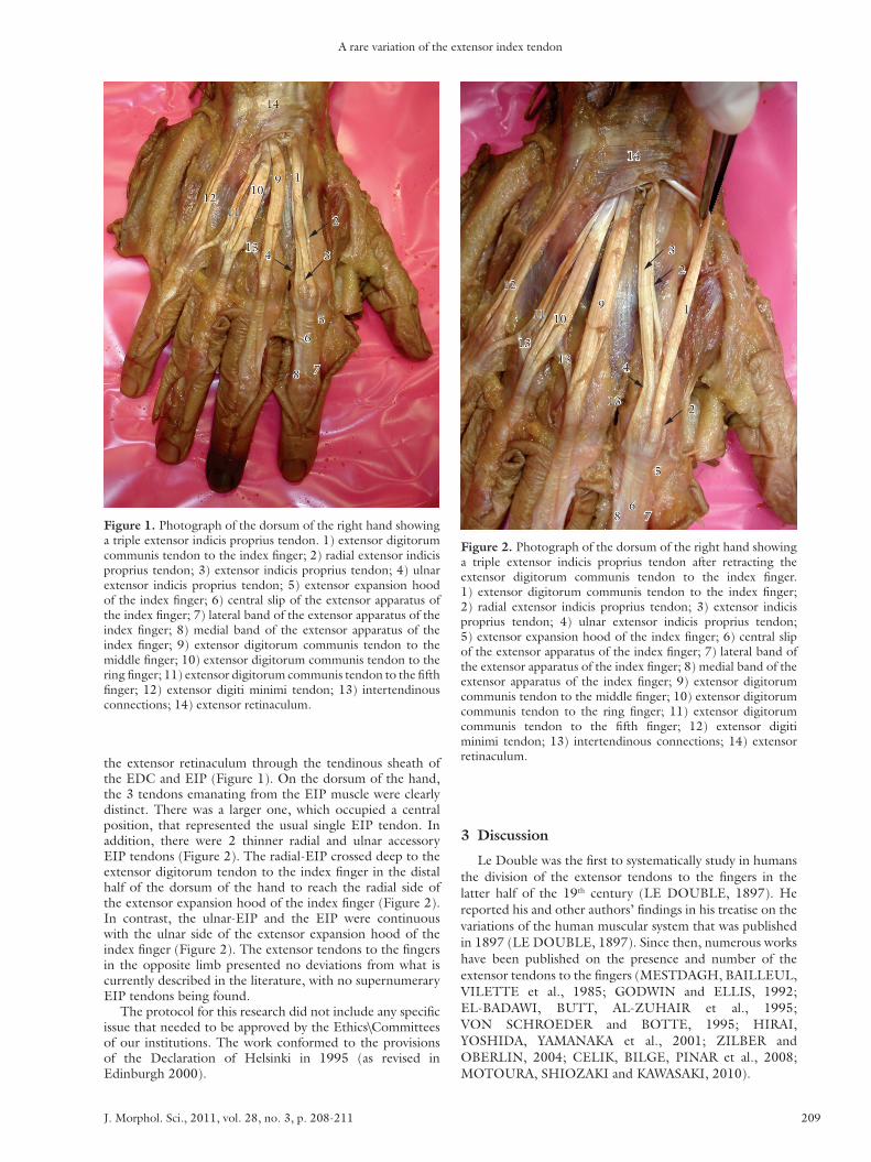
the extensor retinaculum through the tendinous sheath of the EDC and EIP (Figure 1). On the dorsum of the hand, the 3 tendons emanating from the EIP muscle were clearly distinct. There was a larger one, which occupied a central position, that represented the usual single EIP tendon. In addition, there were 2 thinner radial and ulnar accessory EIP tendons (Figure 2). The radial-EIP crossed deep to the extensor digitorum tendon to the index finger in the distal half of the dorsum of the hand to reach the radial side of the extensor expansion hood of the index finger (Figure 2). In contrast, the ulnar-EIP and the EIP were continuous with the ulnar side of the extensor expansion hood of the index finger (Figure 2). The extensor tendons to the fingers in the opposite limb presented no deviations from what is currently described in the literature, with no supernumerary EIP tendons being found.
The protocol for this research did not include any specific issue that needed to be approved by the Ethics\Committees of our institutions. The work conformed to the provisions of the Declaration of Helsinki in 1995 (as revised in Edinburgh 2000).
Figure 2. Photograph of the dorsum of the right hand showing a triple extensor indicis proprius tendon after retracting the extensor digitorum communis tendon to the index finger. 1) extensor digitorum communis tendon to the index finger; 2) radial extensor indicis proprius tendon; 3) extensor indicis proprius tendon; 4) ulnar extensor indicis proprius tendon; 5) extensor expansion hood of the index finger; 6) central slip of the extensor apparatus of the index finger; 7) lateral band of the extensor apparatus of the index finger; 8) medial band of the extensor apparatus of the index finger; 9) extensor digitorum communis tendon to the middle finger; 10) extensor digitorum communis tendon to the ring finger; 11) extensor digitorum communis tendon to the fifth finger; 12) extensor digiti minimi tendon; 13) intertendinous connections; 14) extensor retinaculum.
Casal, D., Pais, D., Bilhim, T. et al.
J. Morphol. Sci., 2011, vol. 28, no. 3, p. 208-211210
Regardless of their cause, anatomical variations of the EIP tendon, as the one described in this paper, are of great anatomical and clinical interest, and should certainly be born in mind by all surgeons when performing tendon repairs, tenoplasties or tendon transfers (ZILBER and OBERLIN, 2004; CELIK, BILGE, PINAR et al., 2008; HANZ, SAINT-CYR, SEMMLER et al., 2008).
Acknowledgements: We appreciate the efforts of all Technical Staff members of the Department of Anatomy, in particular of Mr. Carlos Lopes and of Mr. Marco Costa.
References
CAMPOS, D., NAZER, MB., BARTHOLDY, LM. and SOUZA, PL. Accessory flexor carpi ulnaris muscle: a case report of a rare variation in human. Journal of Morphological Sciences, 2010, vol. 27, p. 30-31.
CELIK, S., BILGE, O., PINAR, Y. and GOVSA, F. The anatomical variations of the extensor tendons to the dorsum of the hand. Clinical Anatomy, 2008, vol. 21, p. 652-659. PMid:18792963. http://dx.doi.org/10.1002/ca.20710
CHEVALLIER, A., KIENY, M. and MAUGER, A. Limb-somite relationship: origin of the limb musculature. Journal of Embryology and Experimental Morphology, 1977, vol. 41, p. 245-258. PMid:591873.
EL-BADAWI, MG., BUTT, MM., AL-ZUHAIR, AG. and FADEL, RA. Extensor tendons of the fingers: arrangement and variations-II. Clinical Anatomy, 1995, vol. 8, p. 391-398. PMid:8713158.
ESPERANÇA-PINA, JA., BENSABAT-RENDAS, A., CORREIA, M., GOYRI-O’NEILL, J. and PAIS, D. Membro Superior. In ESPERANÇA-PINA, JA., ed. Anatomia Geral e Dissecção Humana. Lisboa: Lidel, 2001. p. 133-156.
GODWIN, Y. and ELLIS, H. Distribution of the extensor tendons on the dorsum of the hand. Clinical Anatomy, 1992, vol. 5, p. 394-397. http://dx.doi.org/10.1002/ca.980050506
HANZ, KR., SAINT-CYR, M., SEMMLER, MJ. and ROHRICH, RJ. Extensor tendon injuries: acute management and secondary reconstruction. Plastic and Reconstructive Surgery, 2008, vol. 121, p. 109e-120e. PMid:18317093.
HIRAI, Y., YOSHIDA, K., YAMANAKA, K., INOUE, A., YAMAKI, K. and YOSHIZUKA, M. An anatomic study of the extensor tendons of the human hand. Journal of Hand Surgery (American), 2001, vol. 26, p. 1009-1014. PMid:11721244.
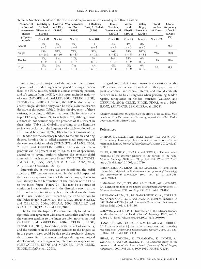
According to the majority of the authors, the extensor apparatus of the index finger is composed of a single tendon from the EDC muscle, which is almost invariably present, and of a tendon from the EIP, which is present in the majority of cases (MOORE and DALLEY, 2006; CELIK, BILGE, PINAR et al., 2008). However, the EIP tendon may be absent, single, double or may even be triple, as in the case we describe in this paper. Table 1 depicts the frequency of these variants, according to different authors. The frequency of a triple EIP ranges from 0%, to as high as 7%, although most authors do not acknowledge the presence of this variant in their series (Table 1). Globally, according to the literature review we performed, the frequency of a triple tendon of the EIP should be around 0,9%. Other frequent variants of the EIP tendon are the accessory tendons to the middle and ring fingers, forming the so called extensor medii proprius, and the extensor digiti annularis (SCHMIDT and LANZ, 2004; ZILBER and OBERLIN, 2004). The extensor medii proprius can be present in up to 3,5 to 13% of specimens, according to several authors, whereas the extensor digiti annularis is much more rarely found (VON SCHROEDER and BOTTE, 1995, 1997; SCHMIDT and LANZ, 2004; ZILBER and OBERLIN, 2004).
Interestingly, in the case we are describing, the radial accessory EIP tendon terminated in the radial aspect of the extensor expansion hood of the index finger, that is to say, laterally to the termination of the tendon of the EDC to the index finger (Figure 2). This may be a source of confusion intraoperatively or in the dissection room, as the EIP tendon has traditionally been identified on the basis of its ulnar location with respect to the EDC tendon to the index finger (SCHMIDT and LANZ, 2004; ZILBER and OBERLIN, 2004; NOLAN, 2006; SHAPIRO and KRAHE, 2010; TARAS and LEE, 2010).
The fact that the triple EIP tendon was only found on the right side is in agreement with recent works that confirm that the extensor tendons to the finger are often not symmetrical (ZILBER and OBERLIN, 2004; CELIK, BILGE, PINAR et al., 2008). In addition, both the lack of symmetry, and the variations in the extensor tendons to the fingers, as in the present case, could be due to the stochastic changes the extensor limb myotomes undergo during ontological development, namely regression, retention, or reappearance (CHEVALLIER, KIENY and MAUGER, 1977; CELIK, BILGE, PINAR et al., 2008).
Table 1. Number of tendons of the extensor indicis proprius muscle according to different authors.Number of tendons of
the extensor indicis
proprius muscle
Mestdagh, Bailleul,
Vilette et al. (1985)
Godwin and Ellis (1992)
Von Schroeder and Botte
(1995)
El-Badawi, Butt, Al-Zuhair
(1995)
Hirai, Yoshida,
Yamana et al. (2001)
Zilber and
Oberlin (2004)
Celik, Bilge,
Pinar et al. (2008)
Total number of Cases
Global frequency
of each variant
(%)N = 150 N = 50 N = 43 N = 181 N = 548 N = 50 N = 54 N = 1076
Absent 1%;n = 1
0%;n = 0
0%;n = 0
1%;n = 2
0%;n = 0
4%;n = 2
0%;n = 0 5 0,5
Single 93%;n = 140
92%;n = 46
77%;n = 33
90%;n = 163
86%;n = 471
78%;n = 39
100%;n = 54 946 88,0
Double 6%;n = 9
8%;n = 4
16%;n = 7
5%;n = 9
14%;n = 77
18%;n = 9
0%;n = 0 115 10,6
Triple 0%;n = 1
0%,n = 0
7%;n = 3
4%;n = 7
0%;n = 0
0%;n = 0
0%;n = 0 10 0,9
A rare variation of the extensor index tendon
J. Morphol. Sci., 2011, vol. 28, no. 3, p. 208-211 211
SHAPIRO, DB. and KRAHE, MA. Repair following traumatic extensor tendon disruption in the hand, wrist and forearm. In HUNT, TRI., ed. Operative Techniques in Hand, Wrist, and Forearm Surgery. 3th ed. Philadelphia: Lippincott Williams and Wilkins, 2010. p. 485-494.
TARAS, JS. and LEE, DJ. 2010. Tendon transfer and grafting for traumatic extensor tendon disruption. In HUNT, TRI., ed. Operative Techniques in Hand, Wrist, and Forearm Surgery. 3th ed. Philadelphia: Lippincott Williams and Wilkins, 2010. p. 495-501.
VON SCHROEDER, HP. and BOTTE, MJ. The functional significance of the long extensors and juncturae tendinum in finger extension. Journal of Hand Surgery [American], 1993, vol. 18, p. 641-647. http://dx.doi.org/10.1016/0363-5023(93)90309-Q
VON SCHROEDER, HP. and BOTTE, MJ. Anatomy of the extensor tendons of the fingers: variations and multiplicity. Journal of Hand Surgery [American], 1995, vol. 20, p. 27-34. http://dx.doi.org/10.1016/S0363-5023(05)80053-X
VON SCHROEDER, HP. and BOTTE, MJ. Functional anatomy of the extensor tendons of the digits. Hand clinics, 1997, vol. 13, p. 51-62. PMid:9048183.
VON SCHROEDER, HP. and BOTTE, MJ. Anatomy and functional significance of the long extensors to the fingers and thumb. Clinical Orthopaedics and Related Research, 2001, vol. 383, p. 74-83. PMid:11210972. http://dx.doi.org/10.1097/00003086-200102000-00010
ZILBER, S. and OBERLIN, C. Anatomical variations of the extensor tendons to the fingers over the dorsum of the hand: a study of 50 hands and a review of the literature. Plastic and Reconstructive Surgery, 2004, vol. 113, p. 214-221. PMid:14707639. http://dx.doi.org/10.1097/01.PRS.0000091163.86851.9C
Received December 1, 2010 Accepted August 22, 2011
JEON, IH., SEOK, JH., PARK, IH., CHOI, JW., MIN, WK., KWON, DS., KIM, HJ. and KIM, PT. An anatomic study on the junctura tendinum in the 4th intermetacarpal space and its clinical implication. Clinical Anatomy, 2010, vol. 23, p. 56-60. PMid:19918878.
LE DOUBLE, AF. Traité des Variations du Système Musculaire de l’Homme et de Leur Signification au Point de Vue de l’Anthropologie Zoologique. Paris: Schleicher Frères, 1897. p. 126-135.
MESTDAGH, H., BAILLEUL, JP., VILETTE, B., BOCQUET, F. and DEPREUX, R. Organization of the extensor complex of the digits. Anatomia Clinica, 1985, vol. 7, p. 49-53. PMid:3994853. http://dx.doi.org/10.1007/BF01654629
MOORE, KL. and DALLEY, AF. Upper limb. In MOORE, KL. and DALLEY, AF., ed. Clinically Oriented Anatomy. 5th ed. Philadelphia: Lippincott Williams and Wilkins, 2006. p. 806-814.
MOTOURA, H., SHIOZAKI, K. and KAWASAKI, K. Anatomical variations in the tendon sheath of the first compartment. Anatomical Science International, 2010, vol. 85, p. 145-151. PMid:20039153. http://dx.doi.org/10.1007/s12565-009-0070-x
NAYAK, SR., MADHAN-KUMAR, SJ., KRISHNAMURTHY, A., PRABHU, LV., RANADE, AV., RAI, R. and RAMANATHAN, L. An additional radial wrist extensor and its clinical significance. Annals of Anatomy, 2007, vol. 189, p. 283-286.
NOLAN, WB. Extensor tendon injuries and reconstruction. In MATHES, SJ., ed. Plastic surgery. 2th ed. Philadelphia: Saunders, 2006. p. 401-421.
ROCKWELL, WB., BUTLER, PN. and BYRNE, BA. Extensor tendon: anatomy, injury, and reconstruction. Plastic and Reconstructive Surgery, 2000, vol. 106, p. 1592-1603. PMid:11129192. http://dx.doi.org/10.1097/00006534-200012000-00024
SCHMIDT, HM. and LANZ, U. Distal forearm, wrist, and dorsum of the hand. In: SCHMIDT, HM. and LANZ, U., ed. Surgical anatomy of the hand. 3th ed. New York: Thime Verlag, 2004. p. 231-247.