Embed Size (px)
Citation preview
A Rare Complication of Percutaneous Nephrolithotomy: Cerebral Infarction and Hemianopsia
Correspondence (İletişim): Abdurrahman İnkaya, M.D. Departmant of Urology, Health Sciences University, Umraniye Training andResearch Hospital, Istanbul, TurkeyPhone (Telefon): +90 216 632 18 18 E-mail (E-posta): [email protected] Date (Başvuru Tarihi): 03.05.2018 Accepted Date(Kabul Tarihi): 22.07.2018
Percutaneous nephrolithotomy (PNL) has become wide-spread since its first description in 1976 and has become
the most prevalently used surgical technique for large and complex stones in the intrarenal collecting system [1]. In this procedure, bleeding, damage to neighboring tissues and organs, infection, positional injuries, thromboembolic events and even death may be observed [2]. Knowing and predicting different complications may be helpful in diag-nosing, and preventing complications.
Case ReportA 58–year-old patient without comorbidity presented with right flank pain, and a visible right flank incision scar. The patient had undergone open surgery for right renal stone, and 7 sessions of shock wave lithıtripsy (ESWL). His hemo-globin (Hb), 12.2 gr/dL; hematocrit (hCT) 41%, creatinine, 0.9 mg/dL, and blood urea nitrogen 20 mg/dL values were as indicated. Whole abdominal computed tomography of the patient demonstrated a stone with dimensions
of 36x23x21 mm in the pelvis of the right grade 3 hy-dronephrotic kidney, and multiple stones in the lower pole of the same kidney the largest one with a diameter of 17 mm (Fig. 1). DMSA (dimercaptosuccinic acid) renal scintig-raphy revealed right and left split renal functions as 34, and 66%, respectively, so PNL application for right kidney was decided. During operation, the lower calyx was accessed with 18 G percutaneous needle and stones in the lower ca-lyx and renal pelvis were fragmented and extracted with pneumatic lithotriptor. Complications did not occur dur-ing the operation which lasted approximately 90 minutes. Residual stones were present in the anterior group of the lower calyx after the operation. Two hours after the opera-tion hb was 11.7 gr/dL, and hct, 36 percent.
Postoperative 12th hour hb was 11.5 gr/dL, and hct 34.6 percent. The patient's consciousness blurred, loss of coop-eration, and impaired orientation developed, he couldn’t give meaningful answers to questions, and repeated the same answer. Right-sided hemianopsia was detected. Dif-
Since its description in 1976, percutaneous nephrolithotomy (PNL) has become the most common surgical technique for the man-agement of large and complex stones in the intrarenal collecting system. A wide variety of complications can be observed, including bleeding associated with this procedure, damage to neighboring tissues and organs, infection, positional injuries, thromboembolic events, and even death. Knowing and predicting the different complications can be useful in preventing, diagnosing and preventing complications. We aimed to present the management of cerebral infarction and hemianopsia after PNL to our clinic as a case report.
Keywords: Hemorrhage; infarction; PCNL; renal stone.
Abdurrahman İnkaya, Ahmet Tahra, Resul Sobay, Uğur Boylu, Eyüp Veli KüçükDepartmant of Urology, Health Sciences University, Umraniye Training and Research Hospital, Istanbul, Turkey
Copyright 2018 Haydarpaşa Numune Medical JournalThis is an open access article under the CC BY-NC license (http://creativecommons.org/licenses/by-nc/4.0/).
Abstract
DOI: 10.14744/hnhj.2018.60252Haydarpasa Numune Med J 2018;58(3):165–167
hnhtipdergisi.com
HAYDARPAŞA NUMUNE MEDICAL JOURNAL
CASE REPORT
166 İnkaya et al., Cerebral Infarction is a Rare Complication of PNL / doi: 10.14744/hnhj.2018.60252
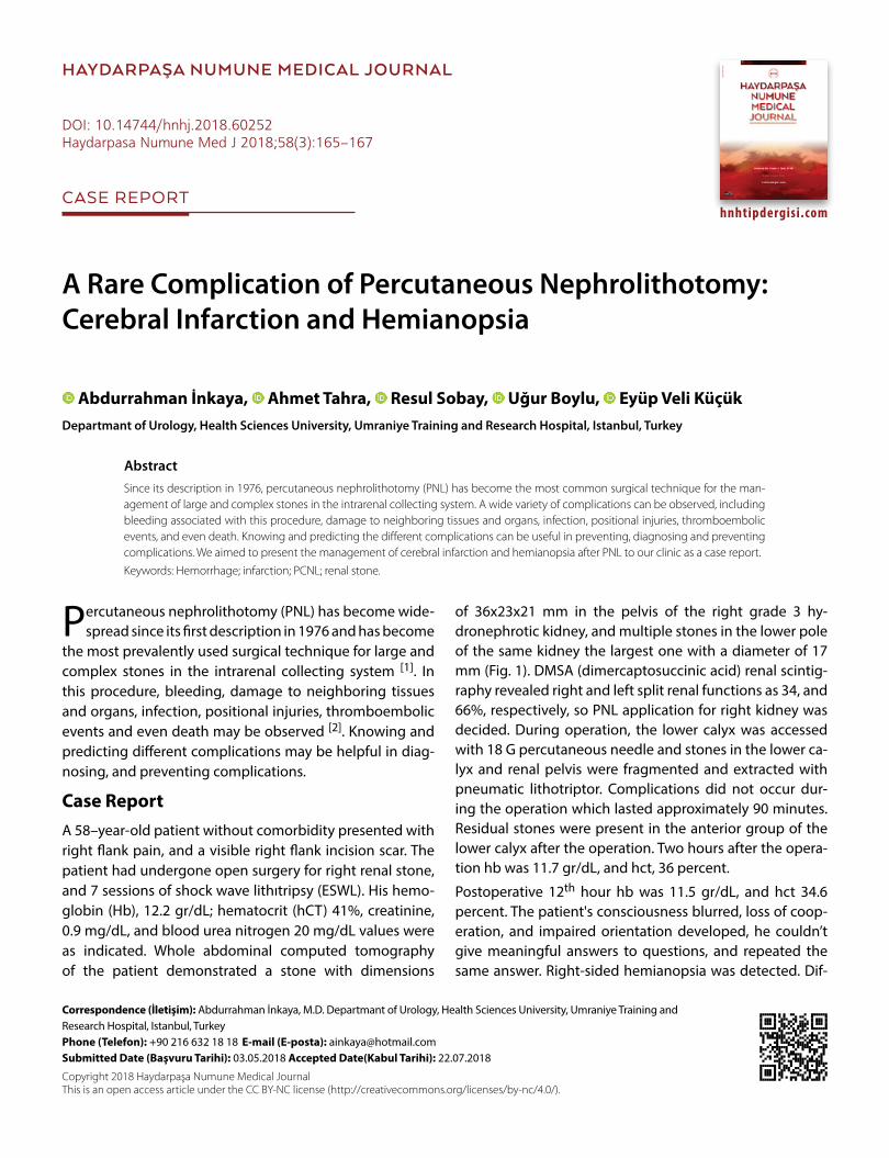
fusion magnetic resonance imaging (MRI) demonstrated patchy areas of acute infarct in the left medial cerebral in-ferior branch, left frontal border zone, and the right frontal segment (Fig. 2), so acetylsalicylic acid (150 mg 1x1) was initiated as a result of neurological consultation. Cardiol-ogy consultation did not indicate any cardiac pathology. Carotid and vertebrobasilar system Doppler ultrasonogra-phy did not reveal any pathology. The patient was started on 2x6000 IU of enoxaparin sodium.
After enoxaparin sodium treatment, macroscopic hema-turia was seen coming from nephrostomy tube, and urethral catheter, so 3-way Foley catheter were delivered through transurethral route, and irrigation of the bladder was started. Despite transfusion, hemoglobin (6 gr/dL), and hematocrit levels (23%) dropped down to indicated levels, therefore selective arterial angiomyolysis was planned. In the depart-ment of interventional radiology pseudoaneurysms were detected in two different segmental arteries, and selective arterial angioembolization was performed.
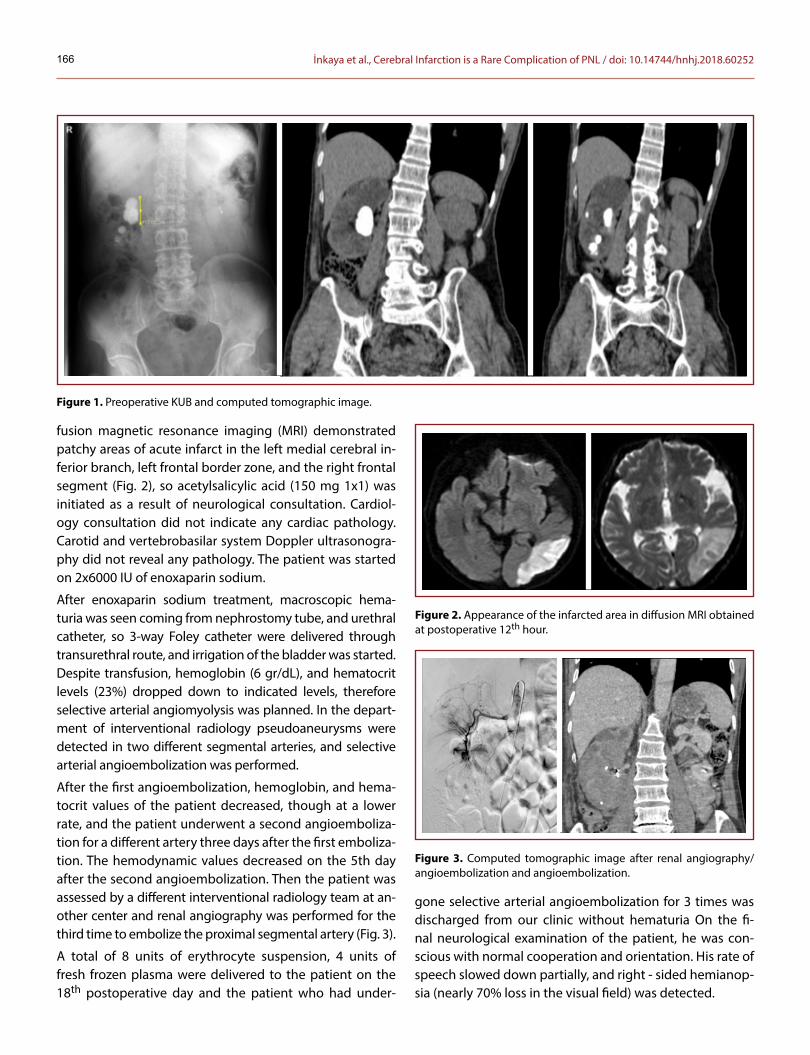
After the first angioembolization, hemoglobin, and hema-tocrit values of the patient decreased, though at a lower rate, and the patient underwent a second angioemboliza-tion for a different artery three days after the first emboliza-tion. The hemodynamic values decreased on the 5th day after the second angioembolization. Then the patient was assessed by a different interventional radiology team at an-other center and renal angiography was performed for the third time to embolize the proximal segmental artery (Fig. 3).
A total of 8 units of erythrocyte suspension, 4 units of fresh frozen plasma were delivered to the patient on the 18th postoperative day and the patient who had under-
gone selective arterial angioembolization for 3 times was discharged from our clinic without hematuria On the fi-nal neurological examination of the patient, he was con-scious with normal cooperation and orientation. His rate of speech slowed down partially, and right - sided hemianop-sia (nearly 70% loss in the visual field) was detected.
Figure 1. Preoperative KUB and computed tomographic image.
Figure 2. Appearance of the infarcted area in diffusion MRI obtained at postoperative 12th hour.
Figure 3. Computed tomographic image after renal angiography/angioembolization and angioembolization.
167İnkaya et al., Cerebral Infarction is a Rare Complication of PNL / doi: 10.14744/hnhj.2018.60252
DiscussionFollowing PNLs complication rates up to 83% have been reported [3]. These complications are usually minor bleed-ings, fever, and extravasation (in 7.2 % of the cases). Hem-orrhagic complications require transfusion in 11.2-17.5% of the cases. Bleedings stemming from formation of pseu-doaneurysms and arteriovenous fistulae are among the most serious complications. Success rates of up to 80% have been reported in the literature after superselective angioembolizations (SAE) performed to stop bleeding [4]. Rarely sepsis (0.3-4.7%), colon injury (0.0-0.8%), and pleural injury (0.0-3.1%) have been observed [5].
The most important factor for achieving acceptable out-comes and success in minimizing major complications in PNL is the selection of the patient who will mostly benefit from this procedure. Use of well-standardized techniques and postoperative follow-up is essential for early detec-tion and management of complications. In PNL in terms of thromboprophylaxis, before proceeding with the surgery, determination of patient-related risk factors as age, pres-ence of comorbidity, family history, systemic diseases, thromboembolic events, increased body mass index, and then initiation of thromboprophylaxis with low-molecular weight heparin has been recommended till complete mo-bilization is achieved [6].
ConclusionWe think that a standardized algorithm for control of bleed-ings, and management of thromboembolism is required for thromboembolic events developed after a kind of con-trolled grade 4 trauma in major renal surgery where collect-
ing system is entered passing through renal parenchyma as in the case of PNL These complications are rare, but man-agement requires a serious and urgent approach.
Informed Consent: Approval was obtained from the patient. Peer-review: Externally peer-reviewed.
Conflict of Interest: None declared.
Authorship Contributions: Concept: A.İ.; Design: A.T.; Data Col-lection or Processing: R.S.; Analysis or Interpretation: E.V.K.; Litera-ture Search: U.B.; Writing: A.İ.
Financial Disclosure: The authors declared that this study re-ceived no financial support.
References1. Fernström I, Johansson B. Percutaneous pyelolithotomy. A new
extraction technique. Scand J Urol Nephrol 1976;10:257–9.2. Kallidonis P, Panagopoulos V, Kyriazis I, Liatsikos E. Complica-
tions of percutaneous nephrolithotomy: classification, man-agement, and prevention. Curr Opin Urol 2016;26:88–94.
3. Michel MS, Trojan L, Rassweiler JJ, Breda A. Complications inpercutaneous nephrolithotomy. Eur Urol 2007;51:899–906.
4. Srivastava A, Singh KJ, Suri A, Dubey D, Kumar A, Kapoor R, et al.Vascular complications after percutaneous nephrolithotomy:are there any predictive factors? Urology 2005;66:38–40.
5. Skolarikos A, de la Rosette J. Prevention and treatment ofcomplications following percutaneous nephrolithotomy. Curr Opin Urol 2008;18:229–34. [CrossRef ]
6. Douketis JD, Spyropoulos AC, Spencer FA, Mayr M, Jaffer AK,Eckman MH, et al. Perioperative management of antithrom-botic therapy: Antithrombotic Therapy and Preventionof Thrombosis, 9th ed: American College of Chest Physi-cians Evidence-Based Clinical Practice Guidelines. Chest2012;141:e326–50S.


![Percutaneous Nephrolithotomy Versus Retrograde Intrarenal Surgery… · RIRS was compared with standard PCNL in four studies [15–18], miniperc in four [13,19–21], and microperc](https://img.pdfslide.us/doc/110x75/5b83a76e7f8b9a866e8d7b94/percutaneous-nephrolithotomy-versus-retrograde-intrarenal-rirs-was-compared.jpg)

![Recent advancement or less invasive treatment of ... · Advancement of percutaneous nephrolithotomy treatment with other less-invasive methods) [5-9]. However, although PCNL is considered](https://img.pdfslide.us/doc/110x75/5ed551a612a6d6201a657edb/recent-advancement-or-less-invasive-treatment-of-advancement-of-percutaneous.jpg)