Embed Size (px)
Citation preview
A Rare Combination of Ribonucleotide Reductases in theSocial Amoeba Dictyostelium discoideum*
Received for publication, December 4, 2012, and in revised form, January 20, 2013 Published, JBC Papers in Press, January 31, 2013, DOI 10.1074/jbc.M112.442434
Mikael Crona‡1, Lotta Avesson§2, Margareta Sahlin‡¶, Daniel Lundin¶, Andrea Hinas§3, Ralph Klose‡4,Fredrik Söderbom§5, and Britt-Marie Sjöberg‡¶6
From the ‡Department of Molecular Biology and Functional Genomics, Arrhenius Laboratories for Natural Sciences, and¶Department of Biochemistry and Biophysics, Stockholm University, SE-10691 Stockholm, Sweden and the §Department ofMolecular Biology, Swedish University of Agricultural Sciences, Uppsala Biomedical Center, SE-75124 Uppsala, Sweden
Background: Dictyostelium discoideum encodes both class I and II RNRs.Results: Both RNRs are expressed in D. discoideum, and depletion of class I RNR drastically reduces spore formation andviability.Conclusion: Class I RNR is central for D. discoideum growth and differentiation, whereas class II function is enigmatic.Significance:Our results underscore the evolutionary selection for this rare set of RNRs in D. discoideum.
Ribonucleotide reductases (RNRs) catalyze the only pathwayfor de novo synthesis of deoxyribonucleotides needed for DNAreplication and repair. The vast majority of eukaryotes encodesonly a class I RNR, but interestingly some eukaryotes, includingthe social amoeba Dictyostelium discoideum, encode both aclass I and a class II RNR. The amino acid sequence of the D.discoideum class I RNR is similar to other eukaryotic RNRs,whereas that of its class II RNR ismost similar to themonomericclass II RNRs found in Lactobacillus spp. and a few other bacte-ria. Here we report the first study of RNRs in a eukaryotic orga-nism that encodes class I and class II RNRs. Both classes of RNRgenes were expressed inD. discoideum cells, although the class Itranscripts were more abundant and strongly enriched duringmid-development compared with the class II transcript. Thequaternary structure, allosteric regulation, and properties of thediiron-oxo/radical cofactor of D. discoideum class I RNR aresimilar to those of the mammalian RNRs. Inhibition of D. dis-coideum class I RNRbyhydroxyurea resulted in a 90% reductionin spore formation and decreased the germination viability ofthe surviving spores by 75%. Class II RNR could not compensatefor class I inhibition during development, and an excess of vita-min B12 coenzyme, which is essential for class II activity, did notimprove spore formation.We suggest that class I is the principal
RNR during D. discoideum development and growth and isimportant for spore formation, possibly by providing dNTPs formitochondrial replication.
The slime mold Dictyostelium discoideum belongs to theAmoebozoa, the closest living relatives to animals and plants(1). This social amoeba lives as solitary cells that feed on bacte-ria and yeasts in decaying forest material (2). However, starva-tion triggers migration of up to 100,000 cells followed by adevelopmental program resulting in a differentiated multicel-lular structure consisting of a long stalk with a ball of sporesready to germinate when nutrients are available (2). The twomajor cell types in the developing organism are prestalk cellsthat differentiate to stalk cells, forming a cellulose-rich stalk,and prespore cells that constitute �80% of all cells and differ-entiate into spores protected from drought and other harshconditions by a spore wall (3).Unlike most eukaryotes D. discoideum encodes genes for
more than one class of the essential enzyme ribonucleotidereductase (RNR)7 (4). RNR catalyzes the only pathway for denovo synthesis of DNA building blocks (deoxyribonucleotidesor dNTPs) by reducing the 2�-hydroxy group of the corre-sponding ribonucleotides using radical chemistry (5). Threedifferent classes of RNRs are currently known that share a com-mon evolutionary origin but differ in radical-generating cofac-tors with specific oxygen dependences. The vast majority ofeukaryotes encode a class I RNR, but D. discoideum encodesboth a class I and a class II RNR. Class I RNRs consist of twosubunits: the larger NrdA subunit contains the active site andusually two different allosteric sites, whereas the smaller NrdBsubunit harbors a stable tyrosyl radical close to a diiron-oxocenter. Together, NrdA and NrdB form �2�2 or larger oligo-meric holoenzyme complexes (6). The tyrosyl radical can bescavenged by hydroxyurea (HU), leading to depletion of the
* This work was supported by the Swedish Research Council (to B. M. S. andF. S.), the Swedish Research Council for Environment, Agricultural Sciencesand Spatial Planning (the Swedish Research Council Formas) (to F. S.),and the European Community (Function of Small RNAs across Kingdoms(FOSRAK) Grant EC005120) (to F. S.).Author’s Choice—Final version full access.
1 Present address: BioThema AB, Handens stationsväg 17, SE-136 40 Handen,Sweden.
2 Present address: Garvan Inst. of Medical Research, 384 Victoria St., Darling-hurst, New South Wales 2010, Australia.
3 Present address: Dept. of Medical Biochemistry and Microbiology, UppsalaUniversity, Box 582, SE-75123 Uppsala, Sweden.
4 Present address: Inst. für Physiologie, University Hospital Essen, UniversityDuisburg-Essen, DE-45122 Essen, Germany.
5 Present address: Dept. of Cell and Molecular Biology, Biomedical Center,Uppsala University, Box 596, SE-75124 Uppsala, Sweden.
6 To whom correspondence should be addressed: Dept. of Biochemistry andBiophysics, Stockholm University, Svante Arrhenius väg 16, SE-10691Stockholm, Sweden. E-mail: [email protected].
7 The abbreviations used are: RNR, ribonucleotide reductase; AdoCbl, 5�-deoxyadenosylcobalamin; GEMMA, gas-phase electrophoretic mobilitymolecular analysis; HU, hydroxyurea.
THE JOURNAL OF BIOLOGICAL CHEMISTRY VOL. 288, NO. 12, pp. 8198 –8208, March 22, 2013Author’s Choice © 2013 by The American Society for Biochemistry and Molecular Biology, Inc. Published in the U.S.A.
8198 JOURNAL OF BIOLOGICAL CHEMISTRY VOLUME 288 • NUMBER 12 • MARCH 22, 2013
by guest on Novem
ber 21, 2018http://w
ww
.jbc.org/D
ownloaded from
class I RNR activity (5). Class II RNRs consist of one proteincomponent, the NrdJ protein that requires the vitamin B12coenzyme (5�-deoxyadenosylcobalamin (AdoCbl)) cofactor.In contrast to the oxygen-requiring class I RNR, the class IIRNR activity is indifferent to oxygen. The only previously stud-ied eukaryotic class II RNR is the Euglena gracilisNrdJ protein(7). In contrast toD. discoideum, E. gracilis encodes only a classII RNR.RNRs provide a balanced dNTP supply via sophisticated
allosteric regulations. Nucleoside triphosphates bound at theallosteric specificity site of RNRs determine which ribonucle-otide will be reduced at a given time: ATP and dATP stimulatethe reduction of cytidine and uridine nucleotides, dTTP stim-ulates guanosine nucleotide reduction, and dGTP stimulatesadenosine nucleotide reduction. Balanced dNTP pools are cru-cial for cells, and abnormal pools result in increasedmutationalrates with downstream negative effects (8–10). Many class IRNRs have a second allosteric site called the overall activity sitethat regulates the total pool of dNTPs by shutting down enzymeactivity in the presence of high dATP levels.Here we report the first study of a eukaryotic organism that
encodes both a class I and a class II RNR. We have cloned,expressed, purified, and characterized theD. discoideum class IRNR.We show that class I RNRmRNAs are present in growingcells and that class I RNR transcripts and proteins are highlyinduced in the tight aggregate stage during development. Inter-estingly, class I RNR activity is important for spore formation.Furthermore, we observed class II RNR expression in growingand developing cells. Together with phylogenetic data fromclose relatives toD. discoideum, this indicates that class II RNRsare functional in Dictyosteliida.
EXPERIMENTAL PROCEDURES
D. discoideum Growth and Development—D. discoideumcells were cultivated in HL5medium and developed at 22 °C onnitrocellulose membranes on top of pads saturated with PDFbuffer (11).Cloning of the D. discoideum nrdA and nrdJ Genes—The
2610-nucleotide-long nrdA (see Table 1 for dictyBase genename) open reading frame (ORF) was amplified by PCR withcDNA synthesized from RNA harvested from growing D. dis-coideum cells (12) as template using theDdAf andDdAr primerpair (5�-ATGATAAGTAATAGTATTAATGTAACTC-3� and5�-TTAACTACCACAAACTAAACAACCTTC-3�, respec-tively, giving an amplicon of 2613 bp). The PCR contained 1�Pfu buffer with 2 mM MgSO4 (Fermentas), 0.2 mM dNTP mix-ture (Fermentas), 1 �M primers, 0.5 �l of 10�-diluted cDNA,1.25 enzymatic units of Pfu polymerase (Fermentas), and 0.05enzymatic unit of Taq polymerase (Fermentas). The PCR wasinitiated by 2min of denaturation at 95 °C followed by 25 cyclesof 30 s of denaturation at 95 °C, 30 s of annealing at 50 °C, and 3min of elongation at 60 °C, and finally a 7-min extra elongationstep at 60 °C was included. The resulting PCR fragment waspurified, and prior to TA cloning, the PCR fragment was incu-bated for 10min at 72 °CwithTaqpolymerase, nucleotides, andTaq buffer to ensure the presence of A overhangs. Subse-quently, the PCR fragment was cloned into a pEXP5-CT/TOPO vector (Invitrogen) according to the manufacturer’s
instructions. Colonies were screened by PCR for nrdA inser-tion, and plasmids were extracted from positive clones usingstandard kits (Qiagen) and sequenced. All clones containedmutations, and the one chosen contained one silent mutationand lacked the stop codon. A correct stop codon (underlinedbelow) was generated by site-directed mutagenesis using theQuikChange XL site-directed mutagenesis kit (Stratagene)according to the manufacturer’s instructions using primersDdAmf and DdAmr (5�-GGTTGTTTAGTTTGTGGTAGT-TAAGGGTCATCATCACCATCACC-3� and 5�-GGTGATG-GTGATGATGACCCTTAACTACCACAAACTAAACAAC-C-3�, respectively) followed by validation by sequencing of theresulting plasmid.The 2274-nucleotide-long nrdJ ORF (see Table 1 for
dictyBase gene name) did not contain introns and was ampli-fied by PCR with genomic DNA derived from growing AX4D. discoideum cells as template using the DdJf and DdJrprimer pair (5�-ATGTTAACTATTAAAAGATTACTTTT-AAATC-3� and 5�-TTATAAATTATTAAGTGTTTCAGAA-TTATCAG-3�, respectively, giving an amplicon of 2277 bp).The PCR mixture contained 1� Taq buffer with (NH4)2SO4, 2mM MgCl4 (Fermentas), 0.2 mM dNTP mixture (Fermentas), 1�M primers, 0.5 �l of 10�-diluted genomic DNA, and 1.25enzymatic units of Taq polymerase (Fermentas). The PCR pro-gram was as for nrdA but 20 cycles and a 48 °C annealing tem-perature were used. The PCR fragment was inserted into apEXP5-CT/TOPO vector (Invitrogen) according to the manu-facturer’s instructions.Expression andPurification of theD. discoideumNrdA,NrdB,
and NrdJ Proteins—All protein expression vectors were trans-formed into the Rosetta-2 Escherichia coli strain (Novagen) forprotein expression. Growth and protein expression were car-ried out in LBmedium supplemented with 100 �g/ml kanamy-cin and 17 �g/ml chloramphenicol.NrdA expression and extraction were essentially as for the
E. coli NrdA (13) except that the NrdA expression cells weremoved to 12 °C before induction with 0.5 mM isopropyl �-D-thiogalactopyranoside and grown for 48 h. After crude proteinextraction, �20% of the NrdA protein (as judged by SDS-PAGE) was left in solution, indicating that the majority of theexpressed NrdA protein was insoluble. The ammonium sulfatepellet was resuspended in 50 mM Tris, pH 7.5 and 2 mM DTT(buffer A) and loaded onto a 1-ml �-aminohexyl-dATP-Sep-harose column (Jena Biosciences) pre-equilibrated with bufferA. The column was washed with 10 ml of buffer A, 10 ml of 0.2M KCl in buffer A, and an additional 10 ml of buffer A. Subse-quently, 10 ml of 10 mM ATP (Sigma-Aldrich) in buffer A wasadded, and the eluate was collected followed by addition of 10ml of 50 mM ATP in buffer A to elute the strongly bound NrdAprotein. The protein-containing fractions eluted after additionof 10 and 50 mM ATP were collected into separate pools, con-centrated, and washed three times in buffer A in Centriconfilters with a 50,000-kDa cutoff (Millipore), and subsequently,buffer A was replaced by 0.75 M ammonium sulfate and 2 mM
DTT (buffer B). Samples were chromatographed on a hydro-phobic column (HiLoad 16/10 phenyl-Sepharose HP, GEHealthcare) as described previously (13) to remove remainingATP molecules. After Centricon filter concentration (50,000-
Ribonucleotide Reductases in D. discoideum
MARCH 22, 2013 • VOLUME 288 • NUMBER 12 JOURNAL OF BIOLOGICAL CHEMISTRY 8199
by guest on Novem
ber 21, 2018http://w
ww
.jbc.org/D
ownloaded from
kDa cutoff; Millipore), SDS-PAGE showed a protein band cor-responding to the theoretical molecular mass of 98 kDa with apurity of �95%.The AX2 nrdB gene cloned in the pET3a vector (Novagen)
was kindly provided by P. Gaudet (see Table 1 for dictyBasegene name; the AX2 strain ORF corresponds to the AX4 rnrB-1). Recombinant expressionwas induced at 0.5A600 by additionof isopropyl �-D-thiogalactopyranoside to 0.5 mM concentra-tion in 37 °C growing cells. Cells were harvested after 3 h, andthe procedure used to extract crude protein was identical to theNrdA procedure except that DTT was excluded from all buf-fers, and the final streptomycin concentration was 3.3%.Approximately 80% of the NrdB protein was soluble as judgedby SDS-PAGE. The ammonium sulfate pellet was dissolved in60% buffer B (without DTT) and 40% buffer A (without DTT)and separated on aHiLoad 16/10 phenyl-SepharoseHP column(GE Healthcare). The NrdB-containing fractions were pooled,concentrated, and desalted on a Centricon filter device with a30,000-kDa cutoff. Subsequently, the buffer was changed to 50mM Tris, pH 7.5 and 1 M NaCl (buffer C) on a NAP25 column(GE Healthcare), and the desalted protein was further purifiedon a strong anion exchange column (HiLoad 16/10 Q-Sephar-ose HP, GEHealthcare). The resulting NrdB protein was�90%pure, and the size corresponded to the theoretical molecularmass of 39 kDa.The NrdJ protein was also recombinantly expressed to high
levels as shown on SDS-PAGE (data not shown). Despite sev-eral expression attempts between 10 and 30 °C and addition ofclass II RNR-specific cofactor AdoCbl all expressed NrdJ pro-tein was insoluble.Reconstitution of theNrdBProtein andUV-visible Absorption
Spectroscopy—Reconstitution of the diiron center inNrdB pro-tein used in activity assays was performed as described previ-ously (14) aiming for 4� iron excess followed by desalting on aNAP5 column (GE Healthcare) to remove excess irons. APerkinElmer Life Sciences Lambda 35 spectrophotometer wasused for recording spectra at 25 °C.Electron Paramagnetic Resonance (EPR) Measurements—
EPR spectra were recorded with Bruker Elexsys 500 seriesX-band spectrometers equipped with either an Oxford ESR900cryostat for temperatures from 10 to 50 K or a liquid nitrogenflow system for temperatures above 90K.The double integral ofthe spectrum was compared with that of a frozen copper stan-dard solution to estimate the radical concentration in the sam-ple. First derivative EPR spectra were recorded at differentmicrowave powers (P) and various temperatures to determinethe microwave power at half-saturation (P1⁄2) for each tempera-ture. The experimental values were fit with the function I �1/(1 � (P/P1⁄2))b/2 where I denotes the normalized intensity ofthe EPR signal and b is a component relating to the type ofrelaxation; the b factor is 1 for a completely inhomogeneousrelaxation and 3 for a completely homogeneous relaxation (15).The temperature dependence ofP1⁄2 gives information about theenvironment of the studied radical. InNrdB proteins the prom-inent environment is the diiron center and its particular prop-erties (16).RNR Enzyme Activity Assays—The standard RNR activity
assay was performed in 50-�l reactions containing 50mMTris-
HCl, pH 7.5, 10 mM DTT, 20 mM magnesium acetate, 5 mM
ATP, and 0.5 mM [3H]CDP (or 0.7 mM [14C]GDP in assays withdTTP as effector), 1 �M NrdB, and 0.125–0.25 �M NrdA, butconditions were varied as seen in Fig. 3. Assays were incubatedat 25 °C for 10 min, and the formed dCDP was analyzed asdescribed in Thelander et al. (17), whereas the procedurereported in Hofer et al. (18) was used for monitoring dGDPformation. Km for the CDP substrate and half-maximum acti-vation (KL) by the ATP allosteric effector were calculated usingtheMichaelis-Menten equation in Prism5 (GraphPad SoftwareInc.). At the least, duplicate samples were performed for allassays.Gas-phase Electrophoretic Mobility Molecular Analysis
(GEMMA)—The general procedure and instrumental setupwere as described (19). In samples containing the NrdA pro-teins, a running buffer consisting of 100mMammoniumacetatebuffer, pH 7.5, 0.005% Tween 20, and 1 mM DTT was used,whereas the NrdB protein in the absence of NrdAwas analyzedin 20 mM ammonium acetate, pH 7.5. The NrdA protein con-centration was 0.04mg/ml (0.2�M dimeric NrdA), whereas theNrdB protein concentration was 0.02 mg/ml (0.26 �M dimericNrdB) when mixtures of NrdA and NrdB were analyzed. Alleffector nucleotide concentrations used were 50 �M in combi-nation with 50 �M Mg2� ions. Capillary pressures of 1.6–2.0p.s.i. were used, and data shown are the sums of two to six scans.Northern Blot—Total RNA was extracted by the TRIzol
method (Invitrogen) from growing cells (0 h) and cells devel-oped for 4, 8, 12, 16, 20, and 24 h. The NorthernMax-Gly kit(Ambion, Applied Biosystems) was used for Northern blottingaccording to the instructions. 10 �g of RNA and 1.25–3.75 �gof High Range RNA ladder (Fermentas) were separated on a0.8% agarose gel. The RNAwas transferred using a vacuumblotonto Hybond-XL nylon membrane (GE Healthcare) followedby UV cross-linking of the RNA. DNA probe templates wereprepared by PCR using the following primer pairs: nrdA,dNrdAf (5�-ATTGCACCAATGCCAACCGC-3�) and dNrdAr(5�-AAAGCACCACGATCAGCAGC-3�) for a 308-bp ampli-con; nrdB, dNrdBf (5�-GGTTTACATTGTGATTTCGC-3�)and dNrdBr (5�-TCCTTGAATTGCAACACCAG-3�) for a336-bp amplicon; nrdB_ps, dNrdB2f (5�-AACACTTGCAAA-TCAATGGG-3�) and dNrdB2r (5�-TAGAAACATTTCGCT-TCTGG-3�) for a 198-bp amplicon; nrdJ, dNrdJf (5�-GCATC-AACTTAAGGATTGGTG-3�) and dNrdJr (5�-GTACTATT-CTCAAAGGCTGGC-3�) for a 273-bp amplicon; and rnlA,dRnlaf (5�-CCAGTAAGGTAAGGGACTAA-3�) and dRnlar(5�-CTGCGCCTATAGTTACTACC-3�) for a 286-bp ampli-con. The PCR program for the production of the probe tem-plates was 2 min of denaturation at 95 °C followed by 28 cyclesof 30 s of denaturation at 95 °C; 30 s of annealing at 56, 49, 49,and 53 °C for the nrdA, nrdB, nrdB_ps, and nrdJ primers,respectively; 45 s of elongation at 72 °C; and finally a 7-minelongation step at 72 °C. The PCRmixture consisted of 1�Taqbuffer with (NH4)2SO4, 2 mM MgCl4 (Fermentas), 0.2 mM
dNTPmixture (Fermentas), 1�Mprimers, 0.5�l of 10� dilutedgenomic DNA from axenic cells as template, and 1.25 enzy-matic units of Taq polymerase (Fermentas). After purificationof the PCR fragments using the PCR purification kit (Qiagen),the PCR fragments were used as template for synthesis of
Ribonucleotide Reductases in D. discoideum
8200 JOURNAL OF BIOLOGICAL CHEMISTRY VOLUME 288 • NUMBER 12 • MARCH 22, 2013
by guest on Novem
ber 21, 2018http://w
ww
.jbc.org/D
ownloaded from
[�-32P]dATP-labeled DNA probes using the HexaLabel DNAlabeling kit (Fermentas) according to the instructions but with25 ng of template. Prehybridization and hybridization wereconducted according to the NorthernMax kit instructions butwith ULTRAhyb buffer supplemented with 2% blocking solu-tion (Roche Applied Science), and hybridization was carriedout overnight at 48 °C. Themembranewaswashed according tothe instructions, and hybridization signals were detected by aPhosphorImager (GE Healthcare). Membrane blotted with thenrdA, nrdB, and rnlA probes was exposed for 1–3 h, and mem-brane blotted with nrdJ and nrdB_ps was exposed for 3 days ormore.Western Blot—D. discoideum AX4 cells were harvested by
centrifugation for 5 min at 300 � g and washed twice in PBSbuffer. Cells were resuspended in 1 ml of radioimmune precip-itation assay buffer and incubated on ice for 20min followed by3 � 5 min of sonication at ice temperature. The supernatantwasmeasured byBradford protein assay (Bio-Rad) after centrif-ugation for 20 min at 13,000 rpm in a table top centrifuge at4 °C. 10 �g of protein was mixed with 2� Laemmli buffer,heated for 10 min at 95 °C, loaded for 8 or 10% SDS-PAGEtogether with Spectra multicolor high range ladder (Fermen-tas), and separated at 85–200 V. Subsequently, the proteinswere blotted to Immobilon-P polyvinylidene fluoride mem-brane (Millipore) using a wet blot apparatus (Bio-Rad). Themembranes were blocked with Tris-buffered saline (TBS)buffer containing 2% bovine serum albumin for 1 h at roomtemperature. Polyclonal peptide antibodies againstD. discoi-deum NrdA, NrdB, and NrdJ were ordered from GenScript,and 1:1000, 1:500, and 1:1000 concentrations were used,respectively. The primary antibodies were added to theblocking solution and incubated with gentle agitation over-night at 4 °C. Subsequently, themembranes were washed 3�5 min in TBS and Tween 20 (TBS-T buffer) followed by onewash in TBS and incubated with a 1:2000 dilution of goatanti-rabbit horseradish peroxidase-conjugated secondaryantibody (Dako) in TBS-T with 5% dry milk for 1 h at roomtemperature. Then the membrane was washed 5 � 5 min inTBS-T. For detection, the Super Signal Femto West(Thermo Scientific) reagent was used according to the
instructions, and the signal was detected in a LAS-4000detector (FujiFilm).Development and Spore Studies—D. discoideum strain AX4
was grown and developed, and when indicated, the bufferedfilter pads were supplemented with 50 mM HU, 2 �g/mlAdoCbl (Sigma-Aldrich), or HU � AdoCbl. All develop-ments were performed in duplicates and were placed in thedark at 22 °C, and photos were taken during development.After 41 h, all cells from each filter were collected in 5 ml of20 mM KPO4, pH 6.2 and 20 mM EDTA buffer (buffer D).Cells were separated from each other by passing through a18–21-gauge needle 10 times, and spore cells versus othercells were counted using a hemocytometer; each duplicatesample was counted at least three times. Subsequently, thesuspensions were mixed 1:1 with buffer D with 0.5% TritonX-100 added and incubated for 1 h at room temperature tobreak all cells except spore cells. The remaining spores werecounted at least three times for each sample. Spore efficiencywas calculated as the percentage of cells originally trans-ferred to the membrane that ended up as spore cells. Subse-quently, 200 spores were plated in duplicates from each sam-ple on SM plates (20) with Klebsiella bacteria. After a fewdays, the number of plaques (reflecting viable spores) werecounted. Two independent experiments were performed asdescribed above. Mean and S.E. were calculated for sporeefficiency and viability and plotted in Fig. 7 where each resultstems from six to nine data points.Phylogeny Estimation—Sequences were aligned with Prob-
cons (21) and analyzed with Zorro (22). Positions with a Zorroscore greater than or equal to 6 were kept for phylogeneticestimation, which was performed with FastTree (23) using theWAG substitution model (24) and Shimodaira-Hasegawabranch support (25).
RESULTS
The Genome of D. discoideum Encodes Both Class I and ClassII RNRs—Four class I RNR genes (nrdA, nrdB1, nrdB2, and thepseudogenenrdB_ps) and one class II RNRgene (nrdJ) are pres-ent in the sequencedD. discoideumAX4 genome (4) (see Table1 for corresponding dictyBase sequence names). For the nrdA
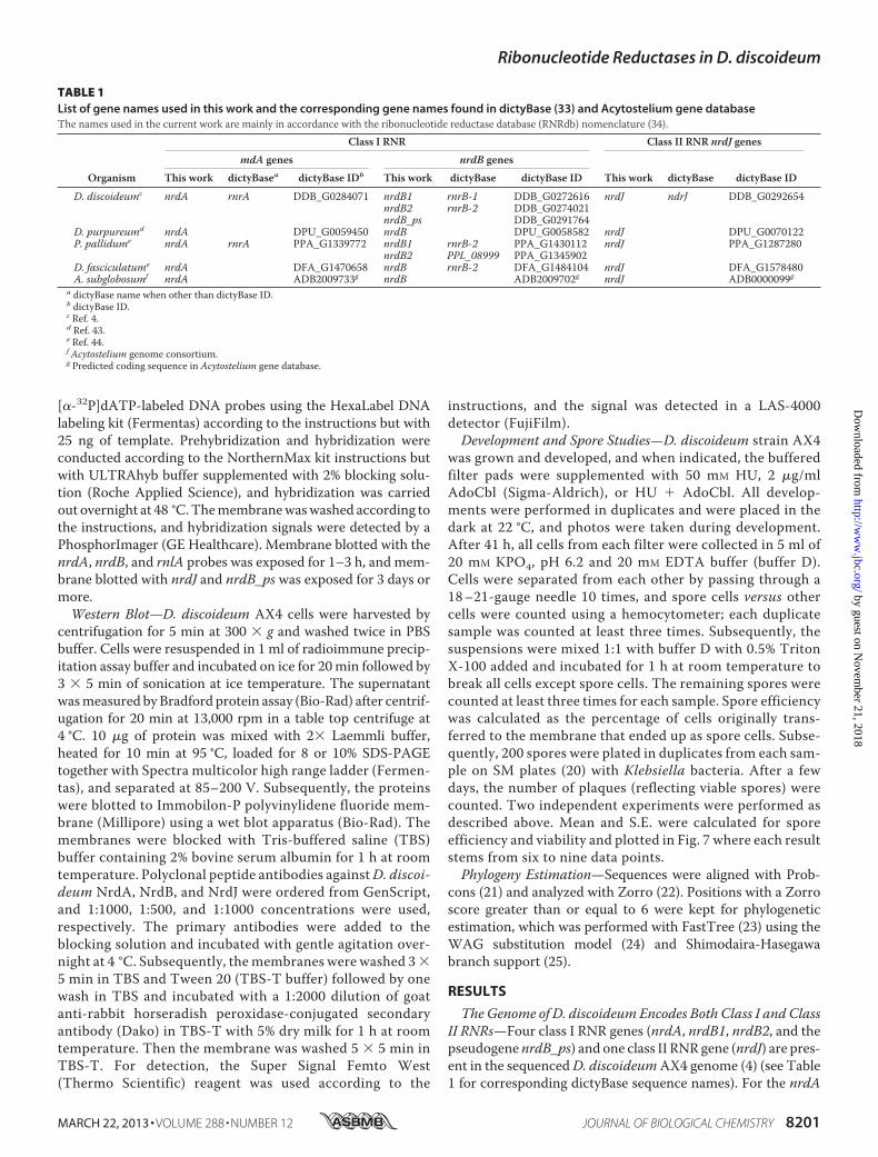
TABLE 1List of gene names used in this work and the corresponding gene names found in dictyBase (33) and Acytostelium gene databaseThe names used in the current work are mainly in accordance with the ribonucleotide reductase database (RNRdb) nomenclature (34).
Organism
Class I RNR Class II RNR nrdJ genes
mdA genes nrdB genes
This work dictyBasea dictyBase IDb This work dictyBase dictyBase ID This work dictyBase dictyBase ID
D. discoideumc nrdA rnrA DDB_G0284071 nrdB1 rnrB-1 DDB_G0272616 nrdJ ndrJ DDB_G0292654nrdB2 rnrB-2 DDB_G0274021nrdB_ps DDB_G0291764
D. purpureumd nrdA DPU_G0059450 nrdB DPU_G0058582 nrdJ DPU_G0070122P. pallidume nrdA rnrA PPA_G1339772 nrdB1 rnrB-2 PPA_G1430112 nrdJ PPA_G1287280
nrdB2 PPL_08999 PPA_G1345902D. fasciculatume nrdA DFA_G1470658 nrdB rnrB-2 DFA_G1484104 nrdJ DFA_G1578480A. subglobosumf nrdA ADB2009733g nrdB ADB2009702g nrdJ ADB0000099g
a dictyBase name when other than dictyBase ID.b dictyBase ID.c Ref. 4.d Ref. 43.e Ref. 44.f Acytostelium genome consortium.g Predicted coding sequence in Acytostelium gene database.
Ribonucleotide Reductases in D. discoideum
MARCH 22, 2013 • VOLUME 288 • NUMBER 12 JOURNAL OF BIOLOGICAL CHEMISTRY 8201
by guest on Novem
ber 21, 2018http://w
ww
.jbc.org/D
ownloaded from
gene on chromosome 4, a class I large subunit NrdA proteinwith �60% amino acid identity to other eukaryotic NrdA pro-teins and 27% identity toE. coliNrdA is predicted. Similarly, forthe nrdB1 gene on chromosome 2, a class I small subunit NrdBprotein with �60% amino acid identity to other eukaryoticNrdB proteins and 25% identity to the corresponding E. coliprotein is predicted. Due to a recent duplication on chromo-some 2, there is an identical second nrdB gene copy in thesequenced AX4 strain (here called nrdB2). In contrast, for thenrdB_ps gene on chromosome 6, a protein with 50 extra N-ter-minal residues compared with NrdB, lacking 120 C-terminalresidues including the region essential for interaction with theNrdA protein, and lacking four of the six crucial metal-ligatingside chains and three of the four side chains involved in radicaltransfer is predicted. Together, this suggests that the nrdB_psgene does not code for a functional RNR component. Phylog-enies estimated from the class I RNR protein sequences areconsistent with established eukaryotic supergroups, placingDictyostelium spp. sequences among other Amoebozoa (26).
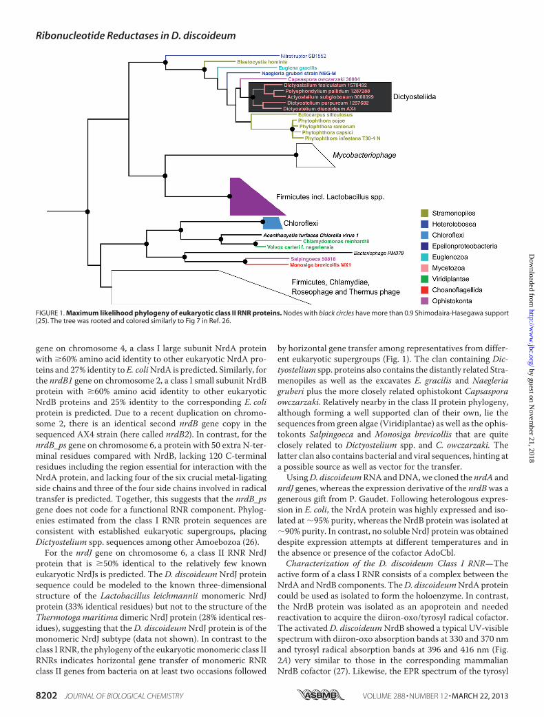
For the nrdJ gene on chromosome 6, a class II RNR NrdJprotein that is �50% identical to the relatively few knowneukaryotic NrdJs is predicted. The D. discoideum NrdJ proteinsequence could be modeled to the known three-dimensionalstructure of the Lactobacillus leichmannii monomeric NrdJprotein (33% identical residues) but not to the structure of theThermotogamaritima dimeric NrdJ protein (28% identical res-idues), suggesting that the D. discoideumNrdJ protein is of themonomeric NrdJ subtype (data not shown). In contrast to theclass I RNR, the phylogeny of the eukaryoticmonomeric class IIRNRs indicates horizontal gene transfer of monomeric RNRclass II genes from bacteria on at least two occasions followed
by horizontal gene transfer among representatives from differ-ent eukaryotic supergroups (Fig. 1). The clan containing Dic-tyostelium spp. proteins also contains the distantly related Stra-menopiles as well as the excavates E. gracilis and Naegleriagruberi plus the more closely related ophistokont Capsasporaowczarzaki. Relatively nearby in the class II protein phylogeny,although forming a well supported clan of their own, lie thesequences from green algae (Viridiplantae) as well as the ophis-tokonts Salpingoeca and Monosiga brevicollis that are quiteclosely related to Dictyostelium spp. and C. owczarzaki. Thelatter clan also contains bacterial and viral sequences, hinting ata possible source as well as vector for the transfer.UsingD. discoideumRNAandDNA,we cloned the nrdA and
nrdJ genes, whereas the expression derivative of the nrdBwas agenerous gift from P. Gaudet. Following heterologous expres-sion in E. coli, the NrdA protein was highly expressed and iso-lated at �95% purity, whereas the NrdB protein was isolated at�90% purity. In contrast, no soluble NrdJ protein was obtaineddespite expression attempts at different temperatures and inthe absence or presence of the cofactor AdoCbl.Characterization of the D. discoideum Class I RNR—The
active form of a class I RNR consists of a complex between theNrdA andNrdB components. TheD. discoideumNrdA proteincould be used as isolated to form the holoenzyme. In contrast,the NrdB protein was isolated as an apoprotein and neededreactivation to acquire the diiron-oxo/tyrosyl radical cofactor.The activatedD. discoideumNrdB showed a typical UV-visiblespectrum with diiron-oxo absorption bands at 330 and 370 nmand tyrosyl radical absorption bands at 396 and 416 nm (Fig.2A) very similar to those in the corresponding mammalianNrdB cofactor (27). Likewise, the EPR spectrum of the tyrosyl
FIGURE 1. Maximum likelihood phylogeny of eukaryotic class II RNR proteins. Nodes with black circles have more than 0.9 Shimodaira-Hasegawa support(25). The tree was rooted and colored similarly to Fig 7 in Ref. 26.
Ribonucleotide Reductases in D. discoideum
8202 JOURNAL OF BIOLOGICAL CHEMISTRY VOLUME 288 • NUMBER 12 • MARCH 22, 2013
by guest on Novem
ber 21, 2018http://w
ww
.jbc.org/D
ownloaded from
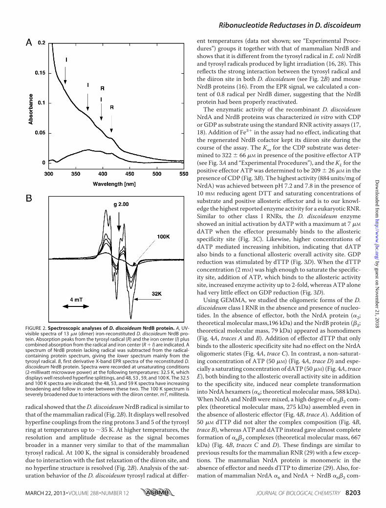
radical showed that theD. discoideumNrdB radical is similar tothat of themammalian radical (Fig. 2B). It displayswell resolvedhyperfine couplings from the ring protons 3 and 5 of the tyrosylring at temperatures up to �35 K. At higher temperatures, theresolution and amplitude decrease as the signal becomesbroader in a manner very similar to that of the mammaliantyrosyl radical. At 100 K, the signal is considerably broadeneddue to interaction with the fast relaxation of the diiron site, andno hyperfine structure is resolved (Fig. 2B). Analysis of the sat-uration behavior of the D. discoideum tyrosyl radical at differ-
ent temperatures (data not shown; see “Experimental Proce-dures”) groups it together with that of mammalian NrdB andshows that it is different from the tyrosyl radical in E. coliNrdBand tyrosyl radicals produced by light irradiation (16, 28). Thisreflects the strong interaction between the tyrosyl radical andthe diiron site in both D. discoideum (see Fig. 2B) and mouseNrdB proteins (16). From the EPR signal, we calculated a con-tent of 0.8 radical per NrdB dimer, suggesting that the NrdBprotein had been properly reactivated.The enzymatic activity of the recombinant D. discoideum
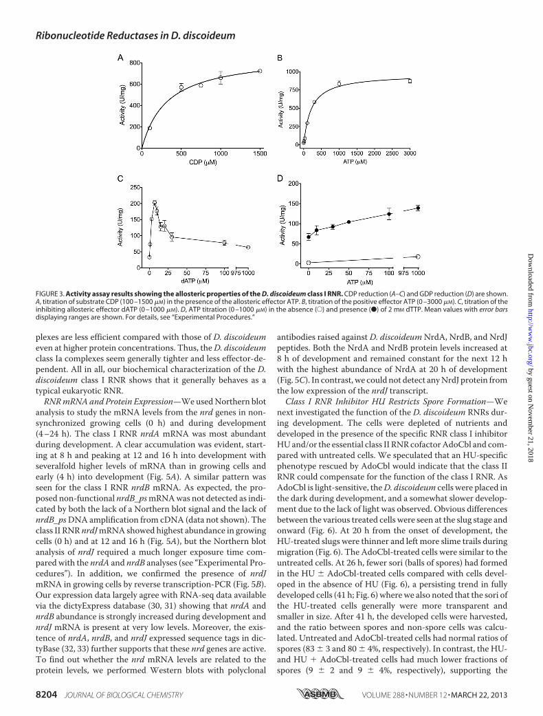
NrdA and NrdB proteins was characterized in vitro with CDPor GDP as substrate using the standard RNR activity assays (17,18). Addition of Fe3� in the assay had no effect, indicating thatthe regenerated NrdB cofactor kept its diiron site during thecourse of the assay. The Km for the CDP substrate was deter-mined to 322 � 66 �M in presence of the positive effector ATP(see Fig. 3A and “Experimental Procedures”), and theKL for thepositive effector ATP was determined to be 209 � 26 �M in thepresence of CDP (Fig. 3B). The highest activity (884 units/mg ofNrdA) was achieved between pH 7.2 and 7.8 in the presence of10 mM reducing agent DTT and saturating concentrations ofsubstrate and positive allosteric effector and is to our knowl-edge the highest reported enzyme activity for a eukaryotic RNR.Similar to other class I RNRs, the D. discoideum enzymeshowed an initial activation by dATP with a maximum at 7 �M
dATP when the effector presumably binds to the allostericspecificity site (Fig. 3C). Likewise, higher concentrations ofdATP mediated increasing inhibition, indicating that dATPalso binds to a functional allosteric overall activity site. GDPreduction was stimulated by dTTP (Fig. 3D). When the dTTPconcentration (2 mM) was high enough to saturate the specific-ity site, addition of ATP, which binds to the allosteric activitysite, increased enzyme activity up to 2-fold, whereas ATP alonehad very little effect on GDP reduction (Fig. 3D).Using GEMMA, we studied the oligomeric forms of the D.
discoideum class I RNR in the absence and presence of nucleo-tides. In the absence of effector, both the NrdA protein (�2;theoretical molecular mass,196 kDa) and the NrdB protein (�2;theoretical molecular mass, 79 kDa) appeared as homodimers(Fig. 4A, traces A and B). Addition of effector dTTP that onlybinds to the allosteric specificity site had no effect on the NrdAoligomeric states (Fig. 4A, trace C). In contrast, a non-saturat-ing concentration of ATP (50 �M) (Fig. 4A, trace D) and espe-cially a saturating concentration of dATP (50�M) (Fig. 4A, traceE), both binding to the allosteric overall activity site in additionto the specificity site, induced near complete transformationinto NrdA hexamers (�6; theoretical molecular mass, 588 kDa).WhenNrdA andNrdB weremixed, a high degree of �2�2 com-plex (theoretical molecular mass, 275 kDa) assembled even inthe absence of allosteric effector (Fig. 4B, trace A). Addition of50 �M dTTP did not alter the complex composition (Fig. 4B,trace B), whereas ATP and dATP instead gave almost completeformation of �6�2 complexes (theoretical molecular mass, 667kDa) (Fig. 4B, traces C and D). These findings are similar toprevious results for themammalian RNR (29) with a few excep-tions. The mammalian NrdA protein is monomeric in theabsence of effector and needs dTTP to dimerize (29). Also, for-mation of mammalian NrdA �6 and NrdA � NrdB �6�2 com-
FIGURE 2. Spectroscopic analyses of D. discoideum NrdB protein. A, UV-visible spectra of 13 �M (dimer) iron-reconstituted D. discoideum NrdB pro-tein. Absorption peaks from the tyrosyl radical (R) and the iron center (I) pluscombined absorption from the radical and iron center (R � I) are indicated. Aspectrum of NrdB protein lacking radical was subtracted from the radical-containing protein spectrum, giving the lower spectrum mainly from thetyrosyl radical. B, first derivative X-band EPR spectra of the reconstituted D.discoideum NrdB protein. Spectra were recorded at unsaturating conditions(2-milliwatt microwave power) at the following temperatures: 32.5 K, whichdisplays well resolved hyperfine splittings, and 48, 53 , 59, and 100 K. The 32.5and 100 K spectra are indicated; the 48, 53, and 59 K spectra have increasingbroadening and follow in order between these two. The 100 K spectrum isseverely broadened due to interactions with the diiron center. mT, millitesla.
Ribonucleotide Reductases in D. discoideum
MARCH 22, 2013 • VOLUME 288 • NUMBER 12 JOURNAL OF BIOLOGICAL CHEMISTRY 8203
by guest on Novem
ber 21, 2018http://w
ww
.jbc.org/D
ownloaded from
plexes are less efficient compared with those of D. discoideumeven at higher protein concentrations. Thus, theD. discoideumclass Ia complexes seem generally tighter and less effector-de-pendent. All in all, our biochemical characterization of the D.discoideum class I RNR shows that it generally behaves as atypical eukaryotic RNR.RNRmRNAand Protein Expression—WeusedNorthern blot
analysis to study the mRNA levels from the nrd genes in non-synchronized growing cells (0 h) and during development(4–24 h). The class I RNR nrdA mRNA was most abundantduring development. A clear accumulation was evident, start-ing at 8 h and peaking at 12 and 16 h into development withseveralfold higher levels of mRNA than in growing cells andearly (4 h) into development (Fig. 5A). A similar pattern wasseen for the class I RNR nrdB mRNA. As expected, the pro-posed non-functional nrdB_psmRNAwas not detected as indi-cated by both the lack of a Northern blot signal and the lack ofnrdB_psDNA amplification from cDNA (data not shown). Theclass II RNR nrdJmRNA showed highest abundance in growingcells (0 h) and at 12 and 16 h (Fig. 5A), but the Northern blotanalysis of nrdJ required a much longer exposure time com-pared with the nrdA and nrdB analyses (see “Experimental Pro-cedures”). In addition, we confirmed the presence of nrdJmRNA in growing cells by reverse transcription-PCR (Fig. 5B).Our expression data largely agree with RNA-seq data availablevia the dictyExpress database (30, 31) showing that nrdA andnrdB abundance is strongly increased during development andnrdJ mRNA is present at very low levels. Moreover, the exis-tence of nrdA, nrdB, and nrdJ expressed sequence tags in dic-tyBase (32, 33) further supports that these nrd genes are active.To find out whether the nrd mRNA levels are related to theprotein levels, we performed Western blots with polyclonal
antibodies raised against D. discoideumNrdA, NrdB, and NrdJpeptides. Both the NrdA and NrdB protein levels increased at8 h of development and remained constant for the next 12 hwith the highest abundance of NrdA at 20 h of development(Fig. 5C). In contrast, we could not detect anyNrdJ protein fromthe low expression of the nrdJ transcript.Class I RNR Inhibitor HU Restricts Spore Formation—We
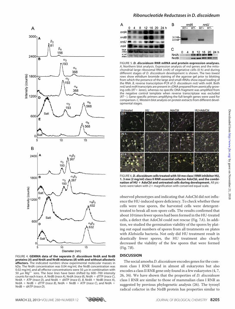
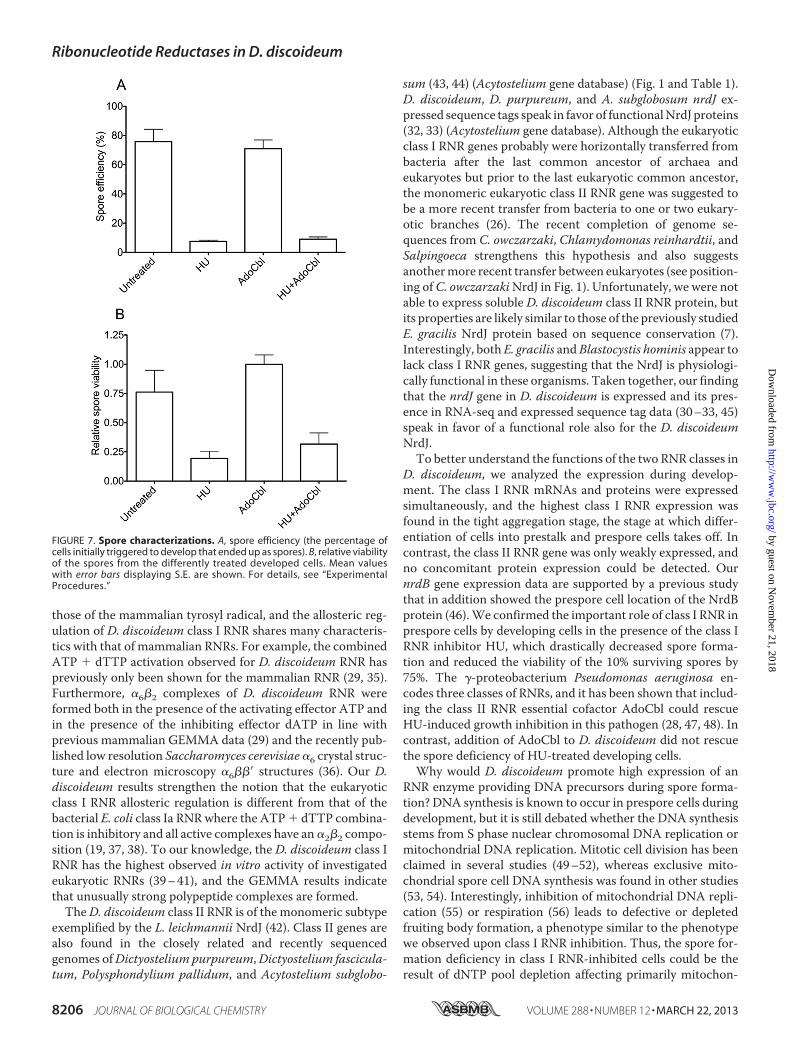
next investigated the function of the D. discoideum RNRs dur-ing development. The cells were depleted of nutrients anddeveloped in the presence of the specific RNR class I inhibitorHUand/or the essential class II RNRcofactorAdoCbl and com-pared with untreated cells. We speculated that an HU-specificphenotype rescued by AdoCbl would indicate that the class IIRNR could compensate for the function of the class I RNR. AsAdoCbl is light-sensitive, theD. discoideum cells were placed inthe dark during development, and a somewhat slower develop-ment due to the lack of light was observed. Obvious differencesbetween the various treated cells were seen at the slug stage andonward (Fig. 6). At 20 h from the onset of development, theHU-treated slugs were thinner and left more slime trails duringmigration (Fig. 6). The AdoCbl-treated cells were similar to theuntreated cells. At 26 h, fewer sori (balls of spores) had formedin the HU � AdoCbl-treated cells compared with cells devel-oped in the absence of HU (Fig. 6), a persisting trend in fullydeveloped cells (41 h; Fig. 6) wherewe also noted that the sori ofthe HU-treated cells generally were more transparent andsmaller in size. After 41 h, the developed cells were harvested,and the ratio between spores and non-spore cells was calcu-lated. Untreated and AdoCbl-treated cells had normal ratios ofspores (83 � 3 and 80 � 4%, respectively). In contrast, the HU-and HU � AdoCbl-treated cells had much lower fractions ofspores (9 � 2 and 9 � 4%, respectively), supporting the
FIGURE 3. Activity assay results showing the allosteric properties of the D. discoideum class I RNR. CDP reduction (A–C) and GDP reduction (D) are shown.A, titration of substrate CDP (100 –1500 �M) in the presence of the allosteric effector ATP. B, titration of the positive effector ATP (0 –3000 �M). C, titration of theinhibiting allosteric effector dATP (0 –1000 �M). D, ATP titration (0 –1000 �M) in the absence (E) and presence (●) of 2 mM dTTP. Mean values with error barsdisplaying ranges are shown. For details, see “Experimental Procedures.”
Ribonucleotide Reductases in D. discoideum
8204 JOURNAL OF BIOLOGICAL CHEMISTRY VOLUME 288 • NUMBER 12 • MARCH 22, 2013
by guest on Novem
ber 21, 2018http://w
ww
.jbc.org/D
ownloaded from
observed phenotypes and indicating that AdoCbl did not influ-ence theHU-induced spore deficiency. To checkwhether thesecells were true spores, the harvested cells were detergent-treated to break all non-spore cells. The results confirmed thatabout 10 times fewer spores had been formed in theHU-treatedcells, a defect that AdoCbl could not rescue (Fig. 7A). In addi-tion, we studied the germination viability of the spores by plat-ing out equal numbers of spores from all treatments on plateswith Klebsiella bacteria. Not only did HU treatment result indrastically fewer spores, the HU treatment also clearlydecreased the viability of the few spores that were formed(Fig. 7B).
DISCUSSION
The social amoebaD. discoideum encodes genes for the com-mon class I RNR found in almost all eukaryotes but alsoencodes a class II RNR gene only found in a few eukaryotes (4, 7,26, 34). We have shown that the properties of D. discoideumclass I RNR are similar to those of mammalian class I RNR assuggested by previous phylogenetic analysis (26). The tyrosylradical cofactor in the NrdB protein has properties similar to
FIGURE 4. GEMMA data of the separate D. discoideum NrdA and NrdBproteins (A) and NrdA and NrdB mixtures (B) with and without allostericeffectors. The indicated numbers show experimental molecular masses inkDa. The NrdA concentration was 0.04 mg/ml, the NrdB concentration was0.02 mg/ml, and all effector concentrations were 50 �M in combination with50 �M Mg2� ions. The base lines have been shifted by 600 –700 intensitycounts for each trace. A, NrdB (trace A), NrdA (trace B), NrdA � dTTP (trace C),NrdA � ATP (trace D), and NrdA � dATP (trace E). B, NrdA � NrdB (trace A),NrdA � NrdB � dTTP (trace B), NrdA � NrdB � ATP (trace C), and NrdA �NrdB � dATP (trace D).
FIGURE 5. D. discoideum RNR mRNA and protein expression analyses.A, Northern blot analysis. Expression analysis of nrd genes and the mito-chondrial large ribosomal RNA (rnlA) of vegetative cells (0 h) and duringdifferent stages of D. discoideum development is shown. The two lowestrows show ethidium bromide staining of the agarose gel prior to blottingfrom which the presence of the large and small rRNAs show equal loading ofthe RNA. B, reverse transcription-PCR of D. discoideum nrdJ with nrdA. BothnrdJ and nrdA transcripts are present in cDNA prepared from axenically grow-ing cells (RT� lanes), whereas no specific DNA fragment was amplified fromthe negative control template when reverse transcriptase was excluded(RT�). Gene-specific primers amplifying the full-length genes were used forcomparison. C, Western blot analysis on protein extracts from different devel-opmental stages.
FIGURE 6. D. discoideum cells treated with 50 mM class I RNR inhibitor HU,1. 3 mM (2 mg/ml) class II RNR essential cofactor AdoCbl, and the combi-nation of HU � AdoCbl and untreated cells during development. All pic-tures were taken with 2� magnification with conserved equal scale.
Ribonucleotide Reductases in D. discoideum
MARCH 22, 2013 • VOLUME 288 • NUMBER 12 JOURNAL OF BIOLOGICAL CHEMISTRY 8205
by guest on Novem
ber 21, 2018http://w
ww
.jbc.org/D
ownloaded from
those of the mammalian tyrosyl radical, and the allosteric reg-ulation of D. discoideum class I RNR shares many characteris-tics with that of mammalian RNRs. For example, the combinedATP � dTTP activation observed for D. discoideum RNR haspreviously only been shown for the mammalian RNR (29, 35).Furthermore, �6�2 complexes of D. discoideum RNR wereformed both in the presence of the activating effector ATP andin the presence of the inhibiting effector dATP in line withprevious mammalian GEMMA data (29) and the recently pub-lished low resolution Saccharomyces cerevisiae�6 crystal struc-ture and electron microscopy �6��� structures (36). Our D.discoideum results strengthen the notion that the eukaryoticclass I RNR allosteric regulation is different from that of thebacterial E. coli class Ia RNRwhere the ATP � dTTP combina-tion is inhibitory and all active complexes have an �2�2 compo-sition (19, 37, 38). To our knowledge, the D. discoideum class IRNR has the highest observed in vitro activity of investigatedeukaryotic RNRs (39–41), and the GEMMA results indicatethat unusually strong polypeptide complexes are formed.TheD. discoideum class II RNR is of the monomeric subtype
exemplified by the L. leichmannii NrdJ (42). Class II genes arealso found in the closely related and recently sequencedgenomes ofDictyostelium purpureum,Dictyostelium fascicula-tum, Polysphondylium pallidum, and Acytostelium subglobo-
sum (43, 44) (Acytostelium gene database) (Fig. 1 and Table 1).D. discoideum, D. purpureum, and A. subglobosum nrdJ ex-pressed sequence tags speak in favor of functionalNrdJ proteins(32, 33) (Acytostelium gene database). Although the eukaryoticclass I RNR genes probably were horizontally transferred frombacteria after the last common ancestor of archaea andeukaryotes but prior to the last eukaryotic common ancestor,the monomeric eukaryotic class II RNR gene was suggested tobe a more recent transfer from bacteria to one or two eukary-otic branches (26). The recent completion of genome se-quences from C. owczarzaki, Chlamydomonas reinhardtii, andSalpingoeca strengthens this hypothesis and also suggestsanothermore recent transfer between eukaryotes (see position-ing ofC. owczarzakiNrdJ in Fig. 1). Unfortunately, we were notable to express soluble D. discoideum class II RNR protein, butits properties are likely similar to those of the previously studiedE. gracilis NrdJ protein based on sequence conservation (7).Interestingly, both E. gracilis andBlastocystis hominis appear tolack class I RNR genes, suggesting that the NrdJ is physiologi-cally functional in these organisms. Taken together, our findingthat the nrdJ gene in D. discoideum is expressed and its pres-ence in RNA-seq and expressed sequence tag data (30–33, 45)speak in favor of a functional role also for the D. discoideumNrdJ.To better understand the functions of the two RNR classes in
D. discoideum, we analyzed the expression during develop-ment. The class I RNR mRNAs and proteins were expressedsimultaneously, and the highest class I RNR expression wasfound in the tight aggregation stage, the stage at which differ-entiation of cells into prestalk and prespore cells takes off. Incontrast, the class II RNR gene was only weakly expressed, andno concomitant protein expression could be detected. OurnrdB gene expression data are supported by a previous studythat in addition showed the prespore cell location of the NrdBprotein (46).We confirmed the important role of class I RNR inprespore cells by developing cells in the presence of the class IRNR inhibitor HU, which drastically decreased spore forma-tion and reduced the viability of the 10% surviving spores by75%. The �-proteobacterium Pseudomonas aeruginosa en-codes three classes of RNRs, and it has been shown that includ-ing the class II RNR essential cofactor AdoCbl could rescueHU-induced growth inhibition in this pathogen (28, 47, 48). Incontrast, addition of AdoCbl to D. discoideum did not rescuethe spore deficiency of HU-treated developing cells.Why would D. discoideum promote high expression of an
RNR enzyme providing DNA precursors during spore forma-tion? DNA synthesis is known to occur in prespore cells duringdevelopment, but it is still debated whether the DNA synthesisstems from S phase nuclear chromosomal DNA replication ormitochondrial DNA replication. Mitotic cell division has beenclaimed in several studies (49–52), whereas exclusive mito-chondrial spore cell DNA synthesis was found in other studies(53, 54). Interestingly, inhibition of mitochondrial DNA repli-cation (55) or respiration (56) leads to defective or depletedfruiting body formation, a phenotype similar to the phenotypewe observed upon class I RNR inhibition. Thus, the spore for-mation deficiency in class I RNR-inhibited cells could be theresult of dNTP pool depletion affecting primarily mitochon-
FIGURE 7. Spore characterizations. A, spore efficiency (the percentage ofcells initially triggered to develop that ended up as spores). B, relative viabilityof the spores from the differently treated developed cells. Mean valueswith error bars displaying S.E. are shown. For details, see “ExperimentalProcedures.”
Ribonucleotide Reductases in D. discoideum
8206 JOURNAL OF BIOLOGICAL CHEMISTRY VOLUME 288 • NUMBER 12 • MARCH 22, 2013
by guest on Novem
ber 21, 2018http://w
ww
.jbc.org/D
ownloaded from
drial DNA replication. Furthermore, the observed increasedlevels of the large mitochondrial rRNA (rnlA) a few hours afterthe induction of class I RNR transcripts (Fig. 5A) might alsofavor the importance of increased mitochondrial activity dur-ing development.The role of the class II RNR in D. discoideum is enigmatic.
Eukaryotes do not synthesize the vitamin B12 coenzymeAdoCbl (57) essential for function of class II RNRs but have torely on external sources. D. discoideum in addition encodestwo other AdoCbl-dependent enzymes, methylmalonyl-CoAmutase and methionine synthase (58), and likely can acquireAdoCbl from phagocytosed bacteria that synthesize the vita-min B12 coenzyme. Perhaps the tolerance to hypoxia is a valu-able class II RNR feature in D. discoideum when growth condi-tions with high nutrient but low oxygen levels are encountered.There is evidence for an oxygen-sensing mechanism in D. dis-coideum (59) as the oxygen level controls the expression of twocytochrome c oxidase isoforms. Clearly, selection for class IIRNR function exists as the class II RNR is expressed in D. dis-coideum, D. purpureum, and A. subglobosum, and the gene ispresent in the genomes of several closely related organisms.
Acknowledgments—We thank Pascale Gaudet and Adrian Tsang forthe kind gift of the plasmid used to produce NrdB protein and AndersHofer for kind support with the GEMMA.
REFERENCES1. Paps, J., Medina-Chacón, L. A., Marshall, W., Suga, H., and Ruiz-Trillo, I.
(2013) Molecular phylogeny of unikonts: new insights into the position ofApusomonads and Ancyromonads and the internal relationships of Opis-thokonts. Protist 164, 2–12
2. Kessin, R. H. (2001) Dictyostelium: Evolution, Cell Biology, and the Devel-opment of Multicellularity, Cambridge University Press, Cambridge, UK
3. Srinivasan, S., Alexander, H., and Alexander, S. (2000) Crossing the finishline of development: regulated secretion ofDictyostelium proteins.TrendsCell Biol. 10, 215–219
4. Eichinger, L., Pachebat, J. A., Glöckner, G., Rajandream, M. A., Sucgang,R., Berriman, M., Song, J., Olsen, R., Szafranski, K., Xu, Q., Tunggal, B.,Kummerfeld, S., Madera, M., Konfortov, B. A., Rivero, F., Bankier, A. T.,Lehmann, R., Hamlin, N., Davies, R., Gaudet, P., Fey, P., Pilcher, K., Chen,G., Saunders, D., Sodergren, E., Davis, P., Kerhornou, A., Nie, X., Hall, N.,Anjard, C., Hemphill, L., Bason, N., Farbrother, P., Desany, B., Just, E.,Morio, T., Rost, R., Churcher, C., Cooper, J., Haydock, S., van Driessche,N., Cronin, A., Goodhead, I., Muzny, D., Mourier, T., Pain, A., Lu, M.,Harper, D., Lindsay, R., Hauser, H., James, K., Quiles,M.,Madan Babu,M.,Saito, T., Buchrieser, C.,Wardroper, A., Felder,M., Thangavelu,M., John-son, D., Knights, A., Loulseged, H.,Mungall, K., Oliver, K., Price, C., Quail,M. A., Urushihara, H., Hernandez, J., Rabbinowitsch, E., Steffen, D., Sand-ers, M., Ma, J., Kohara, Y., Sharp, S., Simmonds, M., Spiegler, S., Tivey, A.,Sugano, S., White, B., Walker, D., Woodward, J., Winckler, T., Tanaka, Y.,Shaulsky, G., Schleicher, M., Weinstock, G., Rosenthal, A., Cox, E. C.,Chisholm, R. L., Gibbs, R., Loomis,W. F., Platzer, M., Kay, R. R.,Williams,J., Dear, P. H., Noegel, A. A., Barrell, B., and Kuspa, A. (2005) The genomeof the social amoeba Dictyostelium discoideum. Nature 435, 43–57
5. Nordlund, P., and Reichard, P. (2006) Ribonucleotide reductases. Annu.Rev. Biochem. 75, 681–706
6. Hofer, A., Crona, M., Logan, D. T., and Sjöberg, B. M. (2012) DNA build-ing blocks: keeping control of manufacture. Crit. Rev. Biochem. Mol. Biol.47, 50–63
7. Torrents, E., Trevisiol, C., Rotte, C., Hellman, U., Martin, W., and Reich-ard, P. (2006) Euglena gracilis ribonucleotide reductase: the eukaryoteclass II enzyme and the possible antiquity of eukaryote B12 dependence.
J. Biol. Chem. 281, 5604–56118. Kunz, B. A., Kohalmi, S. E., Kunkel, T. A., Mathews, C. K., McIntosh,
E. M., and Reidy, J. A. (1994) International Commission for Protectionagainst Environmental Mutagens and Carcinogens. Deoxyribonucleo-side triphosphate levels: a critical factor in the maintenance of geneticstability. Mutat. Res. 318, 1–64
9. Mathews, C. K. (2006) DNA precursor metabolism and genomic stability.FASEB J. 20, 1300–1314
10. Kumar, D., Abdulovic, A. L., Viberg, J., Nilsson, A. K., Kunkel, T. A., andChabes, A. (2011) Mechanisms of mutagenesis in vivo due to imbalanceddNTP pools. Nucleic Acids Res. 39, 1360–1371
11. Sussman, M. (1987) Cultivation and synchronous morphogenesis of Dic-tyostelium under controlled experimental conditions. Methods Cell Biol.28, 9–29
12. Hinas, A., Larsson, P., Avesson, L., Kirsebom, L. A., Virtanen, A., andSöderbom, F. (2006) Identification of the major spliceosomal RNAs inDictyostelium discoideum reveals developmentally regulated U2 variantsand polyadenylated snRNAs. Eukaryot. Cell 5, 924–934
13. Birgander, P. L., Bug, S., Kasrayan, A., Dahlroth, S. L., Westman, M., Gor-don, E., and Sjöberg, B.-M. (2005) Nucleotide-dependent formation ofcatalytically competent dimers from engineered monomeric ribonucle-otide reductase protein R1. J. Biol. Chem. 280, 14997–15003
14. Atkin, C. L., Thelander, L., Reichard, P., and Lang, G. (1973) Iron and freeradical in ribonucleotide reductase. Exchange of iron and Mössbauerspectroscopy of the protein B2 subunit of the Escherichia coli enzyme.J. Biol. Chem. 248, 7464–7472
15. Sahlin, M., Gräslund, A., and Ehrenberg, A. (1986) Determination of re-laxation times for a free radical from microwave saturation studies. J.Magn. Reson. 67, 135–137
16. Sahlin, M., Petersson, L., Gräslund, A., Ehrenberg, A., Sjöberg, B.-M., andThelander, L. (1987)Magnetic interaction between the tyrosyl free radicaland the antiferromagnetically coupled iron center in ribonucleotide re-ductase. Biochemistry 26, 5541–5548
17. Thelander, L., Sjöberg, B. R., and Eriksson, S. (1978) Ribonucleosidediphosphate reductase (Escherichia coli).Methods Enzymol. 51, 227–237
18. Hofer, A., Ekanem, J. T., and Thelander, L. (1998) Allosteric regulation ofTrypanosoma brucei ribonucleotide reductase studied in vitro and in vivo.J. Biol. Chem. 273, 34098–34104
19. Rofougaran, R., Crona, M., Vodnala, M., Sjöberg, B. M., and Hofer, A.(2008) Oligomerization status directs overall activity regulation of theEscherichia coli class Ia ribonucleotide reductase. J. Biol. Chem. 283,35310–35318
20. Sussman, M. (1966) in Methods in Cell Physiology (Prescott, D., ed) pp.397–410, Academic Press, New York
21. Do, C. B., Mahabhashyam, M. S., Brudno, M., and Batzoglou, S. (2005)ProbCons: probabilistic consistency-based multiple sequence alignment.Genome Res. 15, 330–340
22. Wu, M., Chatterji, S., and Eisen, J. A. (2012) Accounting for alignmentuncertainty in phylogenomics. PLoS One 7, e30288
23. Price, M. N., Dehal, P. S., and Arkin, A. P. (2010) FastTree 2—approxi-matelymaximum-likelihood trees for large alignments.PLoSOne 5, e9490
24. Whelan, S., and Goldman, N. (2001) A general empirical model of proteinevolution derived from multiple protein families using a maximum-like-lihood approach.Mol. Biol. Evol. 18, 691–699
25. Shimodaira, H., and Hasegawa, M. (1999) Multiple comparisons of log-likelihoods with applications to phylogenetic inference. Mol. Biol. Evol.16, 1114–1116
26. Lundin, D., Gribaldo, S., Torrents, E., Sjöberg, B. M., and Poole, A. M.(2010) Ribonucleotide reduction—horizontal transfer of a required func-tion spans all three domains. BMC Evol. Biol. 10, 383
27. Mann, G. J., Gräslund, A., Ochiai, E., Ingemarson, R., and Thelander, L.(1991) Purification and characterization of recombinant mouse and her-pes simplex virus ribonucleotide reductase R2 subunit. Biochemistry 30,1939–1947
28. Torrents, E., Poplawski, A., and Sjöberg, B. M. (2005) Two proteins medi-ate class II ribonucleotide reductase activity in Pseudomonas aeruginosa:expression and transcriptional analysis of the aerobic enzymes. J. Biol.Chem. 280, 16571–16578
Ribonucleotide Reductases in D. discoideum
MARCH 22, 2013 • VOLUME 288 • NUMBER 12 JOURNAL OF BIOLOGICAL CHEMISTRY 8207
by guest on Novem
ber 21, 2018http://w
ww
.jbc.org/D
ownloaded from
29. Rofougaran, R., Vodnala, M., and Hofer, A. (2006) Enzymatically activemammalian ribonucleotide reductase exists primarily as an �6�2 octa-mer. J. Biol. Chem. 281, 27705–27711
30. Rot, G., Parikh, A., Curk, T., Kuspa, A., Shaulsky, G., and Zupan, B. (2009)dictyExpress: a Dictyostelium discoideum gene expression database withan explorative data analysis web-based interface. BMC Bioinformatics 10,265
31. Parikh, A., Miranda, E. R., Katoh-Kurasawa, M., Fuller, D., Rot, G., Zagar,L., Curk, T., Sucgang, R., Chen, R., Zupan, B., Loomis,W. F., Kuspa, A., andShaulsky, G. (2010) Conserved developmental transcriptomes in evolu-tionarily divergent species. Genome Biol. 11, R35
32. Gaudet, P., Fey, P., Basu, S., Bushmanova, Y. A., Dodson, R., Sheppard,K. A., Just, E. M., Kibbe, W. A., and Chisholm, R. L. (2011) dictyBaseupdate 2011: web 2.0 functionality and the initial steps towards a genomeportal for the Amoebozoa. Nucleic Acids Res. 39, D620–D624
33. Kreppel, L., Fey, P., Gaudet, P., Just, E., Kibbe, W. A., Chisholm, R. L., andKimmel, A. R. (2004) dictyBase: a new Dictyostelium discoideum genomedatabase. Nucleic Acids Res. 32, D332–D333
34. Lundin, D., Torrents, E., Poole, A. M., and Sjöberg, B. M. (2009) RNRdb, acurated database of the universal enzyme family ribonucleotide reductase,reveals a high level of misannotation in sequences deposited to GenBank.BMC Genomics 10, 589
35. Eriksson, S., Thelander, L., and Akerman, M. (1979) Allosteric regulationof calf thymus ribonucleoside diphosphate reductase. Biochemistry 18,2948–2952
36. Fairman, J.W.,Wijerathna, S. R., Ahmad,M. F., Xu, H., Nakano, R., Jha, S.,Prendergast, J., Welin, R. M., Flodin, S., Roos, A., Nordlund, P., Li, Z.,Walz, T., and Dealwis, C. G. (2011) Structural basis for allosteric regula-tion of human ribonucleotide reductase by nucleotide-induced oligomer-ization. Nat. Struct. Mol. Biol. 18, 316–322
37. Larsson, A., and Reichard, P. (1966) Enzymatic synthesis of deoxyribo-nucleotides. IX. Allosteric effects in the reduction of pyrimidine ribo-nucleotides by the ribonucleoside diphosphate reductase system of Esch-erichia coli. J. Biol. Chem. 241, 2533–2539
38. Brown, N. C., and Reichard, P. (1969) Ribonucleoside diphosphate reduc-tase. Formation of active and inactive complexes of proteins B1 and B2. J.Mol. Biol. 46, 25–38
39. Sauge-Merle, S., Falconet, D., and Fontecave, M. (1999) An active ribonu-cleotide reductase from Arabidopsis thaliana cloning, expression andcharacterization of the large subunit. Eur. J. Biochem. 266, 62–69
40. Domkin, V., Thelander, L., and Chabes, A. (2002) Yeast DNA damage-inducible Rnr3 has a very low catalytic activity strongly stimulated afterthe formation of a cross-talking Rnr1/Rnr3 complex. J. Biol. Chem. 277,18574–18578
41. Wang, J., Lohman, G. J., and Stubbe, J. (2007) Enhanced subunit interac-tions with gemcitabine-5�-diphosphate inhibit ribonucleotide reductases.Proc. Natl. Acad. Sci. U.S.A. 104, 14324–14329
42. Sintchak, M. D., Arjara, G., Kellogg, B. A., Stubbe, J., and Drennan, C. L.(2002) The crystal structure of class II ribonucleotide reductase revealshow an allosterically regulated monomer mimics a dimer. Nat. Struct.Biol. 9, 293–300
43. Sucgang, R., Kuo, A., Tian, X., Salerno,W., Parikh, A., Feasley, C. L., Dalin,E., Tu, H., Huang, E., Barry, K., Lindquist, E., Shapiro, H., Bruce, D.,Schmutz, J., Salamov, A., Fey, P., Gaudet, P., Anjard, C., Babu,M.M., Basu,S., Bushmanova, Y., van der Wel, H., Katoh-Kurasawa, M., Dinh, C.,Coutinho, P. M., Saito, T., Elias, M., Schaap, P., Kay, R. R., Henrissat, B.,Eichinger, L., Rivero, F., Putnam, N. H., West, C. M., Loomis, W. F., Ch-
isholm, R. L., Shaulsky, G., Strassmann, J. E., Queller, D. C., Kuspa, A., andGrigoriev, I. V. (2011) Comparative genomics of the social amoebae Dic-tyostelium discoideum and Dictyostelium purpureum. Genome Biol. 12,R20
44. Heidel, A. J., Lawal, H.M., Felder,M., Schilde, C., Helps, N. R., Tunggal, B.,Rivero, F., John,U., Schleicher,M., Eichinger, L., Platzer,M.,Noegel, A. A.,Schaap, P., and Glöckner, G. (2011) Phylogeny-wide analysis of socialamoeba genomes highlights ancient origins for complex intercellularcommunication. Genome Res. 21, 1882–1891
45. Urushihara, H., Morio, T., Saito, T., Kohara, Y., Koriki, E., Ochiai, H.,Maeda,M.,Williams, J. G., Takeuchi, I., and Tanaka, Y. (2004) Analyses ofcDNAs from growth and slug stages ofDictyostelium discoideum.NucleicAcids Res. 32, 1647–1653
46. Tsang, A., Bonfils, C., Czaika, G., Shtevi, A., and Grant, C. (1996) A pres-pore-specific gene ofDictyostelium discoideum encodes the small subunitof ribonucleotide reductase. Biochim. Biophys. Acta 1309, 100–108
47. Sjöberg, B. M., and Torrents, E. (2011) Shift in ribonucleotide reductasegene expression in Pseudomonas aeruginosa during infection. Infect. Im-mun. 79, 2663–2669
48. Lee, K. M., Go, J., Yoon, M. Y., Park, Y., Kim, S. C., Yong, D. E., and Yoon,S. S. (2012) Vitamin B12-mediated restoration of defective anaerobicgrowth leads to reduced biofilm formation in Pseudomonas aeruginosa.Infect. Immun. 80, 1639–1649
49. Durston, A. J., and Vork, F. (1978) The spatial pattern of DNA synthesis inDictyostelium discoideum slugs. Exp. Cell Res. 115, 454–457
50. Zada-Hames, I. M., and Ashworth, J. M. (1978) The cell cycle and itsrelationship to development in Dictyostelium discoideum. Dev. Biol. 63,307–320
51. Zimmerman,W., andWeijer, C. J. (1993)Analysis of cell cycle progressionduring the development of Dictyostelium and its relationship to differen-tiation. Dev. Biol. 160, 178–185
52. Muramoto, T., and Chubb, J. R. (2008) Live imaging of the Dictyosteliumcell cycle reveals widespread S phase during development, a G2 bias inspore differentiation and a premitotic checkpoint. Development 135,1647–1657
53. Shaulsky, G., and Loomis, W. F. (1995) Mitochondrial DNA replicationbut no nuclear DNA replication during development of Dictyostelium.Proc. Natl. Acad. Sci. U.S.A. 92, 5660–5663
54. Chen, G., Shaulsky, G., and Kuspa, A. (2004) Tissue-specific G1-phasecell-cycle arrest prior to terminal differentiation in Dictyostelium. Devel-opment 131, 2619–2630
55. Chida, J., Yamaguchi, H., Amagai, A., and Maeda, Y. (2004) The necessityof mitochondrial genome DNA for normal development ofDictyosteliumcells. J. Cell Sci. 117, 3141–3152
56. Matsuyama, S. I., and Maeda, Y. (1995) Involvement of cyanide-resistantrespiration in cell-type proportioning duringDictyostelium development.Dev. Biol. 172, 182–191
57. Warren, M. J., Raux, E., Schubert, H. L., and Escalante-Semerena, J. C.(2002) The biosynthesis of adenosylcobalamin (vitamin B12). Nat. Prod.Rep. 19, 390–412
58. Zhang, Y., and Gladyshev, V. N. (2010) General trends in trace elementutilization revealed by comparative genomic analyses of Co, Cu, Mo, Ni,and Se. J. Biol. Chem. 285, 3393–3405
59. Sandonà, D., Gastaldello, S., Rizzuto, R., and Bisson, R. (1995) Expressionof cytochrome c oxidase during growth and development of Dictyoste-lium. J. Biol. Chem. 270, 5587–5593
Ribonucleotide Reductases in D. discoideum
8208 JOURNAL OF BIOLOGICAL CHEMISTRY VOLUME 288 • NUMBER 12 • MARCH 22, 2013
by guest on Novem
ber 21, 2018http://w
ww
.jbc.org/D
ownloaded from
Klose, Fredrik Söderbom and Britt-Marie SjöbergMikael Crona, Lotta Avesson, Margareta Sahlin, Daniel Lundin, Andrea Hinas, Ralph
Dictyostelium discoideumA Rare Combination of Ribonucleotide Reductases in the Social Amoeba
doi: 10.1074/jbc.M112.442434 originally published online January 31, 20132013, 288:8198-8208.J. Biol. Chem.
10.1074/jbc.M112.442434Access the most updated version of this article at doi:
Alerts:
When a correction for this article is posted•
When this article is cited•
to choose from all of JBC's e-mail alertsClick here
http://www.jbc.org/content/288/12/8198.full.html#ref-list-1
This article cites 57 references, 21 of which can be accessed free at
by guest on Novem
ber 21, 2018http://w
ww
.jbc.org/D
ownloaded from