Embed Size (px)
Citation preview
As
MSa
b
c
d
e
a
ARR1AA
KSPCN
1
cSgprp
oe
2
i
pf
(Os
2h
CASE REPORT – OPEN ACCESSInternational Journal of Surgery Case Reports 4 (2013) 515– 517
Contents lists available at SciVerse ScienceDirect
International Journal of Surgery Case Reports
j ourna l ho me page: www.elsev ier .com/ locate / i j scr
rare cause of deep peroneal nerve palsy due to compression ofynovial cyst – Case report
ehmet Erdil a,∗, Korhan Ozkanb, Feyza Unlu Ozkanc, Kerem Bilsel a, Ismail Turkmenb,erkan Senold, Serhan Sarare
Bezmialem Vakif University, Medicine Faculty, Department of Orthopaedics and Traumatology, Istanbul, TurkeyIstanbul Medeniyet Universitiy, Faculty of Medicine, Department of Ortopaedics and Traumatology, Istanbul, TurkeyFatih Sultan Mehmet Research and Training Hospital, Department of Physical Therapy and Rehabilitation, Istanbul, TurkeyIstanbul Medeniyet Universitiy, Faculty of Medicine, Department of Patology, Istanbul, TurkeyIstanbul Medeniyet Universitiy, Faculty of Medicine, Department of Anestesia and Reanimation, Istanbul, Turkey
r t i c l e i n f o
rticle history:eceived 16 August 2012eceived in revised form0 November 2012ccepted 27 November 2012
a b s t r a c t
INTRODUCTION: Synovial cyst is a rare cause of compression neuropathy and its differential diagnosis canbe misleading.PRESENTATION OF CASE: This article presents clinical, radiological, and histological findings of deep per-oneal nerve palsy due to compression of a synovial cyst in a 30-year-old patient admitted with suddendrop foot.
vailable online 13 March 2013eywords:ynovial cysteroneal nerveompressioneuropathy
DISCUSSION: Focal nerve entrapment in lower extremity due to synovial cystis a rare entity. Differentialdiagnosis is important. Surgical excision is the main treatment method with high success rate.CONCLUSION: Synovial cyst compression which can be treated easily with surgical excision should beconsidered in rapidly progressed drop foot.
© 2013 Surgical Associates Ltd. Published by Elsevier Ltd. All rights reserved.
. Introduction
Although synovial cysts are very common soft tissue tumors,ompression neuropathy due to synovial cysts are extremely rare.everal cases of ulnar and median nerve compressions due toanglia have been described.1–3 However, synovial cysts resulteripheral nerve compression in the lower extremities much morearely. Sultan firstly reported a case of compression neuropathy toeroneal nerve by a synovial cyst in 1921.4
In the present case report, a rarely seen compression neuropathyf deep peroneal nerve by a synovial cyst has been presented andvaluated in the light of literature.
. Presentation of case
A 30-year-old man was admitted to our hospital complain-ng of pain in lateral part of his proximal leg and drop foot that
∗ Corresponding author at: Bezmialem Universitesi, Vakıf Gureba Hastanesi Orto-edi ve Travmatoloji AD, 34093 Fatih, Istanbul, Turkey. Tel.: +90 5324249732;ax: +90 2124531700.
E-mail addresses: [email protected], [email protected]. Erdil), [email protected] (K. Ozkan), [email protected] (F.U.zkan), [email protected] (K. Bilsel), [email protected] (I. Turkmen),
[email protected] (S. Senol), [email protected] (S. Sarar).
210-2612/$ – see front matter © 2013 Surgical Associates Ltd. Published by Elsevier Ltdttp://dx.doi.org/10.1016/j.ijscr.2012.11.028
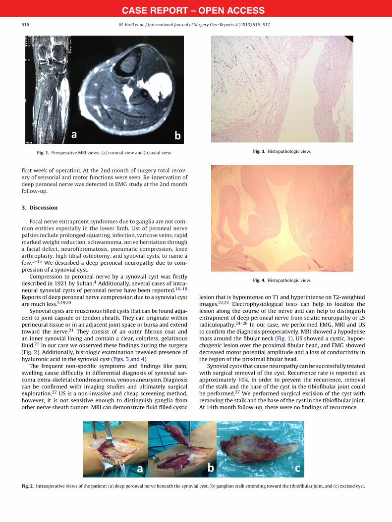
progressed rapidly during the same day in several hours. Onphysical examination, there were no atrophy of the peroneus mus-cles and the tibialis anterior muscle, however, peroneal musclesstrength was 5/5 whereas tibialis anterior, extensor hallucis, andextensor digitorum muscle strengths were 1/5 with reduction ofplantar flexion and stepping gait. Additionally, a cystic mass thatwas approximately 5 cm long and 3 cm width was palpable atthe proximal neck of fibula. Medical record revealed that swellinghad occurred 2 months before and pain with motor deficit pro-gressed rapidly during days. There was no trauma history. He hadbeen diagnosed with lumbar magnetic resonance imaging (MRI),however no pathologic findings had been found. Therefore, elec-tromyography (EMG), MRI, and soft tissue ultrasound (US) wereperformed. A hypodense mass around the fibular neck extend-ing distally to syndesmosis was detected with MRI (Fig. 1a andb). Decreased motor potential amplitude and a loss of conductiv-ity in the region of the proximal fibular head were seen in EMGstudy. These EMG findings were relevant to acute denervation ofdeep peroneal nerve. US showed a cystic, hypoechogenic lesionover the proximal fibular head. Excisional biopsy was performedwith provisional diagnosis of synovial cyst (Fig. 2). Surgical explo-
ration revealed synovial cyst that applied pressure to deep peronealnerve in the 1/3 proximal part of cruris. The presence of hyaluronicacid in the synovial cyst was detected in the histologic examination(Figs. 3 and 4). Motor functions had started to recover within the. All rights reserved.
CASE REPORT – OPEN ACCESS516 M. Erdil et al. / International Journal of Surgery Case Reports 4 (2013) 515– 517
fiedf
3
mpmaafp
dnRa
cptafl(h
scceho
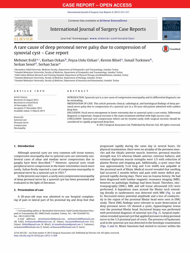
Fig. 3. Histopathologic view.
F
Fig. 1. Preoperative MRI views: (a) coronal view and (b) axial view.
rst week of operation. At the 2nd month of surgery total recov-ry of sensorial and motor functions were seen. Re-innervation ofeep peroneal nerve was detected in EMG study at the 2nd monthollow-up.
. Discussion
Focal nerve entrapment syndromes due to ganglia are not com-on entities especially in the lower limb. List of peroneal nerve
alsies include prolonged squatting, infection, varicose veins, rapidarked weight reduction, schwannoma, nerve herniation through
facial defect, neurofibromatosis, pneumatic compression, kneerthroplasty, high tibial osteotomy, and synovial cysts, to name aew.5–15 We described a deep peroneal neuropathy due to com-ression of a synovial cyst.
Compression to peroneal nerve by a synovial cyst was firstlyescribed in 1921 by Sultan.4 Additionally, several cases of intra-eural synovial cysts of peroneal nerve have been reported.16–18
eports of deep peroneal nerve compression due to a synovial cystre much less.5,19,20
Synovial cysts are muscinous filled cysts that can be found adja-ent to joint capsule or tendon sheath. They can originate withinerineural tissue or in an adjacent joint space or bursa and extendoward the nerve.21 They consist of an outer fibrous coat andn inner synovial lining and contain a clear, colorless, gelatinousuid.21 In our case we observed these findings during the surgeryFig. 2). Additionally, histologic examination revealed presence ofyaluronic acid in the synovial cyst (Figs. 3 and 4).
The frequent non-specific symptoms and findings like pain,welling cause difficulty in differential diagnosis of synovial sar-oma, extra-skeletal chondrosarcoma, venous aneurysm. Diagnosis
an be confirmed with imaging studies and ultimately surgicalxploration.22 US is a non-invasive and cheap screening method,owever, it is not sensitive enough to distinguish ganglia fromther nerve sheath tumors. MRI can demonstrate fluid filled cysticig. 2. Intraoperative views of the patient: (a) deep peroneal nerve beneath the synovial
Fig. 4. Histopathologic view.
lesion that is hypointense on T1 and hyperintense on T2-weightedimages.22,23 Electrophysiological tests can help to localize thelesion along the course of the nerve and can help to distinguishentrapment of deep peroneal nerve from sciatic neuropathy or L5radiculopathy.24–26 In our case, we performed EMG, MRI and USto confirm the diagnosis preoperatively. MRI showed a hypodensemass around the fibular neck (Fig. 1), US showed a cystic, hypoe-chogenic lesion over the proximal fibular head, and EMG showeddecreased motor potential amplitude and a loss of conductivity inthe region of the proximal fibular head.
Synovial cysts that cause neuropathy can be successfully treatedwith surgical removal of the cyst. Recurrence rate is reported asapproximately 10%. In order to prevent the recurrence, removal
of the stalk and the base of the cyst in the tibiofibular joint couldbe performed.27 We performed surgical excision of the cyst withremoving the stalk and the base of the cyst in the tibiofibular joint.At 14th month follow-up, there were no findings of recurrence.cyst, (b) ganglion stalk extending toward the tibiofibular joint, and (c) excised cyst.
– O of Sur
aar
C
F
E
A
psuwwpwibp
R
1
1
1
1
1
1
1
1
1
1
2
2
2
2
2
2
2
OTpc
CASE REPORTM. Erdil et al. / International Journal
This case demonstrates the etiologic cause of sudden drop footnd demonstrates the importance of considering synovial cystround the knee in the differential diagnosis of deep peroneal neu-opathy.
onflicts of interest
None.
unding
None.
thical approval
Obtained.
uthor contributions
Mehmet Erdil, corresponding author of the case, diagnosed theatient and made study design. Korhan Ozkan had performed theurgery with Dr. Erdil, and also worked in data collecting (follow-p data). Feyza Unlu Ozkan, helped in English language editing andriting the manuscript. Kerem Bilsel, had attended the surgeryith Dr. Erdil and Dr. Ozkan. He had taken the intraoperativehotos. Ismail Turkmen worked in data collecting (follow-up data)ith Dr. Ozkan. Serkan Senol worked in data collecting by prepar-
ng and evaluating the histopathologic specimens. Serhan Sarar hadeen in the surgery team as anesthesiologist, also he had helped inreparing the images of the manuscript.
eferences
1. Erkin G, Uysal H, Keles I, Aybay C, Ozel S. Acute ulnar neuropathy at thewrist: a case report and review of the literature. Rheumatology International2006;27:191–6.
2. Strickland JW, Steichen JB. Nerve tumors of the hand and forearm. Journal ofHand Surgery American Volume 1977;2:285–91.
3. Kerrigan JJ, Bertoni JM, Jaeger SH. Ganglion cysts and carpal tunnel syndrome.Journal of Hand Surgery American Volume 1988;13:763–5.
4. Sultan C. Ganglion der Nervenscheide des Nervus Peroneus. Zentralblatt furChirurgie 1921;48:963–5.
5. Mont MA, Dellon AL, Chen F, Hungerford MW, Krackow KA, Hungerford DS. Theoperative treatment of peroneal nerve palsy. Journal of Bone and Joint Surgery:American Volume 1996;78:863–9.
6. Yilmaz E, Karakurt L, Serin E, Güzel H. Peroneal nerve palsy due to rare reasons: areport of three cases. Acta Orthopaedica et Traumatologica Turcica 2004;38:75–8.
2
pen Accesshis article is published Open Access at sciencedirect.com. It is distribermits unrestricted non commercial use, distribution, and reproductredited.
PEN ACCESSgery Case Reports 4 (2013) 515– 517 517
7. Sangwan SS, Marya KM, Kundu ZS, Yadav V, Devgan A, Siwach RC. Compressiveperoneal neuropathy during harvesting season in Indian farmers. Tropical Doctor2004;34:244–6.
8. Schulze J, Troeger H. Fibular nerve compression due to an unusual cause. A casereport. Handchirurgie, Mikrochirurgie, Plastische Chirurgie 2004;36:25–8.
9. Yamamoto N, Koyano K. Neurovascular compression of the common peronealnerve by varicose veins. European Journal of Vascular and Endovascular Surgery2004;28:335–8.
0. Shahar E, Landau E, Genizi J. Adolescence peroneal neuropathy associated withrapid marked weight reduction: case report and literature review. EuropeanJournal of Paediatric Neurology 2007;11:50–4.
1. Mahitchi E, Van Linthoudt D. Schwannoma of the deep peroneal nerve. Anunusual presentation in rheumatology. Praxis (Bern 1994) 2007;96:69–72.
2. Yang LJ, Gala VC, McGillicuddy JE. Superficial peroneal nerve syndrome:an unusual nerve entrapment. Case report. Journal of Neurosurgery2006;104:820–3.
3. Cebesoy O, Tutar E, Isik M, Arpacioglu O. A case of isolated giant plexiform neu-rofibroma involving all branches of the common peroneal nerve. Archives ofOrthopaedic and Trauma Surgery 2007;127:709–12.
4. Fukuda H. Bilateral peroneal nerve palsy caused by intermittent pneumaticcompression. Internal Medicine 2006;45:93–4.
5. Dallari D, Pellacani A, Marinelli A, Verni E, Giunti A. Deep peroneal nerve paresisin a runner caused by ganglion at capitulum peronei. Case report and review ofthe literature. Journal of Sports Medicine and Physical Fitness 2004;44:436–40.
6. Spinner RJ, Ibrahim Elshiekh MA, Tubbs RS, Turner 3rd NS, Amrami KK. Posttrau-matic torsional injury as an indirect cause of fibular intraneural ganglion cysts:case illustrations and potential mechanisms. Clinical Anatomy 2012;25(July(5)):641–6, http://dx.doi.org/10.1002/ca.21290.
7. Al Mufargi YS, Mouch CA, Ziebarth K, Joeris A, Slongo T. An unusual cause ofparalysis of the peroneal nerve: a report of 3 cases. Journal of Pediatric Orthopedics2011;31(July–August (5)):e50–2.
8. Waldschmidt U, Slongo T. An unusual cause of paralysis of the peroneal nerve—acase report. Journal of Pediatric Surgery 2010;45(January (1)):259–61.
9. Rubin DI, Nottmeier E, Blasser KE, Peterson JJ, Kennelly K. Acute onset of deepperoneal neuropathy during a golf game resulting from a ganglion cyst. Journalof Clinical Neuromuscular Disease 2004;6(December (2)):49–53.
0. Wadstein T. Two cases of ganglia in the sheath of the peroneal nerve. ActaOrthopaedica Scandinavica 1931;2:221–31.
1. Mahaley MS. Ganglion of the posterior tibial nerve: case report. Journal of Neu-rosurgery 1974;40:120–4.
2. Masciocchi C, Innacoli M, Cisternino S, Barile A, Rossi F, Passariello R. Myxoidintraneural cysts of external popliteal ischiadic nerve. Report of 2 cases stud-ies with ultrasound, computed tomography and magnetic resonance imaging.European Journal of Radiology 1992;14:52–5.
3. Leon J, Marano G. MRI of peroneal nerve entrapment due to a ganglion cyst.Magnetic Resonance Imaging 1987;5:307–9.
4. Gutmann L. Atypical deep peroneal neuropathy in presence of accessorydeep peroneal nerve. Journal of Neurology, Neurosurgery and Psychiatry1970;33:453–6.
5. Ciucci G, Callegarini C, Stumpo M, Poppi M. Ganglionic cyst of the deep peronealnerve: description of a case. Italian Journal of Neurological Sciences 1996;17:83–6.
6. Antonini G, Bastianello S, Nucci F, Artico M, Bozzao L, Millefiorini M. Ganglionof deep peroneal nerve: electrophysiology and CT scan in the diagnosis. Elec-tromyography and Clinical Neurophysiology 1991;31:9–13.
7. Muckart RD. Compression of the common peroneal nerve by intramuscular gan-glion from the superior tibio-fibular joint. Journal of Bone and Joint Surgery: BritishVolume 1976;58:241–4.
uted under the IJSCR Supplemental terms and conditions, whichion in any medium, provided the original authors and source are