Embed Size (px)
Citation preview
CASE REPORT Open Access
A rare case of difficult airway managementin a Klippel-Feil syndrome pediatric patientwith osseous torticollis undergoneorthopedic surgeryDifficult airway in pediatric patient with torticollisXiaoqing Zhang, Jun Wang, Yajie Liu, Zhengqian Li and Bin Han*
Abstract
Background: Orthopedic surgery for cervical torticollis poses potential threat to airway management both intracheal intubation and extubation. Klippel-Feil syndrome (KFS) is a complex syndrome of osseous and visceralanomalies. The anatomical characteristics of KFS might have significant implications for airway management.
Case presentation: This is a rare case of an 8-year-old boy presenting with osseous torticollis, congenital occipito-atlantal deformity, congenital basilar invagination and KFS undergone elective torticollis correction surgery. Thoughwith difficulty, tracheal intubation was successfully performed. Extubation failed twice on postoperative day 2 and10, and required tracheostomy. Based on radiological findings, we speculated that prolonged airway edemaaccounted for the main reason of the failed extubation, the hypertrophic tonsil and occipito-cervical fusion resultedin reduced oropharyngeal space and limited cervical range of motion. Moreover, the Chiari malformation and KFScomplicated the airway condition and lead to prolonged airway obstruction. The tracheostomy casing wasremoved 1 month later.
Conclusions: Cautions should be taken in extubation of pediatric patients undergone major osseous torticollissurgery. Reintubation should be prepared in case of failed extubation. Severe post-operative airway edema,complicated with hypertrophic tonsil, the structural abnormalities in the oropharyngeal cavity, and occipito-cervicaldeformities constituted the decreased oropharyngeal space and resulted in failed extubation. For severe airwaycompromise and prolonged intubation, tracheostomy should be considered.
Keywords: Difficult extubation, Osseous torticollis, Airway edema, Tonsil hypertrophy, Occipito-cervical fixation,Pediatric, Syndrome
© The Author(s). 2021 Open Access This article is licensed under a Creative Commons Attribution 4.0 International License,which permits use, sharing, adaptation, distribution and reproduction in any medium or format, as long as you giveappropriate credit to the original author(s) and the source, provide a link to the Creative Commons licence, and indicate ifchanges were made. The images or other third party material in this article are included in the article's Creative Commonslicence, unless indicated otherwise in a credit line to the material. If material is not included in the article's Creative Commonslicence and your intended use is not permitted by statutory regulation or exceeds the permitted use, you will need to obtainpermission directly from the copyright holder. To view a copy of this licence, visit http://creativecommons.org/licenses/by/4.0/.The Creative Commons Public Domain Dedication waiver (http://creativecommons.org/publicdomain/zero/1.0/) applies to thedata made available in this article, unless otherwise stated in a credit line to the data.
* Correspondence: [email protected] of Anesthesiology, Peking University Third Hospital, 49 NorthGarden Rd., Haidian District, Beijing, China
Zhang et al. BMC Anesthesiology (2021) 21:121 https://doi.org/10.1186/s12871-021-01341-6

BackgroundCervical torticollis is a rare deformity characterized by alateral head tilt and chin rotation toward the side oppos-ite to the tilt. Klippel-Feil syndrome (KFS) is a cervicalabnormalities in pediatric patients caused by the absenceor fusion of cervical vertebrae. KFS was first reported in1912 as a triad of short neck, restricted motion of theneck, and low posterior hairline. As a consequence offailure in normal segmentation of cervical vertebrae dur-ing the early weeks of fetal development, two or more ofthe seven vertebrae are fused. It has a rare incidence of1:42000 births [1, 2]. The atlanto-occipital joint andspinal canal stenosis could co-exist in KF syndrome [3].The surgical treatment of the cervical osseous deformityposes potential threat to airway management both intracheal intubation and extubation. We reported aunique case of a pediatric patient undergone orthopedicsurgery who need tracheostomy as a result of failed tra-cheal extubation. We will discuss the airway manage-ment of this patient and suggest possible causes in orderto provide strategies for future events. Written informedconsent was obtained from the guardian of the patient.
Case presentationAn 8-year-old boy (weighing 34 kg) was found to be tor-ticollis at age 1. He was diagnosed with osseous torticol-lis in our hospital, and was scheduled for electiveorthopedic surgery.
HistoryThe boy has tonsil hypertrophy and sleep dyspnea in su-pine position and was scheduled for tonsillectomy inlocal hospital 1 year ago. The surgery was cancelled be-cause of failed tracheal intubation. No special birth his-tory was documented. No other dysplasia were reported.
Pre-operative examinationECGSinus arrhythmia by wandering pacemaker with sino-atrial node; incomplete right bundle branch block.Carotid artery and vertebral artery ultrasound: no left
common carotid artery was found (suspected with con-genital variation).
Cervical CT and MRI examinationsAtlanto-occipital fusion; congenital skull base depres-sion; atlantoaxial joint dislocation; cervical spine devel-opmental malformations (C2-3 block vertebrae; C6 leftappendage hypoplasia; C6-7 invisible cleft); sub tonsillarhernia.
Airway evaluationModified Mallampati test was III ~ IV. The mouth open-ing degree (less than 3 fingers) and head and neck
movement (60-120 degree) were limited. No significantnarrowing of tracheal lumen and airway tortuosity werenoticed in preoperative radiological findings (Fig. 1). TheColorado Pediatric Airway Score (COPUR score) withprediction points was 11 (C, points 1; O, points 2: withlimited mouth opening, 20-40 mm; P, points 4: pastfailed intubation history; U, points 2: partially visibleuvula; R, points 2: limited range of cervical motion, 60-120 degree). The glottic view prediction was 3, and theCOPUR prediction points of 11 was indicative of “diffi-cult intubation, fiberoptic less traumatic” [3]. No man-dible/maxilla deformity or other hypoplasia were noted.Therefore, though with expected intubation difficulty,intubation was attempted under full preparation withdifficult airway management tools. An experiencedanesthesiologist with two assistant anesthesiologists werein charge of this case.The patient’s periphery oxygen saturation (SpO2) was
100% breathing room air, in supine position. Dexa-methasone was given to prevent postoperative nauseaand vomiting, as well as airway edema. After sedatedwith propofol (with fractioned dose of 80 mg) followingpreoxygenation, sufficient facemask ventilation was con-firmed. The spontaneous breathing was retained to en-sure the absent of difficult intubation condition. Thepatient’s epiglottis was revealed during sedation. How-ever, after administering 10mg atracurium and 10 μgsufentanyl, the facemask ventilation suddenly becameimpossible. A reinforced endotracheal tube (I.D. of 6.0mm) was tried under glidescope but failed. The SpO2
dropped to 60. Direct laryngoscopy with medium sizedMacintosh blade was tried again by another attendinganesthesiologist after optimizing ventilation maneuvers.A reinforced endotracheal tube with sealing cuff (I.D. of5.5 mm) was placed successfully. The whole intubationduration was approximately 12 min.After prone position was made by orthopedic surgeons
with continuous cervical traction (Mayfield®, IntegraLifeScience. Inc., US), orthopedic surgery was per-formed, including the exploration anteriorly of thewedged C2 and C3-4 intervertebral disk, the fixationposteriorly of occipital-C4 pedicle with iliac bone graft,and correction of cervical torticollis. The surgical dur-ation was 334 min and the hemodynamic status wasstable during the whole procedure. A total of 1700 mlcrystalloid solution was infused, with intraoperativeblood loss of 200 ml, and autologous blood transfusionof 100 ml washed red blood cells. The boy was trans-ferred to the ICU with continuous sedation and anal-gesia without extubation.Extubation was normally performed on post-operative
day 1 in fear of prolonged intubation related complica-tions such as ventilator associated pneumonia or airwaycomplication. After positive result of cuff-leak test, ICU
Zhang et al. BMC Anesthesiology (2021) 21:121 Page 2 of 7

doctors postponed the extubation and invited anesthesi-ologists for consultation the next day. On postoperativeday 2, anesthesiologist and ICU doctors performed cuffleak test. The result was still positive. Considering thesuccess of the first intubation, extubation was attemptedwith preparation of re-intubation. The patient suffocatedimmediately after extubation despite complete con-sciousness and with normal blood-gas analysis results.Inspiratory stridor and cyanosis was noted soon after.Dexamethasone was administered to reduce airwayedema and other possible intubation related damage.The patient’s heart rate (HR) dropped from 125 bmp to80 bpm during re-intubation. Epiglottis and laryngealedema were revealed by video laryngoscope, but theglottis could not be visualized. Tracheal re-intubation fi-nally succeeded 17min later under propofol sedation.CT scanning was made on postoperative day 9 which
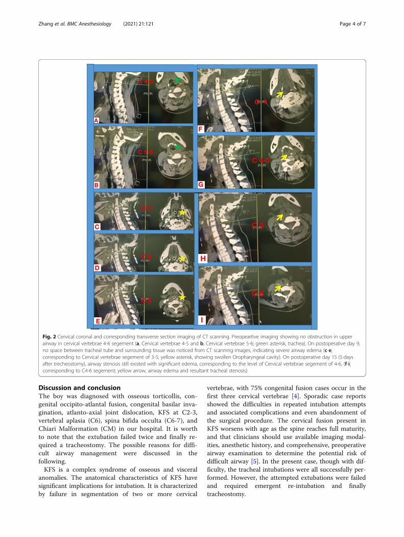
revealed airway edema comparing with preoperativefindings (Fig. 2c-e). The extended intubation durationand long-term sedation would lead to ventilator associ-ated pneumonia and complicated airway management.After Fiber-optical bronchoscope (FOB) inspectionshowing alleviated airway edema on postoperative day 9,and the potential existence of deep cervical fascial spacefrom CT imaging, a proposal of attempted extubation inthe operating room and tracheotomy as the alternative
plan was made by multidisciplinary consultation includ-ing orthopedics, anesthesiologists, otolaryngologists, andICU physicians. On postoperative day 10 in the operat-ing room, the patient was extubated after fully suction ofthe sputum and sufficient oxygenation. However, in-spiratory dyspnea soon ensued. The patient was put inleft lateral head-up position with facemask oxygenationand placement of oropharyngeal airway device. Dyspneawas not alleviated and cyanosis appeared 5min afterextubation. SpO2 dropped to 60% with HR of 70 bmp.Considering the existence of upper airway obstruction,re-intubation was performed immediately using glide-scopy under propofol sedation. Tracheotomy was suc-cessfully performed under general anaesthesia withorthopedists’ assistance with better positioning the headand neck. The patient returned to ICU with vital signsstable and was weaning from mechanical ventilator thenext day. Five days after tracheotomy, CT examinationshowed the still existence of the upper airway edema(Fig. 2f-i). Ten days later, the patient was transferred tothe orthopedic ward.One month after the first operation, the tracheostomy
tube was blocked and no discomfort was complaint.Four days later, the tracheostomy casing was removed.The boy was discharged from hospital 3 days later. Threeyear follow-up revealed satisfactory orthopedic results.
Fig. 1 a, preoperative appearance of the pediatric patients with osseous torticollis. b, a Three-dimensional printed cervical vertebrae model of thepediatric patients with osseous torticollis. c, preoperative X-ray imaging of the lateral view of the cervical in extension position. d, the lateral viewof the cervical X-ray imaging in reflection position. e, CT imaging showing the cervical spine developmental malformations (red arrow: C2-3 blockvertebrae) (C, cervical vertebrae)
Zhang et al. BMC Anesthesiology (2021) 21:121 Page 3 of 7
Discussion and conclusionThe boy was diagnosed with osseous torticollis, con-genital occipito-atlantal fusion, congenital basilar inva-gination, atlanto-axial joint dislocation, KFS at C2-3,vertebral aplasia (C6), spina bifida occulta (C6-7), andChiari Malformation (CM) in our hospital. It is worthto note that the extubation failed twice and finally re-quired a tracheostomy. The possible reasons for diffi-cult airway management were discussed in thefollowing.KFS is a complex syndrome of osseous and visceral
anomalies. The anatomical characteristics of KFS havesignificant implications for intubation. It is characterizedby failure in segmentation of two or more cervical
vertebrae, with 75% congenital fusion cases occur in thefirst three cervical vertebrae [4]. Sporadic case reportsshowed the difficulties in repeated intubation attemptsand associated complications and even abandonment ofthe surgical procedure. The cervical fusion present inKFS worsens with age as the spine reaches full maturity,and that clinicians should use available imaging modal-ities, anesthetic history, and comprehensive, preoperativeairway examination to determine the potential risk ofdifficult airway [5]. In the present case, though with dif-ficulty, the tracheal intubations were all successfully per-formed. However, the attempted extubations were failedand required emergent re-intubation and finallytracheostomy.
Fig. 2 Cervical coronal and corresponding transverse section imaging of CT scanning. Preopeartive imaging showing no obstruction in upperairway in cervical vertebrae 4-6 segement (a, Cervical vertebrae 4-5 and b, Cervical vertebrae 5-6; green asterisk, trachea). On postoperative day 9,no space between tracheal tube and surrounding tissue was noticed from CT scanning images, indicating severe airway edema (c-e,corresponding to Cervical vertebrae segement of 3-5; yellow asterisk, showing swollen Oropharyngeal cavity). On postoperative day 15 (5 daysafter trecheostomy), airway stenosis still existed with significant edema, corresponding to the level of Cervical vertebrae segement of 4-6, (f-i;corresponding to C4-6 segement; yellow arrow, airway edema and resultant tracheal stenosis)
Zhang et al. BMC Anesthesiology (2021) 21:121 Page 4 of 7

The rarely occurred airway obstruction after cervicalspine surgery has been reported with swelling andhematoma. Laryngeal edema presents as inspiratory stri-dor within 30–60 min of extubation, and it is an import-ant cause of post-extubation obstruction [6]. Normally,the degree of swelling peaked on day 2-3 after surgery,and then showed a gradually decreasing tendency [7].Experience from our tertiary center is that the extuba-tion could be safely performed immediately after cervicalsurgery. While for some major orthopedic surgery, itmight take 24-48 h for safe extubation. In the currentcase, the delayed airway obstruction still existed 2 weeksafter surgery and required tracheostomy. The upper air-way edema is obvious from postoperative CT imagingand gradually dissipated after 1 month. For pediatric pa-tients, large adenoids/tonsils and obese are important in-dications for difficult airway management [8].Considering the patient’s past medical history of sleepdyspnea in supine position due to tonsil hypertrophy,the failed tracheal intubation and the canceled tonsillec-tomy, we speculated that this might constitute as an im-portant factor for prolonged extubation whencomplicated with airway edema.Moreover, the occipito-cervical alignment has signifi-
cant impact on the oropharyngeal space. Sporadic casereports revealed that the angle of occipito-cervical (O-C)fusion would be critical for postoperative upper airwayobstruction [9]. A retrospective clinical study showedthere’s a difference in the O-C2 angle (the angles be-tween the inferior endplate of C2 vertebrae body and theMcGregor’s line). The percentage changes in the cross-sectional area of the oropharynx before and after surgery
were linearly correlated. Deceased O-C2 angel may leadto dyspnea and/or dysphagia after surgery [10]. For pa-tients with occipital-cervical fixation surgery, extubationshould be performed with caution.The boy’s congenital cervical skeletal deformity includ-
ing the basal invagination and KFS, as well as the in-ternal fixation may interfere with the accuratemeasuring of O-C2 angel. Therefore, we could onlymade a rough estimation of the O-C4 angel. However,from lateral view of the cervical X-ray in neutral pos-ition, a slight decrease of the O-C4 angel was noticed(Fig. 3). The O-C4 fusion immobilized the head andneck movement, and forced the patient’s head in a slightflexion position when compared with preoperative im-aging, which further compressed the laryngeal space andresulted in the failed extubation. Moreover, pediatric pa-tients with large tonsils, adenoids or obesity were proneto airway collapse during anaesthesia [11]. The de-creased oropharyngeal space caused by the surgery werefurther compressed by the postoperative laryngeal edemaand tonsil hypertrophy which ultimately resulted in diffi-culty breathing and tracheostomy.Chiari Malformation (CM) is the downward herniation
of the caudal part of the cerebellum and/or medullaoblongata into the spinal canal, and is associated withbasilar invagination and can alter neurological functionssuch as upper airway motility and respiratory control,not only central, but also obstructive respiratory events[12]. In this case, the tonsil hypertrophy and CM maynot directly lead to airway obstruction, but may compli-cate the difficulty airway management. The compensa-tory range of the upper airway space in this patient was
Fig. 3 It is hard to make an accurate measurement of O-C4 angle as this patient had complex cervical abnormalities (congenital occipitoatlantalfusion, congenital basilar invagination, atlanto-axial joint dislocation, KFS at C2-3, and the subsequent fixation of O-C4). We made a roughestimation of the O-C4 angle (the angel between the long and short red lines) from lateral X radiography of preoperative (a) and postoperative(b) (2 months after surgery) in neutral position. A slight reflection of the O-C4 was noticed when compared with preoperative imaging (O,occiput; C, cervical vertebrae)
Zhang et al. BMC Anesthesiology (2021) 21:121 Page 5 of 7
limited with the pre-existing tonsil hypertrophy as well aschanged upper airway muscle tension caused by the Chiarideformity. The post-operative decreased O-C4 angel fur-ther worsened the condition and might be an importantconstitutional factor in postoperative dyspnea [13].Extubation should be performed with caution. How-
ever, FOB examination and cuff leak test could only re-flect subglottic airway condition. It is reported from amulticenter evaluation study that several cuff leak testsdisplay limited diagnostic performance for the detectionof post-extubation stridor. Given the high rate of falsepositives, routine cuff leak test may expose to undueprolonged mechanical ventilation [14–17]. Inappropriateselection of tracheal tube would lead to confused result.Therefore, we did not solely depend on this test. For thenarrowing of the oropharyngeal space, more thoroughexamination including CT scanning should be per-formed. Multi-disciplinary consultation is important fordeciding appropriate extubation time. Anaesthesiologistsshould get well prepared for the emergent airway condi-tion. However, there is no pediatric-specific universalextubation guidelines or experts consensus. Current al-gorithms are modifications of adult approaches whichare often inappropriate.We reported a rare case of a pediatric patients with
cervical osseous deformity undergone orthopedic surgeryand need tracheostomy as a result of failed trachealextubation. The prolonged upper airway obstructionafter occipito-cervical fusion have never been reportedin pediatric patients undergone osseous torticollis sur-gery. The causes of prolonged airway obstruction afteroccipito-cervical fusion are multifactorial. The upper air-way edema constituted the major reason, and the hyper-trophic tonsil and the congenital cervical malformationmay further complicated the airway condition with lim-ited occipital-cervical range of motion and decreasedcompensatory space of the oropharyngeal cavity.Cautions should be taken during extubation process in
pediatric patients who undergone major osseous torti-collis surgery. Evaluation of general clinical factors thatmay produce an adverse impact on ventilation after tra-cheal extubation should be comprehensively consideredand optimized. An extubation and possible re-intubationplan in case of failed extubation that can be imple-mented to guarantee adequate ventilation should be for-mulated in advance. We recommend bougie or exchangecatheters to avoid “cannot intubate, cannot ventilate”condition. For severe airway compromise and prolongedintubation, tracheostomy should be considered.The appearance of the patient was significantly improved
at the 3 years follow up after discharge from hospital.
AbbreviationsKFS: Klippel-Feil Syndrome; O-C: Occipito-cervical; FOB: Fiber-opticalbronchoscope; CM: Chiari Malformation
AcknowledgmentsNone.The authors have completed the CARE reporting checklist.
Authors’ contributionsGuarantor of integrity of entire study: JW, XZ, BH, YL, ZL. Literature research:XZ, ZL. Case study and follow-up: XZ, BH. Manuscript preparation: XZ, YL.Manuscript editing and revision: XZ, J W. Manuscript final version approval:JW, BH. all authors have read and approved the manuscript.
FundingThis work was supported by the “Special fund from capital clinical healthdevelopment” from Beijing government to finance the studies of clinicalimportance. Grant No 2016-2-4092.
Availability of data and materialsAll data generated or analyzed during this study are included in thispublished article.
Declarations
Ethics approval and consent to participateThis case was performed according to the ethical guidelines of the HelsinkiDeclaration and was approved by the Human Ethics Committee of PekingUniversity Third Hospital.
Consent for publicationWritten informed consent was obtained from the guardian of the patient.The proof of consent to publish from study participants can be requested atany time.
Competing interestsThe author(s) declare no competing interests.
Received: 13 September 2020 Accepted: 12 April 2021
References1. Gruber J, Saleh A, Bakhsh W, et al. The prevalence of Klippel-Feil syndrome:
a computed tomography-based analysis of 2,917 patients. Spine Deform.2018;6(4):448–53.
2. Moses JT, Williams DM, Rubery PT, et al. The prevalence of Klippel-Feilsyndrome in pediatric patients: analysis of 831 CT scans. J Spine Surg. 2019;5(1):66–71.
3. Raj D, Luginbuehl I. Managing the difficult airway in the syndromic child.Contin Educ Anaesth Crit Care Pain. 2015;15(1):7–13.
4. Guille JT, Sherk HH. Congenital osseous anomalies of the upper and lowercervical spine in children. J Bone Joint Surg Am. 2002;84(2):277–88.
5. Stallmer ML, Vanaharam V, Mashour GA. Congenital cervical spine fusionand airway management: a case series of Klippel-Feil syndrome. J ClinAnesth. 2008;20(6):447–51.
6. Artime CA, Hagberg CA. Tracheal extubation. Respir Care. 2014;59(6):991–1005.
7. Suk KS, Kim KT, Lee SH, et al. Prevertebral soft tissue swelling after anteriorcervical discectomy and fusion with plate fixation. Int Orthop. 2006;30(4):290–4.
8. Engelhardt T, Fiadjoe JE, Weiss M, et al. A framework for the managementof the pediatric airway. Paediatr Anaesth. 2019;29(10):985–92.
9. Tagawa T, Akeda K, Asanuma Y, et al. Upper airway obstruction associatedwith flexed cervical position after posterior occipitocervical fusion. J Anesth.2011;25(1):120–2.
10. Miyata M, Neo M, Fujibayashi S, et al. O-C2 angle as a predictor of dyspneaand/or dysphagia after occipitocervical fusion. Spine (Phila Pa 1976). 2009;34(2):184–8.
11. Engelhardt T, Weiss M. A child with a difficult airway: what do I do next?Curr Opin Anaesthesiol. 2012;25(3):326–32.
12. Luigetti M, Losurdo A, Dittoni S, et al. Improvement of obstructive sleepapneas caused by hydrocephalus associated with Chiari malformation typeII following surgery. J Neurosurg Pediatr. 2010;6(4):336–9.
Zhang et al. BMC Anesthesiology (2021) 21:121 Page 6 of 7
13. Huang M, Gonda DD, Briceño V, et al. Dyspnea and dysphagia from upperairway obstruction after occipitocervical fusion in the pediatric age group.Neurosurg Focus. 2015;38(4):E13.
14. Schnell D, Planquette B, Berger A, et al. Cuff leak test for the diagnosis ofpost-extubation stridor: a Multicenter evaluation study. J Intensive CareMed. 2019;34(5):391–6.
15. Gros A, Holzapfel L, Marqué S, et al. Intra-individual variation of the cuff-leaktest as a predictor of post-extubation stridor. Respir Care. 2012;57(12):2026–631.
16. Kriner EJ, Shafazand S, Colice GL. The endotracheal tube cuff-leak test as apredictor for postextubation stridor. Respir Care. 2005;50(12):1632–8.
17. Deem S. Limited value of the cuff-leak test. Respir Care. 2005;50(12):1617–8.
Publisher’s NoteSpringer Nature remains neutral with regard to jurisdictional claims inpublished maps and institutional affiliations.
Zhang et al. BMC Anesthesiology (2021) 21:121 Page 7 of 7


![[Friederike Klippel] Keep Talking Communicative F(BookFi.org)](https://img.pdfslide.us/doc/110x75/56d6bfe01a28ab3016980726/friederike-klippel-keep-talking-communicative-fbookfiorg.jpg)