Embed Size (px)
Citation preview

Journal of Neuroscwnce Methods, 6 (1982) 369-382 369 Elsevier Biomedical Press
A radioenzymatic technique for the measurement of free and conjugated 3,4-dihydroxyphenylethylene-
glycol in brain tissue and biological fluids
T r e v o r D e n n i s a n d B e r n a r d S c a t t o n l
Synth~labo.L E R S, 31, Avenue Paul Vaillant Couturier, 92220 Bagneux (France)
(Received January 16th, 1982) (Revised version received Apnl 22nd, 1982)
(Accepted Apnl 23rd, 1982)
Key words radioenzymattc assay--3,4-dlhydroxyphenylethyleneglycol--catecholamlnes--rat brain tis- s u e - h u m a n biological f l u I d s - - p l a s m a - - u n n e - - c e r e b r o s p m a l fired
A simple, sensmve and specific radloenzymatzc assay for the measurement of 3,4-dlhydroxyphenyleth- yleneglycol (DOPEG) was developed The assay is based on the conversion of the compound to its O-methylated derlvaUve m the presence of catechol-O-methyltransferase and [JH]S-adenosyl-metluonine. The trltlated 3-methoxy-4-hydroxyphenylethyleneglycol formed is selectwely extracted in orgamc solvents and isolated by thin layer chromatography_ After oxidation to vanlllm the O-methylated compound is extracted and measured by liquid scmtdlatlon spectrophotometry
This assay has been applied to the measurement of free and conjugated DOPEG m a variety of blolog/cal tissues and fluids Both free and conjugated DOPEG were readdy detected in discrete rat brain areas Substantial amounts of free and conjugated DOPEG were also measured m ventncular perfusates from freely moving rats FmaUy, the presence of DOPEG was also demonstrated m human cerebrosplnal fluid, plasma and unne Only the free form of DOPEG was found m cerebrospmal fluid, whereas both unconjugated and conjugated forms were present in plasma and unne
I n t r o d u c t i o n
In the mammahan central nervous system noradrenahne (NA) is metabohzed pnmardy to the free and conjugated glycols 3,4-dlhydroxyphenylethyleneglycol (DOPEG) and 3-methoxy-4-hydroxyphenylethyleneglycol (MOPEG) (Mannarlno et al., 1963; Glowmski and Baldessarini, 1966, Schanberg et al., 1968; Braestrup et al., 1974). DOPEG is a metabohte of NA which is of interest as: (1) both MOPEG and DOPEG are formed in the brain m almost equal amounts (DeMet and Halans, 1979); (2) similarly to MOPEG, DOPEG formation as dependent on central
I To whom correspondence should be addressed.
0165-0270/0000-0000/$03.00 © 1982 Elsevier Blomedxcal Press

370
noradrenerglc neuron activity (Warsh et al., 1981a; Scatton, submitted for pubhca- tlon). The measurement of the brain concentrations of DOPEG would therefore provide a useful index of central NA turnover Fluorometry (Karasawa et al., 1978), gas chromatography with flame ionization or mass spectrometry detection (Kahane et al., 1976, Musloet et al , 1978; Warsh et al., 1981b), and high performance liqmd chromatography (HPLC) with electrochemical detection (Starke et al., 1981) have been successfully employed to measure DOPEG m brain tissue and body fluids. Moreover, combination of enzymatic methylation of DOPEG to MOPEG with subsequent gas chromatographic quantltatlon of the O-methylated NA metabolite (Nielsen and Braestrup, 1976) has also been used. However, these techniques either lack adequate sensitivity (e.g. fluorometry) or are time consurnlng and require complex extraction and derlvatlzatmn procedures (e.g. gas chromatography or HPLC). The present report describes a rapid, accurate and specific radioenzymatlc assay for the measurement of free and conjugated DOPEG The assay is based on the O-methylatlon of DOPEG into MOPEG with the aid of catechol-O-methyltrans- ferase in presence of tritmted S-adenosyl-methionine as a methyl donor Tntiated MOPEG is subsequently isolated and purified by simple analytical procedures This method has been apphed to the measurement of free and conjugated DOPEG m rat brain tissue and ventricular perfusates as well as in human plasma, cerebrosplnal fluid (CSF) and urine.
Materials and Methods
Ammals and sample collection Male Sprague-Dawley rats (COBS CD strain from Charles Raver, France)
weighing 160-180 g were used. Animals were maintained on a 12 h hght /dark cycle (light on at 07.00 h) with free access to food and water. Rats were sacrificed by decapitation and brain regions were dissected out in a cold environment according to Glowinskl and Iversen (1966) and Scatton (1977). 'Limblc areas' represent the septum pooled with nucleus accumbens septi and olfactory tubercle. Tissues were frozen on dry-ice and stored at - 7 0 ° C until analysis.
In some experiments, 2 polypropylene cannulae (external diameter 0.6 mm) were stereotaxically implanted in rats under chloral hydrate (400 mg/kg, i.p.; Fluka) anaesthesia in the anterior and posterior horns of the left lateral ventricle (coordi- nates A 6, L 1.5; H + 2 and A 3 4; L 5; H - 2 ; atlas of Konlg and Khppel, 1963). Seven days after surgery, the lateral ventricles of freely moving rats were perfused with Ringer's medmm (composition in mM: NaC1 118; KCI 4.74; CaC12 1 26; KH2PO 4 1.19; MgSO4 1.18; Na2HPO 4 16) at 37°C at a constant rate of 25 #l / rmn. Fractions were collected every 30 rain in tubes maintained at 0°C.
Blood and urine samples were obtained from normal adult volunteers of both sexes. Blood was collected in heparimzed (100 umts) tubes, centrifuged (1300 g for 10 mln) and plasma was immediately frozen on dry-ice. Cerebrosplnal fluid was obtained with informed consent via lumbar puncture from schizophrenic patients and imme&ately stored at - 8 0 ° C m polypropylene tubes Ventncular fired was

371
obtained by the drainage of the lateral ventricle of patients presenting cerebral oedema.
Reagents Tyramme HC1, DOPEG, MOPEG plperazme salt, dopamme HC1, NA bi-
tartrate, adrenaline bltartrate, epinme HC1, octopamine HC1, isoproterenol HC1, dlhydroxymandehc acid and 6-hydroxydopamine HBr were purchased from Sigma (St Lores, MO, U S.A ). Dlhydroxyphenylalanlne (DOPA) and 3,4-dihydroxyphen- ylacehc acid (DOPAC) were from Fluka (Switzerland) and a-methyl-DOPA from Bonapace (Italy) Alpha-methyl-dopamlne and a-methyl-adrenahne were synthesized by Dr Tlen Duc (Synth+labo, Chemistry Dept) . All of the above-mentioned compounds were dissolved in dlstdled water except DOPA which was dissolved m a few drops of 1 M HC1.
All solvents (analytical grade) and other chemicals were purchased from Fluka (Switzerland)
Sample preparation Ttssues. Frozen brain tissue was homogenized in approximately 20 vols. of
0 2 M HC1 containing 7 mM MgC1 z and 2.6 mM EGTA The homogenate was frozen and thawed before centrlfugation at 20,000 g for 15 mxn From the clear supernatant fraction an ahquot was drawn for the deternunatton of free DOPEG; hydrolysis of DOPEG-SO 4 was carried out by heating the supernatant in sealed tubes at 95°C (Eppendorf heating umt) for 12 mln
Btologwalflutds. One volume 2 M HCI or HC104 containing 70 mM MgC12 and 26 mM EGTA was added to 9 vols. of biological fluid (cerebrosplnal fluid, plasma, unne, t~ssue and ventricular perfusates).
DOPEG assay Catechol-O-methyl transferase (COMT) was isolated from rat liver as described
by Axelrod and Tomchick (1958). The enzyme was stored at - 2 0 ° C in 3ml ahquots. No change in the enzyme activity was found after 6 months of storage. The specific activity of the enzyme preparation was 0.16 unl ts /mg protein (1 umt of actwlty is defined as the amount of enzyme forrmng 1 #mol p roduc t /h at 37°C using adrenaline as the substrate).
Standards were prepared by dissolving DOPEG m dlstdled water and adding 50 gl of a solution containing 0.1-0.5 ng of DOPEG to 250 #1 of the sample or to 250 #1 of 0 2 M HCI (containing MgC12 and EGTA) or Ranger's solution treated with the acid mixture, these being internal and external standards. Appropriate Ranger's solution or acid blanks were also carried through the whole procedure.
Aliquots of supernatant of ussue homogenates or biological fluids (250/~1) treated as described above were placed m conical 12 ml glass-stoppered centrifuge tubes.
The reaction was initiated by the addition of 100 /~1 of the following mixture: Tns-HCI buffer 2.8 M pH 9.6, 50 /~1, 100 mM MgC12 10 /~1, dithxothreltol 1 mg, COMT 25 ~1 (1.3 X 10 -3 units), and 5 /iCi [3H-methyl]-S-adenosyl-L-methlonlne ([3H]SAM) (N.E.N., Boston, MA, spec. act. 13.9 Ci/mmol) . The final pH of the

372
reaction mixture was 9.0-9.1. The tubes were closed and incubated for 15 mm m a shaking water bath at 37°C
After incubation, the reaction was stopped by the addition of 300 #1 of 0.5 M sodium phosphate buffer (pH 6 0) containing 2 mM MOPEG carrier and 10 mM boric acid in an Ice bath. The aqueous phase was extracted with 9.0 ml of
Aliquot (tissue, blologMcal fluid) + reactmn m~xture
J Incubahon 37°C 15 ram.
+ 0 5 M phosphate buffer (containing boric aod + MOPEG) + toluene/isoamyl alcohol
I shake centrifuge
I I aqueous phase organic phase + 8% methylamine d~scarded
I shake centr=fuge
I I organic phase aqueous phase + toluene/isoamyl alcohol discarded
J shake centrifuge
i I orgamc phase aqueous phase discarded
I evaporate
T_L C separation
I 3H-MOPEG oxidat,on (4% Nal04)
I + 10% glycerol -f- 2 M CH3COOH + toluene PPO POPOP
J shake
Radioactivity determination
Fig_ 1 Standard procedure for the radloenzymatlc deterrnmaaon of DOPEG

373
toluene/ lsoamyl alcohol (7:3) by shaking for 5 mln The phases were separated by low speed centrifugatlon (100 g); the aqueous phase was frozen in an alcohol/dry-ice bath and the organic phase transferred into another set of tubes containing 500/~1 of a freshly prepared 8% solution of methylamane After shaking and centrifugatlon, the organic phase was discarded and the aqueous phase was washed with another 5 ml of toluene/ isoamyl alcohol After shaking and centrifugation the organic phase was again discarded and the aqueous phase was evaporated under vacuum by using a Rotavapor (Buchi, Switzerland) in a water bath at 20°C The dry residue was reconsmuted in 40 #1 absolute ethanol and applied to the absorbent band of chromatography plates (Whatman LK6DF).
The plates were developed for 50 man in a freshly prepared solvent maxture of either ch lo ro fo rm/me thano l / ammonia (23 : 6 : 1) or chloroform/acet ic acid/ethanol (16:3.1) . After drying the plates under a fume hood, the spot of MOPEG (Rf. 0.5) was vlsuahzed under UV light (254 nm). The silica gel corresponding to the M O P E G zone was scraped into scintillation vials and the radioactive compound eluted with 1 ml of 1 M ammonium hydroxide. After shaking, 50 #1 of 4% sodium perlodate solution was added to oxadlze the [3H]MOPEG to [3H]vanlllin The 5 man oxidation period was stopped by the addition of 50 /~1 of glycerol solution (10% v/v) . The solution was acidified by addition of 1 ml of 2 M acetic acid and the [3H]vanlllln extracted directly into the scintillation fluid (Toluene, PPO, dimethyl POPOP, Packard) ready for counting after a short time of stabilization
A schematic representation of the above described procedure used for the radloenzymatic determination of DOPEG is given in Fig. 1
Results
Development of the method
Incubatton condtttons The conditions for the O-methylation reaction are those which were developed for
the measurement of catecholammes in our laboratory and derived from those described by Da Prada and Ztircher (1976)
The ratio s ignal /blank is dependent on the amount of enzyme added as well as on the time of incubation; under normal conditions, 15 min of incubation were found to give an optimal ratio of signal/blank. The stabillzang agent dlthiothreltol was introduced to protect catechols from degradation and keep COMT in an actwe reduced form. Dlthlothreltol was found to be superior in this respect to mercaptoethanol. Magnesium was added to the sample homogenate and incubation mixture as a necessary cofactor for COMT. EGTA was added to sample homogenate to chelate calcium ions which inhibit the O-methylation reaction The presence of E G T A was not necessary when assaying brain t~ssue but markedly increased the assay sensitivity for plasma and urine samples. From the specific activity of [3H]SAM and the amount of [3H]MOPEG formed from DOPEG at the end of the incubation procedure we calculated that about 60% of D O P E G was converted into

374
M O P E G under our me thy la t ion condi t ions over the range 10-5000 pg. A shght ly lower convers ion (53%) was ob ta ined using in ternal s t andards ref lect ing a slight m h i b m o n of the O-methy la t lon reac t ion by b ra in tissue. Similar figures were o b t a i n e d by submi t t ing known amount s of D O P E G to the O-methy la t lon 19rocedure and m e a s u n n g the non-me thy la t ed D O P E G by high pressure hquid c h r o m a t o g r a p h y with e lec t rochemical de tec t ion (Semerd j l an -Rouqu le r et al., 1981).
Extraction conditions A varie ty of solvents were tested to ob ta in the best condi t ions of ex t rac t ion of
M O P E G with a m i m m a l con t r ibu t ion of [3H]SAM. The tests were carr ied out under no rma l reac t ion condi t ions in the absence of D O P E G substrate, af ter add i t ion of k n o w n amount s of [3H]SAM and [2-14C]MOPEG (C.E.A., Saclay, France , spec act. 57 m C l / m m o l ) . Al l solvents used were previously sa tu ra ted with water. Of the large n u m b e r of solvent systems tested (e.g. ethyl acetate, buty l acetate, d~ethyl ether, t o l u e n e / l s o a m y l alcohol, butanone) , the one which gave the best ex t rac t ion ef- f iciency for M O P E G with a min ima l contarrunat lon by [3H]SAM was found to be a mixture of toluene and l soamyl alcohol (7:3) . The ex t rac tab ih ty of [14 C ] M O P E G by t o l u e n e / l s o a m y l a lcohol was marked ly dependen t on the p H The ext rac t ion was min ima l at p H 10. Decreas ing the p H toward 4 caused a progress ive increase m ext rac t ion efficiency bu t a concomi tan t rise in b lank values. A p H of 6 was found to give a sa t i s fac tory ext rac t ion of [~4C]MOPEG (72% recovery) with a re la t ively low con t r ibu t ion of [3H]SAM (see Table 1).
The in t roduc t ion of the methylan 'nne ext rac t ion step and subsequent washing of the aqueous methylarrune phase with t o l u e n e / l s o a m y l alcohol were found to de- crease the b lank values wi thout s ignif icant ly reducing the M O P E G recoveries (see Tab le 1). M e t h y l a m i n e proved to be super ior in this respect to o ther chemical ly re la ted amines.
TABLE 1
RECOVERY OF [~4C]MOPEG AT VARIOUS ISOLATION STEPS
[ 14 C]MOPEG (80,000 dpm = 100%) was added at 4°C to the incubation mixture m absence of [3H]SAM and carried out through the whole procedure. [3H]blank was obtained by processing [3H]SAM (5 pCl) through the entire procedure m the absence of DOPEG and expressed as dpm Results are mean with S_E M of 6 determmauons and are expressed as percent of the amount of [ 14 C]MOPEG imtlally added to the samples
Isolation step [ t4 C]MOPEG [ 3 H]blank (% initial radloactxvlty) (dpm)
Toluene/lsoamyl alcohol extraction Methylarmne extraction Toluene/lsoamyl alcohol washing Eluuon from TLC NalO4 oxadat~on
720-- I 0 775826--+7 147 65 7-+0.8 298817-+3094 65 0-+0 1 150031 --+2362
- 18391~ 1 569 38 0-+0,4 349-+ 23

375
Chromatographtc systems A large number of chromatographac systems was lnvesugated. For this study,
[14C]MOPEG and [3H]SAM were spotted directly onto the chromatography plates, with appropriate carriers to enable us to visuahze the spots under UV hght after development of the chromatograms The following solvent mixtures, ch loroform/ methanol /ammonia (23:6 : 1) and chloroform/acetic acid/ethanol (16: 3.1) were found to provide an optimal separation of MOPEG and SAM without being too time consuming and with no overlap between MOPEG and chermcally related compounds.
Recovery and blank evolution The recovery of MOPEG at various steps of the analytical procedure was studied
by processing [ 14C]MOPEG through the entire radioenzymatic assay. The recovery of [~4C]MOPEG and the evolution of the blank values throughout various isolation steps are shown in Table I. The data indicate that 38% of [~4C]MOPEG is still present at the end of multiple isolation steps, whereas the blank values are drasti- cally reduced.
Recovery of endogenous DOPEG from brain tissue and biological flmds was estimated by adding 0.5 ng of DOPEG to 250 /~1 perchlonc acid homogenates (internal standards). The rauo of internal to external (addition of 0 5 ng of DOPEG to the incubation mixture) standards was superior to 90% for all braxn regions
oO
~ , ° f o , t ° 'j"
3O
~o
O.
I0
0 I I I I I I 0 25 50 I00 125 250
' ' I I i 0 250 1000 2500 5000 10000
DOPEG (pg)
F~B 2 Senslt]vaty and Gnearlty of the standard curve of the DOPEG assay Each point represents the mean of 3 samples

376
investigated as well as for ventricular perfusate, cerebrospmal fired and urine, but somewhat lower for blood plasma (70%).
Lmeartty of assay and senslttvtty As shown in Fig 2, the amount of [3H]MOPEG formed radloenzymatlcally from
DOPEG substrate and [3H]SAM was proportional to the amount of DOPEG added over the range of 10 pg to 5000 pg. A slight deviation from hnearity was only noted at 10 ng DOPEG The activity in the DOPEG assay averaged 41 -+ 2 d p m / p g over the above-mentioned range The sensitivity of the assay (dpm twice the blank) was 10 pg (see reset Fig. 2) and the coefficient of variation amounted to 6%.
Specificity of the DOPEG assay Various catecholarmnes and metabohtes which are substrates for COMT as well
as other substrates or molecules which could possibly interfere in the measurement were tested. One nanogram of each substrate or DOPEG itself were added sep- arately to the incubation mixture. Most of the compounds hsted m Table 2 did not interfere in the assay. Tyrarmne, DOPA, 6-OHDA, NA, adrenahne and a-methyl- adrenahne interfered slightly but the interference of each contributed to a neghgable extent (less than 0 3-0.1 dpm/pg) m the DOPEG assay under standard conditions. As the concentrations of DOPEG are far in excess of, or slrmlar to those of DOPA and similar to, or only shghtly lower than those of NA in rat brain tissue (ng /g DOPA 5-8; NA 50-150), human cerebrospinal fluid (pg/ml DOPA 500, NA
TABLE 2
SPECIFICITY OF RADIOENZYMATIC ASSAY FOR DOPEG
Each compound was added separately to the incubation rmxture m the amount of 1 ng Each value is the average of at least 3 determinations
Compound dpm d p m / p g
Blank 314 DOPEG 40 960 40 96 Tyramme 461 0 15 DOPA 600 0 29 a-Methyl-DOPA 439 0 13 Doparmne 388 0 07 a-Methyl-dopamme 267 0 6-Hydroxydopatmne 461 0 15 Noradrenahne 483 0 17 Adrenahne 551 0 24 a-Methyl-adrenahne 465 0 15 Epmme 399 0 09 Octopamlne 286 0 Isoprenahne 218 0 Dlhydroxymandehc acid 272 0 DOPAC 285 0

377
50-150), plasma ( p g / m l DOPA 1400; NA 100-300) and urine (ng /ml DOPA 50) (Ztircher and Da Prada, 1979; Da Prada and Zurcher, 1976; results from our own laboratory), the interference of DOPA and NA m the DOPEG assay in biological samples also appears to be of minor importance
Apphcatlons of the method
The above described radioenzymatic method has been apphed to the measure- ment of D O P E G (free and conjugated forms) in rat brain nssue and ventricular perfusates and human biological flmds
Determmatton of free and conjugated DOPEG m rat brain regions Hydrochloric acid (0.2 M) is sufficiently strong to hydrolyse conjugated D O P E G
by simple heating at 80-95°C for a short period A rapid test to find the ideal time was carried out on hypothalalmc tissue, known to contain large amounts of conjugated DOPEG, by varying the txme of heating of a fixed amount of super- natant The hydrolysis of conjugated D O P E G was found to be maximal within 10-15 mln at 95°C or 30 man at 80°C. More prolonged heating resulted in a destruction of DOPEG. As DOPEG-SO 4 as not commercially available, the recovery of D O P E G at this step could not be evaluated. However, since m complementary experiments incubation of brain perchlonc extracts m the presence of arylsulphatase (data not shown) yielded mmilar levels of D O P E G as after acidic hydrolysis, it is assumed that the latter procedure caused a complete hydrolysis of DOPEG-SO 4 Moreover, experaments carried out with deuterated MOPEG-SO 4 indicated a total hydrolysis of the compound m the absence of a slgmficant degradation of free M O P E G using our acidic hydrolysis condiuons (C. Lee, personal communication)
The stablhty of free D O P E G under the hydrolysis condmons used was evaluated by submitting known amounts (1-10 ng) of D O P E G (either m the absence or in the presence of supernatant from hypothalamac tissue) to the hydrolysis conditions and by measuring the remaining D O P E G by the radloenzymatxc procedure. The recovery of D O P E G was 60% after heating at 95°C for 15 man and 89% after heating at 80°C for 30 rain either in the absence or m the presence of brain tissue
Table 3 shows the concentrations of free, total and conjugated DOPEG in various brain regxons measured by the radioenzymatic procedure Conjugated DOPEG was calculated by subtracting free from total DOPEG. DOPEG was easily detected in all areas assayed. Taking the hmat of sensitivity as being twice the value of the blank, values of free D O P E G m all tissues apart from hippocampus were between 4 and 5 times above the blank. After hydrolysis, all values were between 10 and 15 times above the blank The highest concentrations of free DOPEG were found m the striatum, hmblc areas and bramstem whereas the happocampus had the lowest levels. Conjugated D O P E G concentrations were highest in the hypothalamus and hmbic areas and lowest m the cerebellum. The percentage of DOPEG found m free form vaned widely between the different regions, the ratio of free over conjugated D O P E G being lowest in the hypothalamus (0.05) and highest m the cerebellum (1.1)

378
TABLE 3
FREE, TOTAL AND CONJUGATED DOPEG LEVELS IN VARIOUS RAT BRAIN AREAS
Values are means with S.E_M. of data obtained from 6 ammals_
Brain regaons Free DOPEG Total DOPEG DOPEG-SO 4 Free/SO 4 (ng/g) (ng/g) (ng/g)
Llmblc areas 17 5 -+ 1 6 117 3+3 7 99 8±2 8 0 18 Stnatum 23 1-+3 6 61 8+2 5 38 6±3 3 0 60 Hlppocampus 3_8-+0.4 46 0--+3 l 42 2-+2 8 0 09 Bralnstem 18 8-+2 9 87 0-+3 4 68 3±6 2 0 28 Cerebellum 10 1 -+ 1 8 19 1 -+0_7 8 9--- + 1 2 1 13 Hypothalamus 9 7-+ 1 5 222 2-+5 0 212 5±5 8 0 05 Cerebral cortex 8 1 -+ 0_3 35 3-+ 1 7 27 2 ± 2 0 0 30 Whole brain 8 3-+0 6 38 5-+ 1 4 30 1 -+ 1 7 0 28
Measurement of free and conjugated DOPEG m ventrtcular perfusates F r e e a n d c o n j u g a t e d D O P E G levels we re eas i ly d e t e c t a b l e m p e r f u s a t e s of the
l a t e r a l v e n t r i c l e of f ree ly m o v i n g r a t s p e r f u s e d b y m e a n s of l n d w e l h n g c a n n u l a e
( T a b l e 4). A b o u t 90% of t he N A m e t a b o l i t e was f o u n d to b e c o n j u g a t e d
Measurement of free and conjugated DOPEG tn human btologtcal flutds U s i n g t he a b o v e d e s c r i b e d r a & o e n z y m a t l c p r o c e d u r e the p r e s e n c e o f s u b s t a n t i a l
a m o u n t s of D O P E G in h u m a n p l a s m a , v e n t n c u l a r a n d l u m b a r c e r e b r o s p m a l f lu id
a n d u r i n e was d e m o n s t r a t e d ( T a b l e 4). O n l y free D O P E G c o u l d be d e t e c t e d in the
c e r e b r o s p m a l f l m d w h e r e a s b o t h u n c o n j u g a t e d a n d c o n j u g a t e d f o r m s of D O P E G
w e r e p r e s e n t in p l a s m a a n d u r ine . A b o u t 50 a n d 80% of the D O P E G was c o n j u g a t e d
TABLE 4
CONCENTRATIONS OF FREE AND CONJUGATED DOPEG IN RAT VENTRICULAR PER- FUSATES AND HUMAN BIOLOGICAL FLUIDS
Range of values is m&cated wxtlun brackets
Species n Free DOPEG Conjugated DOPEG (pg/ml) (pg/ml)
Ventncular perfusate Rat
Lateral ventrlcular drainage Man
Lumbar cerebrospmal fired Man
Plasma Man
Urine Man
4 268--+50 4495--+430 (59-632) (2 680-6 900)
8 3 170-+637 0 (1 226-6875)
5 1470-+ 186 0 (1 170-2200)
11 1 1,12-+195 1235-+128 (694-2 890) (860-1 540)
7 155,000-+ 42,000 584,000--+ 133,000 (60,500-378,000) (100,500-1 179,000)
379
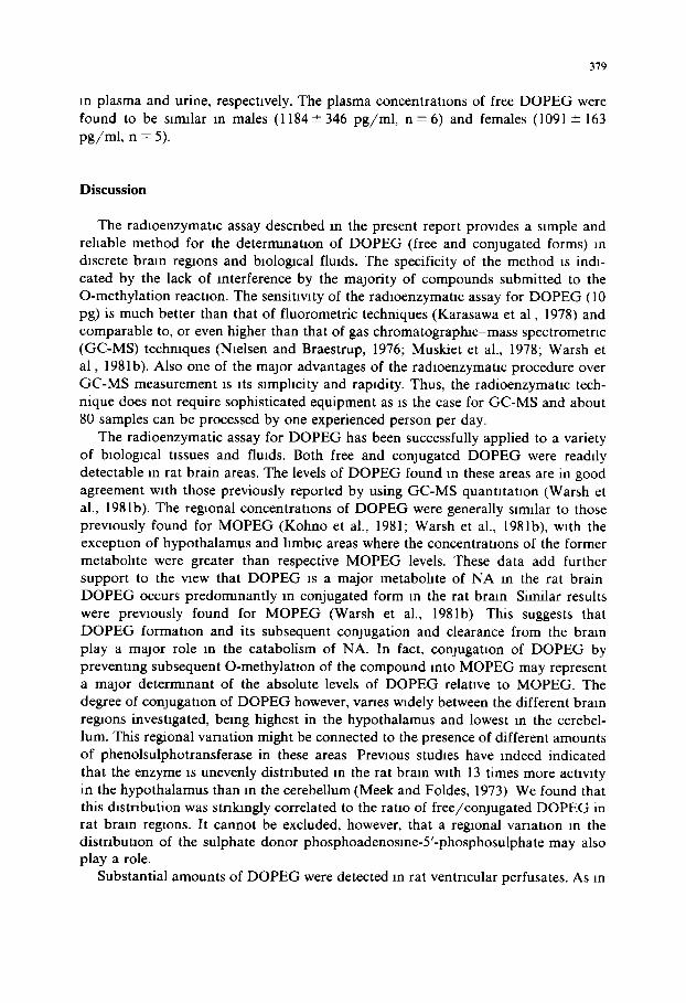
in plasma and urine, respectively. The plasma concentrations of free DOPEG were found to be sirmlar in males (1184+-346 pg /ml , n = 6 ) and females (1091-4-163 pg /ml , n = 5).
Discussion
The radloenzymatic assay described in the present report provides a simple and reliable method for the determination of D O P E G (free and conjugated forms) in discrete brain regions and biological fluids. The specificity of the method is indi- cated by the lack of interference by the majority of compounds submitted to the O-methylation reaction. The sensitivity of the radioenzymatlc assay for D O P E G (10 pg) is much better than that of fluorometric techniques (Karasawa et al , 1978) and comparable to, or even higher than that of gas chromatographic-mass spectrometric (GC-MS) techniques (Nielsen and Braestrup, 1976; Muskiet et al., 1978; Warsh et a l , 1981b). Also one of the major advantages of the radloenzymatlc procedure over GC-MS measurement is its simplicity and rapidity. Thus, the radioenzymatic tech- nique does not require sophisticated equipment as is the case for GC-MS and about 80 samples can be processed by one experienced person per day.
The radioenzymatic assay for D O P E G has been successfully applied to a variety of biological tissues and fluids. Both free and conjugated DOPEG were readily detectable in rat brain areas. The levels of D O P E G found in these areas are in good agreement with those previously reported by using GC-MS quantltatlon (Warsh et al., 1981b). The regional concentrations of D O P E G were generally similar to those previously found for M O P E G (Kohno et al., 1981; Warsh et al., 1981b), with the exception of hypothalamus and hmbic areas where the concentrations of the former metabohte were greater than respective MOPEG levels. These data add further support to the view that DOPEG is a major metabohte of NA in the rat brain D O P E G occurs predormnantly in conjugated form in the rat brain Similar results were previously found for M O P E G (Warsh et al., 1981b) This suggests that D O P E G formation and its subsequent conjugation and clearance from the brain play a major role in the catabolism of NA. In fact, conjugation of DOPEG by preventing subsequent O-methylatlon of the compound into MOPEG may represent a major deterrmnant of the absolute levels of DOPEG relative to MOPEG. The degree of conjugation of D O P E G however, vanes widely between the different brain regions investigated, being highest in the hypothalamus and lowest in the cerebel- lum. This regional variation might be connected to the presence of different amounts of phenolsulphotransferase in these areas Previous studies have indeed indicated that the enzyme is unevenly distributed m the rat brain with 13 times more activity in the hypothalamus than in the cerebellum (Meek and Foldes, 1973) We found that this distribution was str ihngly correlated to the ratio of free/conJugated DOPEG in rat brain re~ons. It cannot be excluded, however, that a regional variation in the distribution of the sulphate donor phosphoadenoslne-5'-phosphosulphate may also play a role.
Substantial amounts of DOPEG were detected in rat ventrlcular perfusates. As in
380
brain tissue, D O P E G occurred primarily in conjugated form in rat ventricular cerebrosplnal fluid.
The presence of free D O P E G was also demonstrated in human cerebrospinal fluid, plasma and urine. As far as plasma-free D O P E G is concerned, no sex difference could be observed The lumbar cerebrospmal fluid, urine and plasma levels of D O P E G found in the present study are in good agreement with those previously reported by others (Kahane et al., 1976; Muskiet et al , 1978; Baker and Johnson, 1981; Vlachakas et al , 1981) To our knowledge this is the first report on the occurrence of D O P E G in human cerebroventricular fluid As is the case for M O P E G (Karoum et al., 1977), the free form of D O P E G is the predominant if not exclusive one in human cerebrosplnal fluid In plasma and urine, D O P E G is found in both unconjugated and conjugated forms, the latter form being predominant in urine. It appears therefore that the degree of conjugation varies widely in the different biological fluids, increasing in the following o rder cerebrospinal fluid > p l a sma>ur ine . It is, however, noteworthy that only 80% of total D O P E G is conjugated in urine as compared with the almost total (97%) conjugation of MOPEG (Karoum et al., 1977) in this body fluid This difference might be connected to the fact that D O P E G being more polar than M O P E G can be excreted without undergo- ing conjugation, a process which aids in the excretion of phenols by converting them to more polar compounds, In man, both the sulphate and glucuronIde conjugates of M O P E G are found in plasma and urine (Karoum et al , 1977). The acidic hydrolysis used in the present study does not allow us to esnmate the respective contribution of the two forms of conjugation for D O P E G However, since the glucuronlde con- jugates of phenolic compounds are in general resistant to the mild acidic hydrolysis presently used, it ~s likely that the levels of conjugated DOPEG found in the present study correspond to the sulphate form. It is of Interest to note that the concentra- tions of total D O P E G are similar in human plasma and cerebrospinal fluid, but much lower in these body fluids than in urine Similar relative concentrations were previously reported for MOPEG (Karoum et al., 1977) This rmght be connected to the existence of a concentrating mechanism (possibly an active transport system) for D O P E G and M O P E G within the kidney. In human cerebrospmal fluid, plasma and urine, DOPEG represents only about 10-20% of the total MOPEG concentrations. Tins predomanance of MOPEG as compared to D O P E G may result from a slower turnover and efflux of CNS DOPEG relatwe to MOPEG a n d / o r differences in the renal clearance of the plasma NA metabohte. Alternatively, the relative predomi- nance of M O P E G might result from the formation of a substantial amount of the compound via oxadatwe deamlnation of normetanephrine
In conclusion, a simple, accurate and specific assay for D O P E G has been developed This method has been applied to the deterrmnation of free and con- jugated D O P E G in brain nssue and body fluids
Acknowledgements
The authors wish to thank Dr. Zarlflan (Hrpl tal St. Anne. Paris), for supply of lumbar cerebrospinal fluid and Prof. Lahoda (Mianich) for supply of lateral ventricu-

lar d r a inage . T h e a u t h o r s a lso wi sh to a c k n o w l e d g e A. S e r r a n o fo r
v e n t r l c u l a r p e r f u s l o n s m f ree ly r n o v m g rats .
381
p e r f o r m i n g
References
Axelrod, J and Tomcluck (1958) Enzymatic O-methylatlon of epmephnne and other catechols, J blol Chem, 233 702-705
Baker, C A and Johnson, G A (1981) Radloenzymatlc assay of dxhydroxyphenylglycol (DOPEG) and dlhydroxyphenylethanol (DOPET) an plasma and cerebrospmal fluid, Life Scl, 29 165-172
Braestrup, C_, Nielsen, M and Krueger, J S (1974) Accumulation and disappearance of noradrenahne and ~ts major metabohtes synthesized from lntraventrlcularly injected (3H) dopamme m the rat brain, J Neurochem_, 23 569-578
Da Prada, M and Zurcher, G (1976) Simultaneous radloenzymattc deterrmnatlon of plasma and tissue adrenahne, noradrenahne and dopamme Wltlun the femtomole range, Life Scl, 19 1161-1174
DeMet, E M and Halans, A E (1979) Ongm and distribution of 3-methoxy-4-hydroxyphenylglycol in body flmds, Bxochem Pharmacol, 28 3043-3050
Glowmsio, J. and Baldessarlnl, R J (1966) Metabolism of norepmephrme m the central nervous system, Pharmacol Rev, 18 1201-1238
Glowmskl, J and Iversen, L L_ (1966) Regional studies of catecholarmnes m rat brain. I DlsposlUon of ~H-norepmephrme, 3H-dopamme and 3H-DOPA in various regions of the brain, J_ Neurochem, 13 655-665
Kahane, Z, Jlndal, S.P and Vestergaard, P (1976) Gas chromatograpluc estimation of 3,4-dlhydroxy- phenylgycol in unne as the dmcetylphenyl-bls(tnmethyl)sdylether, Chn. clum Acta, 73:203-206
Karasawa, T_, Furukawa, K and Slurmzu, M (1978) A sensitive fluorometnc method for the estimation of 3,4-dlhydroxyphenylethyleneglycol sulfate m brain tissue, J Neurochem, 30 1515-1530_
Karoum, F, Moyer-Schwlng, J , Potion, S G and Wyatt, R_J (1977) Presence of free, sulfate and glucuromde conjugated 3-methoxy-4-hydroxyphenylglycol (MHPG) in human brain, cerebrosplnal fluid and plasma, Brain Res, 125 333-339
Kohno, Y, Tanaka, M, Nakagawa, R, Tosluma, N and Nagasaio, N (1981) Regional distribution and production rate of 3-methoxy-4-hydroxyphenylethyleneglycol sulphate (MHPG-SO4) in rat brain, J Neurochem, 36:286-289
Konlg, J F R and Khppel, R A (1963) A Stereotaxac Atlas of the Forebram and Lower Parts of the Brain Stem, Kneger, New York
Mannanno, E, Karshener, N and Nashold, B S, Jr (1963) The metabohsm of 14C-noradrenahne by cat bram in VlVO, J Neurochem_, 10 373-379
Meek, J L and Foldes, A (1973) Sulfate conjugates m the brain In E. Usdln and S H Snyder (Eds), Frontiers m Catecholarmne Research, Pergamon Press, Oxford, pp 167-171
Musioet, F A J, Fremouw-Ottevangers, D C., Nagel, G T, Wolthers, B_G and DeVnes, J A (1978) DeternunaUon of 3-methoxy-4-dihydroxyphenylpyruvlc acid, 3,4-dlhydroxyphenylethyleneglycol and 3,4-dlhydroxyphenylmandehc acid in unne by mass fragmentography, with use of deuterium-labeled internal standards, Clln Chem, 24 2001-2008
Nielsen, M and Braestrup, C (1976) A method for the assay of conjugated 3,4-dlhydroxyphenylglycol, a major noradrenallne metabohte in the rat brain, J. Neurochem, 27 1211-1217
Scatton, B (1977) Differential regtonal development of tolerance to increase in dopamme turnover upon repeated neuroleptlc admimstraUon, Europ. J_ Pharmacol, 46 363-369
Schanberg, S M, Breese, G R, Scluldkraut, J J , Gordon, E K and Kopm, J_J (1968) 3-Methoxy-4-hy- droxyphenylglycol sulfate m braan and cerebrospmal fluid, Blochem Pharmacol, 17 2006-2008
Semerdjlan-Rouquler, L, Bossl, L and Scatton, B. (1981) Deterrmnatlon of 5-hydroxytryptophan, serotomn and 5-hydroxylndoleacetlc acid m rat and human brmn and blologacal fluids by reverse-phase lug, h-performance hquld chromatography with electrochemical detection, J Chromatogr, 218 663-670
Starke, K, Hedler, L and Steppeler, A. (1981) Metabohsm of endogenous and exogenous noradrenahne in guinea ptg atna, Naunyn-Schrmedeberg's Arch Pharmacol, 317 193-198

382
Vlachakls, N_D., Lampano, C., Alexander, N. and Maronde, R F (1981) Catecholammes and their major metabohtes m plasma and cerebrospmal fired of man, Brain Res, 229 67-73,
Warsh, J J , El, P P, Godse, D_D and Cheung, S (1981a) Brain noradrenergtc neuronal activity affects 3,4-dlhydroxyphenylethyleneglycol (DPHG) levels, Lfe Scl, 29 1303-1307
Warsh, J J , Godse, D.D, Cheung, S W and L1, P P (1981b) Rat brain and plasma norepmephnne glycol metabohtes determined by gas chromatography-mass fragmentography, J Neurochem, 36 893-901
Zurcher, G and Da Prada, M (1979) Ra&oenzymatlc assay of femtomole concentraUons of DOPA m tissues and body flmds, J Neurochem, 33- 631-639


![Nanoparticles Based Carbon Paste Electrodes for the ...published for the determination of FLP.2HCl in biological fluids. The reported methods are spectrophotometric analysis [3,4],](https://img.pdfslide.us/doc/110x75/608824875cdca412eb192afb/nanoparticles-based-carbon-paste-electrodes-for-the-published-for-the-determination.jpg)


![Pyrrolo[3,4-g]quinoxaline-6,8-dione-based conjugated](https://img.pdfslide.us/doc/110x75/61cdb1e5909544652e164da7/pyrrolo34-gquinoxaline-68-dione-based-conjugated-.jpg)