Embed Size (px)
Citation preview
Theriognology | Ovulation/ Luteogenesis and Luteolysis | Dr. Isam Bahnan Basheer Page | 1
Dr. Isam Bahnan Basheer, BVMS, MSc, PhD
Lecturer, Department of Surgery and Theriogenology
College of Veterinary Medicine, University of Mosul, Mosul, Iraq
https://orcid.org/0000-0002-6425-913X
https://www.researchgate.net/profile/isam_sharum
Theriogenology | Female Fertility and Diseases| 4th year 2019
Ovulation
Is the process by which the mature oocyte is released from the Graafian follicle. The
number of ovulated eggs varies between species. The exact timing of ovulation is difficult
to establish since the continuous observation of the ovaries would be required. Ovulation
occurs at the average value 8–12 hours after the end of estrus.
Types of Ovulation
1. Spontaneous ovulation
Is the ovulatory process in which the matured follicles secrete ovarian steroids to generate
an LH surge leading to ovulation (independent of copulation). Species in which the
females are spontaneous ovulators include rats, mice, sow, ewe, cow, mare, monkey, and
woman.
2. Induced ovulation
Is the process in which ovulation is induced by genital stimulation during coitus rather
than the increased pre-ovulatory LH surge. Ovulation stimulator might be seminal
plasma, sperms, physical mating even pheromones.
Species in which the females are spontaneous ovulators include cat, rabbit, ferret, and
she-camel.
Theriognology | Ovulation/ Luteogenesis and Luteolysis | Dr. Isam Bahnan Basheer Page | 2
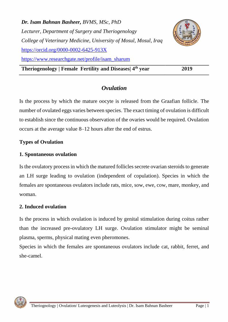
Structure of the Graafian follicle
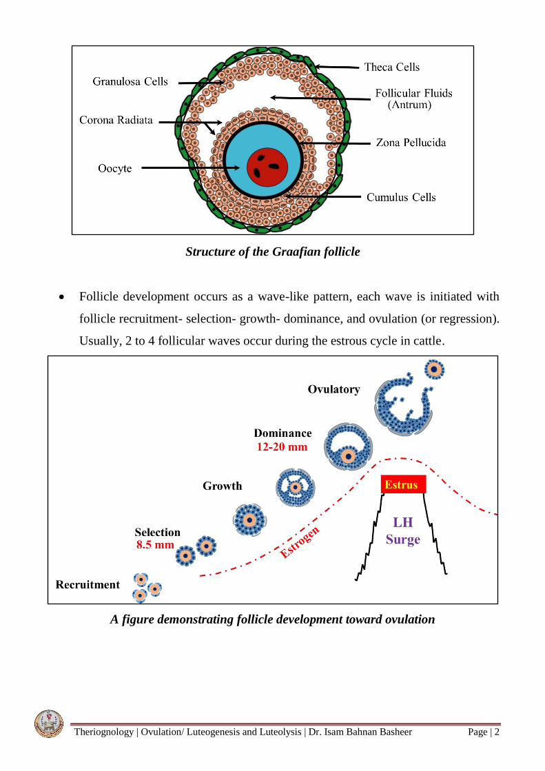
Follicle development occurs as a wave-like pattern, each wave is initiated with
follicle recruitment- selection- growth- dominance, and ovulation (or regression).
Usually, 2 to 4 follicular waves occur during the estrous cycle in cattle.
A figure demonstrating follicle development toward ovulation
Theriognology | Ovulation/ Luteogenesis and Luteolysis | Dr. Isam Bahnan Basheer Page | 3
The elevation in circulating concentrations of estradiol during the late follicular
phase leads to the preovulatory surge of LH.
Local regulation of ovulation involves the interaction of LH and intra-follicular
factors including steroids, prostaglandins, and peptides derived from endothelial
cells, leukocytes, fibroblasts, and steroidogenic cells.
The LH surge stimulates the process of ovulation by:
Activating an inflammatory reaction, which weakens and ruptures the follicle wall.
Initiates luteinization of the granulosa and theca interna cells of the follicle.
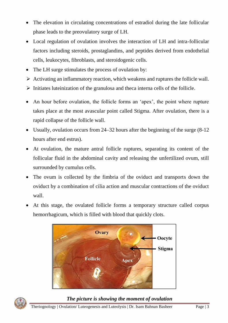
An hour before ovulation, the follicle forms an ‘apex’, the point where rupture
takes place at the most avascular point called Stigma. After ovulation, there is a
rapid collapse of the follicle wall.
Usually, ovulation occurs from 24–32 hours after the beginning of the surge (8-12
hours after end estrus).
At ovulation, the mature antral follicle ruptures, separating its content of the
follicular fluid in the abdominal cavity and releasing the unfertilized ovum, still
surrounded by cumulus cells.
The ovum is collected by the fimbria of the oviduct and transports down the
oviduct by a combination of cilia action and muscular contractions of the oviduct
wall.
At this stage, the ovulated follicle forms a temporary structure called corpus
hemorrhagicum, which is filled with blood that quickly clots.
The picture is showing the moment of ovulation
Theriognology | Ovulation/ Luteogenesis and Luteolysis | Dr. Isam Bahnan Basheer Page | 4
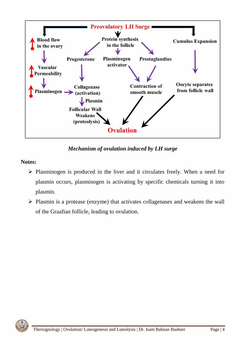
Mechanism of ovulation induced by LH surge
Notes:
Plasminogen is produced in the liver and it circulates freely. When a need for
plasmin occurs, plasminogen is activating by specific chemicals turning it into
plasmin.
Plasmin is a protease (enzyme) that activates collagenases and weakens the wall
of the Graafian follicle, leading to ovulation.
Theriognology | Ovulation/ Luteogenesis and Luteolysis | Dr. Isam Bahnan Basheer Page | 5
Luteogenesis and Luteolysis
The corpus luteum (CL) is a hard-yellow structure resulting from the ovulated follicle
as a temporary reproductive gland that produces and secretes progesterone responsible
for the maintenance of pregnancy.
LH is responsible for the transformation of granulosa and theca interna cells into lutein
cells (Luteinization) which secrete progesterone.
The CL is one of the most highly vascularized organs and receives the greatest rate of
blood flow per gram of tissue of any organ in the body. During the development of
CL, the blood flow surrounding an early corpus luteum gradually increases in parallel
with the increase in CL volume and plasma progesterone concentration.
The increased blood flow is closely associated with the potential to produce and release
of progesterone.
Luteolysis
Is the irreversible structural and functional degradation of the corpus luteum that
occurs at the end of the luteal phase leading to a considerable decrease in progesterone
production. The degraded corpus luteum forms a non-functional structure called
corpus albicans, that is filled with fibrous tissue.
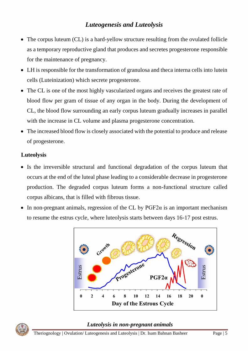
In non-pregnant animals, regression of the CL by PGF2α is an important mechanism
to resume the estrus cycle, where luteolysis starts between days 16-17 post estrus.
Luteolysis in non-pregnant animals
Theriognology | Ovulation/ Luteogenesis and Luteolysis | Dr. Isam Bahnan Basheer Page | 6
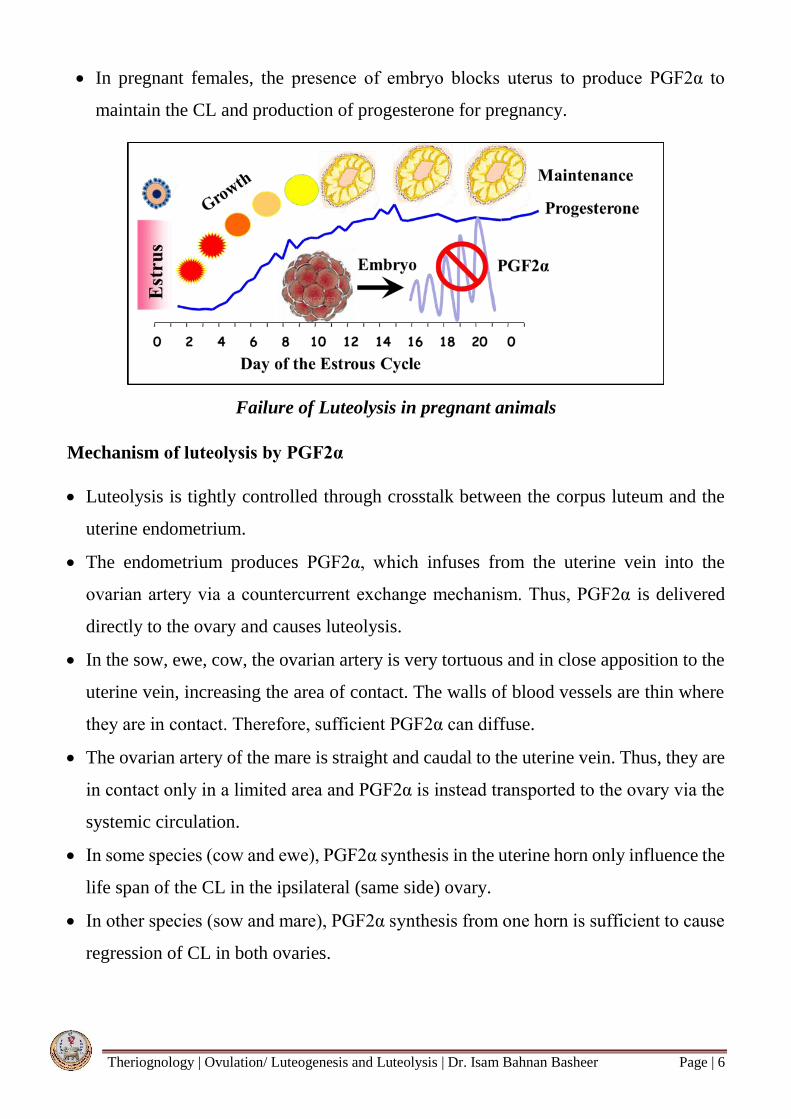
In pregnant females, the presence of embryo blocks uterus to produce PGF2α to
maintain the CL and production of progesterone for pregnancy.
Failure of Luteolysis in pregnant animals
Mechanism of luteolysis by PGF2α
Luteolysis is tightly controlled through crosstalk between the corpus luteum and the
uterine endometrium.
The endometrium produces PGF2α, which infuses from the uterine vein into the
ovarian artery via a countercurrent exchange mechanism. Thus, PGF2α is delivered
directly to the ovary and causes luteolysis.
In the sow, ewe, cow, the ovarian artery is very tortuous and in close apposition to the
uterine vein, increasing the area of contact. The walls of blood vessels are thin where
they are in contact. Therefore, sufficient PGF2α can diffuse.
The ovarian artery of the mare is straight and caudal to the uterine vein. Thus, they are
in contact only in a limited area and PGF2α is instead transported to the ovary via the
systemic circulation.
In some species (cow and ewe), PGF2α synthesis in the uterine horn only influence the
life span of the CL in the ipsilateral (same side) ovary.
In other species (sow and mare), PGF2α synthesis from one horn is sufficient to cause
regression of CL in both ovaries.

Theriognology | Ovulation/ Luteogenesis and Luteolysis | Dr. Isam Bahnan Basheer Page | 7
Countercurrent exchange of PGF2α
The process of luteolysis by PGF2α include:
Reduction of the blood flow in the corpus luteum.
The severe increase in luteal blood flow (day17 after estrus) is a key phenomenon of the
onset of luteal regression. However, PGF2α causes a rapid reduction in luteal blood
supply (day19 after estrus) which is one of the main luteolytic actions.
Blood flow surrounding the corpus luteum in non-pregnant cow

Theriognology | Ovulation/ Luteogenesis and Luteolysis | Dr. Isam Bahnan Basheer Page | 8
The direct action of PGF2α on luteal cells.
1. The continuous exposure of the uterus to progesterone during the diestrus period
causes downregulation of the progesterone receptors in the endometrium. This
allows estrogen (secreted from waves of follicle growth during the luteal phase)
to bind to its receptor leading to stimulation of the oxytocin receptor in the
endometrium.
2. At the end of the estrous cycle, the CL produces oxytocin, which stimulates the
uterus to produce uterine PGF2α, which in turn, stimulates the CL to produce more
oxytocin.
3. Oxytocin binds to its endometrial receptor, activating the synthesis and release of
PGF2α into the utero-ovarian vein.
4. PGF2α reaches the ovary via the countercurrent exchange mechanism. Thus,
PGF2α is directly delivered to the ovary and causes luteolysis.
Note:
In pregnant females, progesterone inhibits both the estrogen and oxytocin receptors
in the endometrium; where both have the potential to stimulate the release of PGF2α.
PR (Progesterone receptor); ES (Estrogen receptor); OR (Oxytocin receptor)
Mechanism of luteolysis drive-by PGF2α