Embed Size (px)
Citation preview
A Prospective Clinical Study on TitaniumImplants in the Zygomatic Arch for ProstheticRehabilitation of the Atrophic Edentulous Maxillawith a Follow-Up of 6 Months to 5 YearsCarlos Aparicio, DDS;*† Wafaa Ouazzani, DDS;* Roberto Garcia, DDS;‡ Xabier Arevalo, DDS;* Rosa Muela;*
Vanessa Fortes*
ABSTRACT
Background: Prosthetic rehabilitation with implant-supported prostheses in the atrophic edentulous maxilla often requiresa bone augmentation procedure to enable implant placement and integration. However, a rigid anchorage can also beachieved by using so-called zygomatic implants placed in the zygomatic arch in combination with regular implants placedin residual bone.
Purpose: The aim of the present study was to report on the clinical outcome of using zygomatic and regular implants forprosthetic rehabilitation of the severely atrophic edentulous maxilla.
Materials and Methods: Sixty-nine consecutive patients with severe maxillary atrophy were, during a 5-year period, treatedwith a total of 69 fixed full-arch prostheses anchored on 435 implants. Of these, 131 were zygomatic implants and 304were regular implants. Fifty-seven bridges were screw-retained and 12 were cemented. The screw-retained bridges wereremoved at the examination appointments and each implant was tested for mobility. In addition, the zygomatic implantswere subjected to Periotest® (Siemens AG, Bensheim, Germany) measurements. The patients had at the time of this reportbeen followed for at least 6 months up to 5 years in loading.
Results: Two regular implants failed during the study period giving a cumulative survival rate of 99.0%. None of the zygo-matic implants was removed. All patients received and maintained a fixed full-arch bridge during the study. Periotest meas-urements of zygomatic implants showed a decreased Periotest values value with time, indictating an increased stability.Three patients presented with sinusitis 14–27 months postoperatively, which could be resolved with antibiotics. Loosen-ing of the zygomatic implant gold screws was recorded in nine patients. Fracture of one gold screw as well as the pros-thesis occurred twice in one patient. Fracture of anterior prosthetic teeth was experienced in four patients.
Conclusions: The results from the present study show that the use of zygomatic and regular implants represents a pre-dictable alternative to bone grafting in the rehabilitation of the atrophic edentulous maxilla.
KEY WORDS: atrophy, clinical study, dental implants, edentulous maxilla, zygomatic arch
114
Prosthetic rehabilitation with implant-supported
dental bridges in the atrophic edentulous maxilla
constitutes a challenge for the treating team. The place-
ment of implants in such cases often results in a biome-
*Private practice, Clinica Aparicio, Barcelona, Spain; †research fellow,Department of Biomaterials, Institute for Surgical Sciences, Sahlgren-ska Academy, Göteborg University, Göteborg, Sweden; ‡private prac-tice, Valladolid, Spain
Reprint requests: Dr.Carlos Aparicio Magallón,Clínica Aparicio,Mitre72-74, 08017 Barcelona, Spain; e-mail: [email protected]
© 2006 Blackwell Publishing, Inc.
DOI 10.1111/j.1708-8208.2006.00009.x
chanically compromised situation due to the association
of risk factors like the use of short implants, the presence
of soft bone, and high loads in the posterior regions.1–3
Various bone augmentation techniques such as
sinus floor augmentation and onlay bone grafting have
been described with the common goal to enable place-
ment and integration of implants.4–6 To date, there still
remain doubts on the need and efficacy of sinus aug-
mentation techniques prior to implant placement. Much
of the available literature describing these techniques
lacks defined implant success and failure criteria,
descriptions of initial bone height, and standardized
Up to 5-Year Follow-Up of Zygomatic Implants 115
radiographic follow up.7–12 Moreover, our experience is
that patient acceptance is restricted due to the complex-
ity of the technique, possible donor site morbidity, and
higher costs. Further, most often the survival rate of
implants is on a 90% level9–11 and should be compared
to 95–98%, which is commonly reported for implants in
none-grafted cases.
The use of iliac crest bone grafts has been proposed
for the treatment of maxillae with severe atrophy, which
fall into classes D and E according to the classification
of Lekholm and Zarb.13 Follow-up studies on this tech-
nique using immediate or delayed implant placement
have reported failure rates in the range of 10–30%.13–16
Studies have reported problems with the placement
of implants in the maxillary tuberosity in a poor-quality
residual bone situation,17 while similar results have been
obtained on alveolar crests from other zones when uti-
lizing an adequate surgical technique for the individual-
ized preparation of the implant bed.2,18 Some authors
have suggested the use of the pterygo-maxillar suture as
an alternative site for implant placement.19–22 Implants
can be effectively harbored in the cortical bone of the
pterygoid process of the sphenoid bone and the pyram-
idal apophysis of the palatal bone, but this treatment
modality is associated with a potential risk of vascular
damage due to the presence of the descending maxillary
artery.23
The placement of implants in an angulated position
has been proposed to avoid the use of bone grafts.24–29
Aparicio and colleagues26 compared angulated (>15°)
and axially placed implants in the posterior maxilla
during a 3- to 7-year follow-up period. The results
showed no differences in the maintenance of the peri-
implant marginal bone height; they suggested that angu-
lated placement of implants can substitute most sinus
lift procedures.
The use of the zygomatic bone for anchorage of
long oral implants was originally developed by Bråne-
mark and colleagues and first described by Aparicio and
colleagues30 for rehabilitation of the atrophied maxillae.
In 1997, Weischer and colleagues31 cited the use of
implants in the zygoma as retaining elements after
hemimaxillectomy. Subsequently, Brånemark and col-
leagues32 presented a study with 77 patients and 156
implants, out of which 24 presented lengths were supe-
rior to the “standard model” and the rest responded to
a specific implant design. The cumulative success rate of
the zygomatic implants was 96.8%. No data of the pros-
theses outcome were reported. More recently, other
authors have reported good results on the use of
zygomatic implants to stabilize a fixed prostheses.33–35
However, because of the novelty of the technique,
there are insufficient prospective studies published that
endorse it. In this prospective study, we present the
preliminary results obtained by a task team on the use
of the zygomatic bone to provide anchorage for oral
implants used to completely rehabilitate the severely
atrophied maxilla.
MATERIALS AND METHODS
Patients
Sixty-nine consecutive patients (22 males and 47
females), aged between 38 and 82 years (average: 56
years) with severely atrophic edentulous (n = 63) or par-
tially dentate (n = 6) maxillae were included in the study.
Forty-five patients had an implant-supported bridge
and 24 patients had their natural dentition in the oppos-
ing mandible. Signs of occlusal abrasion were detected
in 18 patients. Twenty-seven patients smoked 20 or
more cigarettes per day.
The following were the inclusion criteria:
• The presence of residual alveolar crest with less than
4 mm in width and height, immediately distal to the
canine pillar
• The possibility to place a minimum of three
implants per quadrant
The exclusion criteria were general and local health con-
ditions that prevented the use of general anesthesia
and/or intraoral surgery.
Surgery
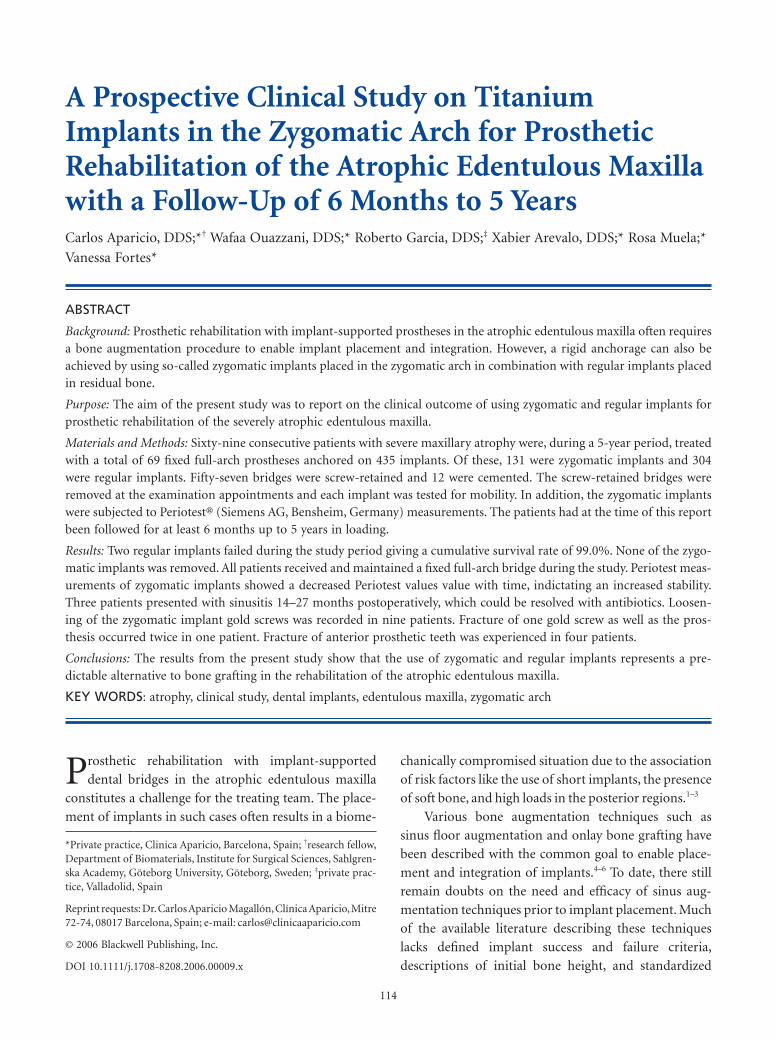
The presurgical radiographic examinations included
computerized tomography scans and orthopantomo-
grams (Figure 1).
Figure 1 Panoramic computerized tomography image of onecase showing severe maxillary atrophy.
116 Clinical Implant Dentistry and Related Research, Volume 8, Number 3, 2006
A total of 435 titanium implants were placed by the
same surgeon between November 1998 and February
2004. Three hundred four implants were regular plat-
form (RP) Brånemark System® (Nobel Biocare AB,
Göteborg, Sweden) with lengths that varied from 7 to
18 mm and diameters from 3.75 to 4 mm. One hundred
thirty-one implants were zygoma fixtures (Nobel
Biocare AB) with lengths from 35 to 52.5 mm (Table 1).
Of the RP implants, 220 were anchored in the
residual bone between the canine pillars including 15
implants that were placed in the anterior nasal spine
and 70 implants intentionally protruded from the nasal
cavity (Table 2). Eighty-four implants were placed in the
pterygoid process of the sphenoid bone and the pyram-
idal apophysis of the palate bone for anchorage. The 131
zygomatic implants were placed in the zygomatic bone.
All the zygomatic implants except five, which rotated,
achieved good primary stability at insertion time.
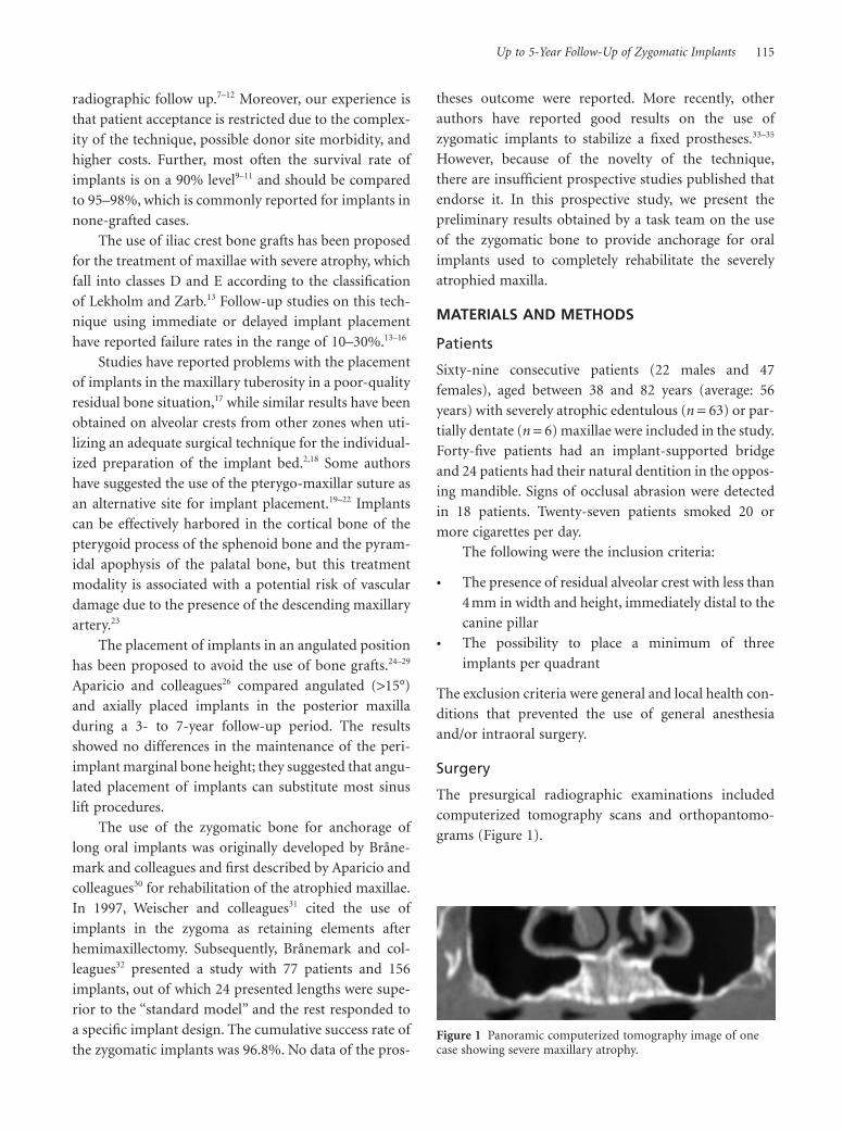
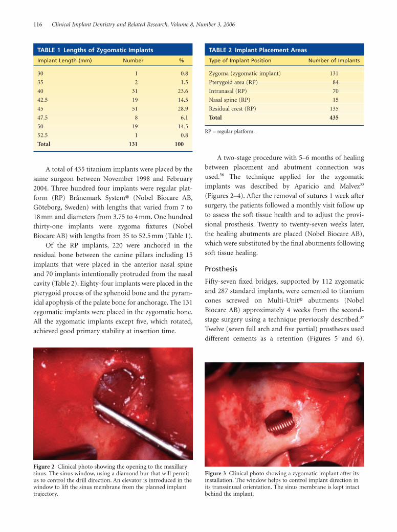
A two-stage procedure with 5–6 months of healing
between placement and abutment connection was
used.36 The technique applied for the zygomatic
implants was described by Aparicio and Malvez33
(Figures 2–4). After the removal of sutures 1 week after
surgery, the patients followed a monthly visit follow up
to assess the soft tissue health and to adjust the provi-
sional prosthesis. Twenty to twenty-seven weeks later,
the healing abutments are placed (Nobel Biocare AB),
which were substituted by the final abutments following
soft tissue healing.
Prosthesis
Fifty-seven fixed bridges, supported by 112 zygomatic
and 287 standard implants, were cemented to titanium
cones screwed on Multi-Unit® abutments (Nobel
Biocare AB) approximately 4 weeks from the second-
stage surgery using a technique previously described.37
Twelve (seven full arch and five partial) prostheses used
different cements as a retention (Figures 5 and 6).
TABLE 1 Lengths of Zygomatic Implants
Implant Length (mm) Number %
30 1 0.8
35 2 1.5
40 31 23.6
42.5 19 14.5
45 51 28.9
47.5 8 6.1
50 19 14.5
52.5 1 0.8
Total 131 100
TABLE 2 Implant Placement Areas
Type of Implant Position Number of Implants
Zygoma (zygomatic implant) 131
Pterygoid area (RP) 84
Intranasal (RP) 70
Nasal spine (RP) 15
Residual crest (RP) 135
Total 435
RP = regular platform.
Figure 2 Clinical photo showing the opening to the maxillarysinus. The sinus window, using a diamond bur that will permitus to control the drill direction. An elevator is introduced in thewindow to lift the sinus membrane from the planned implanttrajectory.
Figure 3 Clinical photo showing a zygomatic implant after itsinstallation. The window helps to control implant direction inits transsinusal orientation. The sinus membrane is kept intactbehind the implant.
Up to 5-Year Follow-Up of Zygomatic Implants 117
Forty-seven bridges were constructed with prefabricated
teeth over a rigid metal structure and the remaining 22
by sintered porcelain over metal (Figure 7, A and B).
Follow Up
The mean follow up was 25.1 months after placement
of the prosthesis with a minimum of 6 months. The
patients were scheduled for checkup 1, 3, 6, and 12
months after prosthesis delivery. After the first year of
function, the patients were examined every 6 months.
The screwed prostheses were removed on every
appointment. The checkup included assessments of oral
hygiene, soft tissue health, prosthesis stability, gold screw
loosening, and other mechanical complications. In addi-
tion, implant stability was evaluated individually on the
57 screwed prostheses using Periotest® (Siemens AG,
Bensheim, Germany) measurements. Standardized
intraoral x-rays of the zygomatic implants could not be
made, and consequently the implants could not be
evaluated with regard to marginal bone resorption. All
implants were classified as either failures or survivals
using the following definitions38,39:
• Failure – implants removed from the patient irre-
spective of the cause
• Survival – stable implant without signs of
pathology
Periotest Measurement
The stability of each implant, supporting a screwed
prostheses, was measured using the Periotest method
according to Olive and Aparicio.40 The measurements
were made at the day of bridge delivery, after 1, 3, and 2
months and thereafter annually. The implants were
measured individually aiming the tip of the device 2 mm
below the rim of the coronal platform. When measur-
ing during the follow-up period, the bridge was removed
to uncover the implant head or abutment. The Periotest
values (PTVs) obtained were compared to the PTV
established previously for implants of a 3.75-mm diam-
eter placed on the maxilla.
RESULTS
The postoperative phase included intense facial edema
and facial hematoma (six cases) that were resolved in 10
days; lip laceration (five cases) due to friction caused by
rotary surgical instruments; and parestesia occurring on
the cheek and paranasal zones (six cases), which sub-
sided 3–8 weeks postoperatively. Moderate nasal bleed-
ing was seen in seven cases for 1–3 days.
After an abutment connection of the zygomatic
implants, swelling of the palate mucosa surrounding the
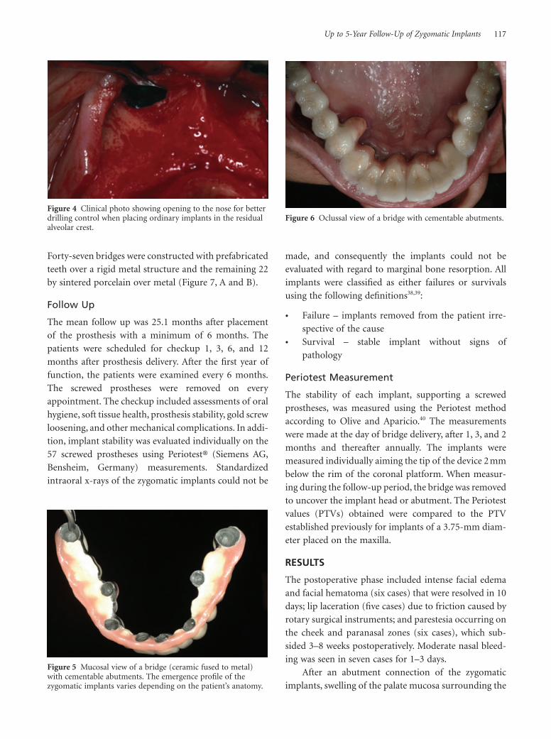
Figure 4 Clinical photo showing opening to the nose for betterdrilling control when placing ordinary implants in the residualalveolar crest.
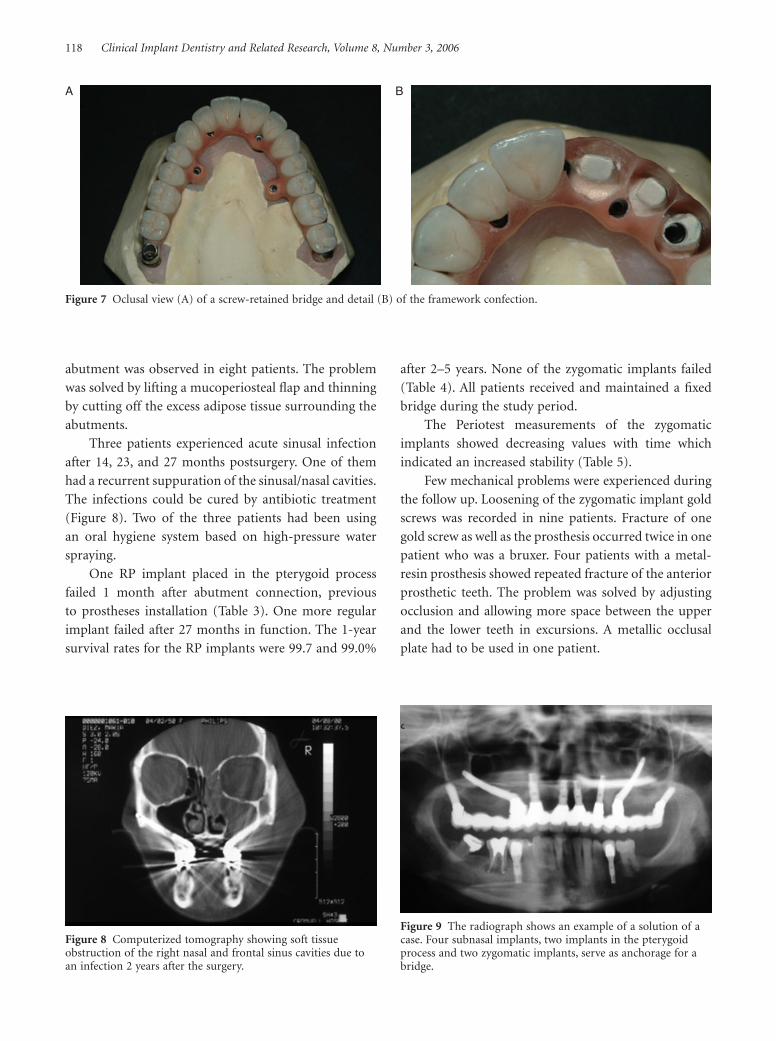
Figure 6 Oclussal view of a bridge with cementable abutments.
Figure 5 Mucosal view of a bridge (ceramic fused to metal)with cementable abutments. The emergence profile of thezygomatic implants varies depending on the patient’s anatomy.
118 Clinical Implant Dentistry and Related Research, Volume 8, Number 3, 2006
abutment was observed in eight patients. The problem
was solved by lifting a mucoperiosteal flap and thinning
by cutting off the excess adipose tissue surrounding the
abutments.
Three patients experienced acute sinusal infection
after 14, 23, and 27 months postsurgery. One of them
had a recurrent suppuration of the sinusal/nasal cavities.
The infections could be cured by antibiotic treatment
(Figure 8). Two of the three patients had been using
an oral hygiene system based on high-pressure water
spraying.
One RP implant placed in the pterygoid process
failed 1 month after abutment connection, previous
to prostheses installation (Table 3). One more regular
implant failed after 27 months in function. The 1-year
survival rates for the RP implants were 99.7 and 99.0%
after 2–5 years. None of the zygomatic implants failed
(Table 4). All patients received and maintained a fixed
bridge during the study period.
The Periotest measurements of the zygomatic
implants showed decreasing values with time which
indicated an increased stability (Table 5).
Few mechanical problems were experienced during
the follow up. Loosening of the zygomatic implant gold
screws was recorded in nine patients. Fracture of one
gold screw as well as the prosthesis occurred twice in one
patient who was a bruxer. Four patients with a metal-
resin prosthesis showed repeated fracture of the anterior
prosthetic teeth. The problem was solved by adjusting
occlusion and allowing more space between the upper
and the lower teeth in excursions. A metallic occlusal
plate had to be used in one patient.
A B
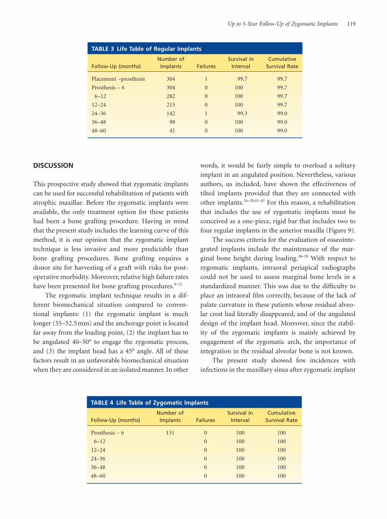
Figure 7 Oclusal view (A) of a screw-retained bridge and detail (B) of the framework confection.
Figure 8 Computerized tomography showing soft tissueobstruction of the right nasal and frontal sinus cavities due toan infection 2 years after the surgery.
Figure 9 The radiograph shows an example of a solution of acase. Four subnasal implants, two implants in the pterygoidprocess and two zygomatic implants, serve as anchorage for abridge.
Up to 5-Year Follow-Up of Zygomatic Implants 119
DISCUSSION
This prospective study showed that zygomatic implants
can be used for successful rehabilitation of patients with
atrophic maxillae. Before the zygomatic implants were
available, the only treatment option for these patients
had been a bone grafting procedure. Having in mind
that the present study includes the learning curve of this
method, it is our opinion that the zygomatic implant
technique is less invasive and more predictable than
bone grafting procedures. Bone grafting requires a
donor site for harvesting of a graft with risks for post-
operative morbidity. Moreover, relative high failure rates
have been presented for bone grafting procedures.9–11
The zygomatic implant technique results in a dif-
ferent biomechanical situation compared to conven-
tional implants: (1) the zygomatic implant is much
longer (35–52.5 mm) and the anchorage point is located
far away from the loading point, (2) the implant has to
be angulated 40–50° to engage the zygomatic process,
and (3) the implant head has a 45° angle. All of these
factors result in an unfavorable biomechanical situation
when they are considered in an isolated manner. In other
words, it would be fairly simple to overload a solitary
implant in an angulated position. Nevertheless, various
authors, us included, have shown the effectiveness of
tilted implants provided that they are connected with
other implants.24–29,41–43 For this reason, a rehabilitation
that includes the use of zygomatic implants must be
conceived as a one-piece, rigid bar that includes two to
four regular implants in the anterior maxilla (Figure 9).
The success criteria for the evaluation of osseointe-
grated implants include the maintenance of the mar-
ginal bone height during loading.38–39 With respect to
zygomatic implants, intraoral periapical radiographs
could not be used to assess marginal bone levels in a
standardized manner. This was due to the difficulty to
place an intraoral film correctly, because of the lack of
palate curvature in these patients whose residual alveo-
lar crest had literally disappeared, and of the angulated
design of the implant head. Moreover, since the stabil-
ity of the zygomatic implants is mainly achieved by
engagement of the zygomatic arch, the importance of
integration in the residual alveolar bone is not known.
The present study showed few incidences with
infections in the maxillary sinus after zygomatic implant
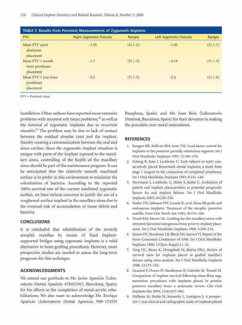
TABLE 3 Life Table of Regular Implants
Number of Survival in CumulativeFollow-Up (months) Implants Failures Interval Survival Rate
Placement –prosthesis 304 1 99.7 99.7
Prosthesis – 6 304 0 100 99.7
6–12 282 0 100 99.7
12–24 215 0 100 99.7
24–36 142 1 99.3 99.0
36–48 98 0 100 99.0
48–60 41 0 100 99.0
TABLE 4 Life Table of Zygomatic Implants
Number of Survival in CumulativeFollow-Up (months) Implants Failures Interval Survival Rate
Prosthesis – 6 131 0 100 100
6–12 0 100 100
12–24 0 100 100
24–36 0 100 100
36–48 0 100 100
48–60 0 100 100
120 Clinical Implant Dentistry and Related Research, Volume 8, Number 3, 2006
installation. Other authors have reported more extensive
problems with intraoral soft tissue problems,44 as well as
the removal of zygomatic implants due to recurrent
sinusitis.45 The problem may be due to lack of contact
between the residual alveolar crest and the implant,
thereby creating a communication between the oral and
sinus cavities. Since the zygomatic implant situation is
unique with parts of the implant exposed to the maxil-
lary sinus, controlling of the health of the maxillary
sinus should be part of the maintenance program. It can
be anticipated that the relatively smooth machined
surface is to prefer in this environment to minimize the
colonization of bacteria. According to the reported
100% survival rate of the current machined zygomatic
surface, we have serious concerns to justify the use of a
roughened-surface implant in the maxillary sinus due to
the eventual risk of accumulation of tissue debris and
bacteria.
CONCLUSIONS
It is concluded that rehabilitation of the severely
atrophic maxillae by means of fixed implant-
supported bridges using zygomatic implants is a valid
alternative to bone grafting procedures. However, more
prospective studies are needed to assess the long-term
prognosis for this technique.
ACKNOWLEDGMENTS
We extend our gratitude to Mr. Javier Aparicio (Labo-
ratorio Dental Aparicio 678435367, Barcelona, Spain)
for his efforts in the completion of metal-acrylic reha-
bilitations. We also want to acknowledge Mr. Enrique
Aparicio (Laboratorio Dental Aparicio, 948–174555
Pamplona, Spain) and Mr. Joan Boix (Laboratorio
Dentinal, Barcelona, Spain) for their devotion in making
the porcelain over metal restorations.
REFERENCES
1. Rangert BR, Sullivan RM, Jemt TM. Load factor control for
implants in the posterior partially edentulous segment. Int J
Oral Maxillofac Implants 1997; 12:360–370.
2. Friberg B, Jemt J, Leckholm U. Early failures in 4,641 con-
secutively placed Branemark dental implants: a study from
stage 1 surgery to the connection of completed prostheses.
Int J Oral Maxillofac Implants 1991; 6:141–146.
3. Herrmann I, Leckholm U, Holm S, Kultje C. Evaluation of
patient and implant characteristics as potential prognostic
factors for oral implant failures. Int J Oral Maxillofac
Implants 2005; 20:220–230.
4. Smiler DG, Johnson PW, Lozada JL, et al. Sinus lift grafts and
endosseous implants. Treatment of the atrophic posterior
maxilla. Dent Clin North Am 1992; 36:151–186.
5. Wood RM, Moore DL. Grafting for the maxillary sinus with
intraoral harvested autogenous bone prior to implant place-
ment. Int J Oral Maxillofac Implants 1988; 3:209–214.
6. Jensen OT, Shoulman LB, Block MS, Iacono VJ. Report of the
Sinus Consensus Conference of 1996. Int J Oral Maxillofac
Implants 1998; 13(Spec Suppl):11–32.
7. Tong DC, Rioux K, Drangsholt M, Bierne ORA. Review of
survival rates for implants placed in grafted maxillary
sinuses using meta-analysis. Int J Oral Maxillofac Implants
1998; 13:175–182.
8. Graziani F, Donos N, Needleman N, Gabriele M, Tonetti M.
Comparison of implant survival following sinus floor aug-
mentation procedures with implants placed in pristine
posterior maxillary bone: a systematic review. Clin Oral
Implants Res 2004; 15(6):677–682.
9. Hallman M, Hedin M, Sennerby L, Lundgren S. A prospec-
tive 1-year clinical and radiographic study of implants placed
TABLE 5 Results from Periotest Measurements of Zygomatic Implants
PTV Right Zygomatic Fixtures Ranges Left Zygomatic Fixtures Ranges
Mean PTV upon −1.95 (4) (−6) −1.95 (3) (−7)
abutment
placement
Mean PTV 1 month −1.7 (5) (−5) −2.18 (5) (−5)
from prostheses
placement
Mean PTV 1 year from −3.5 (3) (−5) −2.4 (3) (−6)
prostheses
placement
PTV = Periotest value.
Up to 5-Year Follow-Up of Zygomatic Implants 121
after maxillary sinus floor augmentation with bovine
hydroxyapatite and autogenous bone. J Oral Maxillofac Surg
2002; 60(3):277–284.
10. Raghoebar GM, Timmenga NM, Vissink A. Maxillary bone
grafting for insertion of endosseous implants: results after
12–124 months. Clin Oral Implants Res COIR 2001; 12(3):
279–286.
11. Wallace SS, Froum SJ. Effect of maxillary sinus augmentation
on the survival of endosseous dental implants. A systematic
review. Ann Periodontol 2003; 8(1):328–343.
12. Regev E, Smith RA, Perrot DH, Pogrel MA. Maxillary sinus
complications related to endosseous implants. Int J Oral
Maxillofac Implants 1995; 10:451–461.
13. Lekholm U, Zarb GA. Patient selection and preparation.
In: Brånemark P-I, Zarb GA, Albrektsson T, eds. Tissue-
integrated prostheses: osseointegration in clinical dentistry.
Chicago, IL: Quintessence, 1985:199–209.
14. Collins TA, Brown GK, Johnson N, Massey JA, Nunn BD.
Team management of atrophic edentulous with autogenous
inlay, veneer, and split grafts and endosseous implants: case
reports. Quintessence Int 1995; 26(2):79–93.
15. Keller EE. Reconstruction of the severely atrophic
edentulous mandible with endosseous implants: a 10-year
longitudinal study. J Oral Maxillofac Surg 1995; 53:317–318.
16. Widmark G, Andersson B, Andrup B, Carlsson GE, Ivanoff
CJ, Lindvall AM. Rehabilitation of patients with severely
resorbed maxillae by means of implants with or without
bone grafts: a 1-year follow-up study. Int J Oral Maxillofac
Implants 1998; 13:474–482.
17. Jaffin RA, Berman CL. The excessive loss of Branemark fix-
tures in type IV bone: a 5-year analysis. J Periodontol 1991;
62:2–4.
18. Lekholm U. The Brånemark implant technique: a standard-
ized procedure under continuous development. In: Laney
WR, Tolman DE, eds. Tissue integration in oral, orthopedic,
and maxillofacial reconstruction. Chicago, IL: Quintessence,
1992:194–199.
19. Tulasne JF. Osseointegrated fixtures in the pterygoid region.
In: Worthington P, Branemark PL, eds. An advanced osseoin-
tegration surgery: applications in the maxillofacial region.
Chicago, IL: Quintessence, 1992:182–188.
20. Raspall G, González J, Bescós S, Hueto JA. Pterigomaxillary
osseointegrated fixture. J Craniomaxillofac Surg 1992; 20:
57–58. (Abstr).
21. Graves SL. The pterigoid plate implant: a solution for restor-
ing the posterior maxilla. Int J Periodontics Restorative Dent
1994; 14:513–523.
22. Fernández Valerón J, Fernández Velázquez J. Placement
of screw type implants in the pterigomaxillary pyramidal
region: surgical procedure and preliminary results. Int J Oral
Maxillofac Implants 1997; 12(6):814–819.
23. Choi J, Park HS. The clinical anatomy of the maxillary artery
in the pterygopalatine fossa. J Oral Maxillofac Surg 2003;
61:72–78.
24. Mattsson T, Köndell P-A, Gynther GW, Fredholm U, Bolin
A. Implant treatment without bone grafting in severely
resorbed edentulous maxillae. J Oral Maxillofac Surg 1999;
57:281–287.
25. Krekmanov L, Kahn M, Rangert B, Lindström H. Tilting of
posterior mandibular and maxillary implants for improved
prosthesis support. Int J Oral Maxillofac Implants 2000;
15:405–414.
26. Aparicio C, Perales P, Rangert B. Tilted implants as an alter-
native to maxillary sinus grafting. J Clin Implant Dent Relat
Res 2001; 3(1):39–49.
27. Aparicio C, Arévalo JX, Ouazzani W, Granados C. Retro-
spective clinical and radiographic evaluation of tilted
implants used in the treatment of the severely resorbed
edentulous maxilla. Applied Osseo Res 2002; 3:17–
21.
28. Fortin Y, Sullivan RM, Rangert BR. The Marius implant
bridge: surgical and prosthetic rehabilitation for the
completely edentulous upper jaw with moderate to severe
resorption: a 5-year retrospective clinical study. Clin Implant
Dent Relat Res 2002; 4:69–77.
29. Calandriello R, Tomates M. Simplified treatment of the
atrophic posterior maxilla via immediate/early function and
tilted implants: a prospective 1-year clinical study. J Clin
Implant Dent Relat Res 2005; 7(Suppl 1):51–62.
30. Aparicio C, Branemark PI, Keller EE, Olive J. Reconstruction
of the premaxilla with autogenous iliac bone in combination
with osseointegrated implants. Int J Oral Maxillofac
Implants 1993; 8:61–67.
31. Weischer T, Schettler D, Mohr CH. Titanium implants in the
zygoma as retaining elements after hemimaxillectomy. Int J
Oral Maxillofac Implants 1997; 12:211–221.
32. The zygomaticus fixture: clinical procedures Nobel Biocare.
Göteborg, Sweden: Nobel-Biocare®, 1998.
33. Aparicio C, Malevez CH. El implante trans–zigomático.
Consejo General de Colegios y Odontólogos y Estomatólo-
gos de España. RCOE 1999; 4:171–184.
34. Bedrossian E, Stumpel LJ. The zygomatic implant; prelimi-
nary data on treatment of severely resorbed maxillae. A
clinical report. Int J Oral Maxillofac Implants 2002; 17:
861–865.
35. Malevez C, Abarca M, Durdu F, Daelemans P. Clinical
outcome of 103 consecutive zygomatic implants: a 6–48
months follow-up study. Clin Oral Implants Res 2004;
15:18–22.
36. Adell R, Lekholm U, Branemark P-I. A 15 year study of
osseointegrated implants in the treatment of the edentulous
jaw. Int J Oral Surg 1981; 10:27–41.
37. Aparicio C. A new method to regularly achieve passive fit of
ceramo-metal prostheses over Branemark osseointegrated
implants: a two-year report. Int J Periodontics Restorative
Dent 1994; 14:405–419.
38. Albrektsson T, Isidor F. Criteria for success and failure
of an implant system. Consensus report. In: Lang NP,
122 Clinical Implant Dentistry and Related Research, Volume 8, Number 3, 2006
42. Rangert B, Krogh PHJ, Langer B, Van Roekel N. Bending
overload and implant fracture: a retrospective clinical
analysis. Int J Oral Maxillofac Implants 1995; 10:326–334.
43. Clelland NL, Gilat A, McGlumphy EA, Brantley WA. A pho-
toelastic and strain gauge analysis of angled abutments for
an implant system. Int J Oral Maxillofac Implants 1993;
8:541–548.
44. Al-Nawas B, Wegener J, Bender C, Wagner W. Critical soft
tissue parameters of the zygomatic implant. J Clin Peri-
odontol 2004; 31:497–500.
45. Becktor JP, Isaksson S, Abrahamsson P, Sennerby L. Evalua-
tion of 31 zygomatic implants and 74 regular dental implants
used in 16 patients for prosthetic reconstruction of the
atrophic maxilla with cross-arch fixed bridges. Clin Implant
Dent Relat Res 2005; 7:159–165.
Karring T, eds. Proceedings of the 1st European Workshop
on Peridontology. Chicago, IL: Quintessence, 1994:243–
244.
39. Albrektsson T, Zarb G, Worthington P, Erikson AR. The
long-term efficacy of currently used dental implants. A
review proposed criteria of success. Int J Oral Maxillofac
Implants 1986; 1:11–25.
40. Olive J, Aparicio C. The Periotest method as a measure of
osseointegrated oral implant stability. Int J Oral Maxillofac
Implants 1990; 5:390–400.
41. Celletti R, Pameijer CH, Bracchetti G, Donath K, Persichetti
G, Visani Y. Histologic evaluation of osseointegrated
implants restored in nonaxial functional occlusion with pre-
angled abutments. Int J Periodontics Restorative Dent 1995;
15:563–573.