Embed Size (px)
Citation preview
© 2016 Nikolis et al. This work is published and licensed by Dove Medical Press Limited. The full terms of this license are available at https://www.dovepress.com/terms. php and incorporate the Creative Commons Attribution – Non Commercial (unported, v3.0) License (http://creativecommons.org/licenses/by-nc/3.0/). By accessing the work
you hereby accept the Terms. Non-commercial uses of the work are permitted without any further permission from Dove Medical Press Limited, provided the work is properly attributed. For permission for commercial use of this work, please see paragraphs 4.2 and 5 of our Terms (https://www.dovepress.com/terms.php).
Chronic Wound Care Management and Research 2016:3 101–111
Chronic Wound Care Management and Research Dovepress
submit your manuscript | www.dovepress.com
Dovepress 101
C l i n i C a l T R i a l R e p o RT
open access to scientific and medical research
Open Access Full Text Article
http://dx.doi.org/10.2147/CWCMR.S104391
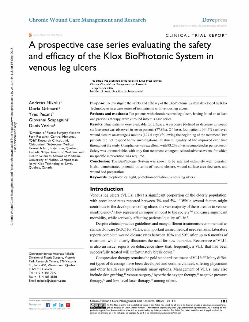
a prospective case series evaluating the safety and efficacy of the Klox BioPhotonic System in venous leg ulcers
andreas nikolis1
Doria Grimard2
Yves pesant3
Giovanni Scapagnini4
Denis Vézina5
1Division of Plastic Surgery, Victoria Park Research Centre, Montreal, 2Q&T Research Chicoutimi, Chicoutimi, 3St-Jerome Medical Research Inc., St-Jerome, Quebec, Canada; 4Department of Medicine and Health Sciences, School of Medicine, University of Molise, Campobasso, italy; 5Klox Technologies, Laval, Quebec, Canada
Purpose: To investigate the safety and efficacy of the BioPhotonic System developed by Klox
Technologies in a case series of ten patients with venous leg ulcers.
Patients and methods: Ten patients with chronic venous leg ulcers, having failed on at least
one previous therapy, were enrolled into this case series.
Results: Nine patients were evaluable for efficacy. A response (defined as decrease in wound
surface area) was observed in seven patients (77.8%). Of these, four patients (44.4%) achieved
wound closure on average 4 months (127.5 days) following the beginning of the treatment. Two
patients did not respond to the investigational treatment. Quality of life improved over time
throughout the study. Compliance was excellent, with 93.2% of visits completed as per protocol.
Safety was unremarkable, with only four treatment-emergent-related adverse events, for which
no specific intervention was required.
Conclusion: The BioPhotonic System was shown to be safe and extremely well tolerated.
It also demonstrated potential in terms of wound closure, wound surface area decrease, and
wound bed preparation.
Keywords: biophotonics, light, photobiomodulation, venous leg ulcers
IntroductionVenous leg ulcers (VLUs) affect a significant proportion of the elderly population,
with prevalence rates reported between 3% and 5%.1–3 While several factors might
contribute to the development of leg ulcers, the vast majority of these are due to venous
insufficiency.4 They represent an important cost to the society5,6 and cause significant
morbidity, while seriously affecting patients’ quality of life.3
Despite clinical practice guidelines and many different treatments recommended as
standard of care (SOC) for VLUs, an important unmet medical need remains. Literature
reports complete wound closure rates between 10% and 50% after up to 6 months of
treatment, which clearly illustrates the need for new therapies. Recurrence of VLUs
is also an issue; reports on dehiscence show that, frequently, a VLU that had been
successfully treated will unfortunately break down.7
Compression therapy remains the gold standard treatment of VLUs.8,9 Many differ-
ent types of dressings have been developed and commercialized, offering physicians
and other health care professionals many options. Management of VLUs may also
include skin grafting,10 venous surgery,7 hyperbaric oxygen therapy,11 negative pressure
therapy,12 and low-level laser therapy,13 among others.
Correspondence: andreas nikolisDivision of Plastic Surgery, Victoria Park Research Centre, 376 Victoria St., Suite 400, Westmount, Quebec, H3Z1C3, CanadaTel +1 514 488 7722Fax +1 514 488 3830email [email protected]
Journal name: Chronic Wound Care Management and ResearchArticle Designation: CLINICAL TRIAL REPORTYear: 2016Volume: 3Running head verso: Nikolis et alRunning head recto: Biophotonic treatment of venous leg ulcersDOI: http://dx.doi.org/10.2147/CWCMR.S104391
C
hron
ic W
ound
Car
e M
anag
emen
t and
Res
earc
h do
wnl
oade
d fr
om h
ttps:
//ww
w.d
ovep
ress
.com
/ by
24.1
14.4
4.11
5 on
16-
Sep
-201
6F
or p
erso
nal u
se o
nly.
Powered by TCPDF (www.tcpdf.org)
1 / 1
Chronic Wound Care Management and Research 2016:3submit your manuscript | www.dovepress.com
Dovepress
Dovepress
102
nikolis et al
Klox Technologies has developed a unique medical
device composed of a gel and a multi-light emitting diode
(LED), which is known to be extremely well tolerated and
efficient in the treatment of acne vulgaris. Preclinical data
also demonstrated many positive attributes of this system,
which was investigated in this case series for the manage-
ment of VLUs.
Materials and methodsExperimental designAn open-label study design was used to investigate the safety
and efficacy of the Klox BioPhotonic System (KBS) in patients
with VLUs. Patients were recruited from three different
sites. Protocol was approved by Health Canada as well as the
Optimum Clinical Research Inc., St-Jerome Hospital Ethics
Review Board (NCT02222467). Patients were first screened
for eligibility criteria and within 2 weeks started the experi-
mental treatment. The BioPhotonic System was administered
twice weekly for 8 weeks and then the investigators could,
depending on patients’ response and their clinical judgment,
either decrease the treatment regimen to once weekly, stay as
is (twice weekly), increase to three times per week or give a
treatment-pause of 1 or 2 weeks. Patients were treated for a
maximum period of 16 weeks or until wound closure. Once
their wound closed, the patients were seen again three times
over an 8-week period to confirm persistence of wound clo-
sure. In all cases, the BioPhotonic System was administered in
addition to SOC. When administered more than once weekly, a
minimum interval of 3 days was required between treatments.
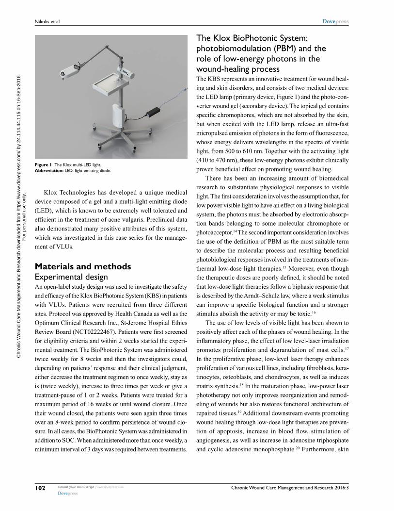
The Klox BioPhotonic System: photobiomodulation (PBM) and the role of low-energy photons in the wound-healing processThe KBS represents an innovative treatment for wound heal-
ing and skin disorders, and consists of two medical devices:
the LED lamp (primary device, Figure 1) and the photo-con-
verter wound gel (secondary device). The topical gel contains
specific chromophores, which are not absorbed by the skin,
but when excited with the LED lamp, release an ultra-fast
micropulsed emission of photons in the form of fluorescence,
whose energy delivers wavelengths in the spectra of visible
light, from 500 to 610 nm. Together with the activating light
(410 to 470 nm), these low-energy photons exhibit clinically
proven beneficial effect on promoting wound healing.
There has been an increasing amount of biomedical
research to substantiate physiological responses to visible
light. The first consideration involves the assumption that, for
low power visible light to have an effect on a living biological
system, the photons must be absorbed by electronic absorp-
tion bands belonging to some molecular chromophore or
photoacceptor.14 The second important consideration involves
the use of the definition of PBM as the most suitable term
to describe the molecular process and resulting beneficial
photobiological responses involved in the treatments of non-
thermal low-dose light therapies.15 Moreover, even though
the therapeutic doses are poorly defined, it should be noted
that low-dose light therapies follow a biphasic response that
is described by the Arndt–Schulz law, where a weak stimulus
can improve a specific biological function and a stronger
stimulus abolish the activity or may be toxic.16
The use of low levels of visible light has been shown to
positively affect each of the phases of wound healing. In the
inflammatory phase, the effect of low level-laser irradiation
promotes proliferation and degranulation of mast cells.17
In the proliferative phase, low-level laser therapy enhances
proliferation of various cell lines, including fibroblasts, kera-
tinocytes, osteoblasts, and chondrocytes, as well as induces
matrix synthesis.18 In the maturation phase, low-power laser
phototherapy not only improves reorganization and remod-
eling of wounds but also restores functional architecture of
repaired tissues.19 Additional downstream events promoting
wound healing through low-dose light therapies are preven-
tion of apoptosis, increase in blood flow, stimulation of
angiogenesis, as well as increase in adenosine triphosphate
and cyclic adenosine monophosphate.20 Furthermore, skin
Figure 1 The Klox multi-LED light.Abbreviation: LED, light emitting diode.
C
hron
ic W
ound
Car
e M
anag
emen
t and
Res
earc
h do
wnl
oade
d fr
om h
ttps:
//ww
w.d
ovep
ress
.com
/ by
24.1
14.4
4.11
5 on
16-
Sep
-201
6F
or p
erso
nal u
se o
nly.
Powered by TCPDF (www.tcpdf.org)
1 / 1
Chronic Wound Care Management and Research 2016:3 submit your manuscript | www.dovepress.com
Dovepress
Dovepress
103
Biophotonic treatment of venous leg ulcers
exposure to low-level light treatment may start a series of
biochemical reactions that result in the production of nitric
oxide, a vasodilator and powerful pain reliever, and an anti-
inflammatory agent.21
Another important factor activated by PBM mechanisms
is the transforming growth factor-β. Transforming growth
factor-β plays a crucial role in the proliferation, resolution,
and remodeling of the wound tissue by promoting keratino-
cytes, endothelial and fibroblast cell migration.22 It has also
been shown that PBM events may also regulate vascular
endothelial growth factor, enhancing the formation of new
vessels and improving the healing of skin.23 In summary, light
treatments can lead to modulation of transcription factors
capable of coordinating a wide range of beneficial responses
in wound healing.
Different clinical studies have been conducted to inves-
tigate low-energy light treatments in the cure of several skin
conditions, including rejuvenation of photoaged skin, acne,
skin inflammation, and wound healing.24–27 Specifically,
a PBM approach has shown promising efficacy in VLU
treatment.28–30
The photo-converter wound gel is presented in two jars,
which have to be mixed together just prior to application.
Typically, upon evaluation, any excess fibrin or necrotic tis-
sue was debrided. The wound was first cleansed with normal
saline and then a 2 mm thick layer of the gel was applied on
the VLU. The wound was then illuminated with the multi-
LED light for 5 minutes. Once the treatment was completed,
the gel was removed from the wound with gentle saline irri-
gation. A nonadherent dressing was then applied to prevent
any contact between the wound and the external environment.
Local SOC was then followed: sharp debridement if excess
of fibrin, cleansing with normal saline water, compression
bandage systems, management of a moist wound environment
with nonadherent dressings, management of wound infection,
and nutritional assessment.
imagery systemAt each study visit, images of the VLUs were taken with the
Aranz Silhouette™ Star System (Aranz Medical, Christ-
church, New Zealand) . This system allows digital pictures
to be taken and offers the calculation of the wound surface
area, perimeter, volume, average, and maximum depth.
participantsPatients had to fulfill all of the following (inclusion) criteria
to be eligible for inclusion into the study: 1) male or female
18 years of age and older; 2) the patients or legal guardian
must have signed an informed consent form; 3) female of
childbearing potential must have a negative pregnancy test
result at baseline, and both male and female patients must be
willing to adhere to a medically accepted birth control method
during the course of the study; 4) willingness to return for all
study visits; 5) proven VLU, clinically defined and confirmed
by duplex, refilling time or venous hypertension; 6) open
VLU for >4 weeks prior to study entry; 7) ulcer surface area
between 5 and 100 cm2 inclusive, with a maximum depth of
1 cm; the maximum diameter of the wound could not exceed
10 cm; 8) wound surface area has not changed by more than
±30% between screening and first study visits; 9) adequate
blood arterial perfusion (ankle–brachial index between 0.7
and 1.3, inclusive).
Patients who met any one of the following (exclusion)
criteria on screening visit were not eligible to participate in
the study: 1) VLU present for >12 months; 2) the ulcer to be
treated was planned for operative debridement; 3) the ulcer
has significant necrotic tissue (eg, >20% of the ulcer surface
area); 4) major uncontrolled medical disorder(s) such as seri-
ous cardiovascular, renal, liver or pulmonary disease, lupus,
palliative care, or sickle cell anemia; 5) severe or significant
hypoalbuminemia (albuminemia <30 g/L, and/or pre-albumin
<5 mg/dL, or hypoproteinemia [proteinemia <55 g/L]); 6)
patients with moderate-to-severe anemia (Hb <90 g/L); 7)
patients currently treated for an active malignant disease;
8) patients with history of malignancy within the wound; 9)
patients with history of radiation therapy to the wound region;
10) patients with prior diagnosis of active malignant disease
who have been disease-free for less than 1 year; 11) patients
with a known osteomyelitis or active cellulitis; 12) patients
who are immunosuppressed or on high-dose chronic steroid
use; 13) patients on systemic corticosteroids (a completion of
corticosteroid course at least 30 days prior to study enrolment
is required); 14) patients with active or systemic infection
(patients could be eligible for rescreening after the systemic
infection has subsided); 15) successful revascularization
surgery of the leg with the ulcer to be treated <8 weeks prior
to screening; 16) patients with severely uncontrolled diabetes
mellitus (defined as A1C >12%); 17) Raynaud disease or
other severe peripheral microvascular disease; 18) dermato-
logic comorbid disease (eg, cutis laxa or collagen vascular
disease); 19) active bleeding; 20) pregnancy, or breast-
feeding; 21) patients with bleeding diathesis; 22) patients
on warfarin or intravenous (IV) heparin; 23) patients having
any physical or psychiatric condition that in the investigator’s
opinion would warrant exclusion from the study or prevent
the patients from completing the study (eg, severe morbid
C
hron
ic W
ound
Car
e M
anag
emen
t and
Res
earc
h do
wnl
oade
d fr
om h
ttps:
//ww
w.d
ovep
ress
.com
/ by
24.1
14.4
4.11
5 on
16-
Sep
-201
6F
or p
erso
nal u
se o
nly.
Powered by TCPDF (www.tcpdf.org)
1 / 1
Chronic Wound Care Management and Research 2016:3submit your manuscript | www.dovepress.com
Dovepress
Dovepress
104
nikolis et al
obesity, recent hip fracture, and suspected noncompliance);
24) patients with ulcers from burns (from exposure to high
heat), pressure ulcers, or diabetic foot ulcers; 25) concurrent
disease or drug(s) known to induce severe photosensitivity
of the skin, such as porphyria; 26) patients have received
biologically based therapy in any wound within 3 months of
screening; 27) concurrent participation in another clinical
trial involving an investigational drug or device that would
interfere with this study; 28) previous participation in another
interventional wound healing clinical investigation within the
last 60 days prior to screening.
Quality of lifeThe Cardiff Wound Impact Schedule® was used to assess
the impact of the experimental treatment on patients’ quality
of life. It was administered at baseline, week 8, the end of
treatment period, as well as the end of the follow-up period.
EfficacyEfficacy was assessed through the following parameters:
1) rate of complete wound closure, 2) time to complete wound
closure, 3) incidence of wound breakdown, 4) wound surface
area reduction over time, 5) wound volume reduction over
time, and 6) health-related quality of life.
SafetySafety was documented via the collection of 1) adverse
events and serious adverse events, 2) device incidents,
3) visual analog scale for pain, 4) clinical laboratory
parameters, 5) vital signs, 6) physical examinations, 7) per-
centage of patients with clinical infection requiring systemic
antimicrobial therapy, and 8) concomitant medications and
treatments.
ResultsEfficacyA total of 21 patients were screened for this study. Five of
them met all eligibility criteria; for five others, waivers were
requested by the investigators. Waivers concerned eligibility
criteria only present in the initial nonamended version of the
clinical protocol, and with the understanding there were no
other treatment modalities available to the treating physician,
they were granted by the sponsor. The nature of waivers is pre-
sented in Table 1. Five males and five females were enrolled,
with an average age of 71.2±10.9 (standard deviation, SD)
years (range 57–87 years). They were all Caucasians and were
affected by a VLU for 50±50.7 (SD) weeks on average (range
9–140 weeks). At screening, the VLUs mean surface area
was 9.3±10.6 (SD) cm2 (range 2.5–38.9 cm2) and 9.0±10.8
(SD) cm2 prior to first treatment (range 2.1–39.2 cm2). All
patients had failed at least one prior treatment for their VLU;
these are described in Table 2. The most frequently reported
Table 1 Description of waivers granted
Patient no.
1803 Deviation description: Patient has a history of diabetes mellitus, with an A1C result of 9.4%.Sponsor comment: Waiver granted, since Protocol Amendment No. 1 allowed A1C ≤12%.
1808 Deviation description: Patient currently taking methotrexate. Had stopped taking it previously, without any improvement in wound condition.Deviation description: patient’s venous leg ulcer is 30 months old. Sponsor comment: Investigator requested a close and frequent follow-up in the management of this patient.
3801 Deviation description: Patient has a body mass index of 61.Deviation description: Ankle–brachial index cannot be performed due to patient being in too much pain. Investigator reports that pulse is adequately present in the feet.Sponsor comment: Doppler requested to be performed at randomization.
4810 Deviation description: patient’s venous leg ulcer is 15 months old.Sponsor comment: Even if the wound is 15 months old, this patient may be enrolled as appropriate standard of care was not continuously delivered during that period, according to the investigator.
4811 Deviation description: Patient is taking Coumadin. Dosing is very stable and patient did not report any bleeding since treatment initiation. Deviation description: patient’s venous leg ulcer is 14 months old.Sponsor comment: Waiver granted on the basis that there has been no bleeding episode since Coumadin initiation. Investigator requested to immediately inform the Sponsor if the dose of Coumadin is modified or in case of a bleeding episode.
Abbreviation: A1C, glycated hemoglobin.
Table 2 Venous leg ulcers’ prior treatments
Description n (%)
Dressings (dry, wet, gels, medicated) 9 (90)Compression (bands, socks) 8 (80)Systemic antibiotherapy 3 (30)Topical antibiotics 2 (20)Other topical antibiotics 2 (20)Surgery 0 (10)negative pressure therapy 0no prior treatment 0
Note: n=10.
C
hron
ic W
ound
Car
e M
anag
emen
t and
Res
earc
h do
wnl
oade
d fr
om h
ttps:
//ww
w.d
ovep
ress
.com
/ by
24.1
14.4
4.11
5 on
16-
Sep
-201
6F
or p
erso
nal u
se o
nly.
Powered by TCPDF (www.tcpdf.org)
1 / 1
Chronic Wound Care Management and Research 2016:3 submit your manuscript | www.dovepress.com
Dovepress
Dovepress
105
Biophotonic treatment of venous leg ulcers
comorbidities were bone and joint therapeutic disorders,
glucose metabolism disorders, arteriosclerosis, and renal
and cardiac disorders.
From the ten enrolled patients, two were discontinued
early: one immediately following his first treatment due to
pneumonia (unrelated to investigational treatment) and the
other following 15 weeks of treatment, due to the need to
take a prohibited medication (antibiotic to treat foot cellulitis,
also unrelated to investigational treatment).
Nine patients were evaluated for efficacy. A response
(defined as a decrease in wound surface area over time) was
observed in seven of these nine patients (77.8% response
rate). From these responders, four (44.4%) went on to
complete wound closure. Time to closure was, on average,
127.5±61.8 (SD) days (range 71–197 days). The three patients
who did not achieve complete wound closure demonstrated a
decrease in wound surface area of 46% (range from 17% to
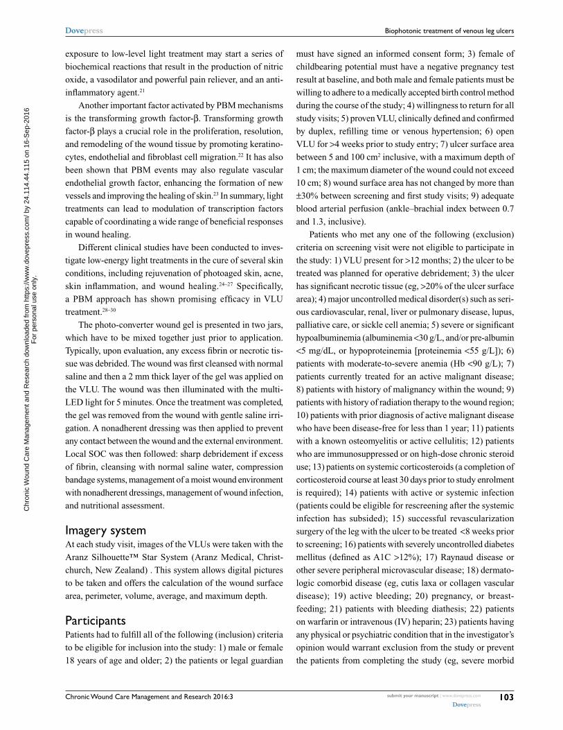
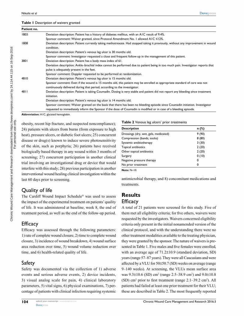
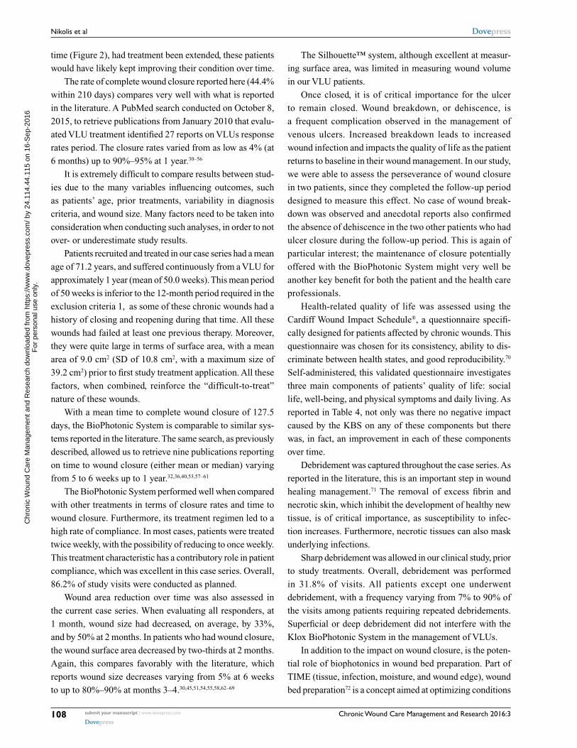
67%). Figure 2 presents wound surface area reduction over
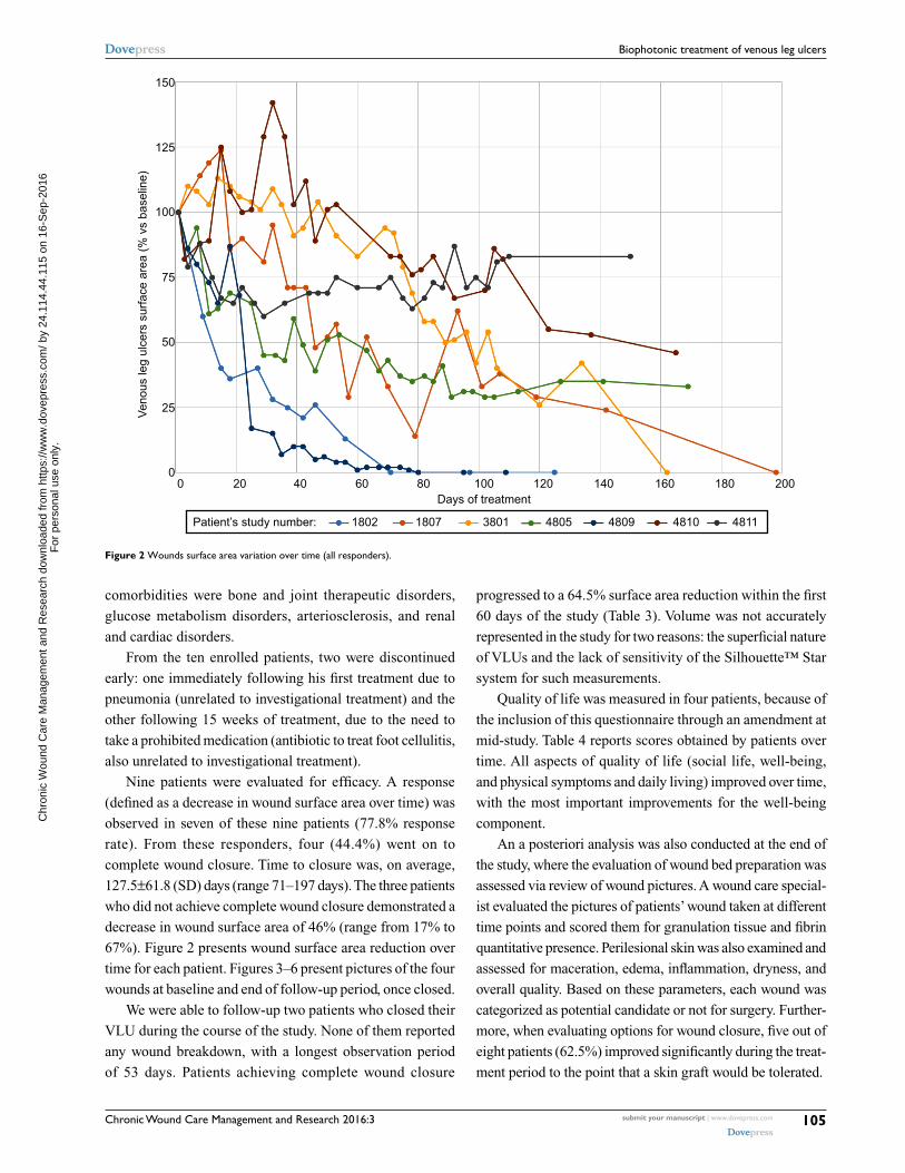
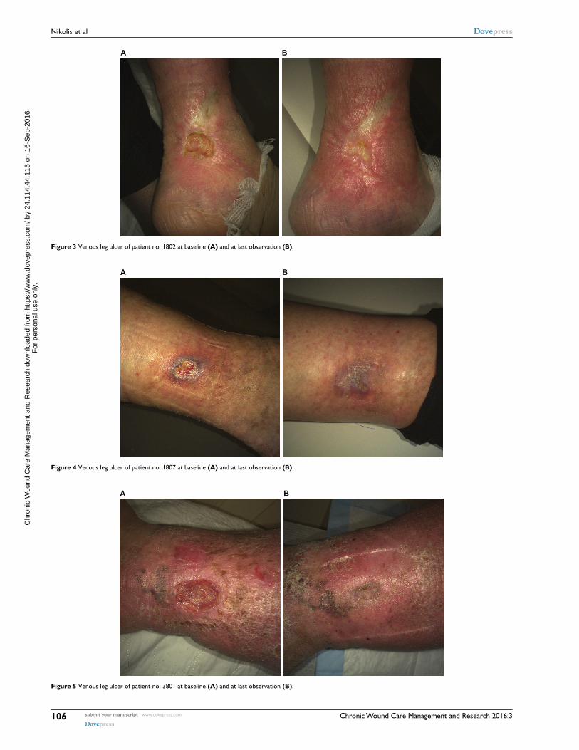
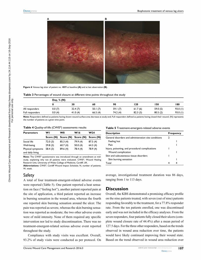
time for each patient. Figures 3–6 present pictures of the four
wounds at baseline and end of follow-up period, once closed.
We were able to follow-up two patients who closed their
VLU during the course of the study. None of them reported
any wound breakdown, with a longest observation period
of 53 days. Patients achieving complete wound closure
progressed to a 64.5% surface area reduction within the first
60 days of the study (Table 3). Volume was not accurately
represented in the study for two reasons: the superficial nature
of VLUs and the lack of sensitivity of the Silhouette™ Star
system for such measurements.
Quality of life was measured in four patients, because of
the inclusion of this questionnaire through an amendment at
mid-study. Table 4 reports scores obtained by patients over
time. All aspects of quality of life (social life, well-being,
and physical symptoms and daily living) improved over time,
with the most important improvements for the well-being
component.
An a posteriori analysis was also conducted at the end of
the study, where the evaluation of wound bed preparation was
assessed via review of wound pictures. A wound care special-
ist evaluated the pictures of patients’ wound taken at different
time points and scored them for granulation tissue and fibrin
quantitative presence. Perilesional skin was also examined and
assessed for maceration, edema, inflammation, dryness, and
overall quality. Based on these parameters, each wound was
categorized as potential candidate or not for surgery. Further-
more, when evaluating options for wound closure, five out of
eight patients (62.5%) improved significantly during the treat-
ment period to the point that a skin graft would be tolerated.
150
125
100
Veno
us le
g ul
cers
sur
face
are
a (%
vs
base
line)
75
50
25
0
Patient’s study number:
0 20 40 60 80 100Days of treatment
1802 1807 3801 4805 4809 4810 4811
120 140 160 180 200
Figure 2 Wounds surface area variation over time (all responders).
C
hron
ic W
ound
Car
e M
anag
emen
t and
Res
earc
h do
wnl
oade
d fr
om h
ttps:
//ww
w.d
ovep
ress
.com
/ by
24.1
14.4
4.11
5 on
16-
Sep
-201
6F
or p
erso
nal u
se o
nly.
Powered by TCPDF (www.tcpdf.org)
1 / 1
Chronic Wound Care Management and Research 2016:3submit your manuscript | www.dovepress.com
Dovepress
Dovepress
106
nikolis et al
A B
Figure 3 Venous leg ulcer of patient no. 1802 at baseline (A) and at last observation (B).
A B
Figure 4 Venous leg ulcer of patient no. 1807 at baseline (A) and at last observation (B).
A B
Figure 5 Venous leg ulcer of patient no. 3801 at baseline (A) and at last observation (B).
C
hron
ic W
ound
Car
e M
anag
emen
t and
Res
earc
h do
wnl
oade
d fr
om h
ttps:
//ww
w.d
ovep
ress
.com
/ by
24.1
14.4
4.11
5 on
16-
Sep
-201
6F
or p
erso
nal u
se o
nly.
Powered by TCPDF (www.tcpdf.org)
1 / 1
Chronic Wound Care Management and Research 2016:3 submit your manuscript | www.dovepress.com
Dovepress
Dovepress
107
Biophotonic treatment of venous leg ulcers
SafetyA total of four treatment-emergent-related adverse events
were reported (Table 5). One patient reported a heat sensa-
tion on face (“feeling hot”), another patient reported pain at
the site of application, a third patient reported an increase
in burning sensation in the wound area, whereas the fourth
one reported skin burning sensation around the ulcer. The
pain was reported as severe, whereas the skin burning sensa-
tion was reported as moderate; the two other adverse events
were of mild intensity. None of them required any specific
intervention nor led to study discontinuation. There was no
treatment-emergent-related serious adverse event reported
throughout the study.
Compliance with study visits was excellent. Overall,
93.2% of study visits were conducted as per protocol. On
average, investigational treatment duration was 86 days,
ranging from 1 to 113 days.
DiscussionOverall, the KBS demonstrated a promising efficacy profile
on the nine patients treated, with seven (out of nine) patients
responding favorably to the treatment, for a 77.8% responder
rate. From the ten patients enrolled, one was discontinued
early and was not included in the efficacy analyses. From the
seven responders, four patients fully closed their ulcers (com-
plete wound closure rate of 44.4%) after a mean period of
127.5 days. For the three other responders, based on the trends
observed in wound area reduction over time, the patients
would have likely continued improving their wound state.
Based on the trend observed in wound area reduction over
A B
Figure 6 Venous leg ulcer of patient no. 4809 at baseline (A) and at last observation (B).
Table 3 percentages of wound closure at different time points throughout the study
Day, % (N)
0 30 60 90 120 150 180
all responders 0.0 (7) 32.4 (7) 50.1 (7) 59.1 (7) 61.7 (6) 59.0 (5) 93.0 (1)Full responders 0.0 (4) 41.0 (4) 66.5 (4) 74.2 (4) 82.3 (3) 80.5 (2) 93.0 (1)
Note: Responders defined as patients having shown wound surface area decrease at study end; Full responders defined as patients having closed their wound. (N) represents the number of patients at a given time point.
Table 4 Quality-of-life (CWIS®) assessments results
Parameters W1 W8 W16 W24
Score (N) Score (N) Score (N) Score (N)
Social life 72.0 (3) 85.3 (4) 79.9 (4) 87.5 (4)Well-being 29.8 (3) 60.7 (4) 50.0 (4) 64.3 (4)physical symptoms and daily living
58.4 (3) 89.6 (4) 78.4 (4) 78.9 (4)
Note: The CWIS® questionnaire was introduced through an amendment at mid-study, explaining why not all patients were evaluated. CWIS®, Wound Healing Research Unit, University of Wales College of Medicine, Cardiff, UK.Abbreviations: CWIS®, Cardiff Wound Impact Schedule; N, number of patients; W, week.
Table 5 Treatment-emergent-related adverse events
Description Frequency
General disorders and administration site conditions 2 Feeling hot 1 pain 1Injury, poisoning, and procedural complications 1 Wound complication 1Skin and subcutaneous tissue disorders 1 Skin burning sensation 1Total 4 4
C
hron
ic W
ound
Car
e M
anag
emen
t and
Res
earc
h do
wnl
oade
d fr
om h
ttps:
//ww
w.d
ovep
ress
.com
/ by
24.1
14.4
4.11
5 on
16-
Sep
-201
6F
or p
erso
nal u
se o
nly.
Powered by TCPDF (www.tcpdf.org)
1 / 1
Chronic Wound Care Management and Research 2016:3submit your manuscript | www.dovepress.com
Dovepress
Dovepress
108
nikolis et al
time (Figure 2), had treatment been extended, these patients
would have likely kept improving their condition over time.
The rate of complete wound closure reported here (44.4%
within 210 days) compares very well with what is reported
in the literature. A PubMed search conducted on October 8,
2015, to retrieve publications from January 2010 that evalu-
ated VLU treatment identified 27 reports on VLUs response
rates period. The closure rates varied from as low as 4% (at
6 months) up to 90%–95% at 1 year.30–56
It is extremely difficult to compare results between stud-
ies due to the many variables influencing outcomes, such
as patients’ age, prior treatments, variability in diagnosis
criteria, and wound size. Many factors need to be taken into
consideration when conducting such analyses, in order to not
over- or underestimate study results.
Patients recruited and treated in our case series had a mean
age of 71.2 years, and suffered continuously from a VLU for
approximately 1 year (mean of 50.0 weeks). This mean period
of 50 weeks is inferior to the 12-month period required in the
exclusion criteria 1, as some of these chronic wounds had a
history of closing and reopening during that time. All these
wounds had failed at least one previous therapy. Moreover,
they were quite large in terms of surface area, with a mean
area of 9.0 cm2 (SD of 10.8 cm2, with a maximum size of
39.2 cm2) prior to first study treatment application. All these
factors, when combined, reinforce the “difficult-to-treat”
nature of these wounds.
With a mean time to complete wound closure of 127.5
days, the BioPhotonic System is comparable to similar sys-
tems reported in the literature. The same search, as previously
described, allowed us to retrieve nine publications reporting
on time to wound closure (either mean or median) varying
from 5 to 6 weeks up to 1 year.32,36,40,53,57–61
The BioPhotonic System performed well when compared
with other treatments in terms of closure rates and time to
wound closure. Furthermore, its treatment regimen led to a
high rate of compliance. In most cases, patients were treated
twice weekly, with the possibility of reducing to once weekly.
This treatment characteristic has a contributory role in patient
compliance, which was excellent in this case series. Overall,
86.2% of study visits were conducted as planned.
Wound area reduction over time was also assessed in
the current case series. When evaluating all responders, at
1 month, wound size had decreased, on average, by 33%,
and by 50% at 2 months. In patients who had wound closure,
the wound surface area decreased by two-thirds at 2 months.
Again, this compares favorably with the literature, which
reports wound size decreases varying from 5% at 6 weeks
to up to 80%–90% at months 3–4.30,45,51,54,55,58,62–69
The Silhouette™ system, although excellent at measur-
ing surface area, was limited in measuring wound volume
in our VLU patients.
Once closed, it is of critical importance for the ulcer
to remain closed. Wound breakdown, or dehiscence, is
a frequent complication observed in the management of
venous ulcers. Increased breakdown leads to increased
wound infection and impacts the quality of life as the patient
returns to baseline in their wound management. In our study,
we were able to assess the perseverance of wound closure
in two patients, since they completed the follow-up period
designed to measure this effect. No case of wound break-
down was observed and anecdotal reports also confirmed
the absence of dehiscence in the two other patients who had
ulcer closure during the follow-up period. This is again of
particular interest; the maintenance of closure potentially
offered with the BioPhotonic System might very well be
another key benefit for both the patient and the health care
professionals.
Health-related quality of life was assessed using the
Cardiff Wound Impact Schedule®, a questionnaire specifi-
cally designed for patients affected by chronic wounds. This
questionnaire was chosen for its consistency, ability to dis-
criminate between health states, and good reproducibility.70
Self-administered, this validated questionnaire investigates
three main components of patients’ quality of life: social
life, well-being, and physical symptoms and daily living. As
reported in Table 4, not only was there no negative impact
caused by the KBS on any of these components but there
was, in fact, an improvement in each of these components
over time.
Debridement was captured throughout the case series. As
reported in the literature, this is an important step in wound
healing management.71 The removal of excess fibrin and
necrotic skin, which inhibit the development of healthy new
tissue, is of critical importance, as susceptibility to infec-
tion increases. Furthermore, necrotic tissues can also mask
underlying infections.
Sharp debridement was allowed in our clinical study, prior
to study treatments. Overall, debridement was performed
in 31.8% of visits. All patients except one underwent
debridement, with a frequency varying from 7% to 90% of
the visits among patients requiring repeated debridements.
Superficial or deep debridement did not interfere with the
Klox BioPhotonic System in the management of VLUs.
In addition to the impact on wound closure, is the poten-
tial role of biophotonics in wound bed preparation. Part of
TIME (tissue, infection, moisture, and wound edge), wound
bed preparation72 is a concept aimed at optimizing conditions
C
hron
ic W
ound
Car
e M
anag
emen
t and
Res
earc
h do
wnl
oade
d fr
om h
ttps:
//ww
w.d
ovep
ress
.com
/ by
24.1
14.4
4.11
5 on
16-
Sep
-201
6F
or p
erso
nal u
se o
nly.
Powered by TCPDF (www.tcpdf.org)
1 / 1
Chronic Wound Care Management and Research 2016:3 submit your manuscript | www.dovepress.com
Dovepress
Dovepress
109
Biophotonic treatment of venous leg ulcers
at the wound bed so as to encourage normal endogenous
healing. The BioPhotonic System, through a retrospective
review of pictures, was shown to have permitted up to 62.5%
of wounds to potentially be treated with skin grafting at one
point during the course of the study, independently of wound
size. It is important to note that all wounds included in this
study had failed previous therapies. The concept of managing
difficult-to-treat wounds with wound bed preparation protocol
in combination with surgery should be evaluated.
This same system (LumiHeal™, Klox Technologies Inc,
Laval, Quebec, Canada) has recently been introduced on
the Italian market, following the CE Mark granted by the
European Community. Real-life experience gathered so far
is similar to what was observed in the present study, that is,
a very good safety profile with great efficacy at reducing
wound area, even up to closure. It is important to note that
the intended use in Europe is not only for the treatment of
VLUs but also for pressure ulcers, diabetic foot ulcers, and
acute wounds, including cosmesis and function.
In this case series, the use of the BioPhotonic System was
coupled with a very favorable safety profile. A total of four
treatment-emergent-related adverse events were reported,
leading to an incidence of one treatment-emergent-related
adverse event over 55 (0.02%) treatments across all patients.
From these, two were reported to be of mild intensity (feeling
hot and skin burning), one was of moderate intensity (skin
burning sensation), whereas pain was reported as severe.
None of them required any specific intervention and did
not lead to study discontinuation. There was no treatment-
emergent-related serious adverse event. Furthermore, the
investigational treatment did not cause any abnormal values
in laboratory analyses (biochemical, hematological, or in
urine). There was no clinically significant impact on vital
signs and no negative impact on any constituents of the
physical examinations.
Of note, during treatment with the BioPhotonic System,
there were no cases that developed a clinical infection requir-
ing systemic antimicrobial therapy. This is particularly impor-
tant when we consider the at-risk nature of an open wound.
Fagerdahl et al73 reported a complication rate of 21% with
negative pressure therapy in a retrospective study involving
87 wounds of different etiologies. To date, ~200 patients
have been treated under different study protocols with the
BioPhotonic platform, with no treatment-related infection
or treatment-related serious adverse event.
Tolerability was also assessed through the use of a visual
analog scale for pain, at each study visit, and before and
after treatment. There was a gradual decrease in quantifiable
pain felt by the patient over time. There was no difference
in terms of pain severity reported before vs immediately
following treatment.
The safety profile reported in this case series is very
similar to what has been observed in two patients with VLUs
treated under the Health Canada Special Access Program,
where no treatment-related serious adverse events were
reported. This same safety profile was also confirmed in other
medical conditions, with similar BioPhotonic Systems. When
compared with other treatments used in the management of
VLUs, the safety of the BioPhotonic System is certainly an
added benefit, offering peace of mind to the treating physician.
There are some limitations in this case series that should
be noted such as the absence of a control group and the
assessment of quality of life. The addition of a control group
would have limited the number of patients in each group and
made recruitment challenging. It was deemed a priority to
evaluate the impact of the technology on VLU in a smaller
proof-of-concept study and use necessary control groups in
a larger trial evaluating efficacy against placebo and possibly
another established modality. Furthermore, strict eligibility
criteria were used to minimize any external factors that may
have influenced the wound healing process.
ConclusionThe use of the BioPhotonic System in the treatment of VLUs
was shown to be extremely safe and very well tolerated by
our patient population. The system was efficacious, with a
responder rate of 77.8% and a closure rate of 44.4%. It also
allowed for the progression of wounds sufficiently for patients
to undergo surgery in 62.5% of cases. Easy to administer, this
medical device has all the required characteristics to become
a treatment of choice in the management of chronic VLUs.
AcknowledgmentsThe authors want to thank the investigational site study per-
sonnel, all the colleagues who collaborated in this project,
and all the study patients for their participation.
DisclosuresAN is consultant with Klox Technologies. DV was an
employee of Klox Technologies. The other authors do not
report any conflicts of interest in this work.
References 1. Margolis DJ, Bilker W, Santanna J, Baumgarten M. Venous leg
ulcer: incidence and prevalence in the elderly. J Am Acad Dermatol. 2002;46(3):381–386.
C
hron
ic W
ound
Car
e M
anag
emen
t and
Res
earc
h do
wnl
oade
d fr
om h
ttps:
//ww
w.d
ovep
ress
.com
/ by
24.1
14.4
4.11
5 on
16-
Sep
-201
6F
or p
erso
nal u
se o
nly.
Powered by TCPDF (www.tcpdf.org)
1 / 1
Chronic Wound Care Management and Research 2016:3submit your manuscript | www.dovepress.com
Dovepress
Dovepress
110
nikolis et al
2. Canadian Institute for Health Information. Compromise Wounds in Canada; 2013
3. Hopman WM, Buchanan M, VanDenKerkhaf EG, Harrison MB. Pain and health-related quality of life in people with chronic leg ulcers. Chronic Dis Inj Can. 2013;33(3):167–174.
4. Collins L, Seraj S. Diagnosis and treatment of venous ulcers. Am Fam Physician. 2010;81(8):989–996.
5. Ruckley CV. Socioeconomic impact of chronic venous insufficiency and leg ulcers. Angiology. 1997;48(1):67–69.
6. Ragnarson Tennvall G, Hjelmgren J. Annual costs of treatment for venous leg ulcers in Sweden and the United Kingdom. Wound Repair Regen. 2005;13(1):13–18.
7. Barwell JR, Davies CE, Deacon J, et al. Comparison of surgery and com-pression with compression alone in chronic venous ulceration (ESCHAR study): randomized controlled trial. Lancet. 2004;363(9424):1854–1859.
8. O’Donnell TF, Passman MA, Marston WA, et al. Management of venous leg ulcers: Clinical practice guidelines of the society for vascular sur-gery® and the American venous forum. J Vasc Surg. 2014;60:3S–59S.
9. Harding K, Dowsett C, Fias L, et al. Simplifying venous leg ulcers management. Consensus recommendations; 2015. Available from: http://www.woundsinternational.com/consensus-documents/view/simplifying-venous-leg-ulcer-management. Accessed July 15, 2016.
10. Jones JE, Nelson EA, Al-Hity A. Skin grafting for venous leg ulcers. Cochrane Database Syst Rev. 2013;30(1):CD001737.
11. Kranke P, Bennett MH, Martyn-St James M, Schnabel A, Debus SE, Weibel S. Hyperbaric oxygen therapy for chronic wounds. Cochrane Database Syst Rev. 2015;6:CD004123.
12. Dumville JC, Land L, Evans D, Peinemann F. Negative pressure wound therapy for treating leg ulcers. Cochrane Database Syst Rev. 2015;7:CD011354.
13. Flemming KA, Cullum NA, Nelson EA. A systematic review of laser therapy for venous leg ulcers. J Wound Care. 1999;8(3):111–114.
14. Sutherland JC. Biological effects of polychromatic light. Photochem Photobiol. 2002;76:164–170.
15. Kim HP. Lightening up light therapy: Activation of retrograde signaling pathway by photobiomodulation. Biomol Ther (Seoul). 2014;22(6):491–496.
16. Huang YY, Sharma SK, Carroll J, Hamblin MR. Biphasic dose response in low level light therapy - an update. Dose Response. 2011;9(4): 602–618.
17. Fathabadie FF, Bayat M, Amini A, Bayat M, Rezaie F. Effects of pulsed infra-red low level-laser irradiation on mast cells number and degranula-tion in open skin wound healing of healthy and streptozotocin-induced diabetic rats. J Cosmet Laser Ther. 2013;15(6):294–304.
18. AlGhamdi KM, Kumar A, Moussa NA. Low-level laser therapy: a use-ful technique for enhancing the proliferation of various cultured cells. Lasers Med Sci. 2012;27(1):237–249.
19. Enwemeka CS, Parker JC, Dowdy DS, Harkness EE, Sanford LE, Wood-ruff LD. The efficacy of low-power lasers in tissue repair and pain con-trol: a meta-analysis study. Photomed Laser Surg. 2004;22(4):323–329.
20. Avci P, Gupta A, Sadasivam M, Vecchio D, Pam Z, Pam N, Hamblin MR. Low-level laser (light) therapy (LLLT) in skin: stimulating, heal-ing, restoring. Semin Cutan Med Surg. 2013;32(1):41–52.
21. Mittermayr R, Osipov A, Piskernik C, et al. Blue laser light increases perfusion of a skin flap via release of nitric oxide from hemoglobin. Mol Med. 2007;13(1–2):22–29.
22. Arany PR, Nayak RS, Hallikerimath S, Limaye AM, Kale AD, Kondaiah P. Activation of latent TGF-beta1 by low-power laser in vitro correlates with increased TGF-beta1 levels in laser-enhanced oral wound healing. Wound Repair Regen. 2007;15:866–874.
23. Cury V, Moretti AI, Assis L, et al. Low level laser therapy increases angiogenesis in a model of ischemic skin flap in rats mediated by VEGF, HIF-1a and MMP-2. J Photochem Photobiol B. 2013;125:164–170.
24. Weiss RA, McDaniel DH, Geronemus RG, Weiss MA. Clinical trial of a novel non-thermal LED array for reversal of photoaging: clinical, histologic, and surface profilometric results. Lasers Surg Med. 2005;36(2):85–91.
25. Goldberg DJ, Russell BA. Combination blue (415 nm) and red (633 nm) LED phototherapy in the treatment of mild to severe acne vulgaris. J Cosmet Laser Ther. 2006;8(2):71–75.
26. Lim W, Lee S, Kim I, et al. The anti-inflammatory mechanism of 635 nm light-emitting-diode irradiation compared with existing COX inhibitors. Lasers Surg Med. 2007;39(7):614–621.
27. Iordanou P, Baltopoulos G, Giannakopoulou M, Bellou P, Ktenas E. Effect of polarized light in the healing process of pressure ulcers. Int J Nurs Pract. 2002;8(1):49–55.
28. Medenica L, Lens M. The use of polarised polychromatic non-coherent light alone as a therapy for venous leg ulceration. J Wound Care. 2003;12(1):37–40.
29. Caetano KS, Frade MA, Minatel DG, Santana LA, Enwemeka CS. Phototherapy improves healing of chronic venous ulcers. Photomed Laser Surg. 2009;27(1):111–118.
30. Landau Z, Migdal M, Lipovsky A, Lubart R. Visible light-induced heal-ing of diabetic or venous foot ulcers: a placebo-controlled double-blind study. Photomed Laser Surg. 2011;29(6):399–404.
31. Brizzio E, Amsler F, Lun B, Blättler W. Comparison of low-strength compression stockings with bandages for the treatment of recalcitrant venous ulcers. J Vasc Surg. 2010;51(2):410–416.
32. Escaleira R, Cardoso M, Rego J, Macedo P, Midões A. Efficacy of a two-component compression system for the therapy of venous leg ulcers. J Wound Care. 2010;19(3):104–109.
33. Leclère FM, Puechguiral IR, Rotteleur G, Thomas P, Mordon SR. A prospective randomized study of 980 nm diode laser-assisted venous ulcer healing on 34 patients. Wound Repair Regen. 2010;18(6):580–585.
34. Milic DJ, Zivic SS, Bogdanovic DC, et al. The influence of different sub-bandage pressure values on venous leg ulcers healing when treated with compression therapy. J Vasc Surg. 2010;51(3):655–661.
35. Ortega-Zilic N, Hunziker T, Läuchli S, et al. EpiDex® Swiss field trial 2004-2008. Dermatology. 2010;221(4):365–372.
36. Romanelli M, Dini V, Bertone MS. Randomized comparison of OASIS wound matrix versus moist wound dressing in the treatment of difficult-to-heal wounds of mixed arterial/venous etiology. Adv Skin Wound Care. 2010;23(1):34–38.
37. Teo TK, Tay KH, Lin SE, et al. Endovenous laser therapy in the treat-ment of lower-limb venous ulcers. J Vasc Interv Radiol. 2010;21(5): 657–662.
38. Hokkam E, El-Labban G, Shams M, Rifaat S, El-Mezaien M. The use of topical phenytoin for healing of chronic venous ulcerations. Int J Surg. 2011;9(4):335–338.
39. Belcaro G, Cesarone MR, Errichi BM, et al. Venous and diabetic ulcerations: management with topical multivalent silver oxide ointment. Panminerva Med. 2010;52(2 Suppl 1):37–42.
40. Watson JM, Kang’ombe AR, Soares MO, et al; VenUS III Team. Use of weekly, low dose, high frequency ultrasound for hard to heal venous leg ulcers: the VenUS III randomised controlled trial. BMJ. 2011;342:d1092.
41. Giuggioli D, Colaci M, Manfredi A, Mariano M, Ferri C. Platelet gel in the treatment of severe scleroderma skin ulcers. Rheumatol Int. 2012;32(9):2929–2932.
42. Kelechi TJ, Mueller M, Hankin CS, Bronstone A, Samies J, Bonham PA. A randomized, investigator-blinded, controlled pilot study to evaluate the safety and efficacy of a poly-N-acetyl glucosamine-derived mem-brane material in patients with venous leg ulcers. J Am Acad Dermatol. 2012;66(6):e209–215.
43. Lazareth I, Moffatt C, Dissemond J, et al. Efficacy of two compression systems in the management of VLUs: results of a European RCT. J Wound Care. 2012;21(11):553–554, 556, 558 passim.
44. Maggio G, Armenio A, Ruccia F, Giglietto D, Pascone M, Ribatti D. A new protocol for the treatment of the chronic venous ulcers of the lower limb. Clin Exp Med. 2012;12(1):55–60.
45. Meaume S, Truchetet F, Cambazard F, et al; CHALLENGE Study Group. A randomized, controlled, double-blind prospective trial with a Lipido-Colloid Technology-Nano-OligoSaccharide Factor wound dressing in the local management of venous leg ulcers. Wound Repair Regen. 2012;20(4):500–511.
C
hron
ic W
ound
Car
e M
anag
emen
t and
Res
earc
h do
wnl
oade
d fr
om h
ttps:
//ww
w.d
ovep
ress
.com
/ by
24.1
14.4
4.11
5 on
16-
Sep
-201
6F
or p
erso
nal u
se o
nly.
Powered by TCPDF (www.tcpdf.org)
1 / 1
Chronic Wound Care Management and Research 2016:3 submit your manuscript | www.dovepress.com
Dovepress
Dovepress
Chronic Wound Care Management and Research
Publish your work in this journal
Submit your manuscript here: https://www.dovepress.com/chronic-wound-care-management-and-research-journal
Chronic Wound Care Management and Research is an international, peer reviewed, open access, online journal publishing original research, reviews, editorials, and commentaries on the causes and management of chronic wounds and the major issues related to chronic wound man-agement. Topics also include chronic wounds as comorbidities to other
conditions, patient adherence to therapy, and the economic burden of chronic wounds. The manuscript management system is completely online and includes a very quick and fair peer review system, which is all easy to use. Visit http://www.dovepress.com/testimonials.php to read real quotes from published authors.
Dovepress
111
Biophotonic treatment of venous leg ulcers
46. Weller CD, Evans SM, Staples MP, Aldons P, McNeil JJ. Randomized clinical trial of three-layer tubular bandaging system for venous leg ulcers. Wound Repair Regen. 2012;20(6):822–829.
47. Wong IK, Andriessen A, Charles HE, Thompson D, Lee DT, So WK, Abel M. Randomized controlled trial comparing treatment outcome of two compression bandaging systems and standard care without compression in patients with venous leg ulcers. J Eur Acad Dermatol Venereol. 2012;26(1):102–110.
48. Chaby G, Senet P, Ganry O, et al; Angio-Dermatology Group of the French Society of Dermatology. Prognostic factors associated with healing of venous leg ulcers: a multicentre, prospective, cohort study. Br J Dermatol. 2013;169(5):1106–1113.
49. Dolibog P, Franek A, Taradaj J, et al. A comparative clinical study on five types of compression therapy in patients with venous leg ulcers. Int J Med Sci. 2013;11(1):34–43.
50. Harding K, Sumner M, Cardinal M. A prospective, multicentre, randomised controlled study of human fibroblast-derived dermal substitute (Derma-graft) in patients with venous leg ulcers. Int Wound J. 2013;10(2):132–137.
51. Humbert P, Mikosinki J, Benchikhi H, Allaert FA. Efficacy and safety of a gauze pad containing hyaluronic acid in treatment of leg ulcers of venous or mixed origin: a double-blind, randomised, controlled trial. Int Wound J. 2013;10(2):159–166.
52. Ashby RL, Gabe R, Ali S, et al. Clinical and cost-effectiveness of compression hosiery versus compression bandages in treatment of venous leg ulcers (Venous leg Ulcer Study IV, VenUS IV): a randomised controlled trial. Lancet. 2014;383(9920):871–879.
53. Finlayson KJ, Courtney MD, Gibb MA, O’Brien JA, Parker CN, Edwards HE. The effectiveness of a four-layer compression bandage system in comparison with class 3 compression hosiery on healing and quality of life in patients with venous leg ulcers: a randomised controlled trial. Int Wound J. 2014;11(1):21–27.
54. Forlee M, Rossington A, Searle R. A prospective, open, multicentre study to evaluate a new gelling fibre dressing containing silver in the management of venous leg ulcers. Int Wound J. 2014;11(4):438–445.
55. Harding K, Aldons P, Edwards H, et al. Effectiveness of an acellular synthetic matrix in the treatment of hard-to-heal leg ulcers. Int Wound J. 2014;11(2):129–137.
56. Arenberger P, Elg F, Petyt J, Cutting K. Expected outcomes from topical haemoglobin spray in non-healing and worsening venous leg ulcers. J Wound Care. 2015;24(5):228, 230–232, 236.
57. Vanscheidt W, Harding K, Téot L, Siebert J. Effectiveness and tissue compatibility of a 12-week treatment of chronic venous leg ulcers with an octenidine based antiseptic--a randomized, double-blind controlled study. Int Wound J. 2012;9(3):316–323.
58. Olyaie M, Rad FS, Elahifar MA, Garkaz A, Mahsa G. High-frequency and noncontact low-frequency ultrasound therapy for venous leg ulcer treatment: a randomized, controlled study. Ostomy Wound Manage. 2013;59(8):14–20.
59. Adderley U, Stubbs N. Stockings or bandages for leg-ulcer compression? Nurs Times. 2014;110(15):19–20.
60. Beheshti A, Shafigh Y, Parsa H, Zangivand AA. Comparison of high-frequency and MIST ultrasound therapy for the healing of venous leg ulcers. Adv Clin Exp Med. 2014;23(6):969–975.
61. Evangelista MT, Casintahan MF, Villafuerte LL. Simvastatin as a novel therapeutic agent for venous ulcers: a randomized, double- blind, placebo-controlled trial. Br J Dermatol. 2014;170(5):1151–1157.
62. Schumann H, Calow T, Weckesser S, Müller ML, Hoffmann G. Water-filtered infrared A for the treatment of chronic venous stasis ulcers of the lower legs at home: a randomized controlled blinded study. Br J Dermatol. 2011;165(3):541–551.
63. Alsina-Gibert M, Pedregosa-Fauste S. Amniotic membrane transplanta-tion in the treatment of chronic lower limb ulcers. Actas Dermosifiliogr. 2012;103(7):608–613.
64. Burkiewicz CJ, Guadagnin FA, Skare TL, do Nascimento MM, Servin SC, de Souza GD. Vitamin D and skin repair: a prospective, double-blind and placebo controlled study in the healing of leg ulcers. Rev Col Bras Cir. 2012;39(5):401–407.
65. Dereure O, Czubek M, Combemale P. Efficacy and safety of hyaluronic acid in treatment of leg ulcers: a double-blind RCT. J Wound Care. 2012;21(3):131–132, 134–136, 138–139.
66. Harding K, Gottrup F, Jawień A, et al. A prospective, multi-centre, randomised, open label, parallel, comparative study to evaluate effects of AQUACEL® Ag and Urgotul® Silver dressing on healing of chronic venous leg ulcers. Int Wound J. 2012;9(3):285–294.
67. Meaume S, Perez J, Rethore V, et al. Management of chronic wounds with an innovative absorbent wound dressing. Wound Care. 2012;21(7): 315–316, 318, 320–322.
68. Arenbergerova M, Engels P, Gkalpakiotis S, Dubská Z, Arenberger P. Topical hemoglobin promotes wound healing of patients with venous leg ulcers. Hautarzt. 2013;64(3):180–186.
69. Ghatnekar GS, Grek CL, Armstrong DG, Desai SC, Gourdie RG. The effect of a connexin43-based peptide on the healing of chronic venous leg ulcers: a multicenter, randomized trial. J Invest Dermatol. 2015;135(1):289–298.
70. Price PE, Harding KG. The Cardiff Wound Impact Schedule: the develop-ment of a condition specific questionnaire to assess health-related quality of life in patients with chronic wounds. Int Wound J. 2004;1(1):10–17.
71. Milne J. Wound-bed preparation: the importance of rapid and effec-tive desloughing to promote healing. Br J Nurs. 2015;24(Suppl. 20): S52–58.
72. EWMA. Position Document: Wound Bed Preparation in Practice. London: MEP Ltd.; 2004.
73. Fagerdahl AM, Bostrom L, Ulfvarson J, Ottosson C. Risk factors for unsuccessful treatment and complications with negative pressure wound therapy. Wounds. 2012;24(6):168–177.
C
hron
ic W
ound
Car
e M
anag
emen
t and
Res
earc
h do
wnl
oade
d fr
om h
ttps:
//ww
w.d
ovep
ress
.com
/ by
24.1
14.4
4.11
5 on
16-
Sep
-201
6F
or p
erso
nal u
se o
nly.
Powered by TCPDF (www.tcpdf.org)
1 / 1