Embed Size (px)
Citation preview

A previously unknown zinc finger protein,DST, regulates drought and salt tolerancein rice via stomatal aperture control
Xin-Yuan Huang,1 Dai-Yin Chao,1 Ji-Ping Gao, Mei-Zhen Zhu, Min Shi, and Hong-Xuan Lin2
National Key Laboratory of Plant Molecular Genetics, Shanghai Institute of Plant Physiology and Ecology, Shanghai Institutes forBiological Sciences, Chinese Academy of Sciences, Shanghai 200032, China
Abiotic stresses, such as drought and salinity, lead to crop growth damage and a decrease in crop yields. Stomatacontrol CO2 uptake and optimize water use efficiency, thereby playing crucial roles in abiotic stress tolerance.Hydrogen peroxide (H2O2) is an important signal molecule that induces stomatal closure. However, the molecularpathway that regulates the H2O2 level in guard cells remains largely unknown. Here, we clone and characterizeDST (drought and salt tolerance)—a previously unknown zinc finger transcription factor that negatively regulatesstomatal closure by direct modulation of genes related to H2O2 homeostasis—and identify a novel pathway for thesignal transduction of DST-mediated H2O2-induced stomatal closure. Loss of DST function increases stomatalclosure and reduces stomatal density, consequently resulting in enhanced drought and salt tolerance in rice. Thesefindings provide an interesting insight into the mechanism of stomata-regulated abiotic stress tolerance, and animportant genetic engineering approach for improving abiotic stress tolerance in crops.
[Keywords: Rice; zinc finger protein; drought tolerance; salt tolerance; stomatal aperture; hydrogen peroxide]
Supplemental material is available at http://www.genesdev.org.
Received April 16, 2009; revised version accepted June 15, 2009.
Drought and salinity are two main abiotic stressesnegatively affecting plant growth and seed production.Plants respond to and adapt to these stresses in order tosurvive. The understanding of plant responses to stressesin physiology, genetics, and molecular biology will begreatly helpful in improving the tolerance of plants toabiotic stresses through genetic engineering. Stomatalpores that are located in the epidermis of plant leaves con-trol the uptake of CO2 for photosynthesis and the waterloss during transpiration, and play a crucial role in abioticstress tolerance (Schroeder et al. 2001a; Hetherington andWoodward 2003). Under drought stress, abscisic acid(ABA)-mediated stomatal closure is a mechanism thatplants use to adapt to water deficiency (Schroeder et al.2001b; Zhu 2002; Fan et al. 2004). Reactive oxygenspecies (ROS) including hydrogen peroxide (H2O2) thatare widely generated under stress have been proposed tofunction as second messengers in ABA signaling in guardcells (McAinsh et al. 1996; Pei et al. 2000; Kohler et al.2003; Bright et al. 2006). Early work has shown that H2O2
can induce stomata closure (McAinsh et al. 1996; Zhanget al. 2001). In guard cells, ABA-stimulated ROS accu-
mulation activates plasma membrane calcium channelsand triggers stomatal closure (Hamilton et al. 2000; Peiet al. 2000).
ABA-induced ROS are mainly generated by NADPHoxidase in guard cells (Kwak et al. 2003). Mutation of twogenes encoding subunits of an NADPH oxidase (AtrbohDand AtrbohF) impairs ABA-induced stomatal closing(Kwak et al. 2003). To cope with increased levels ofROS, plants have evolved at least four kinds of ROSscavenging enzymes including catalase, superoxide dis-mutase, ascorbate peroxidase, and glutathione peroxi-dase. It has been shown that H2O2 is scavenged byglutathione peroxidase in the guard cells (Miao et al.2006). So far, the genes that regulate the expression ofH2O2 scavenging enzymes have not been identified, andthe mechanism of regulation of H2O2 levels, especially inthe stomata, is largely unknown.
Previous studies have shown that the transcriptionalfactors play important roles in plant tolerance to abioticstresses (Stockinger et al. 1997; Liu et al. 1998; Haakeet al. 2002; Yamaguchi-Shinozaki and Shinozaki 2006).The Cys-2/His-2-type (C2H2) zinc finger protein—alsocalled a classical or TFIIIA-type zinc finger—that was firstfound in Xenopus oocytes (Miller et al. 1985) is animportant class of eukaryotic transcription factors. Sev-eral plant members of this family have various regulatoryroles in stress responses and developmental processes
1These authors contributed equally to this work.2Corresponding author.E-MAIL [email protected]; FAX 86-21-54924015.Article is online at http://www.genesdev.org/cgi/doi/10.1101/gad.1812409.
GENES & DEVELOPMENT 23:1805–1817 � 2009 by Cold Spring Harbor Laboratory Press ISSN 0890-9369/09; www.genesdev.org 1805
Cold Spring Harbor Laboratory Press on February 3, 2019 - Published by genesdev.cshlp.orgDownloaded from

(Kim et al. 2001; Sakamoto et al. 2004; de Lorenzo et al.2007; Xu et al. 2007). However, it is still not knownwhether the C2H2-type zinc finger protein is involved inregulating stomatal aperture, although previous studieshave shown that several transcription factors have beenassociated with stomata movement (Cominelli et al.2005; Liang et al. 2005; Hu et al. 2006). In this study, wecharacterized a novel C2H2 zinc finger transcriptionfactor, DST (drought and salt tolerance), that controls sto-matal aperture under drought and salt stress in rice. DSTcontributes to stomata movement via regulation of genesinvolved in ROS homeostasis. These findings will shedlight on the process of stomatal movement in plants andon the engineering of drought and salt tolerance in crops.
Results
Isolation of dst mutant
We performed a large-scale screen for the mutants toidentify genetic loci that affect drought and salt tolerancein rice and isolated a dst mutant line. When treated with20% PEG4000 (simulation of drought stress), the dstmutant exhibited less severe wilting than wild-type(ZH11) plants (Fig. 1A, top panel). Furthermore, ;70%of dst mutant plants survived under 140 mM NaCltreatment, but nearly all of the wild-type plants wilted(Fig. 1A; Supplemental Fig. S1A). The dst mutant ex-hibited better recovery than wild-type plants from bothdrought and salt stresses (Fig. 1A). To accurately evaluate
Figure 1. The dst mutant shows toler-ance to drought and salt stress. (A)PEG4000, NaCl, and drought stress treat-ment of wild-type plants Zhonghua11(ZH11) and the dst mutant. Twenty-day-old plants were treated with 20% PEG4000for 7 d (top panel) or 140 mM NaCl for 12 d(middle panel), and then recovered as in-dicated. (Bottom panel) Thirty days aftertransplantation, plants were subjected todrought stress for 12 or 20 d and thenallowed to recover for 15 d. (B) Leaf ofZH11 and dst mutant. Bar, 0.5 cm. (C)Quantitative measurement of the maxi-mum leaf width of ZH11 and dst mutant(n = 20). (D) Water loss of ZH11 and dst
mutant. For each repeat, 15 fully expandedleaves of 25-d-old plants were used ina triplicate experiment (n = 3). (E) Relativewater content of ZH11 and dst mutanttreated with 18% PEG4000 using the fullyexpanded leaves of 20-d-old plants (n = 9).(F) Osmolality measurement in ZH11 anddst mutant under 18% PEG4000 treat-ment using the fully expanded leaves of25-d-old plants (n = 8). (G) Na+ and K+
contents in the roots and shoots of ZH11and dst mutant plants under non-saltstress and 100 mM NaCl treatment for7 d (n = 3). Data in C–G are presented asmean 6 SEM. (*) P # 0.05; (**) P # 0.01,Student’s t-test.
Huang et al.
1806 GENES & DEVELOPMENT
Cold Spring Harbor Laboratory Press on February 3, 2019 - Published by genesdev.cshlp.orgDownloaded from

the effect of the DST mutation on drought tolerance, weperformed a soil drought experiment in deep polyvinylchloride (PVC) pipes. The dst mutant plants showedstronger drought tolerance than the wild-type plants(Fig. 1A, bottom panel). These results indicated that mu-tation of the DST locus significantly improves droughtand salt tolerance. There were no significant morpholog-ical alterations in dst mutants, except for a markedlywider leaf width (Fig. 1B,C). Most agronomic traits wereunchanged in the mutants, with the exception of thepanicle number per plant and the main panicle length(Supplemental Fig. S2).
Physiological analysis of dst mutant
To elucidate the physiological mechanism of drought andsalt tolerance in the dst mutant, we first investigated thewater status in wild-type and dst mutants. Under de-hydration stress, the dst mutant lost less water andmaintained higher water content than wild-type plants(Fig. 1D,E). The dst mutant also maintained higherosmolality during drought treatment (Fig. 1F). Theseresults indicate that the enhanced drought tolerance ofthe dst mutant is due to an increased ability to maintainwater. Finally, we compared the K+ and Na+ content inthe roots and shoots in response to NaCl stress. Therewas no significant difference in K+ content between theroots of the wild type and dst mutant; however, K+
content was lower in the shoots of dst mutants underboth normal and salt stress conditions (Fig. 1G). NaCltreatment resulted in a lower accumulation of Na+ in theroots and shoots of dst mutant plants (Fig. 1G), indicatingthat the enhanced salt tolerance of dst mutant might bedue to a reduction of Na+ toxicity. These results demon-strate that the DST mutation leads to enhancement ofdrought and salt tolerance in rice plants.
Stomatal movement changes in the dst mutantdue to H2O2 accumulation
Because stomata are involved in responses to abioticstresses (Hetherington and Woodward 2003), we exam-ined the stomatal status of dst mutant and wild-typeplants. The results showed that 42.4% of stomata werecompletely closed in the dst mutant plants, but only16.7% were completely closed in the wild-type plants;also, only 21.2% of stomata were completely open in thedst mutants, but 50.8% were completely open in wild-type plants (Fig. 2A,B). The percentage of partially openstomata was similar in dst mutant and wild-type plants(32.5%–36.4%). These results showed that stomatalmovement is greatly affected in dst mutants. A signifi-cant decrease in stomatal density was also found in dstmutant plants (Fig. 2C), whereas there was no significantdifference in guard cell size between dst mutant and wild-type plants (data not shown). Meanwhile, the stomatalconductance was lower in dst mutant plants than that ofwild-type plants (Fig. 2D). We further investigated thestomatal status of the dst mutant under stress conditions,and found that the stomatal conductance of the dstmutant was significantly lower than that of the wild-type
plants not only in the normal growth condition, but alsoin the salt stress condition (Supplemental Fig. S3). Theseresults demonstrate that the enhanced drought tolerancedisplayed by the dst mutant is primarily due to increasedstomatal closure and decreased stomatal density, whichprevent water loss, and that the enhanced salt toleranceof the dst mutant might be the result of a low transpira-tion rate that reduces Na+ transport from roots to shoots.
The phytohormone ABA induces stomatal closure(Schroeder et al. 2001b; Zhu 2002). However, measure-ment of endogenous ABA content revealed no significantdifference between dst mutant and wild-type plants (Fig.2E). Therefore, the increased stomatal closure of the dstmutant is not the result of ABA accumulation. SinceH2O2 induces leaf stomatal closure (McAinsh et al. 1996;Hamilton et al. 2000; Apel and Hirt 2004; Bright et al.2006), we examined H2O2 content in rice leaves andfound a higher accumulation in dst mutant plants (Fig.2F,G). Furthermore, by using a fluorescence dye, 29,79-dichlorodihydrofluorescein diacetate (H2DCFDA), wefound that dst mutant plants accumulated more H2O2
in the guard cells than those of wild-type plants (Fig.2H,I). These results suggested that accumulation of H2O2
in guard cells probably underlies the increased stomatalclosure in dst mutant plants.
DST encodes a novel zinc finger proteinof unknown function
A map-based cloning approach was used to isolate theDST gene. Since the wide leaf is a typical and visiblephenotype of the dst mutant (Fig. 1B), it was adopted asthe standard dst phenotype. All F1 progeny showeda narrow leaf phenotype similar to that of wild-typeplants (data not shown). In the F2 population, narrowand wide leaf plants segregated in a 3:1 ratio (241 narrowvs. 63 wide; x2 = 2.96 < x2
0.05 = 3.84; P > 0.05), demon-strating that the wide leaf phenotype of the dst mutant iscontrolled by a single recessive nuclear gene. The DSTlocus was initially mapped to the long arm of chromo-some 3 between the markers H2423 and H2519 (Fig. 3A;Supplemental Table S1), and was subsequently fine-mapped to a 14-kb region between H2423 and H2437(Fig. 3B). Only one candidate gene, which encodes a 301-residue polypeptide with unknown function, localized inthis region (Fig. 3C). Protein sequence analysis showedthat DST contains a nuclear localization signal (NLS)peptide and a single C2H2-type zinc finger motif at the Nterminus (Fig. 3C,D). A BLAST search found only onecopy of DST in the rice genome. A comparison of thenucleotide sequences of the gene in dst mutant and wild-type plants revealed that two nucleotide substitutionslead to two amino acid substitutions: asparagine (N69) toaspartic acid (D) and alanine (A162) to threonine (T) (Fig.3C), of which the N69 is a conserved residue of the zincfinger motif.
A complementation test was carried out to confirmthe mapping result. Transfer of a 4.632-kb wild-typeDNA fragment containing the DST promoter region andthe entire ORF into dst mutants conferred phenotypes
DST regulates drought and salt tolerance in rice
GENES & DEVELOPMENT 1807
Cold Spring Harbor Laboratory Press on February 3, 2019 - Published by genesdev.cshlp.orgDownloaded from

similar to those of wild-type plants, including narrow leafshape, sensitivity to drought and salt stresses, stomatalopening level, and H2O2 content (Fig. 3E; SupplementalFig. S1A,B,D,E), whereas a knockout of DST using RNAitechnology produced the dst phenotypes (Fig. 3E; Supple-mental Fig. S1A–E). These results demonstrate that DSTis indeed responsible for the phenotypic changes found indst mutants, and that DST is a negative regulator ofdrought and salt tolerance.
We constructed N69D (M1) and A162T (M2) mutantsto evaluate the role of the two substitutions (Fig. 3C).These two constructs and the dst (M, containing the twomutations) were each transformed into the dst mutant.Only those transformants expressing the M2 constructcomplemented dst phenotypes (Fig. 3F). Therefore, themutation of N69, but not A162 is primarily responsiblefor the phenotypes of the dst mutant.
Subcellular localization and expression pattern of DST
Protein structure prediction showed that DST has a puta-tive NLS (Fig. 3C). We transiently expressed the DST-GFPfusion protein in onion epidermal cells and confirmedthat the fusion protein specifically localizes to the nu-cleus (Fig. 4A). The fluorescence of DST-GFP was alsodetected exclusively in the nucleus in transgenic riceplants that were transformed with a DST-GFP fusionconstruct under the control of the cauliflower mosaicvirus (CaMV) 35S promoter (Supplemental Fig. S4). Thetissue expression pattern of DST was determined bytransforming rice plants with a b-glucuronidase (GUS)construct driven by the DST promoter. Strong GUSsignals were detected in vascular tissues of the roots,internodes, and leaf sheath (Fig. 4B–H). In the leaves, theGUS signal was also detected in the stomata apparatus
Figure 2. H2O2 accumulation induces stomata closingin the dst mutant. (A) Environmental scanning electronmicroscopy images of three levels of stomatal opening.Bar, 10 mm. (B) The percentage of three levels of stoma-tal opening in ZH11 and dst mutant (n = 132 stomata forZH11; n = 126 stomata for dst). (C) Stomatal density ofthe middle leaves of ZH11 and dst mutant (n = 10).Three random scopes were used in each repeat (356.20mm 3 356.20 mm in each scope). (D) Stomatal conduc-tance of ZH11 and dst mutant (n = 12). (E) ABA contentof ZH11 and dst mutant (n = 5 repeats, 16 plants in eachrepeat). (F) DAB staining in the seedling leaves of ZH11(left) and dst mutant (right). (G) Quantitative measure-ment of H2O2 in the seedling leaves of ZH11 and dst
mutant (n = 4 repeats, 16 plants in each repeat). (H)Examination of H2O2 production in the guard cell ofZH11 and dst mutant plant with H2DCFDA. Bar, 5 mm.(I) Quantitative analysis of H2O2 production in the guardcell of ZH11 and dst mutant plant (n = 104 stomata forZH11; n = 99 stomata for dst). Data are presented asmean 6 SEM in (C–E, G, and I). (*) P # 0.05; (**) P #
0.01, Student’s t-test.
Huang et al.
1808 GENES & DEVELOPMENT
Cold Spring Harbor Laboratory Press on February 3, 2019 - Published by genesdev.cshlp.orgDownloaded from

(Fig. 4I,J). To confirm the expression of DST in thestomata apparatus, we generated transgenic rice plantsthat were transformed with a DST-GFP fusion con-struct under the control of the DST native promoter.Confocal microscopic analysis showed that the fluores-cence of DST-GFP was detected in the guard cells andthe subsidiary cells (Fig. 4K; Supplemental Fig. S5),which was consistent with the role of DST in stomataregulation.
We also tested whether the expression of DST isregulated by drought and salt stresses. Real-time PCRanalysis showed that DST is down-regulated rapidly after0.5 h of drought or salt treatment, but that the expressionresumed continuously after 24 h of treatment (Fig. 4L).These data are consistent with its role in regulatingstomatal movement under drought and salt stresses. Toexclude the possibility that DST is regulated by a circa-dian rhythm, we analyzed the rhythmic profiles of DSTand found that DST is not regulated significantly bya circadian rhythm (Supplemental Fig. S6).
DST is a transcription factor withtransactivational activity
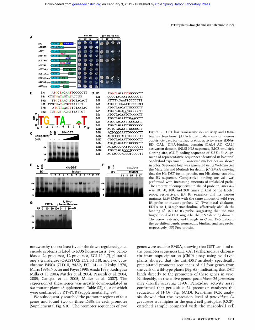
We performed a transcriptional activation assay andfound that expression of the DST-BD (GAL4-binding do-main) fusion protein in yeast resulted in high reportergene expression (Fig. 5A), which revealed that DST hasstrong activity as a transcription activator. Assessment ofthe two amino acid substitutions in the dst mutantshowed that the N69D substitution decreased the levelof transcriptional activation, while the A162T substitu-tion did not alter the activation level. These resultsindicated that the N69 of DST is required for transcrip-tional activation. Interestingly, when the two amino acidsubstitutions were combined, activation was lower thanthat of the N69D mutation alone (Fig. 5A), possibly be-cause the two amino acid substitutions modify DST proteinconfiguration. Further quantitative assays of the b-galacto-sidase activity confirmed these results (SupplementalFig. S7). Deletion analysis shows that the first 72 amino
Figure 3. Map-based cloning of DST and complementa-tion test. (A) The DST locus was mapped to chromosome3 between markers H2423 and H2519 using 310 homo-zygous F2 plants. (B) Fine mapping of the DST locus. TheDST locus was narrowed to a 14-kb region betweenmarkers H2423 and H2437. Numbers below thehorizontal line in A and B are the number of recombi-nants. (C) The protein structure of DST. Two nucleotidesubstitutions in dst lead to an asparagine-to-aspartic acidchange at amino acid 69 and an alanine-to-threoninechange at amino acid 162. (D) Amino acid sequencealignments of the zinc finger domain. Identical andsimilar residues are displayed in colored boxes. Theasterisk marks the cysteine and histidine in the C2H2-type zinc finger domain. The solid triangle denotes thepoint mutation in the zinc finger domain. GenBankaccession numbers are ZFP1 (NP_178188), ZFP2 (NP_200560), ZFP5 (NP_172518), ZFP8 (NP_181725), SGT1-like protein (BAD73077), SUPERMAN (AAC49116),RAMOSA1 (AAY17040), and LIF (BAB58897). (E)Drought and salt treatment of T2 transgenic lines in-cluding vector control, complementation, and RNAi.Twenty-day-old plants (top) were treated with 18%PEG4000 for 10 d (middle) or 100 mM NaCl for 15 d(bottom). (F) Drought and salt treatment of T2 transgeniclines with point mutation constructs. Twenty-day-oldplants (top) were treated with 18% PEG4000 for 15 d(middle) or 100 mM NaCl for 20 d (bottom). M1, M2, andM indicate transgenic plants with N69D, A162T, andboth point mutation constructs, respectively.
DST regulates drought and salt tolerance in rice
GENES & DEVELOPMENT 1809
Cold Spring Harbor Laboratory Press on February 3, 2019 - Published by genesdev.cshlp.orgDownloaded from

acids, containing the zinc finger motif, are required fortranscriptional activation, and that the activation domainis localized in the first 145 N-terminal residues (Fig. 5A).
DST binds to the TGCTANNATTG elementby the zinc finger domain
We used a bacterial one-hybrid system (Meng et al. 2005)to identify the DNA-binding sequence of DST. Amongthe sequences identified, six sequences had a conservedTGNTANN(A/T)T sequence (Fig. 5B). Electrophoreticmobility shift assay (EMSA) showed that the His-DSTfusion protein, but not His alone, could bind to a B3 se-quence that contains this conserved sequence, whereasan excess unlabeled B3 sequence effectively reduced orabolished the His-DST binding to the labeled B3 sequence(Fig. 5C). Meanwhile, the His-DST protein could bind tothe B3 sequence in a dosage-dependent manner (Supple-mental Fig. S8A). These results demonstrated that DSTbinds specifically to this sequence. Individual mutationsof TG (M10), TA (M3), and AT (M5), and simultaneousmutations (M11 and M12) in the TGNTANNAT se-quence markedly reduced or abolished the binding ofHis-DST (Fig. 5D,E). The mutants (M4, M7, M8, M9 andM13) with the conserved nucleotides did not significantlyreduce the binding of His-DST (Fig. 5D–F). However,other mutants (M6 and M14) markedly reduced thebinding of His-DST, showing that TG and C are alsorequired for DST binding (Fig. 5D–F). Taken together,these results showed that DST binds to the TGCTANNATTG sequence, which is a novel cis-element and namedDBS (DST-binding sequence). This was further demon-strated by those mutations (M15, M16, and M17) in thiscore sequence that completely abolished the binding ofHis-DST (Fig. 5D–F). Interestingly, the M8 mutant, whichproduced new ATTG core nucleotides, increased thebinding of His-DST (Fig. 5E; Supplemental Table S2).We also tested whether the mutant DST protein in dstmutants could bind to DBS, and found that the mutantform of DST protein could bind to DBS as the wild-typeprotein did (Supplemental Fig. S8B–D).
Based on previous studies (Wolfe et al. 2000; Sakamotoet al. 2004; Lin et al. 2007), we assumed that the zincfinger motif of DST might be the DNA-binding domain.The preincubation of the His-DST fusion protein withtwo metal chelators—EDTA or 1,10-o-phenanthroline—effectively reduced or abolished the DNA-binding abilityof His-DST, suggesting that DST binds to the DBS ele-ment by the N-terminal zinc finger motif (Fig. 5G). Thissuggestion was further confirmed by EMSA using a trun-cate DST protein with deletion of the zinc finger domain(Supplemental Fig. S8B–D).
DST targets to genes related to H2O2 homeostasis
Expression profile analysis using the Affymetrix Rice Ge-nome Genechip was used to identify DST target genes.We found that among ;50,000 rice genes, the transcrip-tion levels of 60 genes were altered significantly (morethan threefold change) in dst mutant plants (SupplementalTable S3). A high percentage (60%) of these 60 genes hadlower expression levels in dst mutants (SupplementalTable S3), and mutation of the DST protein in dst mu-tants lost its transactivation activity (Fig. 5A), indicatingthat DST might promote expression of these genes. It is
Figure 4. Subcellular localization and expression pattern ofDST. (A) Subcellular localization of DST. GFP and the DST-GFP
fusion gene under the control of the CaMV 35S promoter wereexpressed transiently in onion epidermal cells. The DST-GFPfusion protein was specifically expressed in the nucleus. (B–J) DST
promoter-GUS expression patterns in transgenic rice plants. GUS
expression was observed in the vascular system of roots (B–D),nodes (E,F), leaf sheathes (G,H), and leaves (I). (J) GUS expressionwas also observed in the stomata. The arrow indicates the stoma.Bars: C,D,J, 100 mm. (K) The stoma of the transgenic rice plantsthat were transformed with a DST-GFP fusion construct underthe control of DST native promoter. The GFP fluorescence wasdetected in the guard cells and the subsidiary cells. Bar, 5 mm. (L)Relative expression levels of DST by quantitative real-time PCRin the shoots of plants treated with 18% PEG4000 and 100 mMNaCl. Data are presented as mean 6 SEM (n = 3).
Huang et al.
1810 GENES & DEVELOPMENT
Cold Spring Harbor Laboratory Press on February 3, 2019 - Published by genesdev.cshlp.orgDownloaded from
noteworthy that at least five of the down-regulated genesencode proteins related to ROS homeostasis: two perox-idases (24 precursor, 12 precursor; EC1.11.1.7), glutathi-one S-transferase (OsGSTU2; EC2.5.1.18), and two cyto-chrome P450s (71D10, 94A2; EC1.14.-.-) (Jakoby 1978;Marrs 1996; Noctor and Foyer 1998; Asada 1999; RodriguezMilla et al. 2003; Mittler et al. 2004; Passardi et al. 2004,2005; Campos et al. 2005; Moller et al. 2007). Theexpression of these genes was greatly down-regulated indst mutant plants (Supplemental Table S3), four of whichwere confirmed by RT–PCR (Supplemental Fig. S9).
We subsequently searched the promoter regions of fourgenes and found two or three DBSs in each promoter(Supplemental Fig. S10). The promoter sequences of two
genes were used for EMSA, showing that DST can bind tothe promoter sequences (Fig. 6A). Furthermore, a chroma-tin immunoprecipitation (ChIP) assay using wild-typeplants showed that the anti-DST antibody specificallyprecipitated promoter sequences of all four genes fromthe cells of wild-type plants (Fig. 6B), indicating that DSTbinds directly to the promoters of these genes in vivo.Noticeably, in these five genes, peroxidase 24 precursormay directly scavenge H2O2. Peroxidase activity assayconfirmed that peroxidase 24 precursor catalyzes thereduction of H2O2 (Fig. 6C,D). Real-time PCR analy-sis showed that the expression level of peroxidase 24precursor was higher in the guard cell protoplast (GCP)-enriched sample compared with the mesophyll cell
Figure 5. DST has transactivation activity and DNA-binding functions. (A) Schematic diagrams of variousconstructs used for transactivation activity assay. (DNA-BD) GAL4 DNA-binding domain; (GAL4 AD) GAL4activation domain; (NLS) NLS sequence; (MCS) multiplecloning site; (CDS) coding sequence of DST. (B) Align-ment of representative sequences identified in bacterialone-hybrid experiment. Conserved nucleotides are shownin color. Sequence logo was generated using Weblogo (seethe Materials and Methods for detail). (C) EMSA showingthat the His-DST fusion protein, not His alone, can bindthe B3 sequence. Competitive binding analysis wasperformed with increasing amounts of unlabeled probe.The amount of competitive unlabeled probe in lanes 4–7
was 10, 50, 100, and 200 times of that of the labeledprobe, respectively. (D) B3 sequence and its variousmutants. (E,F) EMSA with the same amount of wild-typeB3 probe or mutant probes. (G) Two metal chelators,EDTA or 1,10-o-phenanthroline, effectively abolish thebinding of DST to B3 probe, suggesting that the zincfinger motif of DST might be the DNA-binding domain.The arrow, asterisk, and triangle in C and E–G indicatethe up-shifted bands, nonspecific binding, and free probe,respectively. (FP) Free protein.
DST regulates drought and salt tolerance in rice
GENES & DEVELOPMENT 1811
Cold Spring Harbor Laboratory Press on February 3, 2019 - Published by genesdev.cshlp.orgDownloaded from

protoplast (MCP)-enriched sample (Fig. 6E). This resultindicated that peroxidase 24 precursor was expressed inguard cells, which was consistent with the expression ofDST in the guard cells. Furthermore, peroxidase 24 pre-cursor was not only down-regulated in the leaf, but also inthe GCP of the dst mutant (Fig. 6F). Meanwhile, undersalt and drought treatment, peroxidase 24 precursor wasdown-regulated and then resumed (Fig. 6G), which wassimilar to the expression pattern of DST. These re-sults indicated that DST might directly regulate theexpression of peroxidase 24 precursor under salt anddrought stresses. Collectively, we propose that accumu-lation of H2O2 in the dst mutant (Fig. 2F–I) may be the
result of down-regulation of the genes related to H2O2
homeostasis.
Discussion
Generally, C2H2-type zinc finger proteins contain morethan one zinc finger motif for DNA binding in theN terminus and separable transcriptional activation (orrepression) domains in the C terminus (Wolfe et al. 2000).The zinc finger motif is thought to recognize and bind itstarget DNA sequence, but has never been shown to berequired for transcriptional activity (Wolfe et al. 2000;Sakamoto et al. 2004; Lin et al. 2007). However, here we
Figure 6. DST targets to genes related to H2O2
homeostasis. (A) EMSA shows that His-DST fusionprotein, but not His alone, can bind to the promotersequences of Peroxidase 24 precursor and Cytochrome
P450 71D10. The 50-bp or 52-bp promoter sequencescontaining DBS were synthesized, biotin-labeled, andused as probes in EMSA. The arrow and triangleindicate the up-shifted bands and free probe, respec-tively. (B) ChIP assay using the wild-type seedlingsshows that DST binds to promoters of four genesrelated to H2O2 homeostasis in vivo. Immunoprecip-itation was performed with anti-DST antibody (Anti-DST) or without antibody (no Ab). (C) Time-courseanalysis of HRP, peroxidase 24 precursor fusion protein(6xHis-PRX), and 6xHis protein in reduction of H2O2.One milliunit per milliliter HRP, 1.34 mg of 6xHis-PRX, and 2.14 mg of 6xHis protein were used in thereaction with excess 2 mM H2O2. HRP and 6xHis wereused as positive control and negative control, respec-tively. Data are presented as mean 6 SEM (n = 3 forHRP and n = 10 for 6xHis-PRX and 6xHis). (D) Relativeenzyme activity of 6xHis and 6xHis-PRX when in-cubated with 2 mM H2O2 for 60 min. Data arepresented as mean 6 SEM (n = 10). (**) P # 0.01,Student’s t-test. (E) Comparison of the expression levelof peroxidase 24 precursor in the MCP and GCP ofZH11. Data are presented as mean 6 SEM (n = 3). (**)P # 0.01, Student’s t-test. (F) Relative expression levelsof peroxidase 24 precursor in the MCP, the GCP, andthe leaf of ZH11 or dst mutant. Data are presented asmean 6 SEM (n = 3). (*) P # 0.05; (**) P # 0.01.Student’s t-test. (G) Relative expression levels ofperoxidase 24 precursor under the treatment with18% PEG4000 or 100 mM NaCl. Data are presentedas mean 6 SEM (n = 3).
Huang et al.
1812 GENES & DEVELOPMENT
Cold Spring Harbor Laboratory Press on February 3, 2019 - Published by genesdev.cshlp.orgDownloaded from

surprisingly found that the single zinc finger motif of DSTis required not only for its DNA-binding activity but alsoits transcriptional activity, indicating that DST is a noveltranscription factor. It brings us brand-new knowledgeand a new understanding about the zinc finger transcrip-tion factor.
H2O2 plays an important role in mediating ABA-inducedstomatal closure (McAinsh et al. 1996; Pei et al. 2000;Kohler et al. 2003; Bright et al. 2006). In Arabidopsisguard cells, H2O2 was scavenged by glutathione peroxi-dase (Miao et al. 2006). Mutation of Arabidopsis thalianaglutathione peroxidase 3 (ATGPX3) leads to the accumu-lation of H2O2 in guard cells. However, direct upstreamregulators of genes related to H2O2 homeostasis, such asATGPX3, have not been identified. Here, we showed thatrice peroxidase 24 precursor has peroxidase activity toscavenge H2O2 (Fig. 6C,D). Meanwhile, EMSA and ChIPresults showed that DST targets to the promoter se-quence of peroxidase 24 precursor (Fig. 6A,B). Therefore,DST is, to our knowledge, the first known direct up-stream regulator of the genes related to H2O2 homeosta-sis in guard cells. Mutation of DST results in the down-regulation of peroxidase 24 precursor (a scavenger ofH2O2), might lead to the accumulation of H2O2 in guardcells and trigger stomatal closure, and enhances droughtand salt tolerance. Recently, Arabidopsis mutants lack-ing either or both a cytosolic and chloroplastic ascorbateperoxidase, which were responsible for H2O2 removal,were found to be more tolerant to salinity stress (Milleret al. 2007). Interestingly, two ROS scavengers (glutathi-one S-transferase GSTU6 and peroxidase 16 precursor)were found to be up-regulated in the dst mutant (Supple-mental Table S3), and they might contribute to theremoval of excess ROS because high levels of ROS havebeen shown to be toxic (Apel and Hirt 2004).
In the last decade, many investigators have made pro-gress in illuminating the regulatory pathways involved inabiotic stress tolerance (Zhu 2002; Shinozaki et al. 2003;Yamaguchi-Shinozaki and Shinozaki 2006). For example,dehydration-responsive element (DRE)/DRE-binding pro-tein (DREB) pathways (Stockinger et al. 1997; Liu et al.1998); the ABA-responsive element (ABRE)/ABRE-bindingprotein ABI5 pathway (Choi et al. 2000; Finkelstein andLynch 2000; Lopez-Molina and Chua 2000; Uno et al.2000); and the MYB recognition sequence (MYBRS)/MYBtranscription factor pathway (Abe et al. 1997, 2003). The
cis-acting and trans-acting regulatory elements in thesepathways have been precisely analyzed at the molecularlevel (Yamaguchi-Shinozaki and Shinozaki 2005). DBS isa novel cis-acting regulatory element that was found inthe promoter sequences of the genes related to ROShomeostasis. DST binds to DBS and regulates the expres-sion of ROS homeostasis-related genes. In the initial stageof dehydration stress, the expression of DST was re-pressed, resulting in the down-regulation of peroxidase24 precursor—whose promoter contains DBS—giving riseto H2O2 accumulation and promoting stomatal closure.
ABA is an important regulatory component of plantresponses to abiotic stresses such as drought and salinity.Many genes that are induced by drought and salt treat-ment also are induced, or not, in response to exogenousABA treatment, suggesting that there are ABA-dependentor ABA-independent stress response pathways (Zhu 2002;Yamaguchi-Shinozaki and Shinozaki 2006). Treatment ofexogenous ABA did not significantly affect the expressionof DST, and there were no significant differences betweenstomatal openings among wild type, the dst mutant, thecomplementation line, and the RNAi line under ABAtreatment (Supplemental Fig. S11A,B), indicating that thepathway that DSTwas involved in was ABA-independent.
Guard cells or stomatal apparatus cells are importantcell types for dehydration tolerance. Although the tran-scriptional network in these cells is quite important inunderstanding plant response mechanisms to dehydra-tion and for improvement of dehydration tolerance ofcrops, progress in this area has been relatively limited.The DST regulatory pathway revealed in this study willlead to better understanding of the transcription networkin the stomatal apparatus. In addition, DST also haspleiotropic effects—at least on stomatal density and leafwidth—implying that DST also is involved in regulatingthe development of tissues or organs. How DST regulatesstomatal density and leaf width is an intriguing questionto be answered in future studies. Based on our data, wesummarized a model to explain the potential role of DSTin the regulation of stomatal status and abiotic stresstolerance (Fig. 7).
Engineering of the stomatal response to reduce waterloss would be a powerful tool for the enhancement ofdrought tolerance in crops (Schroeder et al. 2001b). Down-regulation of DST function, which constitutively reducesthe stomatal opening and stomatal density, would allow
Figure 7. A proposed model for the role of DST inregulation of abiotic stress tolerance. DST binds directlyto the DBS element in the promoter of genes related toH2O2 homeostasis and activates their transcription,thereby inhibiting H2O2 accumulation. Inhibition ofH2O2 accumulation influences stomatal closure and,ultimately, abiotic stress tolerance. However, the path-way for regulation of stomatal density remains to beelucidated in future studies. Under drought and saltstress, the expression of DST was repressed, resulting
in the down-regulation of genes related to H2O2 homeostasis, such as peroxidase 24 precursor, whose promoter contains DBS, givingrise to H2O2 accumulation and promoting stomatal closure, and finally enhancing drought and salt tolerance. This pathway is ABA-independent, and is different from the ABA-induced H2O2 accumulation pathway in controlling stomatal closure.
DST regulates drought and salt tolerance in rice
GENES & DEVELOPMENT 1813
Cold Spring Harbor Laboratory Press on February 3, 2019 - Published by genesdev.cshlp.orgDownloaded from

engineering of crop plants (such as rice) to reduce waterloss, enhance drought tolerance, and reduce water re-quirements in agricultural regions with marginal freshwater availability. Moreover, the down-regulation of DSTdoes not affect rice grain yield (Supplemental Fig. S2F); soalthough the DST mutation results in a smaller stomatalopening and a lower stomatal density (Fig. 2A–C), it issufficient for CO2 influx and growth. This characteristicshows the strong potential value of DST in abiotic stresstolerance engineering. Therefore, our findings not onlyprovide new insights into the molecular mechanisms un-derlying abiotic stress tolerance in plants, but also facil-itate molecular breeding efforts to improve drought andsalt tolerance in staple crops and the conservation ofwater resources in agricultural regions where fresh wateris scarce.
Materials and methods
Plant material and growth conditions
The wild-type plant Zhonghua 11 (ZH11) is a drought- and salt-sensitive japonica variety. We screened >270,000 ethyl meth-anesulfonate (EMS)-mutagenized M2 rice seedlings (;9000 lines)treated with 140 mM NaCl. In the first round of screening, >10lines were isolated as putative salt tolerance mutant plants.Among these mutant lines, one mutant maintained the highestability to survive in the treatment of NaCl. When treated with20% PEG4000 (simulation of drought stress), this mutant ex-hibited less severe wilting than wild-type (ZH11) plants. Thismutant was named as dst. The dst mutant was backcrossed tothe wild-type and then a dst mutant line after propagation for atleast five generations was used for further analysis. Seeds wereplaced for 1 wk or more at 42°C to break any possible dormancy,soaked in the water at room temperature for 3 d, and thengerminated for 1 d at 37°C. The most uniformly germinatedseeds were sown in a 96-well plate from which the bottom wasremoved. The plate was floated on water for 1 d at 37°C in thedark to encourage root growth, before removal to a growthchamber with a 13-h light (24°C)/11-h dark (20°C) photoperiod.Five days later, the seedlings were cultured with Yoshida’s cul-ture solution with or without 1% hygromycin (for the selectionof transgenic plants). For salt treatment, 20-d-old seedlings weretransferred to culture solution containing 100 mM or 140 mMNaCl. For PEG treatment, 20-d-old seedlings were transferred toculture solution containing 18% or 20% (w/v) PEG4000. For soildrought experiments in PVC pipes, 25-d-old seedlings cultured inthe growth chamber were transplanted to PVC pipes and cul-tured in the greenhouse at 24°C–30°C and 50%–60% relativehumidity. Thirty days after transplantation, water was removedfrom the bottom of the PVC pipes for dehydration treatment.
Diaminobenzidine (DAB) staining and quantitative
measurement of H2O2
H2O2 was detected by DAB staining as described previously(ThordalChristensen et al. 1997). The fully expanded leaves of20-d-old wild-type and dst plants were detached and vacuum-infiltrated with the DAB solution. The sampled leaves wereplaced in a growth chamber for 8 h at 28°C and cleared in boil-ing ethanol (96%) for 10 min before photographing. A measure-ment of H2O2 production was performed by extracting H2O2
from leaves according to a previously described method (Raoet al. 2000) and quantifying the molecule with an Amplex Red
Hydrogen/Peroxidase Assay Kit (Molecular Probes) according tothe manufacturer’s instructions.
Detection of H2O2 production in guard cells
H2O2 production in guard cells was detected using H2DCFDA(Molecular Probes) as described previously (Zhang et al. 2001)with modifications. Leaves of 45- to 50-d rice seedlings wereimmerged in 0.01% Tween-20 and vacuum-infiltrated for 5 min.After rinsing twice with distilled water, leaves were incubated in2% (w/v) cellulase RS (Yakult Honsha) for 5 h at 40°C withoutshaking to facilitate peeling off the epidermal strips. Theepidermal strips were peeled off from leaves using tissue forceps.After washing with loading buffer (10 mM Tris-HCl, 50 mM KClat pH 7.2), the epidermal strips were incubated in staining buffer(loading buffer containing 50 mM H2DCFDA) for 10 min at roomtemperature in the dark. The epidermal strips were washed withdistilled water to remove the excess dye. The fluorescence wasexamined using a confocal laser-scanning microscope (CarlZeiss, LAM510) with the following settings: excitation, 488nm; emission, 525 nm; and power, 5%. All confocal images weretaken under identical conditions: 12 bits; scanning speed, 7;frame, 1024 3 1024; pinhole, 300; and detector gain, 750. Forquantifying the gray value (0–4095) in the guard cell, the guardcell region was selected, and the mean gray value was recorded.For dst and wild-type plants, nine independent experiments wereperformed, and >10 stoma were selected randomly for analysis ineach experiment.
Imaging of rice stomata and measurement
of stomatal conductance
Leaves of 25-d-old plants were detached and immediately fixedby liquid nitrogen. The stomatal pictures were obtained using anenvironmental scanning electron microscopy (XT30 ESEM-TMP,Philips). For the stomatal conductance measurement, seedlingswere cultured in a growth chamber for 25 d, and then trans-planted to soil in a greenhouse under natural growth conditions.At tillering stage (;35 d after transplant), the second fullyexpanded leaf, counting from the top of plant, was used forstomatal conductance measurement with a portable gas analysissystem (LI-COR 6400, LI-COR, Inc.). The measurement was per-formed under the following conditions: a constant water concen-tration of 18.85 6 0.02 mmol mol�1, a constant temperature of28.80°C 6 0.02°C, and a constant CO2 concentration of 350.20 6
0.02 mL L�1.
Bacterial one-hybrid analysis and EMSA
Bacterial one-hybrid analysis was performed as described pre-viously (Meng et al. 2005) with some modifications. The full-length coding sequence of DST was PCR-amplified with primerscontaining BstXI and BamHI sites, and then cloned into theAvrII–BamHI sites of pH1B1 (Supplemental Table S2). High-efficiency electrocompetent DH10B was substituted for XL1-blue. The 3-amino-1,2,4-triazole (3-AT) selection plate contained3 mM or 4 mM 3-AT (Sigma). Twenty clones grown on 3 mM or 4mM 3-AT selection plates were selected randomly and se-quenced. Fifteen unique sequences were analyzed using theGibbs Motif Sampler (http://bayesweb.wadsworth.org/cgi-bin/gibbs.12.pl?data_type=DNA). Among them, six sequences con-taining a conserved sequence were identified. The sequence logowas generated using Weblogo 3 (http://weblogo.berkeley.edu/logo.cgi). The plasmids pB1H1 (Addgene plasmid 12611) andpH3U3-mcs (Addgene plasmid 12609) and bacteria strain US0hisB-pyrF- (Addgene plasmid 12614) were obtained from Professor ScotWolfe through Addgene (http://www.addgene.org).
Huang et al.
1814 GENES & DEVELOPMENT
Cold Spring Harbor Laboratory Press on February 3, 2019 - Published by genesdev.cshlp.orgDownloaded from

For EMSA, 6xHis and 6xHis-DST recombinant fusion proteinswere expressed in the Escherichia coli BL21 (DE3) strain andpurified using Ni-NTA resin (Qiagen). The complementary pairsof biotin-labeled oligonucleotides were obtained by PCR ampli-fication using 59-biotin-labeled primers. The PCR products werepurified by electroelution with a GeBAflex tube (Gene Bio-Application Ltd.) according to the manufacturer’s instructions.EMSA was performed with the Light Shift ChemiluminescentEMSA Kit (Pierce) according to the instructions. The bindingreactions—containing 10 mM Tris (pH 7.5), 50 mM KCl, 1 mMDTT, 2.5% glycerol, 0.05% NP-40, 5 mM MgCl2, 0.5 mM EDTA,5 ng/mL poly (dI•dC), 1 mg 6xHis-DST recombinant fusion pro-tein, and 100 fmol biotin-labeled DNA—were kept for 30 min atroom temperature before loading buffer was added. For metalchelation experiments, His-DST fusion protein was preincubatedwith 1,10-o-phenanthroline or EDTA for 30 min before mixingwith the labeled probes. Gel electrophoresis was performed ona 10% native polyacrylamide gel (79:1 acryl/bis). After blottingon a positively charged nylon membrane (Amersham), the DNAwas linked using a UV light cross-linker instrument equippedwith 254-nm bulbs for 0.8 min exposure. The primer sequencesare listed in Supplemental Table S2.
Preparation of His-DST fusion protein and DST antibody
To generate His-DST expression plasmid, the pDST plasmid wasdigested with EcoRI and SalI to release the ORF of DST, whichwas then inserted into the EcoRI–SalI site of pET-32a (Novagen).To construct the truncate DST expression plasmid (His-DST-DZF), a 687-base-pair (bp) coding sequence of DST was PCR-amplified with primers containing BamHI and EcoRI sites, andthen cloned into the BamHI–EcoRI sites of pET-32a (Supplemen-tal Table S2). The His-DST-M protein construct was generated bydigesting the plasmid pDST-M with EcoRI and SalI to release themutation form of the ORF of DST, and then inserted into theEcoRI–SalI site of pET-32a. The His-DST expression plasmid wastransformed into the bacterial strain BL21 (DE3). The trans-formed cells were cultured at 37°C until the OD600 of the cellculture was 0.5, and then induced with 1 mM IPTG (isopropyl-1-thio-b-D-galactopyranoside) for 36 h at 12°C. For extraction ofnative fusion protein, the cultured bacteria cells were lysed byusing a high-pressure cell crusher and the fusion proteins werepurified with Ni-NTA resin (Qiagen).
For preparation of DST antibody, >5 mg of purified His-DSTprotein were used to immune rabbit at 3-wk intervals. DST-specific antibodies were purified from the crude rabbit antiserumusing MBP-DST fusion protein. The MBP-DST plasmid wasconstructed by digesting the pDST plasmid with HindIII and SalIto release the ORF of DST, which was then inserted into theHindIII–SalI site of pMAL-c2X (New England Biolabs). The MBP-DST fusion protein was prepared as described above and purifiedwith anti-MBP magnetic beads (New England Biolabs) accordingto the manufacturer’s instructions.
Preparation of 6xHis-PRX fusion protein and proxidase
activity assay
To generate the 6xHis-PRX, the ORF of peroxidase 24 precursor
was PCR-amplified with the primers listed in SupplementalTable S2. The PCR products were digested with EcoRI andHindIII, and inserted into the EcoRI–HindIII site of pET-32a(Novagen). The 6xHis-PRX expression plasmid was transformedinto the bacterial strain BL21 (DE3) pLysS. The transformed cellswere cultured at 37°C until the OD600 of the cell culture was 0.5,and then induced with 1 mM IPTG for 36 h at 12°C. For ex-traction of native fusion protein, the cultured bacteria cells were
lysed by using a high-pressure cell crusher and the fusion pro-teins were purified with Ni-NTA resin (Qiagen) according to themanufacturer’s instructions.
The peroxidase activity assay was performed using an AmplexRed Hydrogen Peroxide/Peroxidase Assay Kit (Molecular Probes)according to the manufacturer’s instructions. About 1 mg of6xHis-PRX fusion protein was incubated with Amplex Redreaction mixture at 30°C. Absorbance at 560 nm was measuredin a SpectraMax 190 (Molecular Devices) every 1 min for 60 min.Values were subtracted to the background absorbance. About 2 mgof 6xHis protein and 100 mU/mL horseradish peroxidase (HRP)were used as negative and positive controls. For generation of astandard curve, 0, 25, 37.5, 50, 62.5, 75, 87.5, and 100 mU/mL HRPwere incubated with Amplex Red reaction mixture for 60 min at30°C. After subtracting to the background absorbance, the valueswere used to generate a linear regression equation. The relativeactivity of 6xHis-PRX and 6xHis protein was calculated with equalmole protein according to the linear regression equation.
Other experimental methods are described in the Supplemen-tal Material.
Accession numbers
Raw microarray data have been deposited in Gene ExpressionOmnibus (GEO, http://www.ncbi.nlm.nih.gov/geo) with GEO Se-ries accession number GSE11175. The GenBank accession num-ber for DST sequences is GQ178286.
Acknowledgments
We thank Professors D.-Q. Xu and Z.-Z. Piao and Dr. Y. Chen fortechnical assistances; Professor Scot Wolfe for providing thebacterial one-hybrid system; and Arizona Genomics Institutefor providing the BAC clone. This work was supported by grantsfrom the National Natural Science Foundation of China(30730058 and 30821004), the Ministry of Science and Technol-ogy of China (2006CB100100 and 2006AA10A102), the Ministryof Agriculture of China (2009ZX08009-067B), the Chinese Acad-emy of Sciences (KSCX2-YW-N-011), and the Shanghai Scienceand Technology Development Fund (09DJ1400503).
References
Abe H, Yamaguchi-Shinozaki K, Urao T, Iwasaki T, HosokawaD, Shinozaki K. 1997. Role of Arabidopsis MYC and MYBhomologs in drought- and abscisic acid-regulated gene ex-pression. Plant Cell 9: 1859–1868.
Abe H, Urao T, Ito T, Seki M, Shinozaki K, Yamaguchi-Shinozaki K. 2003. Arabidopsis AtMYC2 (bHLH) andAtMYB2 (MYB) function as transcriptional activators inabscisic acid signaling. Plant Cell 15: 63–78.
Apel K, Hirt H. 2004. Reactive oxygen species: Metabolism,oxidative stress, and signal transduction. Annu Rev Plant
Biol 55: 373–399.Asada K. 1999. The water-water cycle in chloroplasts: Scaveng-
ing of active oxygens and dissipation of excess photons.Annu Rev Plant Physiol Plant Mol Biol 50: 601–639.
Bright J, Desikan R, Hancock JT, Weir IS, Neill SJ. 2006. ABA-induced NO generation and stomatal closure in Arabidopsisare dependent on H2O2 synthesis. Plant J 45: 113–122.
Campos EG, Jesuino RS, Dantas Ada S, Brigido Mde M, FelipeMS. 2005. Oxidative stress response in Paracoccidioides
brasiliensis. Genet Mol Res 4: 409–429.Choi H, Hong J, Ha J, Kang J, Kim SY. 2000. ABFs, a family of
ABA-responsive element binding factors. J Biol Chem 275:1723–1730.
DST regulates drought and salt tolerance in rice
GENES & DEVELOPMENT 1815
Cold Spring Harbor Laboratory Press on February 3, 2019 - Published by genesdev.cshlp.orgDownloaded from

Cominelli E, Galbiati M, Vavasseur A, Conti L, Sala T, VuylstekeM, Leonhardt N, Dellaporta SL, Tonelli C. 2005. A guard-cell-specific MYB transcription factor regulates stomatal move-ments and plant drought tolerance. Curr Biol 15: 1196–1200.
de Lorenzo L, Merchan F, Blanchet S, Megias M, Frugier F, CrespiM, Sousa C. 2007. Differential expression of the TFIIIAregulatory pathway in response to salt stress between Med-
icago truncatula genotypes. Plant Physiol 145: 1521–1532.Fan LM, Zhao Z, Assmann SM. 2004. Guard cells: A dynamic
signaling model. Curr Opin Plant Biol 7: 537–546.Finkelstein RR, Lynch TJ. 2000. The Arabidopsis abscisic acid
response gene ABI5 encodes a basic leucine zipper transcrip-tion factor. Plant Cell 12: 599–609.
Haake V, Cook D, Riechmann JL, Pineda O, Thomashow MF,Zhang JZ. 2002. Transcription factor CBF4 is a regulator ofdrought adaptation in Arabidopsis. Plant Physiol 130: 639–648.
Hamilton DW, Hills A, Kohler B, Blatt MR. 2000. Ca2+ channelsat the plasma membrane of stomatal guard cells are activatedby hyperpolarization and abscisic acid. Proc Natl Acad Sci
97: 4967–4972.Hetherington AM, Woodward FI. 2003. The role of stomata in
sensing and driving environmental change. Nature 424: 901–908.
Hu H, Dai M, Yao J, Xiao B, Li X, Zhang Q, Xiong L. 2006.Overexpressing a NAM, ATAF, and CUC (NAC) transcrip-tion factor enhances drought resistance and salt tolerance inrice. Proc Natl Acad Sci 103: 12987–12992.
Jakoby WB. 1978. The glutathione S-transferases: A group ofmultifunctional detoxification proteins. Adv Enzymol Relat
Areas Mol Biol 46: 383–414.Kim JC, Lee SH, Cheong YH, Yoo CM, Lee SI, Chun HJ, Yun DJ,
Hong JC, Lee SY, Lim CO, et al. 2001. A novel cold-induciblezinc finger protein from soybean, SCOF-1, enhances coldtolerance in transgenic plants. Plant J 25: 247–259.
Kohler B, Hills A, Blatt MR. 2003. Control of guard cell ionchannels by hydrogen peroxide and abscisic acid indicatestheir action through alternate signaling pathways. Plant
Physiol 131: 385–388.Kwak JM, Mori IC, Pei ZM, Leonhardt N, Torres MA, Dangl JL,
Bloom RE, Bodde S, Jones JD, Schroeder JI. 2003. NADPHoxidase AtrbohD and AtrbohF genes function in ROS-depen-dent ABA signaling in Arabidopsis. EMBO J 22: 2623–2633.
Liang YK, Dubos C, Dodd IC, Holroyd GH, Hetherington AM,Campbell MM. 2005. AtMYB61, an R2R3-MYB transcriptionfactor controlling stomatal aperture in Arabidopsis thaliana.Curr Biol 15: 1201–1206.
Lin R, Ding L, Casola C, Ripoll DR, Feschotte C, Wang H. 2007.Transposase-derived transcription factors regulate light sig-naling in Arabidopsis. Science 318: 1302–1305.
Liu Q, Kasuga M, Sakuma Y, Abe H, Miura S, Yamaguchi-Shinozaki K, Shinozaki K. 1998. Two transcription factors,DREB1 and DREB2, with an EREBP/AP2 DNA binding do-main separate two cellular signal transduction pathways indrought- and low-temperature-responsive gene expression,respectively, in Arabidopsis. Plant Cell 10: 1391–1406.
Lopez-Molina L, Chua NH. 2000. A null mutation in a bZIPfactor confers ABA-insensitivity in Arabidopsis thaliana.Plant Cell Physiol 41: 541–547.
Marrs KA. 1996. The functions and regulation of glutathioneS-transferases in plants. Annu Rev Plant Physiol Plant Mol
Biol 47: 127–158.McAinsh MR, Clayton H, Mansfield TA, Hetherington AM.
1996. Changes in stomatal behavior and guard cell cytosolicfree calcium in response to oxidative stress. Plant Physiol
111: 1031–1042.
Meng X, Brodsky MH, Wolfe SA. 2005. A bacterial one-hybridsystem for determining the DNA-binding specificity oftranscription factors. Nat Biotechnol 23: 988–994.
Miao Y, Lv D, Wang P, Wang XC, Chen J, Miao C, Song CP. 2006.An Arabidopsis glutathione peroxidase functions as botha redox transducer and a scavenger in abscisic acid anddrought stress responses. Plant Cell 18: 2749–2766.
Miller J, McLachlan AD, Klug A. 1985. Repetitive zinc-bindingdomains in the protein transcription factor IIIA from Xe-nopus oocytes. EMBO J 4: 1609–1614.
Miller G, Suzuki N, Rizhsky L, Hegie A, Koussevitzky S,Mittler R. 2007. Double mutants deficient in cytosolicand thylakoid ascorbate peroxidase reveal a complex modeof interaction between reactive oxygen species, plant de-velopment, and response to abiotic stresses. Plant Physiol
144: 1777–1785.Mittler R, Vanderauwera S, Gollery M, Van Breusegem F. 2004.
Reactive oxygen gene network of plants. Trends Plant Sci 9:490–498.
Moller IM, Jensen PE, Hansson A. 2007. Oxidative modificationsto cellular components in plants. Annu Rev Plant Biol 58:459–481.
Noctor G, Foyer CH. 1998. Ascorbate and glutathione: Keepingactive oxygen under control. Annu Rev Plant Physiol Plant
Mol Biol 49: 249–279.Passardi F, Longet D, Penel C, Dunand C. 2004. The class III
peroxidase multigenic family in rice and its evolution in landplants. Phytochemistry 65: 1879–1893.
Passardi F, Cosio C, Penel C, Dunand C. 2005. Peroxidases havemore functions than a Swiss army knife. Plant Cell Rep 24:255–265.
Pei ZM, Murata Y, Benning G, Thomine S, Klusener B, Allen GJ,Grill E, Schroeder JI. 2000. Calcium channels activated byhydrogen peroxide mediate abscisic acid signalling in guardcells. Nature 406: 731–734.
Rao MV, Lee H, Creelman RA, Mullet JE, Davis KR. 2000.Jasmonic acid signaling modulates ozone-induced hypersen-sitive cell death. Plant Cell 12: 1633–1646.
Rodriguez Milla MA, Maurer A, Rodriguez Huete A, GustafsonJP. 2003. Glutathione peroxidase genes in Arabidopsis areubiquitous and regulated by abiotic stresses through diversesignaling pathways. Plant J 36: 602–615.
Sakamoto H, Maruyama K, Sakuma Y, Meshi T, Iwabuchi M,Shinozaki K, Yamaguchi-Shinozaki K. 2004. Arabidopsis
Cys2/His2-type zinc-finger proteins function as transcriptionrepressors under drought, cold, and high-salinity stress con-ditions. Plant Physiol 136: 2734–2746.
Schroeder JI, Allen GJ, Hugouvieux V, Kwak JM, Waner D.2001a. Guard cell signal transduction. Annu Rev PlantPhysiol Plant Mol Biol 52: 627–658.
Schroeder JI, Kwak JM, Allen GJ. 2001b. Guard cell abscisic acidsignalling and engineering drought hardiness in plants.Nature 410: 327–330.
Shinozaki K, Yamaguchi-Shinozaki K, Seki M. 2003. Regulatorynetwork of gene expression in the drought and cold stressresponses. Curr Opin Plant Biol 6: 410–417.
Stockinger EJ, Gilmour SJ, Thomashow MF. 1997. Arabidopsis
thaliana CBF1 encodes an AP2 domain-containing transcrip-tional activator that binds to the C-repeat/DRE, a cis-actingDNA regulatory element that stimulates transcription inresponse to low temperature and water deficit. Proc Natl
Acad Sci 94: 1035–1040.ThordalChristensen H, Zhang ZG, Wei YD, Collinge DB. 1997.
Subcellular localization of H2O2 in plants. H2O2 accumula-tion in papillae and hypersensitive response during thebarley-powdery mildew interaction. Plant J 11: 1187–1194.
Huang et al.
1816 GENES & DEVELOPMENT
Cold Spring Harbor Laboratory Press on February 3, 2019 - Published by genesdev.cshlp.orgDownloaded from

Uno Y, Furihata T, Abe H, Yoshida R, Shinozaki K, Yamaguchi-Shinozaki K. 2000. Arabidopsis basic leucine zipper tran-scription factors involved in an abscisic acid-dependentsignal transduction pathway under drought and high-salinityconditions. Proc Natl Acad Sci 97: 11632–11637.
Wolfe SA, Nekludova L, Pabo CO. 2000. DNA recognition byCys(2)His(2) zinc finger proteins. Annu Rev Biophys Biomol
Struct 29: 183–212.Xu S, Wang X, Chen J. 2007. Zinc finger protein 1 (ThZF1) from
salt cress (Thellungiella halophila) is a Cys-2/His-2-typetranscription factor involved in drought and salt stress. Plant
Cell Rep 26: 497–506.Yamaguchi-Shinozaki K, Shinozaki K. 2005. Organization of cis-
acting regulatory elements in osmotic- and cold-stress-responsive promoters. Trends Plant Sci 10: 88–94.
Yamaguchi-Shinozaki K, Shinozaki K. 2006. Transcriptionalregulatory networks in cellular responses and tolerance todehydration and cold stresses. Annu Rev Plant Biol 57: 781–803.
Zhang X, Zhang L, Dong F, Gao J, Galbraith DW, Song CP. 2001.Hydrogen peroxide is involved in abscisic acid-inducedstomatal closure in Vicia faba. Plant Physiol 126: 1438–1448.
Zhu JK. 2002. Salt and drought stress signal transduction inplants. Annu Rev Plant Biol 53: 247–273.
DST regulates drought and salt tolerance in rice
GENES & DEVELOPMENT 1817
Cold Spring Harbor Laboratory Press on February 3, 2019 - Published by genesdev.cshlp.orgDownloaded from

10.1101/gad.1812409Access the most recent version at doi: 23:2009, Genes Dev.
Xin-Yuan Huang, Dai-Yin Chao, Ji-Ping Gao, et al. salt tolerance in rice via stomatal aperture controlA previously unknown zinc finger protein, DST, regulates drought and
Material
Supplemental
http://genesdev.cshlp.org/content/suppl/2009/07/09/23.15.1805.DC1
Related Content
Genes Dev. August , 2009 23: 1709-1713
Xian-Jun Song and Makoto MatsuokaBar the windows: an optimized strategy to survive drought and salt adversities
References
http://genesdev.cshlp.org/content/23/15/1805.full.html#related-urls
Articles cited in:
http://genesdev.cshlp.org/content/23/15/1805.full.html#ref-list-1This article cites 50 articles, 20 of which can be accessed free at:
License
ServiceEmail Alerting
click here.right corner of the article or
Receive free email alerts when new articles cite this article - sign up in the box at the top
Copyright © 2009 by Cold Spring Harbor Laboratory Press
Cold Spring Harbor Laboratory Press on February 3, 2019 - Published by genesdev.cshlp.orgDownloaded from