Embed Size (px)
Citation preview
1
2017 Ohio State University Injury Biomechanics Symposium
This paper has not been peer- reviewed.
A Preliminary Exploration of Contribution of the Individual Rib to
Dynamic Response of the Human Thorax
M. M. Murach1, Y-S. Kang1, J. Stammen2, K. Moorhouse2, J. H. Bolte IV1, A. M. Agnew1
1 Injury Biomechanics Research Center, The Ohio State University 2 National Highway Traffic Safety Administration, Vehicle Research and Test Center
ABSTRACT
Rib fractures are still prevalent in motor vehicle crashes and a leading cause of
morbidity and mortality. A large body of work has been undertaken to obtain thoracic and
individual rib properties, but such testing has primarily focused on 50th percentile males and
relies heavily on scaling to apply findings to other populations. Component level testing (i.e.,
individual bones) has the advantage of capturing large amounts of variation in subject level
characteristics (sex, age, stature, etc.). To this end, 318 individual mid-level ribs from 168 post-
mortem human subjects (4-108 years) were tested in a dynamic bending scenario simulating a
frontal impact to the thorax. Although these data have allowed for an extensive exploration of
variation in response of the rib, a gap remains in the ability to understand these findings in the
context of the intact thorax. To address this, a series of non-injurious frontal impacts (20% chest
compression) were conducted on three post-mortem human subjects. Each subject was tested in
four sequential tissue states: intact with upper limbs, intact without upper limbs, denuded
(superficial tissue removed), and eviscerated (superficial tissue and viscera removed). Force
data were used to evaluate differences between the tissue conditions. Mid-level ribs were
removed and tested to failure in the dynamic bending scenario previously described. Preliminary
data presented here reveal that denuded thoraces retain approximately 80% of the intact peak
force and stiffness, and the eviscerated thoraces retain approximately 45% of peak force and
stiffness. Furthermore, the application of a model in which each rib is treated as a spring acting
in parallel was developed in order to use individual rib response data to predict thoracic
response. Initial analyses show the model has potential to predict eviscerated peak force and
stiffness from cumulative rib response data. The ultimate goal is to develop a transfer function
which utilizes the response of the individual rib to predict the response of the thorax from which
it came, allowing for the generation of estimated thoracic response data for all populations.
These data could be used to improve thoracic response targets and help assess the biofidelity of
current anthropomorphic test devices.
2
2017 Ohio State University Injury Biomechanics Symposium
This paper has not been peer- reviewed.
INTRODUCTION
Although great strides have been made in improving frontal crash protection of the
thorax, rib fractures are still prevalent and a leading cause of morbidity and mortality (Lee et al.
2015). To establish and improve response targets and injury thresholds, which are critical to the
design and validation of biofidelic anthropomorphic test devices (ATDs) as well as finite
element (FE) models, extensive thoracic testing must be conducted. Over the past several
decades, a large body of work has obtained thoracic and individual rib properties. However, such
testing has primarily focused on 50th percentile males (Nahum et al. 1971; Nahum et al. 1975;
Ramet & Cesari 1979; Sacreste et al. 1982; Cesari & Bouquet 1990; Cavanaugh et al. 1993;
Morgan et al. 1994; Yoganandan et al. 1995; Kent et al. 2003; Kent & Patrie 2005; Duma et al.
2006; Kemper et al. 2011) and relies heavily on scaling to apply findings to other populations
(Mertz 1984; Maltese et al. 2010; Parent et al. 2010; Moorhouse 2013; Yoganandan et al. 2014).
FE models of the human body are increasingly being used to improve vehicle occupant
protection and allow for manipulation of input data to create a model which represents
populations not tested experimentally. In order to improve model validity, Kent 2008 conducted
a study to determine guidelines which apportioned the contributions of thoracic components
(superficial tissue, rib cage, and viscera) to the overall response. The author found that denuded
thoraces retained approximately 60% of intact stiffness and eviscerated thoraces retained
approximately 30% of their intact stiffness. However, this testing did not investigate the
contribution of the individual rib to the intact thoracic response.
To better understand structural coupling and deformation patterns throughout the ribcage,
Kindig et al. 2010 conducted experiments in which the ribs in a denuded and eviscerated thorax
were loaded individually. The authors proposed a parallel spring model to represent the ribs in
the thorax and found that ribs are more strongly coupled by side than by level. Since they did
not conduct a full thoracic test and all tests were conducted at quasi-static rates, it is difficult to
place rib response into the context of thoracic response, particularly in dynamic loading
scenarios.
Component level testing (i.e., individual bones) has the advantage of capturing large
amounts of human variation. For example, Schafman et al. tested 184 individual ribs from 93
post-mortem human subjects (PMHS) with ages ranging 4-99 years in a dynamic frontal loading
scenario. The authors found that age and sex were only able to explain a small amount of
variation in the structural response of the rib. Subsequent work has also found that most
variation in rib response could not be explained by body size parameters (height, weight, and
BMI) either. However, Murach et al. 2017 found that rib cross-sectional and gross geometry
were able to successfully predict peak force and stiffness, indicating the importance of
understanding the source of variation for individual rib properties.
To date, 318 individual mid-level ribs from 168 PMHS (4-108 years) have been tested,
allowing for extensive exploration in variation between subjects, as well as between and within
ribs. However, a gap remains in the ability to understand these findings in the context of the
intact thorax. Therefore, the objective of this study was to begin preliminary investigations in
order to develop a transfer function which utilizes the response of the individual rib to predict the
response of the thorax from which it came using a hierarchical approach.
3
2017 Ohio State University Injury Biomechanics Symposium
This paper has not been peer- reviewed.
METHODS
Thoracic Hierarchy Testing
A series of non-injurious frontal impacts were conducted on three male PMHS
representing the 50th percentile male for height and weight (Table 1). Prior to impact, the subject
was instrumented with strain gages at 30% and 60% of the rib curve length (measured from the
vertebral end) at levels three through eight. The impacts were delivered using a pneumatic ram at
a speed of 3m/s, corresponding to an approximate average strain rate of 0.5/s.
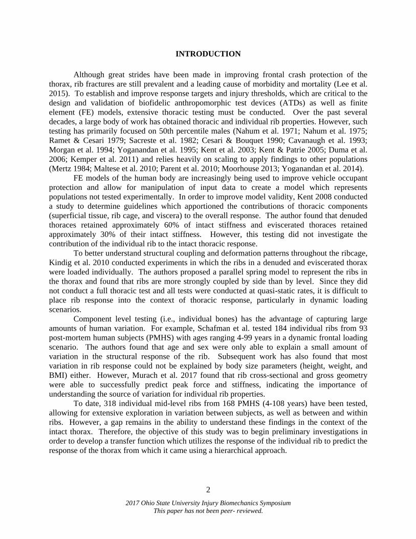
Table1: Subject Demographics Subject A Subject B Subject C
Sex M M M
Age (years) 73 62 55
Stature (cm) 170 173 183
Mass (kg) 62 84 75
BMI (kg/cm2) 21.5 28.3 22.4
aBMD t-score (lumbar) 0.4 1.6 1.1
Chest Depth (cm) 20 24 20
Chest Breadth (cm) 33 34 31
Chest Circumference (cm) 90 108 95
Thoracic Index 0.61 0.71 0.65 *BMI = Body Mass Index (mass/stature2), aBMD = areal bone mineral density as measured by dual-energy X-ray
absorptiometry (DXA)
The ram impactor mass was 23 kg with a 6” high x 12” wide x 0.5” thick rectangular face
centered vertically and horizontally on the sternum. The subject was tested in a fixed-back
scenario for all impacts and chest deflection was calculated from a linear displacement
potentiometer (Celesco CLWG-600-MC4, TE Connectivity Co., Berwyn, PA) attached to the
impactor face. Chest depth was measured prior to each impact and the stroke of the ram was
limited to <20% chest compression, as this is below the currently accepted threshold for thoracic
injury (Kent 2008; Duma et al. 2006; Kemper et al. 2011).
Two 6-axis load cells (Denton 2944JFL, Humanetics, Plymouth, MI) were used to
measure forces at the level of impact, with one attached to the impactor face (anterior) and one
located behind the back plate (posterior). Both load cells were used in order to eventually
compare these data to previous frontal thoracic work i.e., the force in an ATD frontal thorax
impact test is typically measured anteriorly and the force in individual rib testing is measured
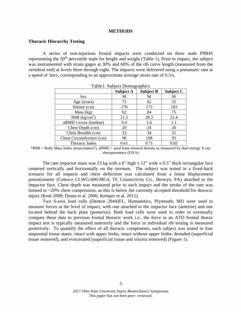
posteriorly. To quantify the effect of all thoracic components, each subject was tested in four
sequential tissue states: intact with upper limbs, intact without upper limbs, denuded (superficial
tissue removed), and eviscerated (superficial tissue and viscera removed) (Figure 1).
4
2017 Ohio State University Injury Biomechanics Symposium
This paper has not been peer- reviewed.
Figure 1: Experimental set-up for thoracic hierarchy testing in the four tissue states: a) intact
with upper limbs b) intact without upper limbs c) denuded and d) eviscerated. Each image is at
initial contact of the impactor face to the thorax.
Individual Rib Testing
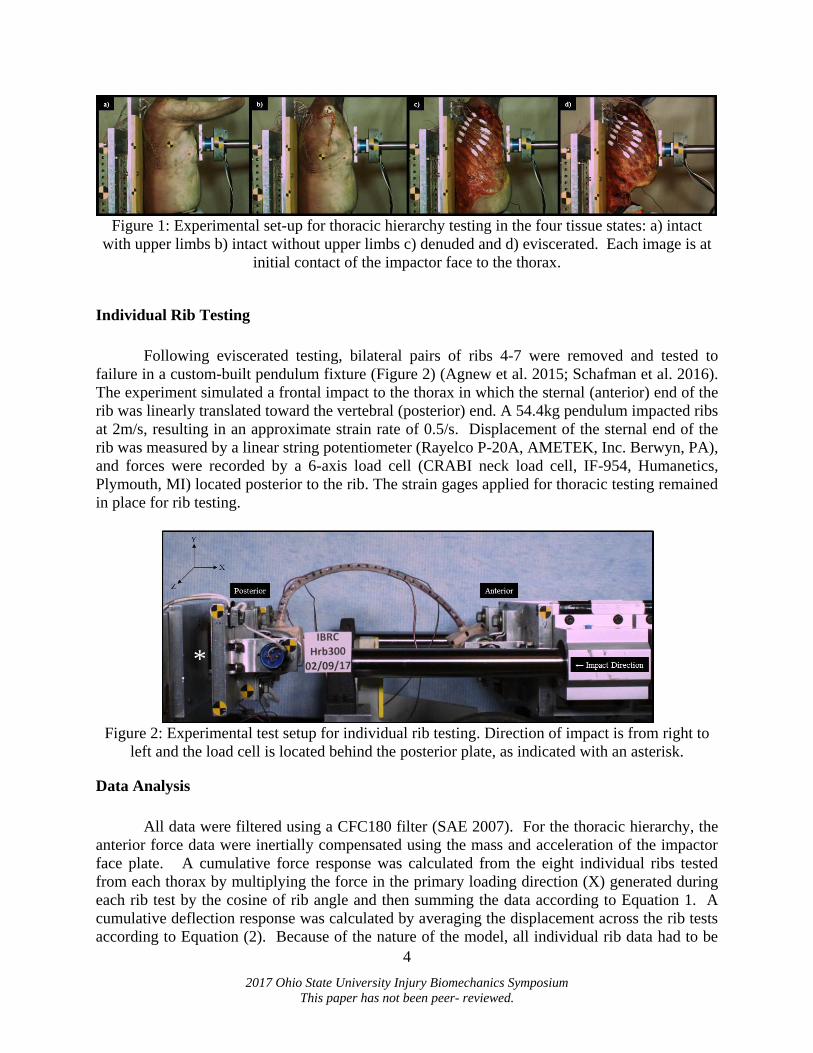
Following eviscerated testing, bilateral pairs of ribs 4-7 were removed and tested to
failure in a custom-built pendulum fixture (Figure 2) (Agnew et al. 2015; Schafman et al. 2016).
The experiment simulated a frontal impact to the thorax in which the sternal (anterior) end of the
rib was linearly translated toward the vertebral (posterior) end. A 54.4kg pendulum impacted ribs
at 2m/s, resulting in an approximate strain rate of 0.5/s. Displacement of the sternal end of the
rib was measured by a linear string potentiometer (Rayelco P-20A, AMETEK, Inc. Berwyn, PA),
and forces were recorded by a 6-axis load cell (CRABI neck load cell, IF-954, Humanetics,
Plymouth, MI) located posterior to the rib. The strain gages applied for thoracic testing remained
in place for rib testing.
Figure 2: Experimental test setup for individual rib testing. Direction of impact is from right to
left and the load cell is located behind the posterior plate, as indicated with an asterisk.
Data Analysis
All data were filtered using a CFC180 filter (SAE 2007). For the thoracic hierarchy, the
anterior force data were inertially compensated using the mass and acceleration of the impactor
face plate. A cumulative force response was calculated from the eight individual ribs tested
from each thorax by multiplying the force in the primary loading direction (X) generated during
each rib test by the cosine of rib angle and then summing the data according to Equation 1. A
cumulative deflection response was calculated by averaging the displacement across the rib tests
according to Equation (2). Because of the nature of the model, all individual rib data had to be
*
5
2017 Ohio State University Injury Biomechanics Symposium
This paper has not been peer- reviewed.
truncated according to the rib with the shortest time to failure. Rib angle was measured by
estimating a line along the rib length on a 2D lateral image of the seated subject in the
eviscerated condition, and then measuring the angle of that line with respect to horizontal. This
method of analysis treats each rib as a spring acting in parallel with the remaining ribs in the
thorax, similar to the analysis presented by Kindig et al. 2010.
𝐹𝑜𝑟𝑐𝑒 = ∑ 𝐹𝑛(𝑡)𝑛𝑖=1 (1)
𝐷𝑖𝑠𝑝𝑙𝑎𝑐𝑒𝑚𝑒𝑛𝑡 = 1
𝑛∑ 𝐷𝑛(𝑡)𝑛
𝑖=1 (2)
Since all subjects were limited to 20% chest deflection, a simplified stiffness value
reflecting the differences in response data was calculated by dividing peak force by peak
deflection. Differences in peak forces and stiffness values within subjects were quantified by
dividing the measured peak force of each tissue condition by the baseline condition (intact with
upper limbs), referred to as the force fraction here. The resulting values are interpreted as the
amount of force or stiffness that each tissue condition retained when compared to the baseline.
The ability of the cumulative rib model to represent the thorax was calculated by dividing the
cumulative rib model peak force (or stiffness) by the eviscerated peak force (or stiffness).
RESULTS
Thoracic Hierarchy Testing
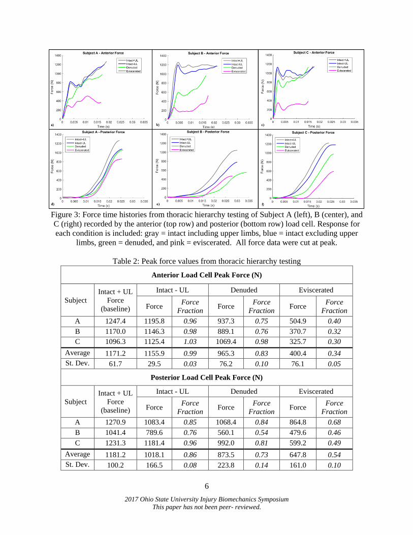
Time histories for forces recorded by the anterior and posterior load cells for all tissue
states of Subjects A-C can be found in Figure 3, and peak force values are shown in Table 2.
Qualitatively, removal of the upper limbs had a minimal effect on the resulting anterior force
data, and subsequent tissue removal resulted in a decrease in the overall response.
6
2017 Ohio State University Injury Biomechanics Symposium
This paper has not been peer- reviewed.
Figure 3: Force time histories from thoracic hierarchy testing of Subject A (left), B (center), and
C (right) recorded by the anterior (top row) and posterior (bottom row) load cell. Response for
each condition is included: gray = intact including upper limbs, blue = intact excluding upper
limbs, green = denuded, and pink = eviscerated. All force data were cut at peak.
Table 2: Peak force values from thoracic hierarchy testing
Anterior Load Cell Peak Force (N)
Subject
Intact + UL
Force
(baseline)
Intact - UL Denuded Eviscerated
Force Force
Fraction Force
Force
Fraction Force
Force
Fraction
A 1247.4 1195.8 0.96 937.3 0.75 504.9 0.40
B 1170.0 1146.3 0.98 889.1 0.76 370.7 0.32
C 1096.3 1125.4 1.03 1069.4 0.98 325.7 0.30
Average 1171.2 1155.9 0.99 965.3 0.83 400.4 0.34
St. Dev. 61.7 29.5 0.03 76.2 0.10 76.1 0.05
Posterior Load Cell Peak Force (N)
Subject
Intact + UL
Force
(baseline)
Intact - UL Denuded Eviscerated
Force Force
Fraction Force
Force
Fraction Force
Force
Fraction
A 1270.9 1083.4 0.85 1068.4 0.84 864.8 0.68
B 1041.4 789.6 0.76 560.1 0.54 479.6 0.46
C 1231.3 1181.4 0.96 992.0 0.81 599.2 0.49
Average 1181.2 1018.1 0.86 873.5 0.73 647.8 0.54
St. Dev. 100.2 166.5 0.08 223.8 0.14 161.0 0.10
7
2017 Ohio State University Injury Biomechanics Symposium
This paper has not been peer- reviewed.
It should be noted that Subject B was found to have incomplete (i.e. not through both
cortices) rib fractures located near the costochondral junction at autopsy. Strain gage data
revealed that these fractures occurred during the intact without upper limbs impact (baseline).
Since these fractures occurred in the first impact, subsequent responses and relative differences
with respect to that initial impact configuration were still compared within Subject B. Subject A
and C did not experience any rib fractures.
Peak forces decrease within each subject as tissue is removed. The differences in peak
force due to the removal of the upper limbs indicates the effect of this procedure is negligible
when using data from the anterior load cell, but not the posterior. On average using either
anterior and posterior load cell, the denuding procedure resulted in the thorax retaining 78% of
its intact force response and the eviscerating procedure resulted in the thorax retaining only 44%
of its intact force response.
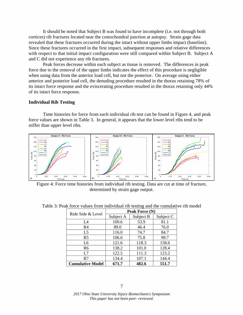
Individual Rib Testing
Time histories for force from each individual rib test can be found in Figure 4, and peak
force values are shown in Table 3. In general, it appears that the lower level ribs tend to be
stiffer than upper level ribs.
Figure 4: Force time histories from individual rib testing. Data are cut at time of fracture,
determined by strain gage output.
Table 3: Peak force values from individual rib testing and the cumulative rib model
Ride Side & Level Peak Force (N)
Subject A Subject B Subject C
L4 100.6 53.9 81.1
R4 89.0 46.4 76.0
L5 116.0 74.7 84.7
R5 106.6 75.8 99.7
L6 121.6 118.3 158.6
R6 138.2 101.0 128.4
L7 122.5 111.3 123.2
R7 134.4 107.1 144.4
Cumulative Model 671.7 482.6 551.7
8
2017 Ohio State University Injury Biomechanics Symposium
This paper has not been peer- reviewed.
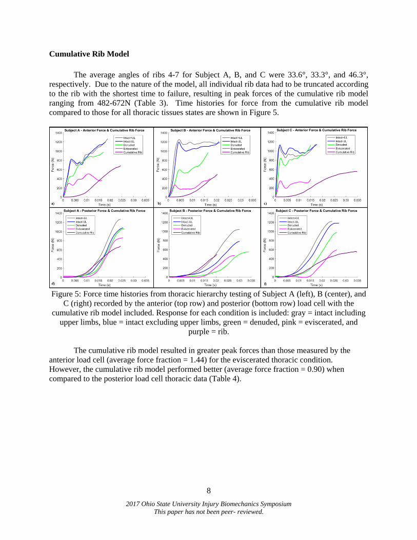
Cumulative Rib Model
The average angles of ribs 4-7 for Subject A, B, and C were 33.6°, 33.3°, and 46.3°,
respectively. Due to the nature of the model, all individual rib data had to be truncated according
to the rib with the shortest time to failure, resulting in peak forces of the cumulative rib model
ranging from 482-672N (Table 3). Time histories for force from the cumulative rib model
compared to those for all thoracic tissues states are shown in Figure 5.
Figure 5: Force time histories from thoracic hierarchy testing of Subject A (left), B (center), and
C (right) recorded by the anterior (top row) and posterior (bottom row) load cell with the
cumulative rib model included. Response for each condition is included: gray = intact including
upper limbs, blue = intact excluding upper limbs, green = denuded, pink = eviscerated, and
purple = rib.
The cumulative rib model resulted in greater peak forces than those measured by the
anterior load cell (average force fraction = 1.44) for the eviscerated thoracic condition.
However, the cumulative rib model performed better (average force fraction = 0.90) when
compared to the posterior load cell thoracic data (Table 4).
9
2017 Ohio State University Injury Biomechanics Symposium
This paper has not been peer- reviewed.
Table 4: Comparison of peak forces from eviscerated thoracic testing and cumulative rib models
Anterior Load Cell Peak Force (N)
Subject Eviscerated Cumulative Rib Force Fraction
A 504.8 671.7 1.33
B 370.7 482.6 1.30
C 325.8 551.7 1.69
Average 400.4 568.7 1.44
St. Dev. 76.0 78.1 0.18
Posterior Load Cell Peak Force (N)
Subject Eviscerated Cumulative Rib Force Fraction
A 864.8 671.7 0.78
B 479.6 482.6 1.01
C 599.1 551.7 0.92
Average 647.8 568.7 0.90
St. Dev. 161.0 78.1 0.09
Since all subjects were limited to 20% chest deflection, a simplified stiffness value
reflecting the differences in response data was calculated by dividing peak force by peak
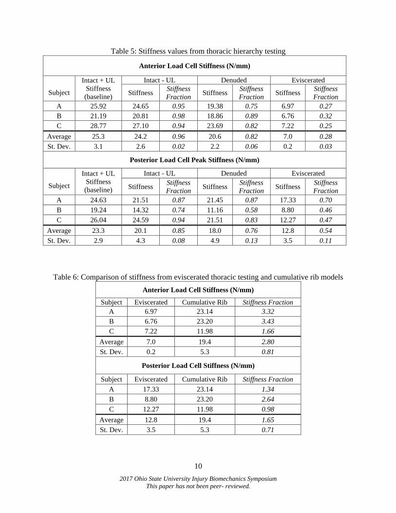
deflection (Table 5). On average from both load cells, denuding resulted in the thorax retaining
80% of its intact stiffness and eviscerating resulted in stiffness retention of 44%. Stiffness values
of the cumulative rib models were typically considerably higher than those obtained from
eviscerated thoracic testing (Table 6).
10
2017 Ohio State University Injury Biomechanics Symposium
This paper has not been peer- reviewed.
Table 5: Stiffness values from thoracic hierarchy testing
Anterior Load Cell Stiffness (N/mm)
Subject
Intact + UL
Stiffness
(baseline)
Intact - UL Denuded Eviscerated
Stiffness Stiffness
Fraction Stiffness
Stiffness
Fraction Stiffness
Stiffness
Fraction
A 25.92 24.65 0.95 19.38 0.75 6.97 0.27
B 21.19 20.81 0.98 18.86 0.89 6.76 0.32
C 28.77 27.10 0.94 23.69 0.82 7.22 0.25
Average 25.3 24.2 0.96 20.6 0.82 7.0 0.28
St. Dev. 3.1 2.6 0.02 2.2 0.06 0.2 0.03
Posterior Load Cell Peak Stiffness (N/mm)
Subject
Intact + UL
Stiffness
(baseline)
Intact - UL Denuded Eviscerated
Stiffness Stiffness
Fraction Stiffness
Stiffness
Fraction Stiffness
Stiffness
Fraction
A 24.63 21.51 0.87 21.45 0.87 17.33 0.70
B 19.24 14.32 0.74 11.16 0.58 8.80 0.46
C 26.04 24.59 0.94 21.51 0.83 12.27 0.47
Average 23.3 20.1 0.85 18.0 0.76 12.8 0.54
St. Dev. 2.9 4.3 0.08 4.9 0.13 3.5 0.11
Table 6: Comparison of stiffness from eviscerated thoracic testing and cumulative rib models
Anterior Load Cell Stiffness (N/mm)
Subject Eviscerated Cumulative Rib Stiffness Fraction
A 6.97 23.14 3.32
B 6.76 23.20 3.43
C 7.22 11.98 1.66
Average 7.0 19.4 2.80
St. Dev. 0.2 5.3 0.81
Posterior Load Cell Stiffness (N/mm)
Subject Eviscerated Cumulative Rib Stiffness Fraction
A 17.33 23.14 1.34
B 8.80 23.20 2.64
C 12.27 11.98 0.98
Average 12.8 19.4 1.65
St. Dev. 3.5 5.3 0.71
11
2017 Ohio State University Injury Biomechanics Symposium
This paper has not been peer- reviewed.
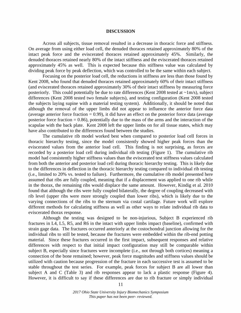
DISCUSSION
Across all subjects, tissue removal resulted in a decrease in thoracic force and stiffness.
On average from using either load cell, the denuded thoraces retained approximately 80% of the
intact peak force and the eviscerated thoraces retained approximately 45%. Similarly, the
denuded thoraces retained nearly 80% of the intact stiffness and the eviscerated thoraces retained
approximately 45% as well. This is expected because this stiffness value was calculated by
dividing peak force by peak deflection, which was controlled to be the same within each subject.
Focusing on the posterior load cell, the reductions in stiffness are less than those found by
Kent 2008, who found that denuded thoraces retained approximately 60% of their intact stiffness
(and eviscerated thoraces retained approximately 30% of their intact stiffness by measuring force
posteriorly. This could potentially be due to rate differences (Kent 2008 tested at ~1m/s), subject
differences (Kent 2008 tested two female subjects), and testing configuration (Kent 2008 tested
the subjects laying supine with a material testing system). Additionally, it should be noted that
although the removal of the upper limbs did not appear to influence the anterior force data
(average anterior force fraction = 0.99), it did have an effect on the posterior force data (average
posterior force fraction = 0.86), potentially due to the mass of the arms and the interaction of the
scapulae with the back plate. Kent 2008 left the upper limbs on for all tissue states, which may
have also contributed to the differences found between the studies.
The cumulative rib model worked best when compared to posterior load cell forces in
thoracic hierarchy testing, since the model consistently showed higher peak forces than the
eviscerated values from the anterior load cell. This finding is not surprising, as forces are
recorded by a posterior load cell during individual rib testing (Figure 1). The cumulative rib
model had consistently higher stiffness values than the eviscerated test stiffness values calculated
from both the anterior and posterior load cell during thoracic hierarchy testing. This is likely due
to the differences in deflection in the thoracic hierarchy testing compared to individual rib testing
(i.e., limited to 20% vs. tested to failure). Furthermore, the cumulative rib model presented here
assumed that ribs are fully coupled, meaning that if a displacement was applied to one rib while
in the thorax, the remaining ribs would displace the same amount. However, Kindig et al. 2010
found that although the ribs were fully coupled bilaterally, the degree of coupling decreased with
rib level (upper ribs were more strongly coupled than lower ribs), which is likely due to the
varying connections of the ribs to the sternum via costal cartilage. Future work will explore
different methods for calculating stiffness as well as other ways to relate individual rib data to
eviscerated thorax response.
Although the testing was designed to be non-injurious, Subject B experienced rib
fractures in L4, L5, R5, and R6 in the intact with upper limbs impact (baseline), confirmed with
strain gage data. The fractures occurred anteriorly at the costochondral junction allowing for the
individual ribs to still be tested, because the fractures were embedded within the rib-end potting
material. Since these fractures occurred in the first impact, subsequent responses and relative
differences with respect to that initial impact configuration may still be comparable within
subject B, especially since fractures were incomplete (i.e., not through both cortices) meaning a
connection of the bone remained; however, peak force magnitudes and stiffness values should be
utilized with caution because progression of the fracture in each successive test is assumed to be
stable throughout the test series. For example, peak forces for subject B are all lower than
subject A and C (Table 3) and rib responses appear to lack a plastic response (Figure 4).
However, it is difficult to say if these differences are due to rib fracture or simply individual
12
2017 Ohio State University Injury Biomechanics Symposium
This paper has not been peer- reviewed.
variation. Surprisingly, Subject B had the highest aBMD t-score, which is the current metric for
bone quality, indicating that this subject should not have experienced fractures.
Although these data offer potential for understanding contributions of individual ribs to
thoracic response, there are several limitations. Thoracic testing was limited to 20% chest
compression to allow for repeated non-injurious impacts, meaning the response data does not
represent a full impact where the ram would be allowed to displace until all input energy is
absorbed. Despite this, fractures still occurred for one subject. Additionally, all tests were
completed using a fixed back scenario, which is not a realistic representation of a car crash
scenario in which the forward inertia of occupants plays an important role. Although the angle
of the rib was factored into the cumulative rib model, the model does not account for off-axis
loading or rotation of the ribs with respect to the spine during thoracic hierarchy testing, both of
which are likely important for establishing an accurate model. Future work will include
additional testing to further improve understanding of the effects of denuding and eviscerating.
Additionally, more complex methods of calculating thoracic stiffness will be investigated, as
well as more intricate thoracic models that incorporate damping and inertial terms. Alternative
cumulative rib models will be explored that incorporate individual rib force, displacement, angle,
and strain.
CONCLUSIONS
Preliminary data presented here reveal that denuded thoraces retain approximately 80%
of the intact peak force and stiffness, and the eviscerated thoraces retain approximately 45% of
peak force and stiffness. The cumulative rib model shows promise when utilized in context of
the posterior load cell force in thoracic testing. These data will be useful for developing a
transfer function to allow for the prediction of thoracic response from individual rib data, to
allow for the generation of thoracic response data for populations for which full-thoracic impacts
are not typically conducted (e.g., pediatric) but individual rib testing can be. These data could be
used to improve thoracic response targets and help assess the biofidelity of ATDs of all sizes and
demographics.
ACKNOWLEDGEMENTS
Funding for this study was provided by National Highway Traffic Safety Administration
(NHTSA). Thank you to all the students and staff of the Injury Biomechanics Research Center,
especially Benjamin Shurtz, David Stark, Arrianna Willis, Julie Bing, Rakshit Ramachandra,
Elina Misicka, Akshara Sreedhar, Scott Stuckey, and Warren Lee for their endless support.
Thank you to Brian Suntay and Colton Thomas from the Transportation Research Center, Inc.
Thank you to Mark and Michelle Whitmer, OSU’s Body Donor Program, Lifeline of Ohio, and
especially the anatomical donors for their generous gifts.
REFERENCES
Agnew, A.M. et al., 2015. The Effect of Age on the Structural Properties of Human Ribs.
Journal of the Mechanical Behavior of Biomedical Materials, 41, pp.302–314.
Cavanaugh, J.M. et al., 1993. Injury and Response of the Thorax in Side Impact Cadaveric Tests.
Stapp Car Crash Conference, 37, pp.199–221.
Cesari, D. & Bouquet, R., 1990. Behavior of Human Surrogates Thorax under Belt Loading. SAE
13
2017 Ohio State University Injury Biomechanics Symposium
This paper has not been peer- reviewed.
International, 902310, pp.73–81.
Duma, S.M. et al., 2006. Non-Censored Rib Fracture Data From Dynamic Belt Loading Tests on
the Human Cadaver Thorax. Biomedical Sciences Instrumentation, 42, pp.148–153.
Kemper, A.R. et al., 2011. Reducing Chest Injuries in Automobile Collisions: Rib Fracture
Timing and Implications for Thoracic Injury Criteria. Annals of Biomedical Engineering,
39(8), pp.2141–2151.
Kent, R., 2008. Frontal Thoracic Response to Dynamic Loading: The Role of Superficial
Tissues, Viscera and the Rib Cage. International Journal of Crashworthiness, 13(3),
pp.289–300.
Kent, R. & Patrie, J., 2005. Chest Deflection Tolerance to Blunt Anterior Loading is Sensitive to
Age but Not Load Distribution. Forensic Science International, 149(2–3), pp.121–128.
Kent, R.W., Patrie, J. & Benson, N., 2003. Restrained Hybrid III Dummy-Based Criteria for
Thoracic Hard Tissue Injury Prediction. In International Conference on the Biomechanics
of Impact. pp. 215–232.
Kindig, M.W. et al., 2010. Structural Response of Cadaveric Ribcages under a Localized
Loading: Stiffness and Kinemamtic Trends. Stapp Car Crash Conference, 54, pp.337–380.
Lee, E.L., Craig, M. & Scarboro, M., 2015. Real-World Rib Fracture Patterns in Frontal Crashes
in Different Restraint Conditions. Traffic Injury Prevention, 16(sup2), pp.S115–S123.
Available at: http://www.tandfonline.com/doi/full/10.1080/15389588.2015.1062888.
Maltese, M.R. et al., 2010. Incorporation of CPR Data into ATD Chest Impact Response
Requirements. Annals of Advances in Automotive Medicine, 54(Figure 1), pp.79–88.
Mertz, H.J., 1984. A Procedure for Normalizing Impact Response Data. Society of Automotive
Engineers.
Moorhouse, K., 2013. An Improved Normalization Methodology for Developing Mean Human
Response Curves. Proc. of 23rd International Technical Conference on the Enhancced
Safety of Vehicles. Paper No. 13-0192.
Morgan, R.M. et al., 1994. Thoracic Trauma Assessment Formulations for Restrained Drivers in
Simulated Frontal Impacts. Stapp Car Crash Journal, 38, pp.15–34.
Murach, M.M. et al., 2017. Rib Geometry Explains Variation in Dynamic Structural Response:
Potential Implications for Frontal Impact Fracture Risk. Annals of Biomedical Engineering.
Nahum, A. et al., 1971. The Biomechanical Basis for Chest Impact Protection: I. Force-
Deflection Characteristics of the Thorax. Journal of Trauma, 11(10), pp.874–882.
Nahum, A., Schneider, D. & Kroell, C., 1975. Cadaver Skeletal Response to Blunt Thoracic
Impact. Stapp Car Crash Conference, 19, pp.259–293.
Parent, D.P. et al., 2010. Comparison of Hybrid III Child Test Dummies to Pediatric PMHS in
Blunt Thoracic Impact Response. Traffic Injury Prevention, 11(4), pp.399–410.
Ramet, M. & Cesari, D., 1979. Behavior of Restrained Dummies and Cadavers in Frontal
Impacts. In International Conference on the Biomechanics of Impact. pp. 210–222.
Sacreste, J. et al., 1982. Proposal for a Thorax Tolerance Level in Side Impacts Based on 62
Tests Performed with Cadavers Having Known Bone Condition. In Stapp Car Crash
Conference. pp. 155–171.
SAE, S. of A.E., 2007. Instrumentation for Impact Test-Part 1-Electronic Instrumentation,
Warrendale, PA.
Schafman, M.A. et al., 2016. Age and sex alone are insufficient to predict human rib structural
response to dynamic A-P loading. Journal of Biomechanics, 49(14), pp.3516–3522.
Available at: http://dx.doi.org/10.1016/j.jbiomech.2016.09.030.
14
2017 Ohio State University Injury Biomechanics Symposium
This paper has not been peer- reviewed.
Yoganandan, N. et al., 1995. Thoracic Deformation and Velocity Analysis in Frontal Impact.
Journal of Biomechanical Engineering, 117(1), pp.48–52.
Yoganandan, N., Arun, M.W.J. & Pintar, F. a., 2014. Normalizing and Scaling of Data to Derive
Human Response Corridors from Impact Tests. Journal of Biomechanics, 47(8), pp.1749–
1756. Available at: http://dx.doi.org/10.1016/j.jbiomech.2014.03.010.