Embed Size (px)
Citation preview

ORIGINAL ARTICLE
A Potential Suppressor of TGF-b Delays Catagen Progressionin Hair Follicles
YumikoTsuji,n Sumiko Denda,nTsutomu Soma,nw Laurel Raftery,wTakashi Momoiz and Toshihiko HibinonnShiseido Life Science Research Center,Yokohama, Japan; wMGH/Harvard Cutaneous Biology Research Center, Charlestown, MA, USA; zNationalInstitute of Neuroscience,Tokyo, Japan
TGF-b plays important roles in the induction of catagenduring the hair cycle. We examined whether TGF-b2could activate a caspase in human hair follicles. Usingactive caspase-9 and -3 speci¢c antibodies, we foundthat TGF-b2 activated these caspases in two regions,the lower part of the hair bulb and the outer layer ofthe outer root sheath. In addition, we searched for aplant extract that can e¡ectively suppress TGF-b action.We found that an extract of Hydrangea macrophyllareduced synthesis of a TGDb-inducible protein.We con-
¢rmed that the extract has a potential to promote hairelongation in the organ culture system. Furthermore, itdelayed in vivo progression of catagen in a mouse mod-el. Our results suggest that the induction of catagen byTGF-b is mediated via activation of caspases and that asuppressor of TGF-b could be e¡ective in preventingmale pattern baldness. Keywords:TGF-�/hair cycle/catageninduction/caspase/apoptosis. JID Symposium Proceedings8:65 ^68, 2003
Hair loss is the result of premature entry into catagendue to various causes, including androgens (Jahoda,1998), anticancer drugs (Paus et al, 1994), and in-£ammatory reactions (Galbraith et al, 1984). Threedistinct phases have been de¢ned for the mamma-
lian hair cycle: anagen (growing phase), catagen (regressingphase), and telogen (resting phase) (Kligman, 1959). It is impor-tant to understand the catagen induction mechanism in order to¢nd ways to prevent hair loss.We have previously reported two important ¢ndings in this
process. First, catagen is characterized by the massive apoptoticcell death of follicular epithelial cells (Soma et al, 1998). Second,TGF-b2 appears in the lower part of the boundary area betweenthe dermal papilla cells and the germinative matrix cells duringanagen-catagen transition phase in vivo. Using hair follicle organculture we have clearly demonstrated that exogenous TGF-bcould induce morphological changes and apoptotic cell death in-distinguishable from that which is seen in human catagen hairfollicles. In progressing stages of catagen, TGF-b2-, TGF-bRII-and TUNEL-positive cells were colocalized at the regressingepithelial strand. Furthermore, we demonstrated that a neutraliz-ing antibody toTGF-b prevented morphological changes and re-sulted in the elongation of hair shafts (Soma, submitted). These¢ndings strongly suggest that TGF-b plays an essential role incatagen induction via activation of an apoptotic pathway.Caspases are well-known expediters of apoptosis and 14 mem-
bers are known in mammals (Kumar, 1999). Once the caspase cas-cade is activated, it inevitably leads to apoptotic cell death. It isnow widely accepted that sequential activation of caspases is re-quired in apoptosis processes.In the present study, we investigated whether TGF-b2 could
activate the expedition of apoptosis, the caspase cascade. In addi-tion, we searched for a plant extract that could e¡ectively suppress
TGF-b action in catagen progression.We identi¢ed such an ex-tract and con¢rmed that it can delay catagen progression in vivo.
MATERIALS ANDMETHODS
Culture of human hair follicles Human scalp skin specimens wereobtained from plastic surgery. Human hair follicles were isolated andcultured according to the method of Philpot et al (1990). In order toanalyze the e¡ect of TGF-b on caspase activation, anagen hair follicleswere incubated in the presence of TGF-b2 (20 ng/ml) for 2 days and then10 mm frozen sections were prepared. To evaluate TGF-b antagonisticmolecules, the length of hair follicles in the presence or absence of testmaterials was measured by light microscopy. Ten hair follicles were usedfor each sample to perform statistical studies.
Double immuno-detection of tunel-positive cells and activecaspases It was important to identify the site of caspase activation forthe better understanding of the catagen induction mechanism. For thispurpose, we used cleavage site-directed antibodies to caspase-9 (Fujitaet al, 2000) and -3 (Kouroku et al, 1998), respectively. These antibodies donot recognize proforms, and only active enzymes can be detected. Usingthese antibodies, we studied the relationship between the appearance ofTUNEL-positive cells and caspase activation. Cryostat sections were ¢xedwith aceton at room temperature for 20 min and incubated with the activecaspase-3 or -9 speci¢c antibodies at 41C overnight. Texas Reds dye-conjugated antirabbit IgG (donkey) was used as a secondary antibody. TheTUNEL reaction was performed using a £uorescein in situ cell deathdetection kit (Roche Diagnostics) according to the manufacturer’sinstructions.
TGF-b suppression activity of plant extracts Dermal papilla (DP)cells were isolated according to the method of Itami et al (1990) and usedwithin 4 passages. DP cells were incubated with plant extracts in thepresence of TGF-b2 (1 ng/ml) for 24 h. TGF-b suppression was assessedby monitoring the amount of plasminogen activator inhibitor 1 (PAI-1), aTGF-b-responsive gene product, in culture medium using TintElize PAI-1(Biopool, CA). Cytotoxicity of every extract was tested using AlamarBlue(Biopool) according to the manufacturer’s instructions.
In vivo test of catagen suppression Female C57BL/6 mice(approximately 8 weeks of age) were used in this study, since the hair
Reprint requests to: Toshihiko Hibino, Shiseido Life Science ResearchCenter, 2-12-1 Fukuura, Kanazawa-ku,Yokohama 236-8643, Japan;E-mail: [email protected]
Accepted for publication February 1, 2003
0022-202X/03/$15.00 . Copyright r 2003 by The Society for Investigative Dermatology, Inc.
65

cycle is well characterized in this mouse (Muller-Rover et al, 2001). Hairinduction was obvious at day 10 after depilation by wax. An extract wastopically applied on the back once a day for 10 days after hair induction.Back skin sections were prepared, stained with HE, and the catagen stageof each hair follicle was scored. Stages of catagen were determined based onthe criteria described by Muller-Rover et al (2001). One hundred hairfollicle sections were graded for each mouse, and 5 mice were used foreach group.
RESULTS
TGF-b2 activates caspases in cultured hair follicles Tounderstand the role of TGF-b2 in relation to apoptosis, humanhair follicles were cultured in the presence of TGF-b2 for 2 dand then examined for the activation of caspase-9 and caspase-3.We focused on these two caspases, since they represent theinitiator caspase and the e¡ector caspase, respectively (Kumar,1999). Using an active caspase-3 in two regions, including thecells in the lower part of the germinative matrix and the outerlayer of the outer root sheath (Fig 1A). Caspase-9 was alsoactivated in similar areas (Fig 1B). Only a few cells were positivefor active caspase-3 in hair follicles cultured without TGF-b2(Fig 1C). Dual staining for active caspase-3 and TUNEL
showed the TUNEL-positive cells consistently overlapped withcaspase-3-positive cells (Fig 1D^1F). Some cells were onlypositive for active caspase-3 (Fig 1E,F). These cells probablylacked nuclear material in the thin section. Control hair follicles(Fig 1G) showed several positive cells around the cortex,suggesting that these are not related to activity of exogenousTGF-b.
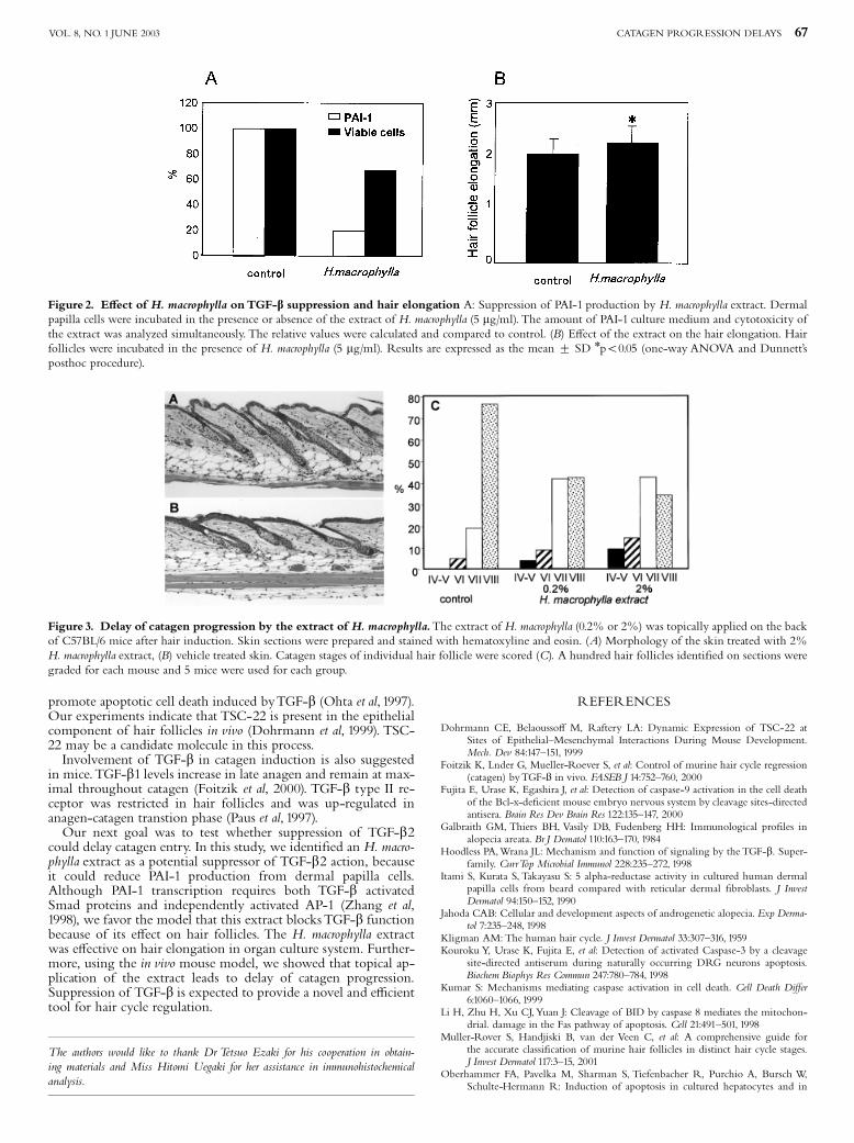
Hydrangea macrophylla, a potential TGF-b2 antagonist, causedelongation of hair follicles Suppression of TGF-b was hypothe-sized to prevent apoptosis and to delay catagen entry. Therefore,we sought a natural inhibitor of TGF-b action.We developed ascreening method for TGF-b suppression by monitoring changesin PAI-1, a TGF-b responsive gene product. (Zhang et al, 1998).Over 400 plant extracts were screened. Among these, the extractfrom Hydrangea macrophylla (tea of Heaven) was most e¡ective insuppressing PAI-1 synthesis (Fig 2A). Using the organ culturesystem, the e¡ect of the H. macrophylla extract on hair follicleelongation was tested. Human hair follicles were cultured in thepresence of the extract for 7 d. The resulting follicles showedsigni¢cant elongation (Fig 2B). Treated follicles showed anaverage of 2.25 mm elongation, compared to an average of 2.0mm for the controls. The di¡erence is signi¢cant to a p-value ofo0.05 as assessed by the one-way ANOVA and Dunnett’s posthocprocedure.
H. macrophylla delayed catagen progression in vivo To con¢rmthe e¡ect of the H. macrophylla extract on catagen progression, anin vivo test was performed using C57BL/6 mice. A new, synchro-nized hair cycle was induced by wax depilation. The plant extractwas topically applied continuously on the back for 10 d from day12 after hair induction. Skin sections were prepared, stained withHE, and the catagen stage of each hair follicle was scored. Stagesof catagen were determined according to the method of Muller-Rover et al (2001). Figure 3A and 3B show the typical features ofhistochemical analyzes. Figure 3C summarizes the ratio of eachcatagen stage for each group. Over half of the hair follicles treatedwith 0.2% H. macrophylla extract were in earlier catagen stagesthan in the control skin. Two percent extract showed a slightlymore potent e¡ect on catagen progression than 0.2%.
DISCUSSION
TGF-b is known to regulate various physiological reactions in-cluding apoptotic cell death (Oberhammer et al, 1992; Ohta et al,1994). Previously we have shown the spatio-temporal localizationof TGF-b isoforms during the human hair cycle, and we sug-gested that up-regulation and speci¢c localization of TGF-b2 inthe anagen-catagen transition may initiate the process of catagen(Soma et al, submitted). In this report, we analyzed activation of acaspase network byTGF-b2 in human hair follicles.We focusedon the initiator caspase-9 and the e¡ector caspase-3. Our resultsclearly demonstrated that TGF-b2 can activate these two caspasesin an organ culture system. This is the ¢rst evidence that TGF-bcan elicit apoptotic cell death through the activation of a caspasenetwork in human hair follicles.We still do not know all the mo-lecules that may play roles in this network activation byTGF-b.Recently it was suggested that caspase-8 is located upstream ofcaspase-9 (Li et al, 1998; Pan et al, 2001). Caspase-8 could also beincluded in apoptosis induced byTGF-b2.Our preliminary data indicate that caspase activation may not
be an immediate early response to TGF-b (unpublished data).Well-known mediators for TGF-b signal transduction includemembers of the Smad family (Hoodless andWrana, 1998). Smad2can speci¢cally mediate TGF-b/activin signal transduction. AfterTGF-b stimulation, phosphorylated Smad2 binds with Smad4and the complex translocates to the nucleus. We speculate thatthe mechanism of caspase activation by TGF-b includes the in-duction of other functional molecules.TSC-22,TGF-b stimulatedclone-22, is a TGF-b responsive gene that was suggested to
Figure1. Caspase activation in hair follicles treated with TGF-b2.Hair follicles were cultured in the presence (A, B, D E and F) or absence(C and G) of TGF-b2 and immuno-stained with the cleavage site-directedantibody to caspase-3 (A) or caspase-9 (B). Caspase activation occured inthe lower part of germinative matrix cells and outer layers of outer rootsheath cells (arrow) in the hair follicles treated with TGF-b2, comparedwith the control (C). Dual staining for active caspase-3 and TUNEL de-monstrated colocalization of these cells (D^F). E and F show magni¢edviews. The anagen hair follicle in the absence of TGF-b2 showed positivesignals around hair cortex, which are not related toTGF-b2 (G). scale bars:100 mm
66 TSUJI ETAL JID SYMPOSIUM PROCEEDINGS
promote apoptotic cell death induced byTGF-b (Ohta et al, 1997).Our experiments indicate that TSC-22 is present in the epithelialcomponent of hair follicles in vivo (Dohrmann et al, 1999). TSC-22 may be a candidate molecule in this process.Involvement of TGF-b in catagen induction is also suggested
in mice.TGF-b1 levels increase in late anagen and remain at max-imal throughout catagen (Foitzik et al, 2000). TGF-b type II re-ceptor was restricted in hair follicles and was up-regulated inanagen-catagen transtion phase (Paus et al, 1997).Our next goal was to test whether suppression of TGF-b2
could delay catagen entry. In this study, we identi¢ed an H. macro-phylla extract as a potential suppressor of TGF-b2 action, becauseit could reduce PAI-1 production from dermal papilla cells.Although PAI-1 transcription requires both TGF-b activatedSmad proteins and independently activated AP-1 (Zhang et al,1998), we favor the model that this extract blocksTGF-b functionbecause of its e¡ect on hair follicles. The H. macrophylla extractwas e¡ective on hair elongation in organ culture system. Further-more, using the in vivo mouse model, we showed that topical ap-plication of the extract leads to delay of catagen progression.Suppression of TGF-b is expected to provide a novel and e⁄cienttool for hair cycle regulation.
The authors would like to thank Dr Tetsuo Ezaki for his cooperation in obtain-ing materials and Miss Hitomi Uegaki for her assistance in immunohistochemicalanalysis.
REFERENCES
Dohrmann CE, Belaousso¡ M, Raftery LA: Dynamic Expression of TSC-22 atSites of Epithelial^Mesenchymal Interactions During Mouse Development.Mech. Dev 84:147^151, 1999
Foitzik K, Lnder G, Mueller-Roever S, et al: Control of murine hair cycle regression(catagen) byTGF-� in vivo. FASEB J 14:752^760, 2000
Fujita E, Urase K, Egashira J, et al: Detection of caspase-9 activation in the cell deathof the Bcl-x-de¢cient mouse embryo nervous system by cleavage sites-directedantisera. Brain Res Dev Brain Res 122:135^147, 2000
Galbraith GM, Thiers BH, Vasily DB, Fudenberg HH: Immunological pro¢les inalopecia areata. Br J Dematol 110:163^170, 1984
Hoodless PA,Wrana JL: Mechanism and function of signaling by the TGF-b. Super-family. CurrTop Microbial Immunol 228:235^272, 1998
Itami S, Kurata S, Takayasu S: 5 alpha-reductase activity in cultured human dermalpapilla cells from beard compared with reticular dermal ¢broblasts. J InvestDermatol 94:150^152, 1990
Jahoda CAB: Cellular and development aspects of androgenetic alopecia. Exp Derma-tol 7:235^248, 1998
Kligman AM:The human hair cycle. J Invest Dermatol 33:307^316, 1959Kouroku Y, Urase K, Fujita E, et al: Detection of activated Caspase-3 by a cleavage
site-directed antiserum during naturally occurring DRG neurons apoptosis.Biochem Biophys Res Commun 247:780^784, 1998
Kumar S: Mechanisms mediating caspase activation in cell death. Cell Death Di¡er6:1060^1066, 1999
Li H, Zhu H, Xu CJ,Yuan J: Cleavage of BID by caspase 8 mediates the mitochon-drial. damage in the Fas pathway of apoptosis. Cell 21:491^501, 1998
Muller-Rover S, Handjiski B, van der Veen C, et al: A comprehensive guide forthe accurate classi¢cation of murine hair follicles in distinct hair cycle stages.J Invest Dermatol 117:3^15, 2001
Oberhammer FA, Pavelka M, Sharman S, Tiefenbacher R, Purchio A, Bursch W,Schulte-Hermann R: Induction of apoptosis in cultured hepatocytes and in
Figure 2. E¡ect of H. macrophylla onTGF-b suppression and hair elongation A: Suppression of PAI-1 production by H. macrophylla extract. Dermalpapilla cells were incubated in the presence or absence of the extract of H. macrophylla (5 mg/ml). The amount of PAI-1 culture medium and cytotoxicity ofthe extract was analyzed simultaneously. The relative values were calculated and compared to control. (B) E¡ect of the extract on the hair elongation. Hairfollicles were incubated in the presence of H. macrophylla (5 mg/ml). Results are expressed as the mean 7 SD npo0.05 (one-way ANOVA and Dunnett’sposthoc procedure).
Figure 3. Delay of catagen progression by the extract of H. macrophylla.The extract of H. macrophylla (0.2% or 2%) was topically applied on the backof C57BL/6 mice after hair induction. Skin sections were prepared and stained with hematoxyline and eosin. (A) Morphology of the skin treated with 2%H. macrophylla extract, (B) vehicle treated skin. Catagen stages of individual hair follicle were scored (C). A hundred hair follicles identi¢ed on sections weregraded for each mouse and 5 mice were used for each group.
CATAGEN PROGRESSION DELAYS 67VOL. 8, NO. 1 JUNE 2003

regressing liver by transforming growth factor-b1. Proceedings of the Natl AcadSci USA 89:5408^5412, 1992
Ohta S,Yanagihara K, Nagata K: Mechanism of apoptotic cell death of human gas-tric carcinoma cells mediated by transforming growth factor beta. Biochem J15:777^782, 1994
Pan J, Xu G,Yeung SC: Cytochrome c release is upstream to activation of caspase-9,caspase-8 and caspase-3 in the enhanced apoptosis of anaplastic thyroid cancercells induced by manumycin and paclitaxel. J Clin Endocrinol Metab 86:4731^4740, 2001
Paus R, Foitzik K, Welker P, Bulfone-Paus S, Eichmuller S: Transforming growthfactor- b receptor type I and type II expression during murine hair follicledevelopment and cycling. J Invest Dermatol 109:518^26, 1997
Paus R, Handjiski B, Eichmuller S, Czarnetzki BM: Chemotherapy-induced alope-cia in mice. Induction by cyclophosphamide, inhibition by cyclosporin A, andmodulation by dexamethasone. AmJ Pathol 144:719^734, 1994
Philpot MP, Green MR, KealeyT: Human hair growth in vitro. J Cell Sci 97:463^471,1990
Soma T, Ogo M, Suzuki J, Takahasi T, Hibino T: Analysis of apoptotic cell death inhuman hair follicles. J Invest Deramtol 112:518^526, 1998
Zhang Y, Feng XH, Derynk R: Smad3 and Smad4 cooperate with c-June/c-Fos tomediate TGFa-induced transcription. Nature 394:909^913, 1998
68 TSUJI ETAL JID SYMPOSIUM PROCEEDINGS