Embed Size (px)
Citation preview

Instructions for use
Title A possible regulatory mechanism of conjugation process in Spirogyra
Author(s) SASAKI, Kimiko
Citation Journal of the Faculty of Science, Hokkaido University. Series 5, Botany, 12(2), 135-145
Issue Date 1980
Doc URL http://hdl.handle.net/2115/26377
Type bulletin (article)
File Information 12(2)_P135-145.pdf
Hokkaido University Collection of Scholarly and Academic Papers : HUSCAP

Journ. Fac. Sci., Hokkaido Univ. Ser. V (Botany), 12 (2): 135-146, 1980.
A possible regulatory mechanism of conjugation
process in Spirogyra
Kimiko SASAKI
For the purpose of examination of sexual regulation of gene expression, soluble
chromatins isolated from Spirogyra sp. in different stages of life cycle were analyzed
chromatographically, spectrophotometrically, and enzymatically.
Fractionation of soluble-, dissociated-, and dehistonized-chromatins showed that
each of these chromatins consisted predominantly of a high molecular weight fraction
and a low one both in vegetative cells (V-cells) and in conjugation and zygote cells
(C, Z-cells). Each of the high and low molecular weight fractions might correspond
with each other between V-cells and C, Z-cells, but both fractions of C, Z-cells
were eluted from a Sepharose 6B and a Sephadex G-200 columns more rapidly
than the respective fractions of V-cells, suggesting that the fractions in C- and
Z-cells had different conformation from or were larger than those in V-cells.
Melting temperature of the Z-cell chromatins was always higher than that of
V-cell chromatins. In opposition to those results, template activity of C- or Z-cell
chromatins was less than that of V-cell chromatins in a presence or absence of
exogenous RNA polymerase. As the dehistonized chromatins still had a large
amount of proteins, we presume that such proteins, probably mating-specific proteins,
may bind to chromatin DNA and regulate sexual process in some manner.
Numerous studies have suggested that nonhistone chromosomal proteins regulate gene expression in eukaryotic cells (STEIN et ai., 1974; PAUL and GILMOUR, 1975). Structural and functional studies of chromatin have made rapid progress (CLARK and FELSENFELD, 1974; ELGIN and WEINTRAUB, 1975; FINCH et ai., 1975, 1977; COMPTON et ai., 1976; STEIN et ai., 1977; FELSENFELD, 1978; MACIEWICZ and LI, 1978; GORDON et ai., 1978). We are attempting to clarify regulatory factors in gene expression for cellular differentiation and mating in Spirogya and Ciosterium. In Spirogyra, we reported that RNA- and DNA-synthesizing activities of the chromatins of C- or Z-cells were much lower than that of V -cells (SASAKI et ai., 1972; SASAKI and TAKAYA, 1972), structure of nucleoprotein complex and respiratory and photochemical activities changed during the life cycle (SASAKI and TAKAYA, 1974; SASAKI, 1977), and nonhistone proteins which included an inhibitor of chromatin-directed RNA synthesis were isolated from purified

136 K. Sasaki
C- and Z-cell chromatins (SASAKI, 1978)_ In this study, the purified chromatins prepared from Spirogyra in different
stages were further solubilized by shearing, and were dissociated or dehistonized. These chromatins were subjected to estimations of template activity and half melting temperature (Tm), and subjected to chromatography. These results and a possible regulatory mechanism of conjugation are described in this paper.
Materials and Methods
Materials: Spirogyra sp. (cell width, ca. 115/-Lm) was collected in the different developmental stages from a pond in the campus of Hokkaido University at 10 to 11 a. m. The cells were thoroughly washed with sterilized deionized water, and frozen at - 20°C until use. The life cycle was conveniently divided into three stages: vegetative growth (V -cells), conjugation (C-cells), and newly-formed zygote (Z-cells) stages. When conjugation proceeded very rapidly, a mixture of C- and Z-cells was used for the mating (C, Z-cells) stage, because of difficulty in separation of C- and Z-cells.
Reagents: Escherichia coli RNA polymerase was prepared by the method of BURGESS (1969). Calf thymus DNA (mol. wt., >2 X 10⁶) was obtained from Boehringer Mannheim. Sepharose 6B, Sephadex G-100 and G-200, and blue dextran (mot. wt., 2 X 10⁶) were purchased from Pharmacia Fine Chemicals. [8-3H] A TP (22.7 Ci/mmol) was from Worthington Biochem. Co.
Preparation of chromatin: Particulate chromatin was isolated and purified by the procedure described in previous papers (SASAKI and TAKA Y A, 1974; BONNER and HUANG, 1963). The chromatin was sheared in 10 mM Tris-HCI (pH 8.0) by vigorous shaking for 1 min, then gentle shaking for 2 hr at 0_4°C in a Vibro Shaker. The sheared chromatin solution was centrifuged at 15,000 X g for 30 min. Resultant supernatant was passed through a Sephadex G-100 column to remove smaller molecular weight components, and used as "soluble chromatin". The shearing procedure was repeated three times, and respective chromatin solutions were named as Exts. 1, 2, and 3.
Preparation of dehistonized chromatin: Histones were removed from ethanol-precipitated chromatin by extraction three times with 0.35 M HCl. Insoluble component in the HCI solution was collected by centrifugation, sheared, dissolved in the dilute saline-citrate solution (DSC, 0.015 M NaCl-0.0015 M trisodium citrate, pH 7.0), and used as "dehistonized chromatin".
Preparation of DNA: DNA was isolated from chromatin by the method of MARMUR (1961), purified by the treatment with chloroform-isoamylalcohol
Chromatin of Spirogyra 137
(8 : 1), RNase, and pronase, and then dissolved in 10 mM Tris-HCI (pH 8.0) or DSC (pH 7.0).
Dissociation of chromatin: To the chromatin solution in 10 ml TrisHCI (pH 8.0) was added NaCI to be 2.6 M, and the solution was kept in a refrigerator (0-4°C) overnight. The dissociated proteins were separated by Sephadex G-200 column chromatography. DNA-containing fractions were collected, desalted, concentrated, and then used as "dissociated chromatin" .
Fractionation of chromatin: The concentrated chromatin solution was loaded on a Sepharose 6 B column (1.5 X 17 cm) equilibrated with 10 mM Tris-HCI (pH 8.0), and eluted with the same buffer at a flow rate of 30 ml/hr. Dehistonized chromatin was fractionated with a Sephadex G-200 column (1.5 X 25 cm) equilibrated with DSC, at a flow rate of 4.5 or 30 ml/hr. Dissociated chromatin was fractionated as follows: the chromatin was loaded on a Sephadex G-200 column (1.5 X 30 cm) equilibrated with 2.6 M NaCI-10 mM Tris-HCI (pH 8.0), eluted with the same solution at a flow rate of 10 or 20 ml/hr.
Absorption spectra: Ultraviolet absorption spectra of soluble chromatins and DNA were measured with a Hitachi EPS-35 recording spectrophotometer.
RNA-synthesizing activity of chromatin: Reaction mixture (0.2 ml) consisted of 50 mM Tris-HCI (pH 8.0), 10 mM MgCI2, 2.5 mM MnCI2, 30 mM ,8-mercaptoethanol, 1 mM each of CTP, GTP, and UTP, 0.01 mM 3H-ATP (10 p.Ci), and chromatin equivalent to 16 p.g DNA. Bentonite (5 pg/ml) was added to the mixture as an inhibitor of RNase. Whole template activity of chromatin was measured in the presence of E. coli RNA polymerase (3.5 pg). After 5 min at 30°C, the reaction was stopped by the addition of 0.2 ml of cold 10% trichloroacetic acid-10 mM sodium pyrophosphate mixture. Acidinsoluble materials were collected, washed, and dried, and then the radioactivity was measured with a Beckman CPM-100 scintillation system.
Melting temperature of chromatin: Melting temperature of chromatin dissolved in DSC was determined by measuring the increased absorbance at 260 nm with elevating temperature in a Hitachi-Perkin-Elmer 139 spectrophotometer connected with a thermostat apparatus (Japan Solidate Co., Ltd.).
Results
Absorption spectra of chromatin and DNA: Chromatin solubilized in 10 mM Tris-HCI (pH 8.0) did not show a characteristic absorption spectrum

138
...J « u ;::: a.. o
K. Sasaki
.75r---------------, A
.25 \ ................ ;-::.}:.:.~< .. . \""---"'/ \'~'~ .....
'~.~.~ ..... .
, ~~"~' .... "'~ ........ .....
~2~O-~~~O~~~~-~200=--~~-~J20 WAVELENGTH (nm)
240 260 2a::J l:lO WAVEL ENGTH ( nm)
Fig. 1. Absorption spectra of soluble chromatins and DNA's. A: Ext. 1. B: Ext. 2. 1, V-cell chromatin; 2, C·cell chromatin; 3, Z-cell chromatin; 4, V-cell DNA; 5, C-cell DNA; 6, Z-cell DNA; 7, control: Calf thymus DNA.
320
of a chromatin (Fig. 1). Absorption peaks of the chromatins in Exts. 1 and 2 were shifted to a longer wave length than 260 nm, and a degree of the shift was higher than in C- and Z-cells than in V -cells. Absorption peak of chromatin DNA from V-cells was observed at 260 nm, whereas absorption peaks of those from C- and Z-cells were shifted to a slightly longer wave length than 260 nm (Fig. 1). Large amount of DNA was eliminated from C- and Z-cell chromatins by deproteinization with chloroform-isoamylalcohol, indicating that considerable amount of protein may be tightly bound in these chromatins.
Fractionation of soluble chromatin: Chromatin solution (Ext. 1 plus 2) was fractionated by Sepharose 6 B column chromatography into three components which were designated as F-I, F-II, and F-III in the order of elution (Fig. 2). Molecular weights of the major components F-I and F-III were more than 2 X 10⁶ and 1 X 10⁵ daltons, respectively, as a rough estimate. Ratios of O. D. 260 nm to 280 nm in F-I and F-III of V-cell chromatins and in F - I of C- and Z-cell chromatins were near 1.0, but in F - III of the latter was much higher than 1.0. All these components of C- and Z-cell

Chromatin of Spirogyra
A
·2
F-[
.1
E ~ N
0 Z ct
EO c
0 <t> F-IJI N
B !;t .2 UJ u z < III
~ <D «
.1
OI~~O~----~3~O------~6~O----"9~O-----"12~O ELUTION VOLUME eml)
Fig. 2. Sepharose 6B column chromatography of soluble chromatin (Ext. 1 plus 2). A: V-cell chromatin. B: C, Z-cell chromatins. --, absorbance at 260 nm; ----, absorbance at 280 nm. Arrow, elution peak of blue dextran (mol. wt., 2 X 10).
139
chromatins were eluted more rapidly than the respective components of V-cell chromatin (Fig. 2).
Chromatin solution prepared from another strain of Spirogyra (SASAKI and TAKA Y A, 1974) showed a similar elution profile as taken from a Sepharose 6 B column as the present results show (data not shown).
Fractionation of dissociated chromatin: Dissociated chromatin solution (Ext. 1, 2, or the mixture) was fractionated by Sephadex G-200 column chromatography. Dissociated chromatin of Ext. 1 contained mainly a high molecular weight component (DF -I) and a minor component of low molecule (DF-II) (Fig. 3), and that of Ext. 2 mainly a low molecular weight component (data not shown). Molecular weights of DF -I and DF -II in Cand Z-cells seemed to be higher than those of DF-I and DF-II in V-cells,
140 K. Sasaki
A OF·!
.2 ~
E c
~.1 0
OF·. z <I:
E gO N
!;( B
lj.2 t F'( z <I: III
~.
~.1 OF·n
" ." ... - ... - ...
0 0 20 ~O 60 80
ELUTION VOLUME (ml 1
Fig. 3. Sephadex G-200 column chromatography of dissociated chromatin (Ext. 1). Flow rate was 20 mt/h. A: V-cell chromatin. B: C, Z-cell chromatins. --, absorbance at 260 nm; ----, absorbance at 280 nm. Arrow, elution peak of blue dextran.
OF'( .2 A
E c.l ~ !il N
0 Z <I:
E c
0 )!l 0 I-<I: .2 a lIJ u z OF-( i! 0:: @ til -< OF·m
.1
0L-~~----~30~----~60~----~90~----~1~~ ELUTION VOLUME (mIl
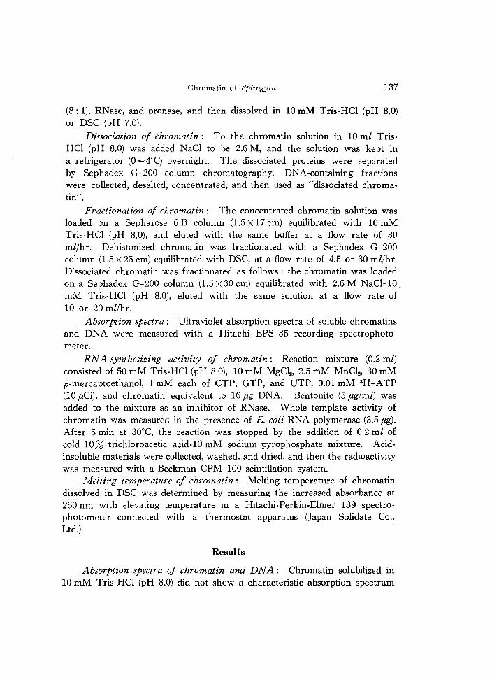
Fig. 4. Sephadex G-200 column chromatography of dissociated chromatin (Exts. 1 plus 2). Flow rate was 20 mt/h. A: V-cell chromatin. B: C, Z-cell chromatins. --, absorbance at 260 nm; ----, absorbance at 280 nm. Arrow, elution peak of blue dextran.

Chromatin of Spirogyra
.4
A OF·!
.3
I I
E " c I
g I I I I
~ .2 I I I I
Z I I « I I
E I I OF·l! I \ c \
0 , <D I N I I- .1 « \fl w u , z , ..., « m
0 a: 0 \fl m « B
.1 OF·! OF'l!
I' I "
I ~ . I ,-- ....
~ -------_...-' ---------~
0 0 20 40 60 8)
ELUT!ON VOLUME ( ml)
Fig. 5. Sephadex G-200 column chromatography of dehistonized chromatin (Exts. 1 and 2) of V-cells. Flow rate was 30 ml/h (A) or 4.5 ml/h (B). -, absorbance at 260 nm; ----, absorbance at 280 nm
141
judging from the elution profile (Fig. 3). Dissociated chromatin III the mixture of Exts. 1 and 2 was separated into two major components (DF-I, and DF-IU) and one minor component (DF-II) (Fig. 4). Molecular weights of these components were higher in those originated from C- and Z-cells than in those originated from V-cells as similar as the above experiments with Ext. 1 (Fig. 3).
Fractionation of dehistonized chromatin: Dehistonized chromatin of the mixture of Exts. 1 and 2 was separated into two major components by Sephadex G-200 column chromatography (Fig. 5). Ratios of O. D. 260 nm to 280 nm of these components (DF-I and DF-II) were 1.1 and 1.4 (or 1.6), respectively (Fig. 5 A or 5 B). The ratio of 1.1 indicates that this component binds a large amount of tightly-bound nonhistone proteins.
RNA-synthesizing activity: As shown in Table 1, RNA-synthesizing activity of chromatin in Exts. 2 and 3 was higher than that of chromatin in Ext. 1 not only in the presence of external RNA polymerase but also

142 K. Sasaki
TABLE 1. RNA-synthesizing activity in soluble chromatin
Chromatin RNA synthesis
Solution" Origin Mass ratio of (cpm 3H-AMP incorp./5 min)
protein/DNAc Without E. coli With E. coli RNA polymerase RNA polymeraseb
V-cell 1.38 740 1310 Ext. 1
CZ-cell 2.00 350 683
V-cell 1.00 3285 5160 Ext. 2, 3
CZ-cell 1.12 2425 3153
a. Chromatin concentration was adjusted to equivalent to 16 p.g DNA. b. 3.5 p.g was added. Detail are given in the Materials and Methods. c. Protein and DNA were assayed by the methods of LOWRY et at. (1951) and
CERIOTTI (1952), respectively.
TABLE 2. Melting temperature of chromatins and DNA
Chromatin Tm values in DSC (0C)
Solution Cells Soluble Dissociated F-I chromatin chromatin Component DNA DNA·Hl DNA·H3
V (68),78 68, 78 (68),79 68 79 72
Ext. 1 C (72),84
Z (71),84 71,84 (71),84 70 80 70
V 68,(78)
Ext. 2 C 71, (79)
Z 72,(80)
Ext. 3 The same as Ext. 2
DNA·Hl and DNA·H3 are reconstituted complexes of DNA and histone HI and of DNA and histone H3, respectively. Tm values of minor components are in parentheses.
in the absence of it. Furthermore, the activities of these chromatin solutions were always higher in V -cells than in C- and Z-cells.
Melting temperature: Melting temperatures of soluble chromatin, dissociated chromatin, F -I component of soluble chromatin, purified DNA, and DNA-histone complexes were measured as described in "Materials and Methods". As seen in Table 2, Tm of the soluble chromatins of C- and Z-cells was higher than that of V -cells regardless of either Ext. 1 or Ext. 2, and Tm's of the other samples were always higher in Z-cells than in V-cells. Tm values of DNA's coincided with the results reported previously (SASAKI and TAKAYA, 1974).

Chromatin of Spirogyra 143
Discussion
As shown in Fig. 1, the absorption spectra of chromatins, Exts. 1 and 2, prepared from V -cells and C- or Z-cells did not show the typical absorption
spectrum of a chromatin, especially in the absorption peak at 260 nm. If a chromatin contains a high quantity of proteins, its absorption peak must be shifted from 260 nm to 280 nm. Based upon this consideration, it could be concluded that the chromatin in C- or Z-cells binds more proteins than that in V -cells (see Fig. 1). The conclusion was also supported by the findings that chromatographically separated compounds of chromatins were always eluted from Sepharose 6 Band Sephadex G-200 columns faster in C- or Z-cells than in V-cells (Figs. 2, 3, and 4), and the melting temperatures of C- or Z-cell chromatins were always higher than those of V -cell chromatins (Table 2). Results on the ratio of protein to DNA proved it directly, but the chromatins of Z-cells having higher ratios of protein/DNA had less activity of RNA synthesis in a presence or absence of exogenous RNA polymerase (Table 1). The ratio of the rapidly eluted component of dehistonized chromatins in V -cells was about 1.1 (Fig. 5), suggesting that a large amount of nonhistone proteins may bind to these chromatins.
MONTAGNA et al. (1977) fractionated rat liver chromatin into templateactive (euchromatin) and template-inactive (heterochromatin) fractions and found that euchromatin contained 3.7 times more loosely-bound nonhistone proteins than did heterochromatin, but the latter contained twice as much residual nonhistone proteins (those not extracted by 2.0 M NaCI). Our results very much resembled their results with respect to relationship between template activity and tightly-bound proteins in chromatins. KOSTRABE et al. (1977) reported that a nonhistone chromosomal protein isolated from Ehrlich ascite tumor cells inhibited DNA-dependent RNA polymerase II. We have isolated a nonhistone protein from C, Z-cell chromatin, which inhibits transcriptional activity of V -cell chromatin (SASAKI, 1978). Although we have not succeeded in purifying the conjugation-specific nonhistone protein and to clarify the factor which regulates synthesis of this nonhistone protein, we presume that particular nonhistone proteins are synthesized at some stages of the life cycle, probably before the pairing stage. The proteins bind to chromatins and may regulate the level of transcription such as repression of RNA synthesis for vegetative growth during the mating process.
In this study, a large amount of DNA and proteins were lost from chromatins during purification procedures in spite of effort to minimize denaturation and degradation of any chromatin-bound components (unpublished results). The purified DNA had a lower molecular weight than calf thymus

144 K. Sasaki
DNA. Accordingly, we presume that naked DNA IS very easily nicked during removal of protein, so that the tightly-bound protein has a role in protection of DNA from nicking enzymes.
References
BONNER, J. & HUANG, R. C. C. 1963. Properties of chromatin and nucleohistone.
J. Mol. BioI. 6: 169-174.
BURGESS, R. R. 1969. A new method for the large scale purification of E. coli DNA
dependent RNA polymerase. J. BioI. Chern. 244: 6160-6167.
CERIOTTI, G. 1952. A microchemical determination of deoxyribonucleic acid. J. BioI. Chern. 198: 297-303.
CLARK, R. J. & FELSENFELD, G. 1974. Chemical probes of chromatin structure.
Biochemistry 13: 3622-3628.
COMPTON, J. L., BELLARD, M. & CHAMBON, P. 1976. Biochemical evidence of vari
ability in the DNA repeat length in the chromatin of higher eukaryotes.
Proc. Nat. Acad. Sci. 73: 4382-4386.
ELGIN, S. C. R. & WEINTRAUB, H. 1975. Chromosomal proteins and chromatin
structure. Ann. Rev. Biochem. 44: 726-774. FELSENFELD, G. 1978. Chromatin. Nature 271: 115-122.
FINCH, J. T., LUTTER, L. C., RHODES, D., BROWN, R. S., RUSHTON, B., LEVITT, M.
& KLUG, A. 1977. Structure of nucleosome core particles of chromatin.
Nature 269: 29-34.
FINCH, J. T., NOLL, M. & KORNBERG, R. D. 1975. Electron microscopy of defined
length of chromatin. Proc. Nat. Acad. Sci. 72: 3320-3322.
GORDON, V. C., KNOBLER, C. M., OLINS, D. E. & SCHUMAKER, V. N. 1978. Confor
mational changes of the chromatin subunit. Proc. Nat. Acad. Sci. 75: 660-
663.
KOSTRABE, N. C., NEWMAN, R. S. & WANG, T. V. 1977. Selective inhibition of
transcription by a nonhistone protein isolated from Ehrlich ascites tumor
chromatin. Arch. Biochem. Biophys. 179: 100-105.
LOWRY, O. H., ROSENGROUGH, N. J., F ARR, A. L. & RANDALL, R. J. 1951. Protein
measurement with the Folin phenol reagent. J. BioI. Chern. 193: 265-275.
MACIEWICZ, H. & LI, J. 1978. Effects of shearing on chromatin structure. Bio
chemistry 17: 962-967.
MARMUR, J. 1961. A procedure for the isolation of deoxyribonuclic acid from
micro-organisms. J. Mol. BioI. 5: 109-118.
MONTAGNA, R. A., RODRIGUEZ, L. V. & BECKER, F. B. 1977. A comparative study
of the nonhistone proteins of rat liver euchromatin and heterochromatin.
Arch. Biochem. Biophys. 179: 617-624.
PAUL, J. & GILMOUR, R. S. 1975. The regulatory role of nonhistone proteins in
RNA synthesis. In The Structure and Function of Chromatin, ed. by D. W.
FITZSIMONS & G. E. W. WOLSTENHOLME, Elsevier, Amsterdam, pp.181-198.
SASAKI, K. 1977. Changes in respiratory and photochemical activities during the

Chromatin of Spirogyra 145
conjugation process of Spirogyra. J. Fac. Sci., Hokkaido Vniv., Ser. V 10: 209-218.
SASAKI, K. 1978. Non-histone proteins associated with chromatin of Spirogyra during the conjugation process. Physiol. Plant. 42: 257-260.
SASAKI, K. & TAKA Y A, K. 1972. Suppression of nucleic acid synthesis in mito
chondrial and chloroplast fractions of Spirogyra during conjugation process. Plant & Cell Physiol. 13: 737-740.
SASAKI, K. & TAKA Y A, K. 1974. Thermal denaturation properties of deoxyribonu
cleic acid and chromatin during the life cycle of Spirogyra. Physiol. Plant. 30: 103-107.
SASAKI, K., VENO, T. & TAKA Y A, K. 1972. Suppression of nucleic acid synthesis
in chromatin of Spirogyra during conjugation process. Plant & Cell Physiol.
13: 601-608.
STEIN, A., BINA-STEIN, M. & SIMPSON, R. T. 1977. Crosslinked histone octomer
as a model of the nucleosome core. Proc. Nat. Acad. Sci. 74: 2780-2784.
STEIN, G. S., SPELBERG, T. C. & KLEINSMITH, L. T. 1974. Nonhistone chromosomal
proteins and gene regulation. Science 183: 817-824.