Embed Size (px)
Citation preview
A Portable Immersive Surgery TrainingSystem Using RGB-D Sensors
Xinqing GUO a, Luis D. LOPEZ a, Zhan YU a, Karl V. STEINER a,Kenneth E. BARNER a, Thomas L. BAUER b and Jingyi YU a
a University of Delawareb Christiana Care Health Services
Newark, Delaware
Abstract. Surgical training plays an important role in assisting residents to developcritical skills. Providing effective surgical training, however, remains as a challeng-ing task. Existing videotaped training instructions can only show imagery from afixed viewpoint that lacks both depth perception and interactivity. We present a newportable immersive surgical training system that is capable of acquiring and dis-playing high fidelity 3D reconstructions of actual surgical procedures. Our solu-tion utilizes a set of Microsoft Kinect sensors to simultaneously recover the partic-ipants, the surgical environment, and the surgical scene itself. We then develop aspace-time navigator to allow the trainees to witness and explore a prior procedureas if they were there. Preliminary feedback from residents shows that our system ismuch more effective than conventional videotaped system.
Keywords. RGB-D Sensor, Microsoft Kinect, Immersive Surgery Training, 3DReconstruction, Stereoscopic Display
Introduction
In the U.S., surgeons require longer education and training than other specialists: onlyafter four years of medical school and a minimum of five years of extensive training willthey qualify. The satisfaction of surgical residents with their training program determinesits output. The task of providing effective surgical training and re-training, however, isinherently challenging: the number of high quality educators is rather limited and bothinstructors and trainees are over-constrained by time. The problem is further deterioratingas new surgical procedures are becoming increasingly complex and often require usingnew devices and protocols.
In traditional surgical training, videotaped instruction has long served as a workhorsefor teaching surgical procedures. However, they are marginally effective: videotapes onlyprovide 2D imagery that lacks depth perception and the trainee cannot freely changeviewpoints as the inputs are captured from a fixed location. To address these issues, thepioneering work of 3D telepresence [1,2,3,4] aims to emulate remote medical proce-dures. At its core are acquisition, reconstruction, and display of the complete 3D geome-try in room-sized surgical environments. Most existing approaches [5,2,6,7,8], e.g., fromFuchs’s group at UNC, Bajcsy’s group at Penn, Kanade’s group at CMU, and Gross’sgroup at ETH, have pioneered the use of a “sea of cameras” around a room. Their sem-
inal work has led to great advances in telemedicine and provides useful insights on thesystem design and processing algorithms.
However, there is not a single educational environment that comes close to replacingthe traditional apprenticeship environment of the Operating Room (OR) for two mainreasons. On the system front, it is literally impractical to mount “a sea of cameras” withinan OR. Most existing multi-camera systems (including the immersive solutions men-tioned above) require using multiple workstations just for data transmission and storage.The system infrastructure, such as camera mountings, interconnects, and workstations,is bulky, making them unsuitable for on-site tasks. On the reconstruction front, recover-ing 3D scene geometry from images is still one of the open problems in computer vi-sion [9,10]. To make the problem tractable, many existing algorithms tend to make sim-plified assumptions about scenes, such as Lambertian surface and distant light sources.However, in surgical environments, we simply cannot assume these factors. For example,specular highlights and changing lighting are the norm in surgical environments (withbody fluid, metal instruments, head-mounted lights, etc.), easily causing classical com-puter vision algorithms, such as binocular stereo or shape-from-shading to break down.
In this paper, we present a new immersive surgical training system. Our proposedsolution resolves both the system and reconstruction problems by leveraging emerging3D imaging technologies and multi-modal fusion algorithms. Instead of using a largenumber of cameras, we use a small number (2 ∼ 4) of 3D sensors, namely MicrosoftKinect. These sensors are uniformly controlled by a single workstation and their rangeand imagery data are fused via a companion computer vision algorithm for robustlyrecovering the 3D surgical scene. We further develop a user interface to allow the traineesto navigate the 3D environment in both space and time.
Our preliminary experiments, conducted at the Virtual Education and SimulationTechnology (VEST) Center at Christiana Care Health System (CCHS), show that oursystem can effectively capture and reconstruct 3D surgical procedures performed by anexpert. These three-dimensional recordings can be presented in a virtual operation theaterin which medical students can perceive solid stereoscopic views without glasses (e.g. onan autostereo display) or with special glasses on a commercial 3D TV, as if they werepresent in the room.
1. Methods and Materials
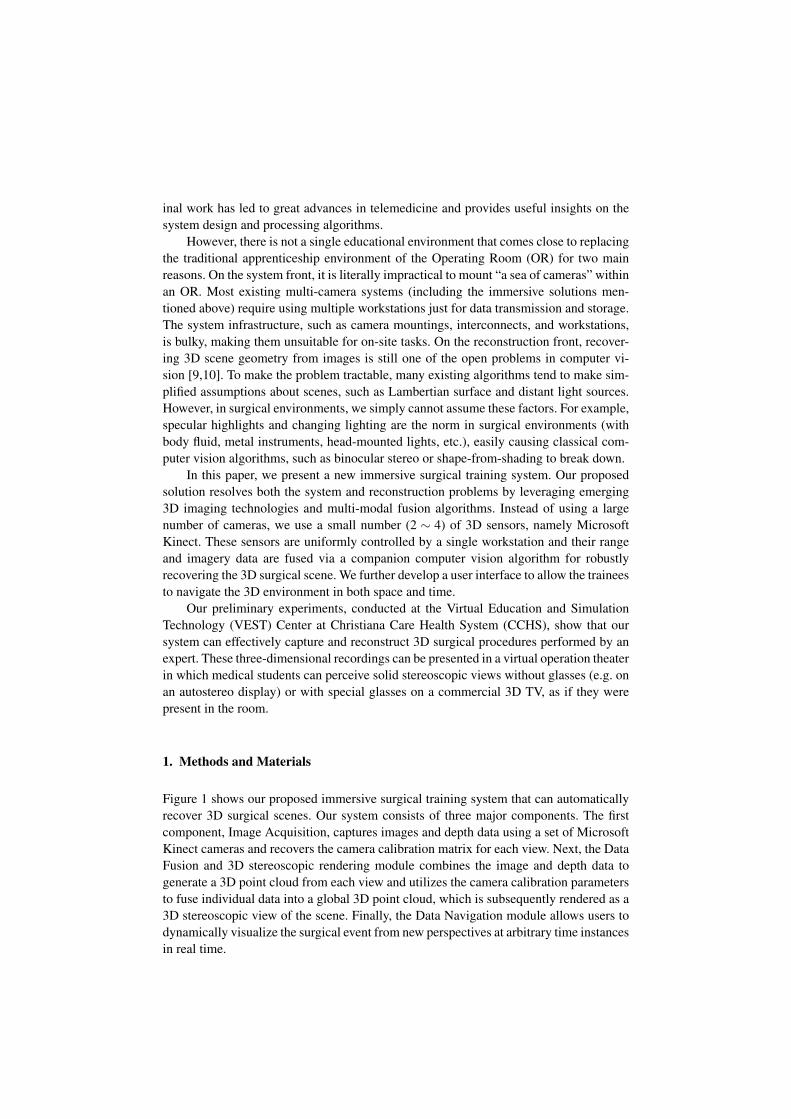
Figure 1 shows our proposed immersive surgical training system that can automaticallyrecover 3D surgical scenes. Our system consists of three major components. The firstcomponent, Image Acquisition, captures images and depth data using a set of MicrosoftKinect cameras and recovers the camera calibration matrix for each view. Next, the DataFusion and 3D stereoscopic rendering module combines the image and depth data togenerate a 3D point cloud from each view and utilizes the camera calibration parametersto fuse individual data into a global 3D point cloud, which is subsequently rendered as a3D stereoscopic view of the scene. Finally, the Data Navigation module allows users todynamically visualize the surgical event from new perspectives at arbitrary time instancesin real time.
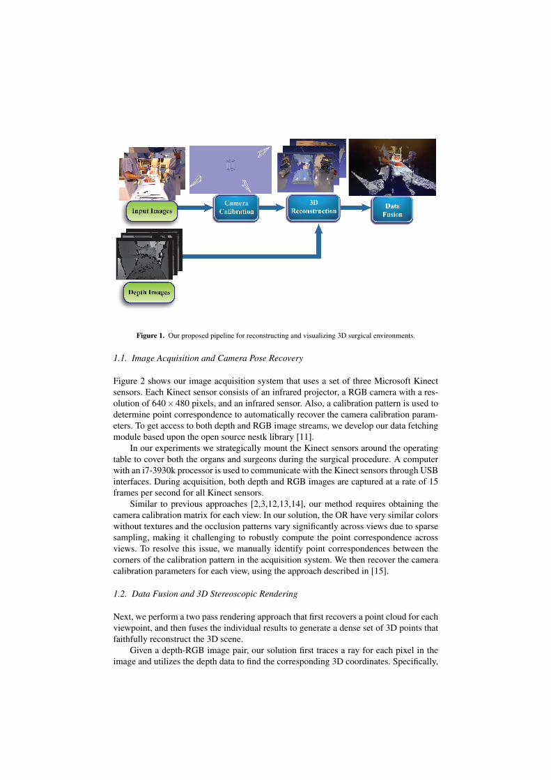
Figure 1. Our proposed pipeline for reconstructing and visualizing 3D surgical environments.
1.1. Image Acquisition and Camera Pose Recovery
Figure 2 shows our image acquisition system that uses a set of three Microsoft Kinectsensors. Each Kinect sensor consists of an infrared projector, a RGB camera with a res-olution of 640×480 pixels, and an infrared sensor. Also, a calibration pattern is used todetermine point correspondence to automatically recover the camera calibration param-eters. To get access to both depth and RGB image streams, we develop our data fetchingmodule based upon the open source nestk library [11].
In our experiments we strategically mount the Kinect sensors around the operatingtable to cover both the organs and surgeons during the surgical procedure. A computerwith an i7-3930k processor is used to communicate with the Kinect sensors through USBinterfaces. During acquisition, both depth and RGB images are captured at a rate of 15frames per second for all Kinect sensors.
Similar to previous approaches [2,3,12,13,14], our method requires obtaining thecamera calibration matrix for each view. In our solution, the OR have very similar colorswithout textures and the occlusion patterns vary significantly across views due to sparsesampling, making it challenging to robustly compute the point correspondence acrossviews. To resolve this issue, we manually identify point correspondences between thecorners of the calibration pattern in the acquisition system. We then recover the cameracalibration parameters for each view, using the approach described in [15].
1.2. Data Fusion and 3D Stereoscopic Rendering
Next, we perform a two pass rendering approach that first recovers a point cloud for eachviewpoint, and then fuses the individual results to generate a dense set of 3D points thatfaithfully reconstruct the 3D scene.
Given a depth-RGB image pair, our solution first traces a ray for each pixel in theimage and utilizes the depth data to find the corresponding 3D coordinates. Specifically,
(a) (b)
Figure 2. (a) Microsoft Kinect has a microphone array, an infrared projector, an infrared sensor and a VGAcamera. (b) Acquisition system consists of a set of three Microsoft Kinect cameras.
for each pixel in the input image we trace a ray originating at the center of projection Ctoward the image plane. Let r̄ denote a ray originating from C toward pixel (u,v) in theimage plane. The trajectory of the ray can be described as
r̄ = C+λ d̄ (1)
where d̄ is the direction vector. In camera coordinate system, the direction vector d̄ canbe written in terms of camera image plane axis d̄x, d̄y and the optical axis d̄z as:
d̄ = ud̄x + vd̄y + f d̄z (2)
Here (u,v) is the pixel coordinate in the image plane and f is the focal length of thecamera. Therefore, the original equation can be described as
r̄ = C+λ (ud̄x + vd̄y + f d̄z) (3)
Notice that the ray intersects the image plane when λ = 1. Since the depth imagecontains a measure of depth along the optical axis, we can conveniently determine λ foreach pixel. Thus, for each Kinect sensor we can compute a 3D textured point from eachinput pixel in the 2D image.
Next, we use the camera calibration parameters to transform the point cloud of eachKinect sensor from local coordinate into a global coordinate system. Then we fuse mul-tiple point clouds into one global point cloud representation. Notice that Kinect is de-signed as a stand-alone solution. While a single Kinect sensor delivers quite robust depthmaps, simultaneously running multiple sensors may lead to deteriorated results.
With the generated point cloud, we set out to render a 3D stereoscopic view of thescene. Traditional 3D rendering generates a single perspective view by synthesizing apinhole camera image in the scene. We extend this approach by simultaneously settingtwo cameras in the scene with a user specified baseline. In a single frame, two camerascapture two views of the point cloud and render them with red-cyan anaglyph. We also
(a)
(b) (c) (d)
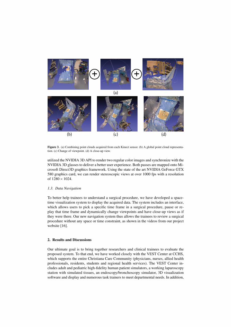
Figure 3. (a) Combining point clouds acquired from each Kinect sensor. (b) A global point cloud representa-tion. (c) Change of viewpoint. (d) A close-up view.
utilized the NVIDIA 3D API to render two regular color images and synchronize with theNVIDIA 3D glasses to deliver a better user experience. Both passes are mapped onto Mi-crosoft Direct3D graphics framework. Using the state of the art NVIDIA GeForce GTX580 graphics card, we can render stereoscopic views at over 1000 fps with a resolutionof 1280×1024.
1.3. Data Navigation
To better help trainees to understand a surgical procedure, we have developed a space-time visualization system to display the acquired data. The system includes an interface,which allows users to pick a specific time frame in a surgical procedure, pause or re-play that time frame and dynamically change viewpoints and have close-up views as ifthey were there. Our new navigation system thus allows the trainees to review a surgicalprocedure without any space or time constraint, as shown in the videos from our projectwebsite [16].
2. Results and Discussions
Our ultimate goal is to bring together researchers and clinical trainees to evaluate theproposed system. To that end, we have worked closely with the VEST Center at CCHS,which supports the entire Christiana Care Community (physicians, nurses, allied healthprofessionals, residents, students and regional health services). The VEST Center in-cludes adult and pediatric high-fidelity human patient simulators, a working laparoscopystation with simulated tissues, an endoscopy/bronchoscopy simulator, 3D visualizationsoftware and display and numerous task trainers to meet departmental needs. In addition,
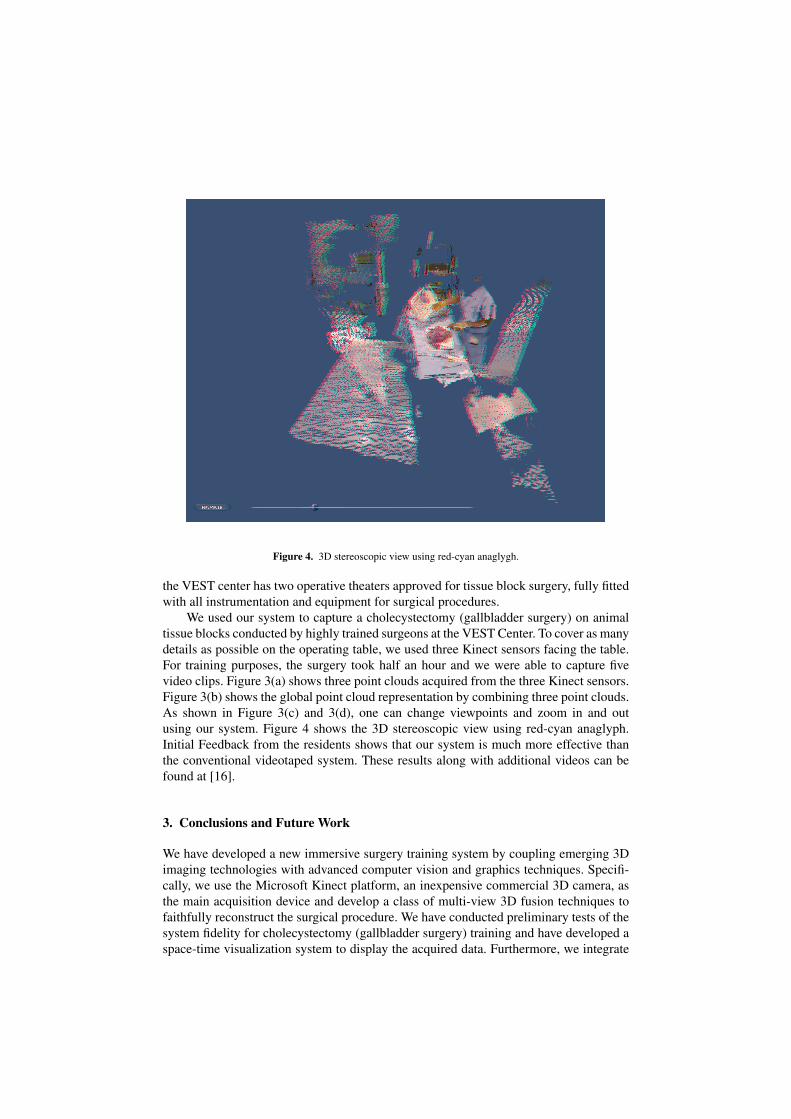
Figure 4. 3D stereoscopic view using red-cyan anaglygh.
the VEST center has two operative theaters approved for tissue block surgery, fully fittedwith all instrumentation and equipment for surgical procedures.
We used our system to capture a cholecystectomy (gallbladder surgery) on animaltissue blocks conducted by highly trained surgeons at the VEST Center. To cover as manydetails as possible on the operating table, we used three Kinect sensors facing the table.For training purposes, the surgery took half an hour and we were able to capture fivevideo clips. Figure 3(a) shows three point clouds acquired from the three Kinect sensors.Figure 3(b) shows the global point cloud representation by combining three point clouds.As shown in Figure 3(c) and 3(d), one can change viewpoints and zoom in and outusing our system. Figure 4 shows the 3D stereoscopic view using red-cyan anaglyph.Initial Feedback from the residents shows that our system is much more effective thanthe conventional videotaped system. These results along with additional videos can befound at [16].
3. Conclusions and Future Work
We have developed a new immersive surgery training system by coupling emerging 3Dimaging technologies with advanced computer vision and graphics techniques. Specifi-cally, we use the Microsoft Kinect platform, an inexpensive commercial 3D camera, asthe main acquisition device and develop a class of multi-view 3D fusion techniques tofaithfully reconstruct the surgical procedure. We have conducted preliminary tests of thesystem fidelity for cholecystectomy (gallbladder surgery) training and have developed aspace-time visualization system to display the acquired data. Furthermore, we integrate
our system with 3D stereoscopic displays to enhance the user experience. For the nextstage, We will explore possible integrations with the Visible Human [17] and the DigitalAnatomist [18] projects.
4. Acknowledgments
This project is supported by the Delaware INBRE under a grant from NIGMS (8P20GM103446) at NIH.
References
[1] H. Fuchs and U. Neumann. A vision of telepresence for medical consultation and other applications. InProceedings of the Sixth International Symposium on Robotics Research, pages 565–571, 1993.
[2] Ramesh Raskar, Greg Welch, Matt Cutts, Adam Lake, Lev Stesin, and Henry Fuchs. The office of the fu-ture: a unified approach to image-based modeling and spatially immersive displays. ACM SIGGRAPH,pages 179–188, 1998.
[3] L.-Q. Xu, B. Lei, and E. Hendriks. Computer vision for a 3-d visualisation and telepresence collaborativeworking environment. BT Technology Journal, 20(1):64–74, January 2002.
[4] E. Trucco, K Plakas, Nicole Brandenburg, Peter Kauff, Michael Karl, and Oliver Schreer. Real-timedisparity maps for immersive 3-d teleconferencing by hybrid recursive matching and census transform.In ICCV, Proceeding of Workshop on Video Registration, 2001.
[5] Henry Fuchs, Gary Bishop, Kevin Arthur, Leonard McMillan, Henry Fuchs Gary Bishop, Ruzena Ba-jcsy, Sang Wook Lee, Hany Farid, and Takeo Kanade. Virtual space teleconferencing using a sea ofcameras. In Proc. First International Conference on Medical Robotics and Computer Assisted Surgery,pages 161–167, 1994.
[6] Oliver G. Staadt, Markus H. Gross, Andreas Kunz, and Markus Meier. The blue-c (poster session): inte-grating real humans into a networked immersive environment. In Proceedings of the third internationalconference on Collaborative virtual environments, CVE ’00, pages 201–202, 2000.
[7] Kok lim Low, Adrian Ilie, Greg Welch, and Anselmo Lastra. Combining head-mounted and projector-based displays for surgical training. In in Proceedings of IEEE Virtual Reality 2003. Los, pages 110–117,2003.
[8] Greg Welch, Andrei State, Adrian Ilie, Kok-Lim Low, Anselmo Lastra, Bruce Cairns, Herman Towles,Henry Fuchs, Ruigang Yang, Sascha Becker, Dan Russo, Jesse Funaro, and Andries van Dam. Immersiveelectronic books for surgical training. IEEE MultiMedia, 12(3):22–35, July 2005.
[9] Motilal Agrawal and Larry S. Davis. A probabilistic framework for surface reconstruction from multipleimages. In CVPR (2), pages 470–476, 2001.
[10] P. J. Narayanan, Peter W. Rander, and Takeo Kanade. Constructing virtual worlds using dense stereo. InProceedings of the Sixth International Conference on Computer Vision, ICCV ’98, pages 3–10, 1998.
[11] Nestk library. https://github.com/nburrus/nestk.[12] Yuanjie Zheng, Chandra Kambhamettu, Jingyi Yu, Thomas Bauer, and Karl Steiner. Fuzzymatte: A
computationally efficient scheme for interactive matting. In CVPR, 2008.[13] Yuanjie Zheng, Jingyi Yu, Chandra Kambhamettu, Sarah Englander, Mitchell D. Schnall, and Dinggang
Shen. De-enhancing the dynamic contrast-enhanced breast mri for robust registration. In Proceedingsof the 10th international conference on Medical image computing and computer-assisted intervention -Volume Part I, MICCAI’07, pages 933–941, 2007.
[14] Yuanjie Zheng, Karl Steiner, Thomas Bauer, Jingyi Yu, Dinggang Shen, and Chandra Kambhamettu.Lung nodule growth analysis from 3d ct data with a coupled segmentation and registration framework.In ICCV, pages 1–8, 2007.
[15] Q.-T. Luong and O. D. Faugeras. Self-calibration of a moving camera from pointcorrespondences andfundamental matrices. Int. J. Comput. Vision, 22(3):261–289, March 1997.
[16] Immersive Surgery Training System. http://www.eecis.udel.edu/∼xinqing/inbre/.[17] The Visible Human Project. http://www.nlm.nih.gov/research/visible.[18] Digital Anatomist Project. http://sig.biostr.washington.edu/projects/da/index.html.