Embed Size (px)
Citation preview

A Plant Germline-Specific Integrator of SpermSpecification and Cell Cycle ProgressionLynette Brownfield1, Said Hafidh1, Michael Borg1, Anna Sidorova1, Toshiyuki Mori2, David Twell1*
1 Department of Biology, University of Leicester, Leicester, United Kingdom, 2 Miyagishima Initiative Research Unit, Advance Science Institute, RIKEN, Wako, Saitama,
Japan
Abstract
The unique double fertilisation mechanism in flowering plants depends upon a pair of functional sperm cells. During malegametogenesis, each haploid microspore undergoes an asymmetric division to produce a large, non-germline vegetativecell and a single germ cell that divides once to produce the sperm cell pair. Despite the importance of sperm cells in plantreproduction, relatively little is known about the molecular mechanisms controlling germ cell proliferation and specification.Here, we investigate the role of the Arabidopsis male germline-specific Myb protein DUO POLLEN1, DUO1, as a positiveregulator of male germline development. We show that DUO1 is required for correct male germ cell differentiationincluding the expression of key genes required for fertilisation. DUO1 is also necessary for male germ cell division, and weshow that DUO1 is required for the germline expression of the G2/M regulator AtCycB1;1 and that AtCycB1:1 can partiallyrescue defective germ cell division in duo1. We further show that the male germline-restricted expression of DUO1 dependsupon positive promoter elements and not upon a proposed repressor binding site. Thus, DUO1 is a key regulator in theproduction of functional sperm cells in flowering plants that has a novel integrative role linking gametic cell specificationand cell cycle progression.
Citation: Brownfield L, Hafidh S, Borg M, Sidorova A, Mori T, et al. (2009) A Plant Germline-Specific Integrator of Sperm Specification and Cell CycleProgression. PLoS Genet 5(3): e1000430. doi:10.1371/journal.pgen.1000430
Editor: Gregory P. Copenhaver, The University of North Carolina at Chapel Hill, United States of America
Received January 8, 2009; Accepted February 18, 2009; Published March 20, 2009
Copyright: � 2009 Brownfield et al. This is an open-access article distributed under the terms of the Creative Commons Attribution License, which permitsunrestricted use, distribution, and reproduction in any medium, provided the original author and source are credited.
Funding: This work was funded by the UK Biotechnology and Biological Sciences Research Council (Grant no. BB/C004205/1). The funders had no role in studydesign, data collection and analysis, decision to publish, or preparation of the manuscript.
Competing Interests: The authors have declared that no competing interests exist.
* E-mail: [email protected]
Introduction
The gametes of flowering plants are formed by discrete haploid
gametophyte structures consisting of only a few cells that develop
within the diploid reproductive floral organs. During spermato-
genesis, each single haploid microspore divides asymmetrically to
produce a larger vegetative cell that eventually gives rise to the
pollen tube and a smaller germ, or generative, cell (Figure S1;
reviewed in [1,2]). In contrast to germline cells in metozoans [3],
angiosperm male germ cells do not undergo regenerative stem cell
divisions, but divide once to form a pair of sperm cells. These
sperm cells are delivered to the embryo sac via the pollen tube,
where they fuse with egg and central cells to produce embryo and
endosperm respectively. This process of double fertilization
depends upon two functional sperm cells and is considered one
of the major advances in the evolutionary success of flowering
plants. Despite this importance, the molecular mechanisms
underlying many component processes, including the production
of both male and female gametes, remain largely unknown.
Recent transcriptomic analysis of isolated Arabidopsis sperm cells
shows that sperm cells express a distinct and diverse set of genes
[4] and there is evidence for extensive male germ cell gene
expression in maize and lily [5,6]. Several male germline-specific
genes have been characterized in Arabidopsis including AtMGH3,
encoding a histone H3.3 variant [7,8], AtGEX2, encoding a
putative membrane associated protein [9], and AtGCS1 (HAP2),
encoding a sperm cell surface protein required for fertilisation
[10,11]. Homologues of AtGCS1 are found in many genera
[5,12,13] that include the green alga Chlamydomonas and the rat
malarial parasite Plasmodium berghei, where they are required for
gamete interactions and membrane fusion [13]. Although gene
expression in angiosperm sperm cells is extensive and essential for
gamete functions little is known about its regulation. A
transcriptional derepression mechanism, in which expression of
male germline expressed genes is repressed in all non-germline
cells by a protein called Germline Restrictive Silencing Factor
(GRSF), has recently been proposed [14]. A binding site for the
GRSF protein was identified in the promoter region of the Lily
male germline gene LGC1, and mutations in this sequence led to
the ectopic activation of the LGC1 promoter in non-germline cells
in lily and Arabidopsis. Although similar binding sites have been
found in the promoter regions of several germline genes in
Arabidopsis, including the germline-specific transcription factor
gene DUO1 [14], a functional role for these sites or of GRSF
activity in regulating gene expression in Arabidopsis pollen has not
been shown.
Germ cell division resulting in the sperm cell pair in each pollen
grain, is essential for double fertilization and recent data supports
the capacity of both sperm cells to fertilize the egg cell in Arabidopsis
[15]. Several mutants have been described in which germ cell
division is disrupted [16–18]. Mutations in the conserved cell cycle
regulator CDKA1 [16,17] and in the F-BOX protein FBL17 [18]
prevent germ cell division and result in mature pollen with a single
germ cell. Defects in Chromosome Assembly Factor 1 (CAF1) can
PLoS Genetics | www.plosgenetics.org 1 March 2009 | Volume 5 | Issue 3 | e1000430
also disrupt germ cell division [19]. Interestingly, the single germ
cells in these mutants are capable of fertilization, with cdka1 and
fbl17 mutant germ cells fertilizing the egg cell to produce an
embryo that aborts early in development due to the lack of
endosperm production. These mutations clearly demonstrate that
germ cell division and specification can be uncoupled, but do not
identify how these processes may be coordinated to produce twin
sperm cells competent for double fertilization.
DUO POLLEN1 (DUO1) is a unique male germ cell-specific
R2R3 Myb protein that is also required for germ cell division in
Arabidopsis [20]. Unlike cdka1 and fbl17 single germ cells, duo1 germ
cells do not lead to successful fertilization, suggesting that in
addition to germ cell cycle defects, key features of gamete
differentiation and function are impaired in duo1. Here we further
characterize DUO1 as an essential, positive regulator of sperm cell
production in plants. We use various molecular markers and
ectopic expression assays to show that DUO1 is both necessary
and sufficient for the expression of male germline genes. We show
that DUO1 is required for the expression of the Arabidopsis G2/M
regulator CyclinB1;1 (AtCycB1;1) in the male germline and that
AtCycB1:1 can partially rescue defective germ cell division in duo1.
Our findings reveal a novel integrative role for the germline-
specific DUO1 protein, in cell specification and cell cycle
progression necessary for twin sperm cell production. Further-
more, we show that restriction of DUO1 expression to the male
germline is not dependent on a putative GRSF binding site but
involves positive elements in the promoter.
Results/Discussion
DUO1 Is a Key Regulator of Sperm Cell SpecificationTo investigate the potential role of DUO1 in regulating sperm
specification we examined the expression of three male germline
markers, AtMGH3, AtGEX2 and AtGCS1, in mutant duo1 pollen.
We exploited marker lines with promoter regions of these germline
genes linked to GFP. First we characterised the expression of these
markers in a coordinated manner using confocal laser scanning
microscopy (CLSM) throughout development of wild-type pollen
(Figure 1A–C), and compared their profiles with the expression of
a DUO1:mRFP fusion protein under control of the DUO1
promoter (DUO1-DUO1::mRFP; Figure 1D). The expression of
all three germ cell markers is undetectable in free microspores
when DUO1 is not expressed (Figure 1, Panel 1). Fluorescence is
first detected in the germ cell during or soon after engulfment by
the vegetative cell, appearing at a similar time to the expression of
DUO1 (Figure 1, Panel 2). As the pollen matures the level of GFP
accumulates in germ cells before mitosis and remains high in
mature sperm cells (Figure 1A–C, Panels 3–5). The accumulation
of GFP in progressive stages is illustrated by the reduced
autofluorescence signal arising from the pollen wall, reflecting
the reduced exposure needed to capture a relatively unsaturated
germ cell GFP signal. DUO1 expression persists during pollen
development, although its abundance does not obviously increase
in tricellular and mature pollen (Figure 1D). Our analysis shows
that in common with AtMGH3 and AtGEX2, the expression of
AtGCS1, previously thought to be sperm cell-specific in Arabidopsis
[11], is detected in germ cells soon after asymmetric division
(Figure 1C). The expression of all male germ cell markers shortly
after the asymmetric division shows that sperm cell specification
begins early after inception of the germline prior to passage of
germ cells through mitosis.
The three male germline markers were introduced into
heterozygous duo1 plants that produce 50% wild type pollen and
50% mutant pollen, and GFP expression was scored. Virtually all
the wild type pollen showed GFP fluorescence in twin sperm cells
while there was no fluorescence, or rarely a weak GFP signal, in
the single germ cell in duo1 pollen (Figure 1E–G, I–K; Table S1).
When these markers were introduced into the cdka;1 mutant in
which the arrested germ cell is able to fertilize the egg cell,
fluorescence was observed in the single germ cells in mutant pollen
(Figure 1E–G, Table S1). This result confirms that germ cell
division and cell fate specification are uncoupled in cdka;1 mutant
pollen, similar to the observed expression of germ cell markers in
arrested but functional germ cells in CAF1 mutants [19]. The
absence of GFP in mutant duo1 germ cells demonstrates that
DUO1 is necessary for the expression of several germline-
expressed genes, and explains why duo1 pollen is infertile (it lacks
proteins including AtGCS1 that are essential for fertilization). In
contrast, when the DUO1 promoter was used to express a nuclear-
targeted histone H2B::mRFP marker protein, fluorescence was
detected in mutant duo1 germ cells, similar to its expression in wild
type sperm cells and in cdka;1 germ cells (Figure 1H,L; Table S1),
indicating that DUO1 promoter activation does not depend upon
DUO1 itself.
To independently confirm the regulation of germline genes by
DUO1 we ectopically expressed DUO1 in seedlings, and in pollen
vegetative cells, where AtMGH3, AtGEX2 and AtGCS1 are not
normally expressed. As DUO1 contains a recognition site for
microRNA159 we used a resistant DUO1 cDNA (mDUO1) with an
altered nucleotide sequence at the miR159 binding site, but
encoding the native amino acid sequence [21]. Transgenic
seedlings in which the mDUO1 cDNA was placed under the
control of an estradiol inducible promoter [22] showed mDUO1
induction when exposed to estradiol (Figure 2A). Expression of the
male germline genes, AtMGH3, AtGEX2 and AtGCS1, was also
induced, with high levels of transcripts present only in plants
exposed to estradiol and containing mDUO1 (Figure 2A). Similarly,
when a DUO1::mRFP fusion was ectopically expressed in pollen
vegetative cells using the LAT52 promoter [23], we observed
ectopic expression of the AtMGH3 marker in vegetative cell nuclei
(Figure 2B,C; Table S2). Thus ectopic expression of DUO1 is
sufficient for activation of germ cell-specific gene expression in a
range of non-germline cells.
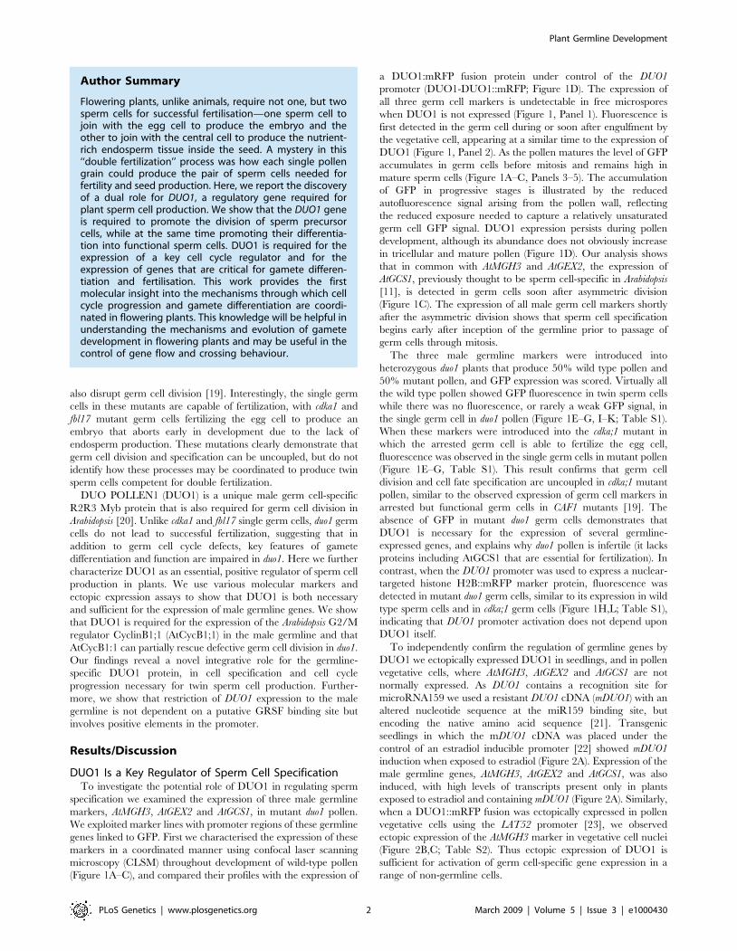
Author Summary
Flowering plants, unlike animals, require not one, but twosperm cells for successful fertilisation—one sperm cell tojoin with the egg cell to produce the embryo and theother to join with the central cell to produce the nutrient-rich endosperm tissue inside the seed. A mystery in this‘‘double fertilization’’ process was how each single pollengrain could produce the pair of sperm cells needed forfertility and seed production. Here, we report the discoveryof a dual role for DUO1, a regulatory gene required forplant sperm cell production. We show that the DUO1 geneis required to promote the division of sperm precursorcells, while at the same time promoting their differentia-tion into functional sperm cells. DUO1 is required for theexpression of a key cell cycle regulator and for theexpression of genes that are critical for gamete differen-tiation and fertilisation. This work provides the firstmolecular insight into the mechanisms through which cellcycle progression and gamete differentiation are coordi-nated in flowering plants. This knowledge will be helpful inunderstanding the mechanisms and evolution of gametedevelopment in flowering plants and may be useful in thecontrol of gene flow and crossing behaviour.
Plant Germline Development
PLoS Genetics | www.plosgenetics.org 2 March 2009 | Volume 5 | Issue 3 | e1000430
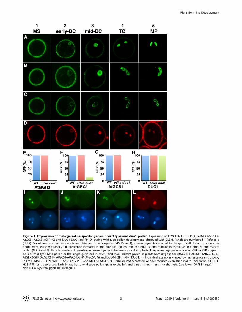
Figure 1. Expression of male germline-specific genes in wild type and duo1 pollen. Expression of AtMGH3-H2B::GFP (A), AtGEX2-GFP (B),AtGCS1-AtGCS1::GFP (C) and DUO1-DUO1::mRFP (D) during wild type pollen development, observed with CLSM. Panels are numbered 1 (left) to 5(right). For all markers, fluorescence is not detected in microspores (MS; Panel 1), a weak signal is detected in the germ cell during or soon afterengulfment (early-BC; Panel 2), fluorescence increases in mid-bicellular pollen (mid-BC; Panel 3) and remains in tricellular (TC; Panel 4) and maturepollen (MP; Panel 5). (E–L) Expression of germline expressed genes in heterozygous duo1 plants. The percentage pollen showing GFP or RFP in spermcells of wild type (WT) pollen or the single germ cell in cdka;1 and duo1 mutant pollen in plants homozygous for AtMGH3-H2B::GFP (AtMGH3, E),AtGEX2-GFP (AtGEX2, F), AtGCS1-AtGCS1::GFP (AtGCS1, G) and DUO1-H2B::mRFP (DUO1, H). Individual examples viewed by fluorescence microscopyin I to L. AtMGH3-H2B::GFP (I), AtGEX2-GFP (J) and AtGCS1-AtGCS1::GFP (K) are not expressed, or have reduced expression in duo1 pollen while DUO1-H2B::RFP (L) is expressed. Each image has a wild type pollen grain to the left and a duo1 mutant grain to the right (see lower DAPI images).doi:10.1371/journal.pgen.1000430.g001
Plant Germline Development
PLoS Genetics | www.plosgenetics.org 3 March 2009 | Volume 5 | Issue 3 | e1000430

DUO1 Is Required for AtCycB1;1 Expression in the MaleGermline
The phenotype of duo1 shows that in addition to the activation
of male germline genes, DUO1 is required for germ cell division.
Mutant duo1 germ cells complete DNA synthesis (S) phase but fail
to enter mitosis (M) [20,24], suggesting that DUO1 may regulate
the expression of essential G2/M factors. As the Arabidopsis CDK
regulatory subunit AtCycB1;1 shows enhanced expression at G2/
M [25,26] and is expressed in developing pollen (Figure S2), we
investigated AtCycB1;1 as a potential downstream target of DUO1.
To monitor the expression of AtCycB1;1 we used the pCDG
marker which contains the AtCycB1;1 promoter region and mitotic
destruction box fused to the b-glucuronidase (GUS) reporter [25].
First we analysed the marker in wild type pollen (Figure 3A–F).
Individual pollen grains at different stages of development (as
determined by DAPI staining) were analysed for GUS activity,
which results in the formation of indigo microcrystals. Microspores
and bicellular pollen shortly after mitosis contain numerous indigo
crystals, with the number peaking close to mitosis (Figure 3A–C),
indicating that expression of AtCycB1;1 is linked to asymmetric
division. Expression is then abolished in bicellular pollen
(Figure 3D). Close to germ cell mitosis, single indigo crystals are
present specifically in germ cells (located by DAPI staining;
Figure 3E) indicating expression of AtCycB1;1 in the germ cell
before division. The protein is degraded after mitosis and is absent
in tricellular pollen (Figure 3F).
We then counted the number of pollen grains with GUS
staining at different stages of development in wild type and
heterozygous duo1 plants. In both wild type and heterozygous duo1
plants, polarized microspores and vegetative cells shortly after
asymmetric division showed almost 100% staining, indicating
expression of AtCycB1:1 (Figure 3G). Thereafter vegetative cell
staining declined and was absent from late-bicellular stage pollen
(Figure 3G). Germ cell staining was subsequently observed in
,100% of pollen from wild type plants close to mitosis, but was
reduced by approximately half in heterozygous duo1 plants at this
stage (Figure 3H). As half of the pollen population is mutant in
heterozygous duo1 plants, and wild type pollen show GUS staining,
this reduction in staining is consistent with a lack of AtCycB1;1
expression in mutant duo1 pollen. This indicates that DUO1 is
required for the expression of AtCycB1;1 in male germ cells.
We then analysed the expression of AtCycB1;1 transcripts in
seedlings after steroid induction of mDUO1. In contrast to the
germline markers, AtCycB1;1 was expressed at a low level in
seedlings not exposed to estradiol and the presence of DUO1 did
not affect the level of AtCycB1;1 transcripts (Figure 2A). Thus,
although DUO1 is required for germline expression of AtCycB1;1
the presence of DUO1 is not sufficient to induce AtCycB1;1 mRNA
in seedlings. Transcription of the AtCycB1;1 gene is known to be
regulated by a number of factors, including activators such as three
repeat [27] or other Myb proteins [28] and TCP20 [29] and
repressors such as TOUSLED [30]. Thus, DUO1 may be unable
to overcome these controls in seedlings, and may affect AtCycB1;1
transcription in the male germline through an indirect mechanism
or through effects on AtCycB1;1 protein stability.
To investigate the role of AtCycB1;1 in the failure of duo1 male
germ cells to enter mitosis we determined whether AtCycB1;1 is
sufficient to rescue the germ cell mitosis defect in duo1 pollen. We
used the DUO1 promoter to drive AtCycB1;1 expression in the male
germline. The proportion of bicellular or tricellular pollen grains
from heterozygous duo1 plants either not transformed or
transformed with either of two control constructs (MGH3-
AtCycB1;1::GFP, which is not expressed in mutant pollen, and
LAT52-AtCycB1;1, which is expressed only in the vegetative cell)
did not vary significantly from 50% (Chi2 p,0.05) (Figure 3I,
Table S3). In contrast, in heterozygous duo1 plants transformed
with DUO1-AtCycB1:1 the majority of lines (31/49) showed a
significantly reduced frequency of bicellular pollen and a
corresponding increase in tricellular pollen (Figure 3I, Table S3).
This suggests that restoring AtCycB1;1 in duo1 mutant germ cells is
Figure 2. Ectopic expression of DUO1 results in expression ofmale germline specific genes. (A) RT-PCR analysis of mDUO1,AtMGH3, AtGEX2, AtGCS1 and AtCycB1;1 expression in whole seedlingstransformed with the mDUO1 cDNA (see methods) under the control ofan estradiol inducible promoter grown on media without estradiol (2)or with estradiol (+). Histone H3 was used as a control. (B, C) Maturepollen grains showing AtMGH3-H2B::GFP expression specifically insperm cells in the absence of LAT52-DUO1::mRFP (B), or in both thevegetative cell nucleus and sperm cells in the presence of LAT52-DUO1::mRFP (C). Left and right panels correspond to RFP and GFPsignals viewed by CLSM.doi:10.1371/journal.pgen.1000430.g002
Plant Germline Development
PLoS Genetics | www.plosgenetics.org 4 March 2009 | Volume 5 | Issue 3 | e1000430

sufficient to promote mitosis in a proportion of the population.
Complementation was however incompletely penetrant, which
may result from the use of the DUO1 promoter that may not
produce native amounts of AtCycB1;1. It is also possible that other
factors with a role in G2/M transition, such as other AtCycB
family members that are also expressed during pollen development
[31], may also be absent in duo1 pollen.
To determine if the presence of DUO1-AtCycB1;1 in duo1
pollen restored only the ability to proceed through mitosis or
germline specification as well, we analysed expression of the
AtMGH3 and AtGCS1 markers in duo1 plants showing partial
complementation (Figure 3J, Table S4). In contrast to plants
without DUO1-AtCycB1;1 where almost all tricellular pollen
expresses GFP, plants displaying partial complementation produce
,10% of pollen that is tricellular but does not express the markers.
As there is also a ,10% decrease in bicellular pollen, this new class
of tricellular pollen is most likely duo1 pollen in which the division
defect has been complemented by the DUO1-AtCycB1;1
construct, but in which the markers have not been activated.
Consistent with this, DUO1-AtCycB1;1 complemented duo1
pollen showed no male transmission (Table S5). Thus, comple-
mentation of the bicellular phenotype by AtCycB1;1 only affects
cell division and does not restore expression of germline gene
expression and sperm cell function.
Figure 3. AtCycB1;1 expression in developing pollen. (A–F), pCDG-dependent GUS staining (upper panel) and DAPI staining (lower panel) inisolated spores: (A, B), unicellular microspores, (C, D, E), early, mid-and late bicellular pollen and (F), tricellular pollen. (G, H) The frequency of pCDG-dependent GUS staining in microspores and vegetative cells close to mitosis is similar in duo1 heterozygotes and wild type plants (G), whereas GUSstaining in germ cells, is reduced by approximately half in duo1 heterozygotes, where 50% of the pollen is WT and the other 50% mutant (H). Thestage of pollen development is indicated below each graph and the approximate time of mitosis is indicated by grey squares with a dashed line. (I)DUO1-AtCycB1;1 is able to partially complement the bicellular phenotype of duo1 pollen. The amount of tricellular pollen (T) increases and theamount of bicellular pollen (B) decreases when heterozygous duo1 plants are transformed with DUO1-AtCycB1;1 (n = 31 T1 lines) compared withplants either not transformed (n = 3 individuals) or transformed with control constructs AtMGH3-AtCycB1;1::GFP (n = 17 T1 lines) or LAT52-AtCycB1;1(n = 17 T1 lines). Bars represent the average percentage of pollen with error bars showing standard deviation. (J) Germline markers are not activatedin the complemented tricellular pollen. In non-complemented plants ,50% of the pollen is tricellular (T) with marker expression and ,50% isbicellular (B) without marker expression. When the bicellular phenotype is partially complemented by DUO1-AtCycB1;1, ,10% of pollen is tricellularwithout marker expression, while there is a decrease in the amount of bicellular pollen. Bars represent the average percentage of pollen from 3–6individual plants with the error bars showing standard deviation.doi:10.1371/journal.pgen.1000430.g003
Plant Germline Development
PLoS Genetics | www.plosgenetics.org 5 March 2009 | Volume 5 | Issue 3 | e1000430

DUO1 Expression Is Restricted to the Male GermlineIndependent of a Putative GRSF Binding Site
Closer examination of mature pollen grains ectopically
expressing DUO1 in the vegetative cell revealed a distinctive
morphology with reduced cytoplasmic density, larger vacuoles and
numerous large cytoplasmic inclusions (Figure S3). This phenotype
was only found in pollen containing vegetative nucleus GFP (Table
S6) and analysis of pollen viability revealed up to 50% non-viable
pollen with the aberrant pollen not being viable (Figure S3E, F,
Table S7). Similar phenotypes are not seen in pollen of plants
transformed with LAT52-H2B::GFP where the transgene is
transmitted normally (data not shown). Furthermore, Arabidopsis
plants constitutively expressing DUO1 (driven by the 35S
promoter) show severe seedling patterning defects, twisted and
curled leaves and floral defects [21]. These phenotypes demon-
strate the importance of restricting high level expression of DUO1
to male germ cells.
Such restriction may partially rely upon degradation of DUO1
mRNA by microRNA159 [21] in certain cell types but promoter
elements are also likely to be important. As such, restriction of
DUO1 expression to the male germline has been proposed to rely
on the repressor protein GRSF due to a putative GRSF binding
site in the DUO1 promoter [14]. Mutagenesis of similar sequences
in the LGC1 promoter led to ectopic activation of the LGC1
promoter in non-germ line cells in tobacco and Arabidopsis [14].
However, when we specifically mutated the putative GRSF
binding site in the DUO1 promoter this did not affect the
germline-specific expression of DUO1 (Figure 4A–D). Moreover,
sequences in the 150 bp proximal DUO1 promoter, excluding
putative GRSF binding sites, were sufficient for germline-specific
expression (Figure 4E). Although factors that bind to the lily LGC1
silencer appear to be present in non-germline cells in Arabidopsis
[14], the germline-restricted activation of DUO1 does not appear
to involve GRSF mediated repression. Since the DUO1 promoter
appears to be active only after asymmetric division in the newly
formed germ cell and that activation does not depend upon
DUO1 itself (see Figure 1), activation of the DUO1 promoter may
depend on proximal region-binding transcription factors that are
inherited and/or segregated during asymmetric division of the
microspore.
ConclusionsWe have shown that DUO1 is both necessary and sufficient for
the expression of several male germline genes including AtGCS1
that is required for gamete fusion [13], thus DUO1 has a major
role in the specification of functional male gametes. DUO1 is not
involved in regulating microspore division and is first expressed in
germ cells after asymmetric division. DUO1 is also required for
the entry of male germ cells into mitosis and for the germline
expression of the G2/M regulator AtCycB1;1. Thus, the germ cell
programme under DUO1 control has an important role in
regulating core cell cycle machinery specifically in the male
germline. The discovery of the dual role of DUO1 as a positive
regulator in male germline specification and cell cycle progression
is a major advance in uncovering the molecular mechanisms
involved in plant sexual reproduction. DUO1 is currently the only
regulatory factor that has been shown to be required for gamete
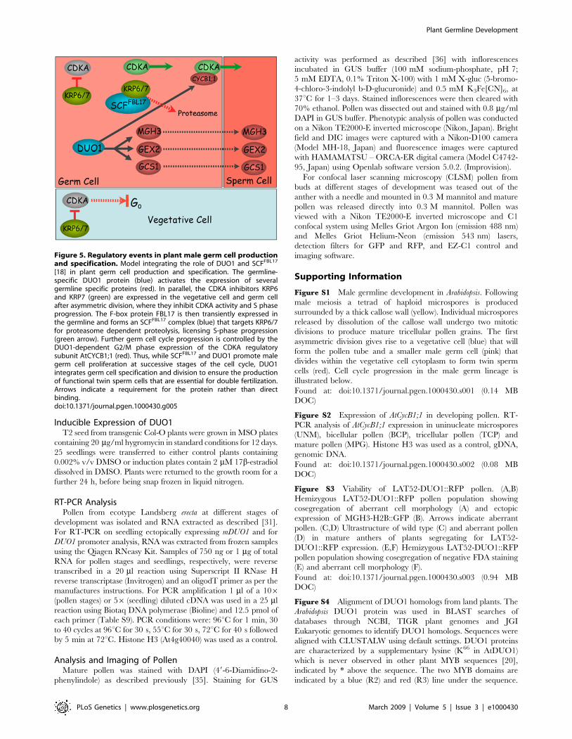
specification in plants. Recently we described an independent
mechanism for male germ cell cycle regulation where the F-BOX
protein FBL17 controls germ cell entry into S-phase via the
degradation of the CDKA inhibitors KRP6 and 7 [4]. Taken
together these data establish a molecular framework for twin
sperm cell production in flowering plants (Figure 5).
Further analysis will shed light on how DUO1 activates its
targets, and how DUO1 itself is activated specifically in the male
germline. The identification of the role of DUO1 in germ cell
specification also provides an exciting platform to develop a
detailed regulatory network for male gametogenesis and for
comparative studies of the control of sperm cell production.
DUO1 homologs are found throughout the land plants from the
non-flowering plants Selaginella moellendorffii and Physcomitrella patens
(moss) through to the monocots and dicots (Figure S4). Exploring
the functional conservation of DUO1 in different species will
reveal if DUO1 has a conserved role in male gamete production,
in terms of both of germline mitosis and specification, where
DUO1 may regulate the expression of a similar suite of genes such
as the conserved GCS1 protein. Such studies may shed light on the
evolution of regulatory mechanisms in plant germline develop-
ment and their significance in double fertilization in flowering
plants.
Materials and Methods
Plant Material and TransformationArabidopsis plants were grown at 21uC with a 16 h-light and 8 h-
dark cycle or with 24 h light, with variable humidity. Experiments
were conducted in the duo1-1 (in No-0) or the No-0 backgrounds,
except for those involving the inducible ectopic expression of
mDUO1 and analysis of the DUO1 promoter that were
conducted in Col-0. The AtGCS1-AtGCS1::GFP, AtGEX2-GFP
and CDG marker lines are also in Col-0. Plants were transformed
with Agrobacterium tumefaciens (GV3101) using a standard floral
dipping method. Transformants were selected either on Mura-
shige and Skoog (MS) agar containing 50 mg/ml kanamycin or
20 mg/ml hygromycin or on soil with 30 mg/ml BASTA
(glufosinate ammonium, DHAI PROCIDA) fed by sub-irrigation.
Vector ConstructionGateway single and multi-site construction (Invitrogen) was used
to generate most vectors. DNA was amplified from genomic DNA,
cDNA or plasmid DNA by PCR with high fidelity Phusion DNA
polymerase (Finnzymes) and primers with suitable attachment site
(attB) adapters (Table S8, attB adapters in italics). Full-length attB
sites were added to each fragment in a second high fidelity PCR.
For site-directed mutagenesis of the putative GRSF binding site in
the DUO1 promoter a two-step recombinant PCR approach was
taken. Two overlapping PCR fragments were generated contain-
ing the mutated sequence (underlined in Table S8) and the two
fragments joined in a stitching PCR. PCR fragments were cloned
into pDONR vectors (Invitrogen; pDONR207 for AtCycB1;1
cDNA or pDONR221 for H2B and DUO1 and mDUO1 cDNA,
pDONRP4P1R for promoter regions and pDONRP2RP3 for
GFP and RFP) via a BP reaction using BP Clonase II (Invitrogen).
The product of BP reactions was transformed into alpha-select
chemically competent cells (Bioline) and all clones were verified by
sequencing.
A multipart LR reaction using LR Clonase plus (Invitrogen) and
the destination vector pK7m34GW [32] was used to generate the
AtMGH3 marker, AtMGH3-H2B::GFP. This contains the region
upstream of the AtMGH3 coding region [7] driving expression of a
H2B::GFP fusion protein, with the H2B used to give a nuclear
GFP signal. The GCS1-GCS1::GFP marker was constructed by
inserting a PCR fragment of GFP (primers in Table S8) into an
AflII site in the 16th exon of a Arabidopsis GCS1 genomic DNA
fragment in the previously described binary vector [10]. The
AtGEX2-GFP marker, with the GEX2 promoter region driving
GFP expression was kindly provided by Shelia McCormick [9].
Plant Germline Development
PLoS Genetics | www.plosgenetics.org 6 March 2009 | Volume 5 | Issue 3 | e1000430

The vectors DUO1-DUO1::mRFP, DUO1-H2B::mRFP and
LAT52-DUO1::mRFP were also generated using gateway multi-
site cloning and the vectors pK7m34GW or pB7m34GW [32].
DUO1-DUO1::mRFP uses the DUO1 promoter region to drive
expression of a DUO1::mRFP fusion (used to follow the DUO1
protein during pollen development) while DUO1-H2B::GFP uses
the DUO1 promoter to produce a H2B::mRFP fusion protein (used
to follow the activity of the DUO1 promoter in duo1 pollen). The
LAT52 promoter is active in the vegetative cell [23] so was used to
ectopically express DUO1::mRFP in the vegetative cell. Vectors to
analyse the DUO1 promoter region were also constructed using
gateway multisite cloning.
The DUO1 mRNA contains a functional recognition site for the
microRNA miR159 [21], so for inducible expression of DUO1 a
miR159 resistant version of the DUO1 cDNA was used containing
silent mutations in the miR159 binding site. This was cloned in the
vector pMDC7 [33] that contains the XVE estradiol inducible
promoter system [22], using a single part LR reaction and LR
Clonase II (Invitrogen).
For experiments examining the ability of AtCycB1;1 to
complement the duo1 division phenotype the vectors pB2GW7
and pH2GW7 [34] were modified to contain the DUO1 and LAT52
promoters respectively. The 1.2 kb DUO1 and 609 bp LAT52
promoter fragments were amplified from cloned sequences using
restriction tagged oligonucleotide primer pairs (Table S8). A single
part gateway reaction was then used to clone AtCycB1;1 into the
vectors creating DUO1-CycB1;1 and LAT52-CycB1;1. MGH3-
CycB1;1::GFP was generated using a multipart gateway reaction.
Figure 4. Male germline specificity of DUO1 does not depend on putative GRSF binding sites. (A) Schematic of the DUO1 promoterregion illustrating the mutagenized putative GRSF binding site. (B,C) Expression of H2B::GFP in pollen driven by the native (B) or mutagenized DUO1(C) promoters. Top panels show GFP signal, lower panels show DAPI staining. (D) RT-PCR analysis of native and mutagenized DUO1 promoter activityin seedlings. PCR was conducted on cDNA from wild type plants (1), control plants transformed with a constituitive HistoneH3 promoter-H2B::GFPfusion (2), and plants transformed with the native (3), or mutagenized (4), DUO1 promoters driving H2B::GFP expression. The primers used werespecific for GFP (upper panel) or native Histone H3 transcripts (lower panel). The native or mutagenized DUO1 promoters showed no sporophyticexpression of GFP transcripts. (E) Schematic representation of the of the DUO1 promoter 59 deletion series used to drive expression of H2B::GFP. Thefirst four deletions, including deletion 3 in which the putative GRSF binding site is removed, showed a similar expression pattern to that of the full-length DUO1 promoter, with GFP signal only observed in sperm cell nuclei. The same expression pattern was observed in all independent linesexamined (n). GFP expression was not observed in any transformants harbouring the shortest promoter fragment (deletion 5).doi:10.1371/journal.pgen.1000430.g004
Plant Germline Development
PLoS Genetics | www.plosgenetics.org 7 March 2009 | Volume 5 | Issue 3 | e1000430
Inducible Expression of DUO1T2 seed from transgenic Col-O plants were grown in MSO plates
containing 20 mg/ml hygromycin in standard conditions for 12 days.
25 seedlings were transferred to either control plants containing
0.002% v/v DMSO or induction plates contain 2 mM 17b-estradiol
dissolved in DMSO. Plants were returned to the growth room for a
further 24 h, before being snap frozen in liquid nitrogen.
RT-PCR AnalysisPollen from ecotype Landsberg erecta at different stages of
development was isolated and RNA extracted as described [31].
For RT-PCR on seedling ectopically expressing mDUO1 and for
DUO1 promoter analysis, RNA was extracted from frozen samples
using the Qiagen RNeasy Kit. Samples of 750 ng or 1 mg of total
RNA for pollen stages and seedlings, respectively, were reverse
transcribed in a 20 ml reaction using Superscript II RNase H
reverse transcriptase (Invitrogen) and an oligodT primer as per the
manufactures instructions. For PCR amplification 1 ml of a 106(pollen stages) or 56 (seedling) diluted cDNA was used in a 25 ml
reaction using Biotaq DNA polymerase (Bioline) and 12.5 pmol of
each primer (Table S9). PCR conditions were: 96uC for 1 min, 30
to 40 cycles at 96uC for 30 s, 55uC for 30 s, 72uC for 40 s followed
by 5 min at 72uC. Histone H3 (At4g40040) was used as a control.
Analysis and Imaging of PollenMature pollen was stained with DAPI (49-6-Diamidino-2-
phenylindole) as described previously [35]. Staining for GUS
activity was performed as described [36] with inflorescences
incubated in GUS buffer (100 mM sodium-phosphate, pH 7;
5 mM EDTA, 0.1% Triton X-100) with 1 mM X-gluc (5-bromo-
4-chloro-3-indolyl b-D-glucuronide) and 0.5 mM K3Fe[CN]6, at
37uC for 1–3 days. Stained inflorescences were then cleared with
70% ethanol. Pollen was dissected out and stained with 0.8 mg/ml
DAPI in GUS buffer. Phenotypic analysis of pollen was conducted
on a Nikon TE2000-E inverted microscope (Nikon, Japan). Bright
field and DIC images were captured with a Nikon-D100 camera
(Model MH-18, Japan) and fluorescence images were captured
with HAMAMATSU – ORCA-ER digital camera (Model C4742-
95, Japan) using Openlab software version 5.0.2. (Improvision).
For confocal laser scanning microscopy (CLSM) pollen from
buds at different stages of development was teased out of the
anther with a needle and mounted in 0.3 M mannitol and mature
pollen was released directly into 0.3 M mannitol. Pollen was
viewed with a Nikon TE2000-E inverted microscope and C1
confocal system using Melles Griot Argon Ion (emission 488 nm)
and Melles Griot Helium-Neon (emission 543 nm) lasers,
detection filters for GFP and RFP, and EZ-C1 control and
imaging software.
Supporting Information
Figure S1 Male germline development in Arabidopsis. Following
male meiosis a tetrad of haploid microspores is produced
surrounded by a thick callose wall (yellow). Individual microspores
released by dissolution of the callose wall undergo two mitotic
divisions to produce mature tricellular pollen grains. The first
asymmetric division gives rise to a vegetative cell (blue) that will
form the pollen tube and a smaller male germ cell (pink) that
divides within the vegetative cell cytoplasm to form twin sperm
cells (red). Cell cycle progression in the male germ lineage is
illustrated below.
Found at: doi:10.1371/journal.pgen.1000430.s001 (0.14 MB
DOC)
Figure S2 Expression of AtCycB1;1 in developing pollen. RT-
PCR analysis of AtCycB1;1 expression in uninucleate microspores
(UNM), bicellular pollen (BCP), tricellular pollen (TCP) and
mature pollen (MPG). Histone H3 was used as a control, gDNA,
genomic DNA.
Found at: doi:10.1371/journal.pgen.1000430.s002 (0.08 MB
DOC)
Figure S3 Viability of LAT52-DUO1::RFP pollen. (A,B)
Hemizygous LAT52-DUO1::RFP pollen population showing
cosegregation of aberrant cell morphology (A) and ectopic
expression of MGH3-H2B::GFP (B). Arrows indicate aberrant
pollen. (C,D) Ultrastructure of wild type (C) and aberrant pollen
(D) in mature anthers of plants segregating for LAT52-
DUO1::RFP expression. (E,F) Hemizygous LAT52-DUO1::RFP
pollen population showing cosegregation of negative FDA staining
(E) and aberrant cell morphology (F).
Found at: doi:10.1371/journal.pgen.1000430.s003 (0.94 MB
DOC)
Figure S4 Alignment of DUO1 homologs from land plants. The
Arabidopsis DUO1 protein was used in BLAST searches of
databases through NCBI, TIGR plant genomes and JGI
Eukaryotic genomes to identify DUO1 homologs. Sequences were
aligned with CLUSTALW using default settings. DUO1 proteins
are characterized by a supplementary lysine (K66 in AtDUO1)
which is never observed in other plant MYB sequences [20],
indicated by * above the sequence. The two MYB domains are
indicated by a blue (R2) and red (R3) line under the sequence.
Figure 5. Regulatory events in plant male germ cell productionand specification. Model integrating the role of DUO1 and SCFFBL17
[18] in plant germ cell production and specification. The germline-specific DUO1 protein (blue) activates the expression of severalgermline specific proteins (red). In parallel, the CDKA inhibitors KRP6and KRP7 (green) are expressed in the vegetative cell and germ cellafter asymmetric division, where they inhibit CDKA activity and S phaseprogression. The F-box protein FBL17 is then transiently expressed inthe germline and forms an SCFFBL17 complex (blue) that targets KRP6/7for proteasome dependent proteolysis, licensing S-phase progression(green arrow). Further germ cell cycle progression is controlled by theDUO1-dependent G2/M phase expression of the CDKA regulatorysubunit AtCYCB1;1 (red). Thus, while SCFFBL17 and DUO1 promote malegerm cell proliferation at successive stages of the cell cycle, DUO1integrates germ cell specification and division to ensure the productionof functional twin sperm cells that are essential for double fertilization.Arrows indicate a requirement for the protein rather than directbinding.doi:10.1371/journal.pgen.1000430.g005
Plant Germline Development
PLoS Genetics | www.plosgenetics.org 8 March 2009 | Volume 5 | Issue 3 | e1000430

Species: At = Arabidopsis thaliana, Rc = Ricinus communis (Castor
bean), Pt = Populus trichocarpa (Poplar), Nt, Nicotiana tabacum
(Tobacco), Os = Oryza sativa (Rice), Ll = Lilium longiflorum (Lily),
Sm = Selaginella moellendorffii and Pp = Physcomitrella patens (moss).
There are two DUO1-related proteins, named A and B, in P.
patens and S. moellendorffii.
Found at: doi:10.1371/journal.pgen.1000430.s004 (1.61 MB
DOC)
Table S1 Expression of male germline marker constructs in wild
type, cdka and duo1 pollen. Mature pollen from heterozygous cdka and
duo1 plants that were homozygous for individual marker constructs
was stained with DAPI and observed by fluorescence microscopy.
The phenotype of each pollen grain was determined and the
presence (+) or absence (2) of GFP or RFP in the germline scored.
Found at: doi:10.1371/journal.pgen.1000430.s005 (0.03 MB
DOC)
Table S2 Analysis of LAT52-DUO1::RFP pollen. Mature
pollen from plants homozygous for MGH3-H2B::GFP and
heterozygous for LAT52-DUO1::RFP (three separate T1 lines,
A1–A3) was analysed by fluorescence microscopy for GFP and
RFP expression. Control plants homozygous for MGH3-
H2B::GFP show 100% sperm cell-specific GFP signal (SC GFP).
Approximately 50% of pollen from each hemizygous LAT52-
DUO1::RFP line showed GFP signal in the vegetative nucleus
(VN GFP). RFP was also detected in the vegetative nucleus (VN
RFP) of these lines, although its detection levels varied between
individual lines. Data for each marker is presented as a percentage,
with the number of pollen grains indicated in parentheses.
Found at: doi:10.1371/journal.pgen.1000430.s006 (0.03 MB
DOC)
Table S3 Complementation of duo1 pollen by AtCycB1;1. DAPI
stained pollen from heterozygous duo1 individuals either not
transformed, transformed with AtMGH3-AtCycB1;1::GFP (a con-
trol not expressed in duo1 pollen, see Figure 1), LAT52-AtCycB1;1 (a
control expressed in the vegetative cell but not the germline) or
DUO1-AtCycB1:1 were counted to analyse the proportion of
tricellular and bicellular (duo1) pollen. The Chi-square test was
applied to determine if the ratio of wild type to mutant pollen was
significantly different from the expected 1:1 ratio (ns = not signifi-
cantly different (p,0.05); * = significantly different (p,0.05).)
Found at: doi:10.1371/journal.pgen.1000430.s007 (0.12 MB
DOC)
Table S4 Marker expression in duo1 pollen complemented with
DUO1-AtCycB1;1. Pollen from plants homozygous for AtMGH3-
H2B::GFP (AtMGH3) or AtGCS1-AtGCS1::GFP (AtGCS1)
without the DUO1-AtCycB1;1 construct (control) or showing
partial complementation by DUO1-AtCycB1;1 was stained with
DAPI and observed by fluorescence microscopy. The phenotype
of each pollen grain was determined and the presence (+) or
absence (2) of GFP in the germline scored.
Found at: doi:10.1371/journal.pgen.1000430.s008 (0.03 MB
DOC)
Table S5 Transmission of the duo1 allele after introduction of
DUO1-AtCycB1:1. When duo1 heterozygotes are selfed the F1
progeny display a 1:1 ratio of WT to duo1 plants. A similar ratio is
observed when duo1 heterozygotes partially complemented by
DUO1-AtCycB1;1 (+/duo1DC) are selfed. The duo1 allele is not
transmitted through the male in either heterozygous duo1 or duo1-
complemented plants (+/duo1DC). The DUO1-AtCycB1;1 trans-
gene (pptR) is transmitted as a single locus in selfed individuals and
normally through the male when crossed to wild type female.
TEmale represents the transmission efficiency of duo1 through
pollen (mutant/wild type X 100); na = not applicable.
Found at: doi:10.1371/journal.pgen.1000430.s009 (0.03 MB
DOC)
Table S6 Aberrant morphology of pollen containing LAT52-
DUO1::RFP. GFP and RFP signals along with cell morphology
were analysed for each pollen grain from line A3 (Table S2), that
was homozygous for MGH3-H2B::GFP and hemizygous for
LAT52-DUO1::RFP. Approximately 50% of pollen possessed
aberrant morphology (see Figure S3), and of these pollen grains all
were positive for vegetative nucleus GFP (+ VN GFP), indicating
the presence of LAT52-DUO1::RFP. Data is presented as
numbers of pollen grains scored from the population.
Found at: doi:10.1371/journal.pgen.1000430.s010 (0.03 MB
DOC)
Table S7 Viability of hemizygous LAT52-DUO1::RFP pollen.
Viability of mature pollen from plants homozygous for MGH3-
H2B::GFP and heterozygous for LAT52-DUO1::RFP (three
separate T1 lines, A1–A3) was analysed by fluorescence micros-
copy after FDA staining. Pollen from control plants homozygous
for MGH3-H2B::GFP is almost all viable. Pollen viability is
reduced by up to 50% in hemizygous LAT52-DUO1::RFP lines.
Data for each marker is presented as a percentage, with the
number of pollen grains counted indicated in parentheses.
Found at: doi:10.1371/journal.pgen.1000430.s011 (0.03 MB
DOC)
Table S8 Primers used for vector construction.
Found at: doi:10.1371/journal.pgen.1000430.s012 (0.05 MB
DOC)
Table S9 Primers used in RT-PCR analyses.
Found at: doi:10.1371/journal.pgen.1000430.s013 (0.03 MB
DOC)
Acknowledgments
We thank Sheila McCormick and Peter Doerner for GEX2-GFP and
pCDG marker lines, Javier Palatnik for mDUO1 cDNA and Nam-Hai
Chua for the estradiol inducible promoter. We acknowledge Prof.
Tsuneyoshi Kuroiwa for supporting TM in his work with GCS1::GFP.
Author Contributions
Conceived and designed the experiments: LB SH MB DT. Performed the
experiments: LB SH MB AS. Analyzed the data: LB SH MB DT.
Contributed reagents/materials/analysis tools: TM DT. Wrote the paper:
LB DT.
References
1. McCormick S (2004) Control of male gametophyte development. Plant Cell 16
Suppl: S142–53.
2. Honys D, Oh SA, Twell D (2006) Pollen development, a genetic and
transcriptomic view. In The Pollen Tube: A Cellular and Molecular Perspective
Malho R, ed. Springer-Verlag- Berlin. Plant Cell Monographs. Volume 3: 15–45.
3. Strome S, Lehmann R (2007) Germ versus soma decisions: lessons from flies and
worms. Science 316: 392–393.
4. Borges F, Gomes G, Gardner R, Moreno N, McCormick S, et al. (2008)
Comparative Transcriptomics of Arabidopsis Sperm Cells. Plant Physiol 148:
1168–1181.
5. Engel ML, Chaboud A, Dumas C, McCormick S (2003) Sperm cells of Zea mays
have a complex complement of mRNAs. Plant J 34: 697–707.
6. Okada T, Bhalla PL, Singh MB (2006) Expressed sequence tag analysis of Lilium
longiflorum generative cells. Plant Cell Physiol 47: 698–705.
Plant Germline Development
PLoS Genetics | www.plosgenetics.org 9 March 2009 | Volume 5 | Issue 3 | e1000430

7. Okada T, Endo M, Singh MB, Bhalla PL (2005) Analysis of the histone H3 gene
family in Arabidopsis and identification of the male-gamete-specific variantAtMGH3. Plant J 44: 557–568.
8. Ingouff M, Hamamura Y, Gourgues M, Higashiyama T, Berger F (2007)
Distinct dynamics of HISTONE3 variants between the two fertilization productsin plants. Curr Biol 17: 1032–1037.
9. Engel ML, Holmes-Davis R, McCormick S (2005) Green Sperm. Identi-fication of male gamete promoters in Arabidopsis. Plant Physiol 138: 2124–
2133.
10. Mori T, Kuroiwa H, Higashiyama T, Kuroiwa T (2006) GENERATIVE CELLSPECIFIC 1 is essential for angiosperm fertilization. Nat Cell Biol 8: 64–71.
11. von Besser K, Frank AC, Johnson MA, Preuss D (2006) Arabidopsis HAP2(GCS1) is a sperm-specific gene required for pollen tube guidance and
fertilization. Development 133: 4761–4769.12. Hirai M, Arai M, Mori T, Miyagishima S, Kawai S, et al. (2008) Male fertility of
malaria parasite is determined by GCS1, a plant-type reproduction factor. Curr
Biol 18: 607–613.13. Liu Y, Tewari R, Ning J, Blagborough AM, Garbom S, et al. (2008) The
conserved plant sterility gene HAP2 functions after attachment of fusogenicmembranes in Chlamydomonas and Plasmodium gametes. Genes Dev 22:
1051–1068.
14. Haerizadeh F, Singh MB, Bhalla PL (2006) Transcriptional repressiondistinguishes somatic from germ cell lineages in a plant. Science 313: 496–499.
15. Ingouff M, Sakata T, Li J, Sprunck S, Dresselhaus T, et al. (2009) The two malegametes share equal ability to fertilize the egg cell in Arabidopsis thaliana. Curr Biol
19: R19–R20.16. Iwakawa H, Shinmyo A, Sekine M (2006) Arabidopsis CDKA;1, a cdc2
homologue, controls proliferation of generative cells in male gametogenesis.
Plant J 45: 819–831.17. Nowack MK, Grini PE, Jakoby MJ, Lafos M, Koncz C, et al. (2006) A positive
signal from the fertilization of the egg cell sets off endosperm proliferation inangiosperm embryogenesis. Nat Genet 38: 63–67.
18. Kim HJ, Oh SA, Brownfield L, Hong SH, Ryu H, et al. (2008) Control of plant
germline proliferation by SCFFBL17 degradation of cell cycle inhibitors. Nature455: 1134–1137.
19. Chen Z, Tan JLH, Ingouff M, Sundaresan V, Berger F (2008) Chromatinassembly factor 1 regulates the cell cycle but not cell fate during male
gametogenesis in Arabidopsis thaliana. Development 135: 65–73.20. Rotman N, Durbarry A, Wardle A Yang WC, Chaboud A, et al. (2005) A novel
class of MYB factors controls sperm-cell formation in plants. Curr Biol 15:
244–248.21. Palatnik JF, Wollmann H, Schommer C, Schwab R, Boisbouvier J, et al. (2007)
Sequence and expression differences underlie functional specialization ofArabidopsis microRNAs miR159 and miR319. Dev Cell 13: 115–125.
22. Zuo J, Niu QW, Chua NH (2000) An estrogen receptor-based transactivator
XVE mediates highly inducible gene expression in transgenic plants. Plant J 24:
265–273.
23. Twell D, Wing R, Yamaguchi J, McCormick S (1989) Isolation and expression
of an anther-specific gene from tomato. Mol Gen Genet 217: 240–245.
24. Durbarry A, Vizir I, Twell D (2005) Male germ line development in Arabidopsis
duo pollen mutants reveal gametophytic regulators of generative cell cycle
progression. Plant Physiol 137: 297–307.
25. Colon-Carmona A, You R, Haimovitch-Gal T, Doerner P (1999) Technical
advance: spatio-temporal analysis of mitotic activity with a labile cyclin-GUS
fusion protein. Plant J 20: 503–508.
26. Menges M, Murray JAH (2002) Synchronous Arabidopsis suspension cultures for
analysis of cell-cycle gene activity. Plant J 30: 203–212.
27. Ito M, Araki S, Matsunaga S, Itoh T, Nishihama R, et al. (2001) G2/M-phase-
specific transcription during the plant cell cycle is mediated by c-Myb-like
transcription factors. Plant Cell 13: 1891–1905.
28. Planchais S, Perennes C, Glab N, Mironov V, Inze D, et al. (2002)
Characterization of cis-acting element involved in cell cycle phase-independent
activation of Arath;CycB1;1 transcription and identification of putative
regulatory proteins. Plant Mol Biol 50: 111–127.
29. Li C, Potuschak T, Colon-Carmona A, Gutierrez RA, Doerner P (2005)
Arabidopsis TCP20 links regulation of growth and cell division control pathways.
PNAS 102: 12978.
30. Ehsan H, Reichheld JP, Durfee T, Roe JL (2004) TOUSLED kinase activity
oscillates during the cell cycle and interacts with chromatin regulators. Plant
Physiol 134: 1488–1499.
31. Honys D, Twell D (2004) Transcriptome analysis of haploid male gametophyte
development in Arabidopsis. Genome Biol 5: R85.
32. Karimi M, DeMeyer B, Hilson P (2005) Modular cloning in plant cells. Trends
Plant Sci 10: 103–105.
33. Curtis MD, Grossniklaus U (2003) A gateway cloning vector set for high-
throughput functional analysis of genes in planta. Plant Physiol 133: 462.
34. Karimi M, Inze D, Depicker A (2002) GATEWAY vectors for Agrobacterium-
mediated plant transformation. Trends Plant Sci 7: 193–195.
35. Park SK, Howden R, Twell D (1998) The Arabidopsis thaliana gametophytic
mutation gemini pollen1 disrupts microspore polarity, division asymmetry and
pollen cell fate. Development 125: 3789–3799.
36. Honys D, Oh SA, Renak D, Donders M, Solcova B, et al. (2006) Identification
of microspore-active promoters that allow targeted manipulation of gene
expression at early stages of microgametogenesis in Arabidopsis. BMC Plant Biol
6: 31.
Plant Germline Development
PLoS Genetics | www.plosgenetics.org 10 March 2009 | Volume 5 | Issue 3 | e1000430