Embed Size (px)
Citation preview

JOURNAL OF BACTERIOLOGY,0021-9193/01/$04.0010 DOI: 10.1128/JB.183.10.3117–3126.2001
May 2001, p. 3117–3126 Vol. 183, No. 10
Copyright © 2001, American Society for Microbiology. All Rights Reserved.
A Phosphopantetheinyl Transferase Homolog Is Essential forPhotorhabdus luminescens To Support Growth andReproduction of the Entomopathogenic Nematode
Heterorhabditis bacteriophoraTODD A. CICHE,† SCOTT B. BINTRIM,‡ ALEXANDER R. HORSWILL, AND JERALD C. ENSIGN*
Department of Bacteriology, University of Wisconsin, Madison, Wisconsin 53706
Received 3 August 2000/Accepted 28 February 2001
The bacterium Photorhabdus luminescens is a symbiont of the entomopathogenic nematode Heterorhabditisbacteriophora. The nematode requires the bacterium for infection of insect larvae and as a substrate for growthand reproduction. The nematodes do not grow and reproduce in insect hosts or on artificial media in theabsence of viable P. luminescens cells. In an effort to identify bacterial factors that are required for nematodegrowth and reproduction, transposon-induced mutants of P. luminescens were screened for the loss of the abilityto support growth and reproduction of H. bacteriophora nematodes. One mutant, NGR209, consistently failedto support nematode growth and reproduction. This mutant was also defective in the production of siderophoreand antibiotic activities. The transposon was inserted into an open reading frame homologous to Escherichiacoli EntD, a 4*-phosphopantetheinyl (Ppant) transferase, which is required for the biosynthesis of the catecholsiderophore enterobactin. Ppant transferases catalyze the transfer of the Ppant moiety from coenzyme A to aholo-acyl, -aryl, or -peptidyl carrier protein(s) required for the biosynthesis of fatty acids, polyketides, ornonribosomal peptides. Possible roles of a Ppant transferase in the ability of P. luminescens to supportnematode growth and reproduction are discussed.
Photorhabdus luminescens (Enterobacteriaceae) bacteria aresymbiotic with entomopathogenic rhabditid nematodes of thefamily Heterorhabditidae, with which they cooperate in infect-ing a wide variety of insect larvae (38, 45; for reviews, seereferences 25 and 26). The nematode requires P. luminescensfor insect pathogenicity (34), while the bacteria depend on thenematodes for transmission between insect prey. The infectivejuvenile (IJ)-stage nematodes specifically retain symbiotic P.luminescens cells in their gut mucosa, and transmission of thebacteria is a requisite for insect pathogenicity (31, 32, 34). Thenematodes require P. luminescens cells as a substrate forgrowth and reproduction (2, 21, 22, 30). It was suggested pre-viously that symbiotic P. luminescens cells provide favorablenutritional conditions for Heterorhabditis bacteriophora nema-todes to grow and reproduce (45).
During prolonged laboratory culture, P. luminescens strainsshow a tendency to undergo an apparent phase variation phe-nomenon (8, 9, 36). The native form of the bacteria, termedprimary phase, is isolated from the IJ stage of the nematode.The secondary-phase variants appear at high frequency duringprolonged culturing, while more rare is the generation of pri-mary-phase cells from secondary phase (6). The secondary-phase cells differ from the primary-phase cells in colony mor-phology, cell size, and dye uptake characteristics (6, 7, 9, 52).Also, typical primary-phase characteristics such as biolumines-
cence, pigment synthesis, phospholipase and siderophore ac-tivities, and production of intracellular crystalline inclusionproteins are depressed or absent in secondary-phase cells. Themechanism and role of phase variation in P. luminescens areunknown. Particularly significant for the subject of this inves-tigation is the inability of secondary-phase variant cells to sup-port nematode growth and reproduction (22, 30).
Because H. bacteriophora nematodes have a strict require-ment for P. luminescens for growth and reproduction, it seemslikely that P. luminescens provides some nutrients and/or otherfactors to the nematode. Dead cells or culture supernatants ofP. luminescens cells do not provide the nutrients and/or factorsrequired for nematode growth and reproduction (34; T. A.Ciche, personal observation), suggesting that actively metabo-lizing P. luminescens cells are required. To better understandthe contribution of P. luminescens to nematode growth andreproduction, we developed a genetic screen to identify P.luminescens genes necessary for nematode growth and repro-duction.
Genetic studies of P. luminescens have been limited, proba-bly because of a low frequency of transformation (7) and thecurrent inability to introduce recombinant DNA into P. lumi-nescens by conjugation (52). Success has been achieved in usingallelic exchange to construct disruptions in the genes encodingintracellular inclusion proteins CipA and CipB (7) and insec-ticidal toxin genes tca, tcb, tcc, and tcd (10) of P. luminescens.
Here we describe the construction of a mini-Tn5-basedtransposon and a delivery vector for efficient mutagenesis andgene characterization in P. luminescens. We used this system toidentify genes that are required for P. luminescens to supportgrowth and reproduction of its nematode host, H. bacterio-phora. We identified a transposon mutant that has lost the
* Corresponding author. Mailing address: Department of Bacteriol-ogy, University of Wisconsin, Madison, WI 53706. Phone: (608) 262-7877. Fax: (608) 262-9865. E-mail: [email protected].
†Present address: Hopkins Marine Station of Stanford University,Pacific Grove, CA 93950.
‡Present address: DowAgroSciences, Indianapolis, IN 46268-1054.
3117
Dow
nloa
ded
from
http
s://j
ourn
als.
asm
.org
/jour
nal/j
b on
18
Oct
ober
202
1 by
116
.127
.106
.76.

ability to support nematode growth and reproduction, and wedescribe the analyses of the disrupted gene region.
MATERIALS AND METHODS
Microbiological methods. Sources of strains and plasmids are listed in Table 1.Dye reagents, Tween types, and antibiotics were purchased from Sigma ChemicalCorp. (St. Louis, Mo.), and bacteriological growth media were purchased fromDifco (Detroit, Mich.). Cells of P. luminescens were grown in 2% ProteosePeptone 3 (PP3), with 1.5% agar added when required, at 28°C in the dark.Kanamycin (15 mg/ml), streptomycin (25 mg/ml), spectinomycin (25 mg/ml), andsucrose (7.5% [wt/vol]) were added when required. Escherichia coli strains weregrown in Luria-Bertani (LB) broth or on LB agar (1.5% agar) at 37°C withampicillin (100 mg/ml), kanamycin (50 mg/ml), streptomycin (25 mg/ml), specti-nomycin (25 mg/ml), 5-bromo-4-chloro-3-indolyl-b-D-galactopyranoside (X-Gal)(40 mg/ml), chloramphenicol (35 mg/ml), and sucrose (5% [wt/vol]) added whenrequired.
Secondary-phase (NC1/2) cells of P. luminescens were obtained and distin-guished from the primary-phase (NC1/1) cells as described previously (7). Sid-erophore activity was determined using chrome azurol S (CAS) agar (49), andlipase activity was determined on spirit blue agar containing 0.5% (vol/vol)Tween 20, Tween 40, Tween 60, Tween 80, or Tween 85 (50). Antibiotic activitywas determined by placing a 5-mm-diameter plug, taken 5 mm away fromconfluent growth of a 96-h culture of P. luminescens on PP3 agar, onto a plate ofantibiotic medium 3 (Difco) that had been inoculated with Micrococcus luteuscells.
Nematode propagation. IJ nematodes were propagated by infecting greaterwax moth larvae, Galleria mellonella (Ja-Da Bait Co., Antigo, Wis.), or by addingapproximately 20 IJ nematodes to lawns of P. luminescens cells on lipid agar (LA)(53) (2.5% nutrient broth, 1.5% agar, 1% corn oil), nematode growth medium(12), or liquid culture medium (LCM) (22) as described previously (7).
The IJ nematodes were collected by flooding the LA plates or infected larvaeat the time of IJ release and separated from adult nematodes by the watertrap method of Wouts (53). Alternatively, IJ nematodes were grown on P.luminescens cells that were seeded onto LA or nematode growth medium con-tained on one side of a divided petri dish (Fisher Scientific, Pittsburgh, Pa.).After 14 days, newly formed IJ nematodes migrated into the sterile saline con-tained on the other side. IJ nematodes were surface sterilized as describedpreviously (41).
Axenic nematodes. Axenic nematodes were obtained using a modification ofthe procedure of Han and Ehlers (33). H. bacteriophora nematodes were prop-agated on a P. luminescens strain, Meg/1, that was isolated from Heterorhabditismegidis nematodes. The H. bacteriophora nematodes grow and reproduce nor-mally on Meg/1 bacteria, but the resulting surface-sterilized IJ nematodes do notretain Meg/1 bacteria and are therefore axenic (33).
Retention of bacteria by nematodes. The numbers of P. luminescens cells in theintestine of IJ nematodes were determined. For some experiments, 50 to 100surface-sterilized nematodes were disrupted using an 0.1-ml microtissue grinder(Kontes, Vineland, N.J.). The homogenate was then serially diluted and platedon PP3 agar. Alternatively, a 10-ml sample of a water suspension containing 10to 50 IJ nematodes was placed in the depression of a sterile hanging drop slideand dried in a laminar flow hood for 5 to 10 min, and then each nematode wasdisrupted with a sterile scalpel while being examined under a 403 dissectingmicroscope. The disrupted nematodes were suspended in 0.1 ml of PP3 broth,and the scalpel blade was rinsed in this suspension. The material was thentransferred to a tube containing 0.9 ml of PP3 broth. The slide depression andscalpel were rinsed three times before plating serial dilutions of the tube ontoPP3 agar. Colonies were counted following incubation at 28°C for 3 days.
DNA manipulations. Plasmid purification from P. luminescens and E. coli wascarried out using Wizard Mini and Midi preps (Promega Corp., Madison, Wis.).Restriction enzymes and T4 ligase were used according to the manufacturer’sinstructions (Promega Corp.). When required, DNA fragments were extracted
TABLE 1. Strains and plasmids used in this study
Strain or plasmid Relevant characteristic(s) Source or reference
Nematode strainsH. bacteriophora Strain NC1, symbiotic with NC1/1 bacteria 38H. megidis Strain Meg H. Kaya (46)
Bacterial strainsP. luminescens
NC1/1 Primary phase, isolated from H. bacteriophora (identical to ATCC 29304, strain NC-19) 38NC1/2 Secondary phase, isolated from NC1/1 cells This studyMeg/1 Primary phase, isolated from H. megidis 46NP394 NC1/1 with pUB394 This studyNGR209 NC1/1 ngrA::mini-Tn5 This studyNGR209A NC1/1 with allelic-exchange vector p209A This studyNGR209C NC1/1 ngrA::mini-Tn5 via allelic exchange This studyNPngrA::VKm NC1/1 ngrA::VKm This study
E. coliDH5a Cloning strain Gibco BRLEC393 DH5a with pUB394 This studyEC209R DH5a with p209, Kmr This study
Plasmid vectorspGEM-3Z Cloning vector PromegapGEM-7Zf(1) Cloning vector PromegapBC SK(1) Cloning vector StratagenepUT-mTn5 Mini-Tn5; transposase 19, 35pSU19 P15A replicon, Cmr 5, 43pSU39 P15A replicon, Kmr 5, 43pSB101 pBC SK(1) with 2.6-kb PstI fragment containing sacR/sacB 7pHP45V Source of interposon containing streptomycin resistance 24pHP45V-Km Source of interposon containing kanamycin resistance 24pG325 pGEM-7 containing Spr Smr and sacB/sacR This studypUB394 Mini-Tn5 delivery vector, Kmr Smr Spr sucroses This studyp209 Retrieved plasmid with mini-Tn5 and flanking P. luminescens DNA This studyp209A p209 with Spr Smr and sacB/sacR PstI fragment into single NsiI site of p209 This studypngrA::VKm pBCSK containing ngrA, Smr Spr, and sacB/sacR PstI fragment This studypngrA pBCSK containing ngrA This study
3118 CICHE ET AL. J. BACTERIOL.
Dow
nloa
ded
from
http
s://j
ourn
als.
asm
.org
/jour
nal/j
b on
18
Oct
ober
202
1 by
116
.127
.106
.76.

from agarose gels using the Qiagen Gel extraction kit (Qiagen Inc., Valencia,Calif.). The bacterial DNA was purified using a modified cetyltrimethylam-monium bromide (CTAB) method (14). Southern hybridization was performedunder high-stringency conditions using the Genius kit (Boehringer MannheimCorp., Indianapolis, Ind.). Transformation of E. coli and P. luminescens was doneby electroporation using a Bio-Rad gene pulser according to the conditionssuggested for E. coli by the supplier (Bio-Rad Laboratories, Hercules, Calif.).
Construction of pUB394. The structure of the transposon delivery vector,pUB394, is shown in Fig. 1A. A pSU39 plasmid (5, 43) was inserted between theI and O ends that define the termini of the mini-Tn5 by removing the chloram-phenicol resistance gene from the mini-Tn5 of pUT-mTnCm (24) by digesting itwith SmaI and replacing it with a HincII and SmaI fragment of pSU39. Theresulting plasmid is named pUS39. The correct orientation of the insert wasdetermined. Because P. luminescens organisms are resistant to ampicillin andconjugation techniques have not been established with these bacteria, the am-picillin resistance and Mob RP4 genes were removed by BamHI and SfiI diges-tion of pUS39 followed by self-ligation, resulting in pUSF39. pGTn, pGHP, andpGHS were constructed to isolate the transposase, streptomycin and spectino-mycin resistance, and levansucrase genes, respectively. Plasmid pGTn was con-structed by removing a 1.5-kb SalI fragment containing the transposase genefrom pUT-mTn5Cm (19, 35) and inserting it into pGEM-7Zf(1) (Promega).The orientation of the insert was verified. The transposase-encoding gene wasinserted into the XbaI site located outside the I-end inverted repeat of themini-Tn5 of pUB39 as a XbaI fragment from plasmid pGTn. The resultingplasmid is named pUGS394. The orientation of the insert was verified. pUGS394was digested with BamHI to remove the ampicillin resistance and Mob RP4genes and self-ligated. The resulting plasmid is named pUS394. The streptomy-cin and spectinomycin resistance gene was removed as a 2.0-kb HindIII fragmentfrom pHP45V (24) and inserted into pGEM-7Zf(1). The resulting plasmid isnamed pGHP. Plasmid pGHS was constructed by inserting a 2.6-kb XbaI frag-ment from pBS101 (7) containing sacB/sacR (27, 47) into XbaI-digested pGHP.The orientation of the insert was verified. The streptomycin and spectinomycinresistance and the sacB/sacR sucrose sensitivity genes were removed as a 4.6-kbBamHI fragment from pGHS and inserted into a BamHI site of pUS394, yieldingpUB394. A second, alternative O end was located 1.7 kb 59 to the O end of themini-Tn5. Between the two O ends are the R6K ori and DNA encoding theN-terminal (amino acids 1 to 202) region of the Tn5 transposase-encoding genes.No difference was observed in the stability of these and normal O-end mini-Tn5insertions.
Transposon mutagenesis. The use of pUB394 for transposon mutagenesis andretrieval of DNA containing mini-Tn5 insertions is shown in Fig. 1B. Lowtransformation efficiency and the inability to conjugate or transduce DNA in P.luminescens make the standard transposon mutagenesis techniques utilizing sui-cide plasmids inefficient. The sacB gene, conferring sucrose sensitivity, allowsselection against cells containing pUB394 and, when used with selection for thetransposon (resistance to kanamycin), allows cells containing insertions, but notpUB394, to be selected. Cells of P. luminescens NC1/1 were transformed with thetransposon delivery vector pUB394 to create strain NP394. As a consequence ofpUB394 replication in NP394, mini-Tn5 insertions may accumulate. NP394 con-taining a mini-Tn5 insertion and pUB394 will cause, at high frequency (1023),kanamycin- and sucrose-resistant cells by loss of pUB394. To select mini-Tn5mutants that have lost pUB394, cells of NP394 (checked for the absence of amini-Tn5 insertion prior to mutant generation) were grown overnight in PP3containing kanamycin, and 1021 and 1022 dilutions were plated on PP3 agarcontaining kanamycin and sucrose to select for cells containing the mini-Tn5 butnot pUB394. The mutants were transferred to PP3 agar containing kanamycin toverify the resistance to kanamycin, PP3 agar containing streptomycin and spec-tinomycin to verify the absence of pUB394, M9 minimal medium to determineauxotrophy, and eosin-methylene blue to determine the phase state of the mu-tants. Mutant cells unable to grow on M9 medium were assumed to be auxo-trophs, and those not accumulating dye on eosin-methylene blue were assumedto be secondary-phase cells. Secondary-phase cells do not support nematodegrowth and reproduction and were not characterized, because they are likely tobe spontaneous phase variants of NP394 and not transposon-induced secondary-phase cells.
Screening for the ability of mutants to support growth and reproduction of H.bacteriophora nematodes. The screen for the ability of bacteria to support nem-atode growth and reproduction is shown in Fig. 2A. Individual colonies oftransposon-induced mutants of P. luminescens were inoculated into 0.25 ml ofPP3 with 10 mg of kanamycin/ml and were incubated statically overnight at 28°C.Samples of 0.05 ml were added to individual wells of 24-well tissue culture plates(Falcon 1143; Becton Dickinson Labware, Lincoln Park, N.J.) with 1.5 ml ofLA-containing kanamycin. Following incubation overnight at 28°C, an average of
12 axenic IJ nematodes was added to each well. Bacteria able to support nem-atode growth and reproduction were detected by the appearance of a white massof nematodes 21 days later. NP394 and secondary-phase (NC1/2) P. luminescenscontaining pUB394 were included in the assay as positive and negative controls,respectively. Putative nematode growth and reproduction mutants were verifiedby repeating the nematode growth and reproduction experiments twice, eachwith 12 replicates. Mutants were named NGR for nematode growth and repro-duction mutants.
Retrieval of DNA flanking the mini-Tn5 of NGR209. DNA from mutantNGR209 was purified, restriction enzyme digested with NsiI (the mini-Tn5 con-tains no NsiI site), intramolecularly and ethanol precipitated, and transformed byelectroporation into E. coli DH5a (Fig. 1B). Transformants containing the mini-Tn5 were selected by being resistant to kanamycin. The retrieved plasmid, p209,was purified and restriction enzyme digested with NsiI and SfiI to verify that theplasmid contained a single NsiI restriction fragment and the mini-Tn5 (deter-mined by the presence of a 2.9-kb SfiI restriction fragment), respectively.
Causality determination of the mini-Tn5 insertions. A 4.6-kb PstI fragmentfrom pG325 (Table 1) containing the sacB/sacR genes and a gene conferringresistance to streptomycin and spectinomycin was ligated into the NsiI site of theretrieved mini-Tn5 plasmids to create the allelic-exchange plasmid p209C. Theplasmid was transformed into wild-type NC1/1 cells. Allelic exchange was se-lected for by growing NC1/1 cells containing the allelic-exchange plasmid over-night in PP3 containing kanamycin and then plating the cells on PP3 containingkanamycin and sucrose. The mutants were tested for sensitivity to streptomycinand spectinomycin to verify the loss of the allelic-exchange plasmid. The phe-notype of the allelic-exchange mutant, NGR209A, was compared to that of theoriginal mini-Tn5 mutant, NGR209.
Sequence analysis of p209. The sequence of DNA flanking the transposoninsertion of p209 was obtained by using M13 forward and reverse primers located60 or 40 bp from the inverted repeat termini of the transposon and by primerwalking. If the sequence obtained from the M13 forward primer revealed thealternative O-end insertion, the DNA sequence flanking the alternative O endwas obtained by using the oligonucleotide primer (59 TAAGCGCCTTCCTGCATGGCTT 39). Dye terminator cycle sequencing using ABI terminator mix wasperformed using the conditions suggested by the supplier (Perkin-Elmer Corp.,Foster City, Calif.), and then the reaction products were analyzed on an ABI 377automated sequencer (Perkin-Elmer Corp.) at the University of Wisconsin Bio-technology Center. Comparison of the DNA sequence to database sequenceswas done using BLAST programs using nonredundant databases (4).
Complementation of NGR209 with pNgrA. The disrupted allele of NGR209was designated ngrA. Intact ngrA was obtained by PCR amplification using theoligonucleotide primers Edf (59 ATTAAGTATAGACTGTAGGATA 39) andEdr (59 TGATCAGGGACGGTATCAGCT 39) and Pfu polymerase (StratageneCloning Systems, La Jolla, Calif.). The Edf primer was designed to include theintergenic region between ngrA and the phfB gene that might contain a promoterelement (see Fig. 4). An 0.8-kb band was extracted from an agarose gel and bluntend ligated into pBC SK2 (Stratagene) that had been treated with HincII andshrimp alkaline phosphatase (United States Biochemical Corp., Cleveland,Ohio). Clones containing intact ngrA were obtained by screening transformantson LB medium containing chloramphenicol and X-Gal. Plasmid preparationswere made on white colonies, resulting in a clone containing an 0.8-kb insert witha sequence identical to ngrA of p209 and in the same orientation as the lacZ geneto allow the lac promoter to be utilized for transcription of ngrA. The resultingplasmid, pNgrA, was transformed into NGR209, and the phenotype of theresulting clone was determined.
Phenotypic characterization. Analyses of phase-dependent characteristics andthe ability of the cells to support nematode growth and reproduction or to beretained by IJ nematodes were performed as described above. The pathogenicityof bacteria for insects was determined as described previously (11). Positiveinsect pathogenicity was defined 72 h postinjection as 50% mortality of insectlarvae resulting from a dose of less than 30 cells. To test for oral insecticidalactivity (11), growth liquors from 72-h PP3 cultures were filter sterilized andconcentrated 15 times using a Microcon 40 (Amicon, Inc., Beverly, Mass.) mi-croconcentrator. An 0.05-ml sample of retentate was added to a 1.0-g portion ofgypsy moth diet (ICN Pharmaceuticals Inc., Costa Mesa, Calif.), and a singlefirst- or second-instar larva of Manduca sexta was then added. Larvae wereobserved for weight gain and/or death following 72 h of incubation. Experimentswere performed twice with 12 replicates each.
Nucleotide sequence accession number. The GenBank accession number forDNA sequence flanking the mini-Tn5 insertion of mutant NGR209 is AF288077.
VOL. 183, 2001 SYMBIOTIC Ppant TRANSFERASE OF P. LUMINESCENS 3119
Dow
nloa
ded
from
http
s://j
ourn
als.
asm
.org
/jour
nal/j
b on
18
Oct
ober
202
1 by
116
.127
.106
.76.
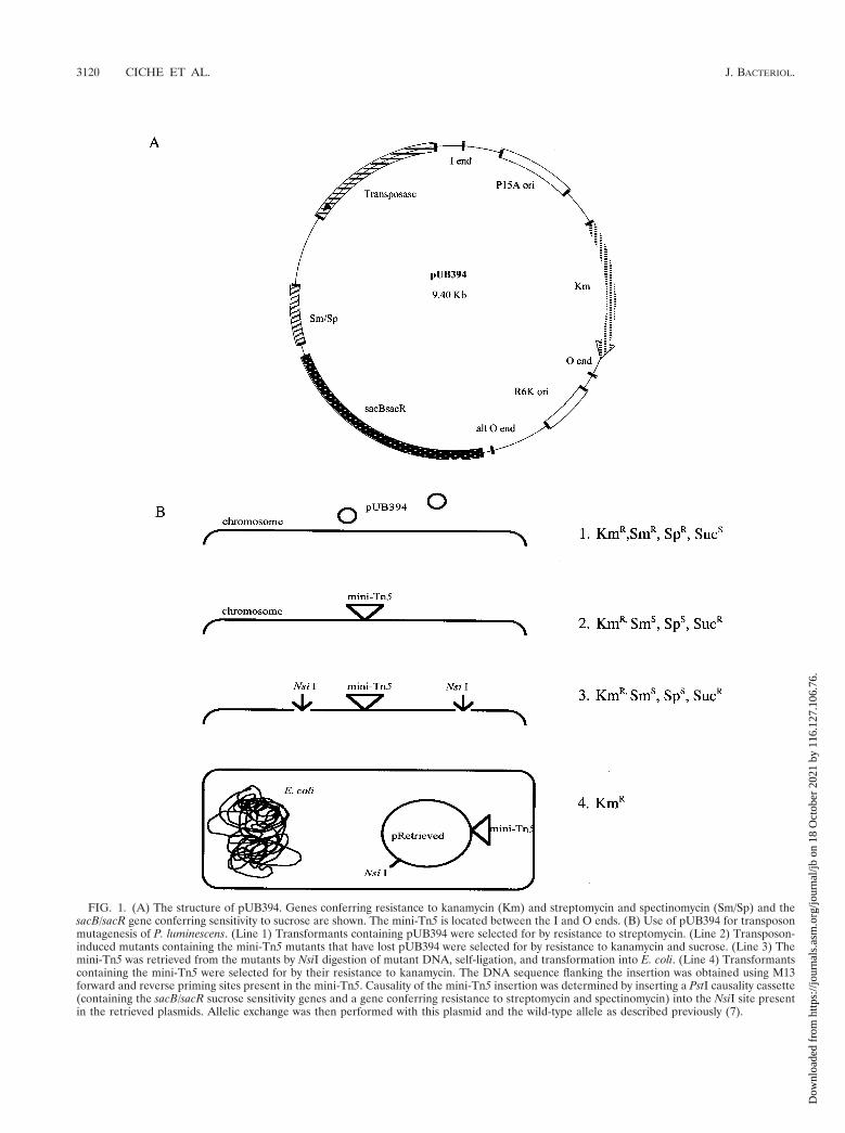
FIG. 1. (A) The structure of pUB394. Genes conferring resistance to kanamycin (Km) and streptomycin and spectinomycin (Sm/Sp) and thesacB/sacR gene conferring sensitivity to sucrose are shown. The mini-Tn5 is located between the I and O ends. (B) Use of pUB394 for transposonmutagenesis of P. luminescens. (Line 1) Transformants containing pUB394 were selected for by resistance to streptomycin. (Line 2) Transposon-induced mutants containing the mini-Tn5 mutants that have lost pUB394 were selected for by resistance to kanamycin and sucrose. (Line 3) Themini-Tn5 was retrieved from the mutants by NsiI digestion of mutant DNA, self-ligation, and transformation into E. coli. (Line 4) Transformantscontaining the mini-Tn5 were selected for by their resistance to kanamycin. The DNA sequence flanking the insertion was obtained using M13forward and reverse priming sites present in the mini-Tn5. Causality of the mini-Tn5 insertion was determined by inserting a PstI causality cassette(containing the sacB/sacR sucrose sensitivity genes and a gene conferring resistance to streptomycin and spectinomycin) into the NsiI site presentin the retrieved plasmids. Allelic exchange was then performed with this plasmid and the wild-type allele as described previously (7).
3120 CICHE ET AL. J. BACTERIOL.
Dow
nloa
ded
from
http
s://j
ourn
als.
asm
.org
/jour
nal/j
b on
18
Oct
ober
202
1 by
116
.127
.106
.76.

FIG. 2. Illustration of the in vitro screen for the ability of mutants to support nematode growth and reproduction. (A) Transposon-inducedmutants were grown overnight in liquid culture, and a sample was placed onto LA in titer dishes and incubated overnight. An average of 12 IJnematodes was added to the wells. Following incubation for 20 days, the wells were then observed for the presence of nematode growth. Mutantcells in wells not showing nematode growth were further characterized. (B and C) Photographs of the surface of LA wells showing growth ofnematodes on NC1/1 cells (B) and no growth on NGR209 cells (C). Bar, 1 cm.
VOL. 183, 2001 SYMBIOTIC Ppant TRANSFERASE OF P. LUMINESCENS 3121
Dow
nloa
ded
from
http
s://j
ourn
als.
asm
.org
/jour
nal/j
b on
18
Oct
ober
202
1 by
116
.127
.106
.76.

RESULTS
Transposon mutagenesis. The frequency of transpositionand loss of the pUB394 delivery vector, defined as the ratio ofcells resistant to kanamycin and sucrose to total cells present,was 1.7 3 1027. A large number of strains with putative mini-Tn5 insertions were obtained. Southern analysis of 20 ran-domly picked mutants showed that more than 90% of theputative transposon mutants contained single insertions ondifferent locations of DNA (data not shown). Less than 2% ofthe strains with putative insertions were resistant to strepto-mycin and spectinomycin. This suggests that resistance to su-crose in these rare strains occurred by a mechanism other thanloss of the delivery vector pUB394. Approximately 1 to 3% ofthe mutants were auxotrophic. This frequency would be ex-pected if the mini-Tn5 was inserted randomly into the genomeof P. luminescens, assuming the genome to be approximatelythe same size as that of E. coli.
Mutant screening. Of 2,800 transposon-induced mutantsscreened, a mutant (NGR209) was obtained that consistentlyfailed to support nematode growth and reproduction. A whitemass of nematodes is evident when nematodes are grown on
lawns of NC1/1 (Fig. 2B), while no adult nematodes are seenon lawns of NGR209 (Fig. 2C).
Cloning and analyses of DNA flanking the mini-Tn5 inser-tion of mutant NGR209. A 10.6-kb plasmid containing themini-Tn5 was retrieved from NGR209. The ngrA gene, dis-rupted by the mini-Tn5 in mutant NGR209, appears to encodea 49-phosphopantetheinyl (Ppant) transferase enzyme. The de-duced protein product shows a significant degree of similarityto the two Ppant transferase motifs (boxed residues) charac-teristic of these proteins (39) (Fig. 3). NgrA is most similar tothe Ppant transferases EntD from Salmonella enterica serovarTyphimurium and E. coli and VibD from Vibrio cholerae, whichare required for the biosynthesis of the catechol siderophoresenterobactin (16, 39) and vibriobactin (54), respectively (resi-dues identical to NgrA are shaded). Ppant transferases transferthe Ppant moiety from coenzyme A to acyl carrier proteins(ACP), aryl carrier proteins, and peptidyl carrier proteins (39).The Ppant-modified carrier proteins are required for the bio-synthesis of lipids, lipoproteins, polyketide, and nonribosomalpeptides, many with siderophore, antibiotic, or pharmacologi-cal activities (37, 42).
FIG. 3. Alignment of NgrA with Ppant transferase proteins. Shaded residues indicate identity to NgrA. Boxed are the two Ppant transferasemotifs (39). Proteins were aligned using Clustal W (DNAStar, Madison, Wis.): ACPS (accession no. sp P24224); EntD, enterobactin synthetasecomponent D (accession no. P19925), E. coli; VibD, phosphopantetheinyl transferase (accession no. AAD48884), V. cholerae.
FIG. 4. Physical structure of DNA flanking the mini-Tn5 insertion of mutant NGR209. Shown are the insertion site of the mini-Tn5 transposon,the location of two potential iron boxes, the site of an VKm insertion, and NsiI sites at the termini of the retrieved DNA.
3122 CICHE ET AL. J. BACTERIOL.
Dow
nloa
ded
from
http
s://j
ourn
als.
asm
.org
/jour
nal/j
b on
18
Oct
ober
202
1 by
116
.127
.106
.76.

The sequence of the entire 5.9 kb of p209 flanking themini-Tn5 was analyzed. The physical map of this DNA (Fig. 4)shows that three open reading frames similar to those forfimbrial proteins, phfB, phfA, and phfD, are located 59 and onthe same strand as ngrA. The phfB gene is located 54 bp fromngrA and is 1,014 bp in length, with residues 215 to 337 of thepredicted protein product being 37% identical and 53% similarto the type I fimbrial subunit (FimA/PapA family) (spP12903)(29). The next gene 59 of ngrA, phfA, is 966 bp in length withresidues 115 to 320 of the predicted protein product being 28%identical and 42% similar to the fimbrial adhesin MrkD fromKlebsiella pneumoniae (spP21648) (3). The phfD gene is at least2,397 bp in length (the start codon was not retrieved). Itsprotein product is 38% identical and 58% similar to the S-fimbrial usher SfaF (prf1713397E) (48). Two possible ironboxes (ferric iron uptake [Fur] regulator-binding sites) (13, 18)were identified. Iron box 1 is located between phfA and phfBand contains 15 bases identical, with a 3-bp insertion, to thepalindromic 19-bp consensus iron box (13, 17, 23). Iron box 2is located between phfB and ngrA and contains 10 bases iden-tical to the 19-bp consensus iron box.
Phenotypic characterization of mutant NGR209. The phe-notype of NGR209 was compared to those of the primary(NC1/1)- and secondary (NC1/2)-phase cells and to NGR209reconstituted with pNgrA (complementation of NGR209 withintact ngrA) (Table 2). NGR209 is identical to NC1/1 in allproperties except in not supporting nematode growth and re-production or producing siderophore and antibiotic activities.Complementation of NGR209 with pNgrA restored theseproperties. The restoration of these properties was not due togene replacement of the mini-Tn5-disrupted ngrA with intact
ngrA, because NGR209 cured of pNgrA reverted to theNGR209 phenotype.
The causality of the transposon for the phenotype ofNGR209 was demonstrated by performing allelic exchangewith the mini-Tn5-disrupted ngrA gene into the wild-type ngrAgene of P. luminescens cells. The resulting mutant, NGR209C,had all the phenotypic characteristics of NGR209 (data notshown). An omega cassette disruption of ngrA, NPngrA::VKm,also had a phenotype identical to that of NGR209 (data notshown).
The results of the analyses of growth, reproduction, andmortality of nematodes when grown on cells of NC1/1 andNGR209 and retention of NC1/1 and NGR209 by nematodesare shown in Table 3. Nematode development from the IJ tothe J4 stage was reduced significantly when nematodes weregrown on LA medium seeded with cells of NGR209 comparedto the equivalent nematodes propagated on NC1/1 cells. Theinability of nematodes to reproduce on NGR209 cells wasshown by the observation that no IJ nematodes were presentafter 20 days of growth on the NGR209 cells. In contrast, largenumbers of nematodes were produced by nematodes propa-gated on NC1/1 cells. The NGR209 cells are not toxic to thenematodes, as indicated by a similar percent mortality of IJnematodes after incubation for 3 days with NGR209 or NC1/1cells. Also, the numbers of IJ nematodes were essentially thesame following 10 to 14 days of growth on 1:1 mixtures ofNGR209 and NC1/1 or NGR209 and Meg/1 cells, again indi-cating that NGR209 cells are not inhibitory for nematodegrowth and reproduction.
The addition of large numbers (250) of IJ nematodes did notovercome the inability of NGR209 cells to support nematode
TABLE 2. Phenotypic characterization of NC1/1, NC1/2, NGR209, and NGR209 reconstituted with pNgrA
Phenotype assayedReaction of strain:
NC1/1 (primary) NC1/2 (secondary) NGR209 NGR209 1 pNgrA
Dye absorptionEosin Y-methylene blue 1 2 1 1Neutral red 1 2 1 1Bromthymol blue 1 2 1 1
Bioluminescence 1 2 1 1
Extracellular productsLipase activity 1 2 1 1Hemolytic activity 1 2 1 1Protease activity 1 2 1 1Antibiotic activity 1 2 2 1Siderophore activity 1 2 2 1
Colony morphology Convex, mucoid Flat, nonmucoid Convex, mucoid Convex, mucoid
Pigmentation 1 2 1 1
CipA and CipB production 1 2 1 1
Insect pathogenicityInjected cells 1 1 1 1Oral cell-free activity 1 NDb 1 1
Support of nematode growth and reproductiona 1 2 2 1
a Nematode growth and reproduction on LA and LCM and in G. mellonella hosts.b ND, not determined.
VOL. 183, 2001 SYMBIOTIC Ppant TRANSFERASE OF P. LUMINESCENS 3123
Dow
nloa
ded
from
http
s://j
ourn
als.
asm
.org
/jour
nal/j
b on
18
Oct
ober
202
1 by
116
.127
.106
.76.

growth and reproduction. The addition of 110 IJ nematodes toNGR209 cells caused the development of IJ to J4 nematodesto increase from 1 to 5.7 (standard deviation, 2.1; n 5 3).Although this is approximately equal to the number of J4nematodes seen on NC/1 cells (Table 3), the J4 nematodesgrowing on NGR209 cells did not develop to hermaphrodites.This suggests that the decreased development from IJ to J4observed on NGR209 cells was not the only cause for theinability of NGR209 to support nematode growth and repro-duction. Furthermore, a mixture of developmental stages (J1to adult) added to NGR209 cells did not grow and reproduce.Some nematode development and reproduction were initiallyseen but quickly ceased. This again indicates that the defect ofNGR209 for nematode growth and reproduction does not in-volve only the development of IJ nematodes to reproductivehermaphrodites; other stages of nematodes were also unableto develop and reproduce.
Because of the close proximity of fimbrial genes to ngrA, it isconceivable that the defect in NGR209 for growth and repro-duction might also affect colonization by NGR209 of the IJnematode intestine. Therefore, the ability of NGR209 to beretained by the IJ nematodes was determined. To do this, theH. bacteriophora nematodes were first propagated on Meg/1bacteria. This resulted in IJ nematodes that were nearly axenic(approximately one Meg/1 cell per 200 IJ nematodes). Thenematodes were incubated for 10 to 14 days on LA inoculatedwith NC1/1 cells, a 1:1 (vol/vol) mixture of NGR209 and Meg/1cells, and a 1:1 mixture of NGR209 and NC1/1 cells. The IJnematodes cultured on NC1/1 cells retained an average of 106NC1/1 cells (Table 3). The IJ nematodes retained an averageof 78 NGR209 cells and no Meg/1 cells when cultured on amixture of NGR209 and Meg/1. This is essentially the same asthe amount of NC1/1 cells retained by IJ nematodes. However,IJ nematodes retained an average of 83 NC1/1 and no
NGR209 cells when propagated on a mixture of NC1/1 andNGR209. At day 20, the proportions of bacteria on the LAwere about the same as those added initially. Thus, competi-tion of the bacterial strains on the LA prior to IJ retention isnot the cause for the differential retention in the IJ nematodes.It is evident that the mini-Tn5 insertion in ngrA causesNGR209 not to be retained in the presence of NC1/1 cells butto be retained in the absence of NC1/1 and the presence ofMeg/1 cells.
DISCUSSION
The entomopathogenic nematode H. bacteriophora will growand reproduce only when feeding on living cells of its symbioticbacterium, P. luminescens. Spontaneous phase variants of thebacterium that have lost expression of multiple characteristicswill not support nematode growth. A screen of 2,800 transpo-son mutants of P. luminescens yielded only one mutant,NGR209, which lost the ability to support nematode growthand reproduction while retaining most primary-phase charac-teristics. The transposon is inserted into a gene, ngrA, whichdatabase analyses show to be most similar to the entD gene thatencodes the enzyme Ppant transferase. The enzyme transfersthe Ppant moiety from coenzyme A to EntB and EntF, whichare required for the biosynthesis of the siderophore enterobac-tin (16, 28, 39).
The nematode growth and reproduction mutation alsocaused loss of detectable antibiotic and siderophore produc-tion. It is unlikely that loss of these properties is involved in thenematode growth phenotype, because adding growth liquorfrom a P. luminescens culture, which contained both activities,to the nematode growth medium did not overcome the growthdefect. In addition, we isolated a transposon mutant of P.luminescens producing no detectable siderophore activity, andthis mutant supports nematode growth and reproduction (15).
The ngrA gene is more likely to be involved in biosynthesis ofa hormone or signal regulator of nematode development thanin that of a nutritional factor. The gene is probably not in-volved in the biosynthesis of fatty acids or lipids because thePpant transferase of E. coli that activates ACP required forfatty acid biosynthesis is essential (40, 51). Other possiblefunctions of NgrA are the biosynthesis of polyketide or non-ribosomally synthesized peptide molecules (39) that are goodcandidates for hormonal or signal molecules. One possiblecandidate is the quorum-sensing homoserine lactone mole-cules that require ACP and ACP synthase (ACPS) for biosyn-thesis (44). It is unlikely that NgrA is involved in homoserinelactone biosynthesis because NgrA is not equivalent to ACPSbased on amino acid similarity.
The NgrA product might be very unstable or active at acritical threshold level, since adding growth liquor of exponen-tial- or stationary-phase cultures of P. luminescens to LA doesnot restore nematode growth. It is also possible that the puta-tive signal molecule is produced by the bacteria only when theyare grown in the presence of the nematode.
The ngrA mutant cells retain most of the characteristics ofthe parent and have clearly not been converted to the second-ary-phase variant. The parent and ngrA mutant cells producetwo crystalline inclusion proteins, CipA and CipB (7). Thesecondary-phase cells do not produce them. Inactivation of
TABLE 3. Ability of NC1/1 and NGR209 to associatewith H. bacteriophora
CharacteristicValue for straing:
NC1/1 NGR209
Nematode developmenta 5.1 (2.5) 1 (1.1)
Nematode yieldsb 2,730 (1,750) 0 (0)
% Nematode mortalityc 13.7 (12.0) 7.7 (7.0)
Retention by IJ nematodesd
Grown on NC1/1 106.6 (102) —e
Grown on NGR209 and Meg/1 — f 78 (35)Grown on NGR209 and NC1/1 83.5 (70) 0
a Average numbers of J4 or adult nematodes observed on LA 4 days followingthe addition of an average of 12 IJ nematodes (n 5 12).
b Average numbers of IJ nematodes observed on LA at day 20 (same culturesas for nematode development; n 5 12).
c Percent nonviable IJ nematodes per milliliter of LCM 5 days after additionof 150 IJ nematodes per ml, determined by observing no movement 1 min afterthe addition of 1.0% commercial bleach (n 5 3).
d IJ nematodes were propagated on LA seeded with bacteria on one half ofdivided petri dishes as described in Materials and Methods. Results show theaverage number of bacteria per disrupted IJ nematode (n 5 3).
e NGR209 was not present in the experiment.f NC1/1 was not present in the experiment.g Values in parentheses are standard deviations.
3124 CICHE ET AL. J. BACTERIOL.
Dow
nloa
ded
from
http
s://j
ourn
als.
asm
.org
/jour
nal/j
b on
18
Oct
ober
202
1 by
116
.127
.106
.76.

either cipA or cipB by omega cassettes resulted in cells exhib-iting secondary-phase characteristics (7). These mutants didnot support nematode growth and reproduction. It thus seemsclear that the ngrA mutant is specifically related to nematodegrowth and is not involved in the secondary-phase variationphenomenon.
The genes located near ngrA, having putative functions in-volving fimbrial biogenesis and adhesion and the iron boxes(Fig. 4), might be relevant to the nematode-bacterium symbi-osis. Fur is a global regulator in E. coli and regulates somevirulence genes (17). Fimbriae are often responsible for spe-cific binding of bacterial cells to eukaryotic cells (20), whichcan signal changes in gene expression in both bacteria and hostcells (1). Knowing the nucleotide sequence of these genes willallow us to specifically disrupt the genes to determine theirpossible roles in the symbiotic association. Our isolation of themini-Tn5 insertion in the ngrA gene provides a starting pointfor genetic and physiological analysis of this symbiotic relation-ship.
ACKNOWLEDGMENTS
This research was partially supported by the S. C. Johnson WaxDistinguished Scientist Fellowship awarded to T.A.C. and funds fromDowAgroSciences and the College of Agriculture and Life Sciences atthe University of Wisconsin—Madison.
REFERENCES
1. Abraham, S., A. B. Jonsson, and S. Normark. 1998. Fimbriae-mediatedhost-pathogen cross-talk. Curr. Opin. Microbiol. 1:75–81.
2. Akhurst, R. J., R. G. Mourant, L. Baud, and N. E. Boemare. 1996. Pheno-typic and DNA relatedness study between nematode-symbiotic and clinicalstrains of the genus Photorhabdus (Enterobacteriaceae). Int. J. Syst. Bacteriol.43:249–255.
3. Allen, B. L., G. F. Gerlach, and S. Clegg. 1991. Nucleotide sequence andfunctions of mrk determinants necessary for expression of type 3 fimbriae inKlebsiella pneumoniae. J. Bacteriol. 173:916–920.
4. Altschul, S. F., W. Gish, W. Miller, E. W. Myers, and D. J. Lipman. 1990.Basic local alignment search tool. J. Mol. Biol. 215:403–410.
5. Bartolome, B., Y. Jubete, E. Martinez, and F. de la Cruz. 1991. Constructionand properties of a family of pACYC184-derived cloning vectors compatiblewith pBR322 and its derivatives. Gene 102:75–78.
6. Bintrim, S. B. 1994. A study of the crystalline inclusion proteins of Photor-habdus luminescens. Ph.D. thesis. University of Wisconsin, Madison.
7. Bintrim, S. B., and J. C. Ensign. 1998. Insertional inactivation of genesencoding the crystalline inclusion proteins of Photorhabdus luminescens re-sults in mutants with pleiotropic phenotypes. J. Bacteriol. 180:1261–1269.
8. Bleakley, B., and K. H. Nealson. 1988. Characterization of primary andsecondary forms of Xenorhabdus luminescens strain Hm. FEMS Microbiol.Ecol. 53:241–250.
9. Boemare, N. E., and R. J. Akhurst. 1988. Biochemical and physiologicalcharacterization of colony form variants in Xenorhabdus spp. (Enterobacte-riaceae). J. Gen. Microbiol. 134:1835–1845.
10. Bowen, D., T. A. Rocheleau, M. Blackburn, O. Andreev, E. Golubeva, R.Bhartia, and R. H. ffrench-Constant. 1998. Insecticidal toxins from thebacterium Photorhabdus luminescens. Science 280:2129–2132.
11. Bowen, D. J., and J. C. Ensign. 1998. Purification and characterization of ahigh-molecular-weight insecticidal protein complex produced by the ento-mopathogenic bacterium Photorhabdus luminescens. Appl. Environ. Micro-biol. 64:3029–3055.
12. Brenner, S. 1974. The genetics of Caenorhabditis elegans. Genetics 77:71–94.13. Calderwood, S. B., and J. J. Mekalanos. 1988. Confirmation of the Fur
operator site by insertion of a synthetic oligonucleotide into an operatorfusion plasmid. J. Bacteriol. 170:1015–1017.
14. Chan, J. W. Y. F., and P. H. Goodwin. 1994. Extraction of genomic DNAfrom extracellular polysaccharide-synthesizing Gram-negative bacteria. Bio-Techniques 18:419–422.
15. Ciche, T. A. 2000. Symbiotic interactions between the bacterium Photorhab-dus luminescens and the entomopathogenic nematode Heterorhabditis bacte-riophora. Ph.D. thesis. University of Wisconsin, Madison.
16. Coderre, P. E., and C. F. Earhart. 1989. The entD gene of the Escherichia coliK12 enterobactin gene cluster. J. Gen. Microbiol. 135:3043–3055.
17. Crosa, J. H. 1997. Signal transduction and transcriptional and posttranscrip-tional control of iron-regulated genes in bacteria. Microbiol. Mol. Biol. Rev.61:319–336.
18. de Lorenzo, V., S. Wee, M. Herrero, and J. B. Neilands. 1987. Operatorsequences of the aerobactin operon of plasmid ColV-K30 binding the ferricuptake regulator (fur) repressor. J. Bacteriol. 169:2624–2630.
19. de Lorenzo, V., M. Herrero, U. Jakubzik, and K. H. Timmis. 1990. Mini-Tn5transposon derivatives for the insertion mutagenesis, promoter probing, andchromosomal insertion of cloned DNA in gram-negative eubacteria. J. Bac-teriol. 172:6568–6572.
20. Edwards, R. A., and J. L. Puente. 1998. Fimbrial expression in entericbacteria: a critical step in intestinal pathogenesis. Trends Microbiol. 6:282–287.
21. Ehlers, R. D., S. Lunau, K. Krasomil-Osterfeld, and J. H. Osterfeld. 1998.Liquid culture of the entomopathogenic nematode-bacterium-complex Het-erorhabditis megidis/Photorhabdus luminescens. BioControl 43:77–86.
22. Ehlers, R. D., S. Stoessel, and U. Whyss. 1990. The influence of phasevariants of Xenorhabdus spp. and Escherichia coli (Enterobacteriaceae) onthe propagation of entomopathogenic nematodes of the genera Steinernemaand Heterorhabditis. Rev. Nematol. 13:417–424.
23. Escolar, L., J. Perez-Martın, and V. de Lorenzo. 1998. Binding of Fur (ferricuptake regulator) repressor of Escherichia coli to arrays of GATAAT se-quence. J. Mol. Biol. 283:537–547.
24. Fellay, R., J. Frey, and H. Krisch. 1987. Interposon mutagenesis of soil andwater bacteria: a family of DNA fragments designed for in vitro insertionalmutagenesis of Gram-negative bacteria. Gene 52:147–154.
25. Forst, S., and K. H. Nealson. 1996. Molecular biology of the symbiotic-pathogenic bacteria Xenorhabdus spp. and Photorhabdus spp. Microbiol.Rev. 60:21–43.
26. Forst, S., B. Dowds, N. Boemare, and E. Stackebrandt. 1997. Xenorhabdusspp. and Photorhabdus spp.: bugs that kill bugs. Annu. Rev. Microbiol.51:47–72.
27. Gay, R., D. Le Coq, M. Steinmetz, T. Berkelman, and C. I. Kado. 1985.Positive selection procedure for entrapment of insertion sequence elementsin gram-negative bacteria. J. Bacteriol. 164:918–921.
28. Gehring, A. M., K. A. Bradley, and C. T. Walsh. 1997. Enterobactin biosyn-thesis in Escherichia coli: isochorismate lyase (EntB) is a bifunctional enzymethat is phosphopantetheinylated by EntD and then acylated by EntE usingATP and 2,3-dihydroxybenzoate. Biochemistry 36:8495–8503.
29. Gerlach, G. F., S. Clegg, and B. L. Allen. 1989. Identification and charac-terization of the genes encoding type 3 and type 1 fimbrial adhesin ofKlebsiella pneumoniae. J. Bacteriol. 171:1262–1270.
30. Gerritsen, L. J. M., and P. H. Smits. 1993. Variation in pathogenicity ofrecombinations of Heterorhabditis and Xenorhabdus luminescens strains. Fun-dam. Appl. Nematol. 16:367–373.
31. Han, R., W. M. Wouts, and L. Li. 1990. Development of Heterorhabditis spp.strains as characteristics of possible Xenorhabdus luminescens subspecies.Rev. Nematol. 13:411–415.
32. Han, R., W. M. Wouts, and L. Li. 1991. Development and virulence ofHeterorhabditis spp. strains associated with different Xenorhabdus lumine-scens isolates. J. Invertebr. Pathol. 58:27–32.
33. Han, R. C., and R.-U. Ehlers. 1998. Cultivation of axenic Heterorhabditis spp.dauer juveniles and their response to non-specific Photorhabdus luminescensfood signals. Nematologica 44:425–435.
34. Han, R. C., and R.-U. Ehlers. 2000. Pathogenicity, development, and repro-duction of Heterorhabditis and Steinernema carpocapsae under axenic in vivoconditions. J. Invertebr. Pathol. 75:55–58.
35. Herrero, M., V. de Lorenzo, and K. H. Timmis. 1990. Transposon vectorscontaining non-antibiotic resistance selection markers for cloning and stablechromosomal insertion of foreign genes in gram-negative bacteria. J. Bacte-riol. 172:6557–6567.
36. Hurlbert, R. E., J. Xu, and C. L. Small. 1989. Colonial and cellular poly-morphism in Xenorhabdus luminescens. Appl. Environ. Microbiol. 55:1136–1143.
37. Keating, T. A., and C. T. Walsh. 1999. Initiation, elongation, and terminationstrategies in polyketide and polypeptide antibiotics. Curr. Opin. Chem. Biol.3:598–606.
38. Khan, A., and W. M. Brooks. 1976. A chromogenic bioluminescent bacte-rium associated with the entomophilic nematode Chromonema heliothidis.J. Invertebr. Pathol. 29:253–261.
39. Lambalot, R. H., A. M. Gehring, R. S. Flugel, P. Zuber, M. LaCelle, M. A.Marahiel, R. Reid, C. Khosla, and C. T. Walsh. 1996. A new enzyme super-family—the phosphopantetheinyl transferases. Chem. Biol. 3:923–936.
40. Lambalot, R. H., and C. T. Walsh. 1995. Cloning, overproduction, andcharacterization of the Escherichia coli holo-acyl carrier protein synthase.J. Biol. Chem. 270:24658–24661.
41. Lunau, S., S. Stoessal, A. J. Schmidt-Peisker, and R.-U. Ehlers. 1993. Es-tablishment of monoxenic inocula for scaling up in vitro cultures of theentomopathogenic nematodes Steinernema spp. and Heterorhabditis spp.Nematologica 39:385–399.
42. Marahiel, M. A., T. Stachelhaus, and H. D. Mootz. 1997. Modular peptidesynthetases involved in nonribosomal peptide synthesis. Chem. Rev. 97:2651–2674.
43. Martinez, E., B. Bartomole, and F. de la Cruz. 1988. pACYC184-derivedcloning vectors containing the multiple cloning site and lacZa reporter gene
VOL. 183, 2001 SYMBIOTIC Ppant TRANSFERASE OF P. LUMINESCENS 3125
Dow
nloa
ded
from
http
s://j
ourn
als.
asm
.org
/jour
nal/j
b on
18
Oct
ober
202
1 by
116
.127
.106
.76.
of pUC8/9 and pUC18/19 plasmids. Gene 68:159–162.44. More, M. I., L. Finger, L. David, J. L. Stryker, C. Fuqua, A. Eberhard, and
S. C. Winans. 1998. Enzymatic synthesis of a quorum-sensing autoinducerthrough use of defined substrates. Science 272:1655–1658.
45. Poinar, G. O., Jr., G. M. Thomas, and R. Hess. 1977. Characteristics of thespecific bacterium associated with Heterorhabditis bacteriophora (Hetero-rhabditidae: Rhabditida). Nematologica 23:97–102.
46. Poinar, G. O., T. Jackson, and M. Klein. 1987. Heterorhabditis megidis sp. n.(Heterorhabditae: Rhabditida), parasitic in the japanese beetle, Popillia ja-ponica (Scarabaeidae: Coleoptera), in Ohio. Proc. Helminthol. Soc. Wash.54:53–59.
47. Reid, J. L., and A. Collmer. 1987. An nptI-sacB-sacR cartridge for construct-ing directed, unmarked mutations in Gram-negative bacteria by markerexchange-eviction mutagenesis. Gene 57:239–246.
48. Schmoll, T., J. Morschhaeuser, J. M. Ott, B. Ludwig, I. Van Die, and J.Hacker. 1990. Complete genetic organization and functional aspects of theEscherichia coli S fimbrial adhesin determinant. Nucleotide sequence ofgenes sfaB, C, D, E, F. Microb. Pathol. 9:331–343.
49. Schwyn, B., and J. B. Neilands. 1987. Universal chemical assay for thedetection and determination of siderophores. Anal. Biochem. 160:47–56.
50. Sierra, G. 1957. A simple method for the detection of lipolytic activity ofmicroorganisms and some observations on the influence of the contact be-tween cells and fatty acid substrates. J. Microbiol. Serol. 23:15–22.
51. Takiff, H. E., T. Baker, T. Copeland, S. M. Chen, and D. L. Court. 1992.Locating essential Escherichia coli genes by using mini-Tn10 transposons: thepdxJ operon. J. Bacteriol. 174:1544–1553.
52. Wang, H., and B. C. A. Dowds. 1993. Phase variation in Xenorhabdus lumi-nescens: cloning and sequencing of the lipase gene and analysis of its expres-sion in the primary and secondary phases of the bacterium. J. Bacteriol.175:1665–1673.
53. Wouts, W. M. 1981. Mass production of the entomogenous nematode Het-erorhabditis bacteriophora on artificial media. J. Nematol. 13:467–469.
54. Wyckoff, E. E., J. A. Stoebner, K. E. Reed, and S. M. Payne. 1997. Cloning ofa Vibrio cholerae gene cluster: identification of genes required for early stepsin siderophore biosynthesis. J. Bacteriol. 179:7055–7062.
3126 CICHE ET AL. J. BACTERIOL.
Dow
nloa
ded
from
http
s://j
ourn
als.
asm
.org
/jour
nal/j
b on
18
Oct
ober
202
1 by
116
.127
.106
.76.

![Transcript Abundance of Photorhabdus Insect-Related (Pir) Toxin …centaur.reading.ac.uk/67414/1/Transcript Abundance of Photorhabdu… · another [4,5]. Once a suitable insect host](https://img.pdfslide.us/doc/110x75/5f8e2c986fb3a1288f4e6734/transcript-abundance-of-photorhabdus-insect-related-pir-toxin-abundance-of-photorhabdu.jpg)