Embed Size (px)
Citation preview
Fall 2016
A Patient’s Guide to Trigeminal Neuropathies
MyofascialPain
TemporalMandibular
Disorder PersistentIdiopathicFace Pain
BurningMouth
Glossopharyngeal
Journal of The Facial Pain AssociationTh
e Fa
cial
Pai
n A
ssoc
iatio
n40
8 W
. Uni
vers
ity A
venu
e,
Suite
402
G
aine
svill
e FL
326
01-3
248

Eating. Talking. Smiling. Simple facial movements like these aren’t supposed to hurt, but when you have trigeminal neuralgia, you know how extreme the pain can be.
We understand. That’s why the experts at Northwell Health’s Neuroscience Institute are proud to offer a wide range of options that use the latest technology, including
– Gamma Knife® stereotactic radiosurgery – Neuromodulation procedures – CyberKnife® robotic radiosurgery – Microvascular decompression – Percutaneous rhizotomy
You don’t have to live with the pain of trigeminal neuralgia — make an appointment at one of our New York City or Long Island offices today.
Freedom from the pain of trigeminal neuralgia
Care Provided by Northwell Physician Partners:
Amir R. Dehdashti, MD, FACSNorth Shore University HospitalManhasset, NY (516) 562-3026
Mark B. Eisenberg, MDNorth Shore University HospitalManhasset, NY (516) 773-7737
Robert G. Kerr, MD, PhD, FRCPSCHuntington HospitalHuntington, NY (631) 351-4840
Mitchell E. Levine, MDLenox Hill HospitalNew York, NY (212) 434-3900
Michael Schulder, MD, FAANSNorth Shore University HospitalManhasset, NY (516) 941-1260
20108 Trigeminal Neuralgia - print ad_8.5x11.indd 1 3/22/16 2:09 PM
Fall 2016 -------------- 1
From the Chairman of the Board
THE MAB CORNERFrom the Chairman of the FPA’s Medical
Advisory Board
YPC on how the brain can adjust to chronic pain and a young patient’s profile
Memorial and Honorary Tribute Fund New Members
FEATURES
IN EVERY Q
A Patient’s Guide to Trigeminal Neuropathies
MyofascialPain
TemporalMandibular
Disorder PersistentIdiopathicFace Pain
BurningMouth
Glossopharyngeal
CoverOur feature article written by FPA Medical Advisory Board member Steven Graff-Rafford, DDS, and Bahareh Safaie, DDS a resident at the UCLA Orofacial Pain Clinic.
2 3
23
24
28
The Quarterly asks the head of the Face Pain Center at Cedars-Sinai Medical Center to prepare a Patient Guide to managing trigeminal neuropathies
In response to many patient requests, an FPA staffer conducts some research on medical marijuana and chronic pain.
Long time FPA associate and patient advocate Dr. Leesa Morrow writes about stress and systemic illness.
5 15 17
Journal of The Facial Pain Association

2 -------------- Quarterly: Journal of the Facial Pain Association
I have had the pleasure of serving as Chairman of the Board for four years and every one of these quarterly letters, so far, has been about what the FPA is doing to help those with facial nerve pain to find information, world-leading experts in treatment and help them manage life outside of a doctor’s office. Much is underway. This letter is about the people that make it happen; our Staff, Support Group Leaders, Medical Advisory Board, and some visionary supporters too.
For FPA’s staff in Gainesville FL, it is more than a job. Led by CEO John Koff, our staff works with passion and creativity. They know that their efforts make a large and positive difference in the lives of those in pain and those who are caregivers too. Call our office anytime for assistance and to speak with someone who has personal experience in managing the pain of trigeminal neuralgia. Our staff provides national infrastructure for regional support groups, provides assistance to the Young Patient’s Committee for those younger than 40, and does all the work to put on several regional conferences each year and a biannual national conference. Our staff put the FPA on the internet years ago and keeps the site up-to-date as technology improves and visits increase. They built fpa-support.org into the global “go to” site for information on facial nerve pain and referrals to experts in diagnosis and treatment. They built FPA’s own social media Facial Pain Network and they are active on Facebook and Twitter too. They publish the TN Newswire several times each month with information about pain-related research and new treatments, they have built this Quarterly into a high-quality journal with substantive articles and personal stories that keep us in touch. Standing back, our staff puts a lot of thought into how FPA needs to evolve as social interaction continues to move from physical meetings and mailed newsletters to heavy use of internet-
based resources and social media. All of that is a lot of challenging work.
Support Group Leaders organize regular meetings with informative speakers and a chance for discussion among attendees. In many cases, they are at the front of the shift in social interaction mentioned above. They work to keep the meetings convenient and useful. A source of speakers at Support Group meetings and FPA conferences, a powerhouse of expertise, is the Medical Advisory Board. The neurosurgeons, dentists and other healthcare professionals on the MAB are world experts in diagnosing and treating facial nerve pain. The MAB keeps the FPA grounded in science and it provides information to members by speaking at conferences, meeting one-on-one with attendees at conferences, writing articles and now producing webinars too.
Membership dues, annual donations and advertising are important sources of revenue that help the FPA pay its bills. Join me in renewing your membership and in supporting the FPA. The FPA is also fortunate that a few visionary supporters have provided funding for extra programs. The results include regional conferences that help people now and research that, in the future, may lead to improved diagnosis and treatments for facial nerve pain.
On behalf of the Board of Directors, I cannot overstate how much we appreciate the essential efforts of our Staff, Support Group Leaders, MAB and visionary supporters. Thank you.
From the Chairman of the Board
Jeff Bodington, Chairman of the BoardTNA – The Facial Pain Association
Fall 2016 -------------- 3
Managing EditorJohn Koff Editor/Circulation ManagerNancy Oscarson Contributing EditorsAnne CiemneckiPam Neff, RN
Research EditorCindy Ezell
Art and DesignCaren Hackman
QUARTERLY is published four times per year by The Facial Pain Association, 408 W. University Avenue, Suite 402Gainesville FL 32601-3248
800-923-3608
www.facepain.org
Do I have anesthesia dolorosa?
Fortunately, this is an uncommon question that I am asked in my office and my answer is always the same- “That’s not a question that will lead to a helpful answer.”
Why is that?
“Anesthesia dolorosa” is one of those fancy Latin terms in medicine that medical students memorize because it is so hard to do and so unnecessary to know once learned. I have yet to discover the origin of the term (that’s unnecessary also) but the textbook definition is that there is pain at a site of “complete” numbness. But when is numbness “complete”? What if the numbness is “near complete”?
The more appropriate way to frame the question is to define the pain as “neuropathic,” then use a questionnaire that has been validated through years of use that measures the quality of one’s pain as well as the severity. The McGill Pain Questionnaire is ideal for this application. “Neuropathic pain” refers to pain that occurs as a consequence of injury to a nerve, the spinal cord or brain rather than injury to the body that is detected by the nervous system. Neuropathic pain always has an electrical quality to it.
When the nerve injury is severe enough, patients will have constant pain that they describe as “burning” or “searing.” Associated with it, in the same location will be loss of sensation also from the nerve injury. Whether it is complete or not is not the important thing. What is important is the severity of the pain. The questionnaire helps define the psychological elements of the pain with words like “torturing,” for example when it is most severe.
Patients understandably fear the diagnosis of “anesthesia dolorosa” because it translates to an untreatable disorder. That’s not a helpful label to use.
My answer to that question, “Do I have anesthesia dolorosa,” is best put as:
“Let’s look at the McGill Pain Questionnaire” and see how severe your pain is and what the quality of it is, then we can begin to come up with a plan on how to treat it.”
The M
AB Corner
Jeffrey A. Brown, MDChairman,
Medical Advisory Board Facial Pain Association

4 -------------- Quarterly: Journal of the Facial Pain Association

Fall 2016 -------------- 5
This review serves as a guide in the evaluation and management of facial neuropathic pain. Many facial pains mimic a neuropathy and the patient and clinician need a systematic and careful approach to diagnosis. Common conditions such as myofascial pain, temporomandibular disorder (TMD), dental pathology (caries or gum disease), cracked teeth, infectious causes, sinusitis, or neurovascular causes, like migraine headache must be considered in the differential diagnosis. Myofascial Pain and TMD are very common and will also be reviewed.
It is not uncommon for a dentist to mistake the pain as dental-related and proceed with unnecessary dental treatments, which can worsen a neuropathy. Similarly the diagnosis of sinus pathology or atypical facial pain (a wastebasket term) by a physician may lead to unnecessary therapies and additional frustration and suffering for the patient. Through better medical understanding by the
patient, such unnecessary treatments and misdiagnoses can hopefully be avoided and prompt referral to the proper orofacial pain specialist can be made. Likewise, patients should be made aware of the various new treatment options available to them. One reason for poor response to treatment may be inaccurate diagnosis. Differentiating between the various types of facial pains and mechanisms associated with neuropathies can sometimes be confusing, especially if the symptoms are complex and don’t fall into any one specific category.
Neuropathies Defined
Neuralgia is defined as pain in the distribution of a nerve or nerves. Simply put, pain involved in the nervous system is considered a neuralgia. Stimulation of these nerves, by mechanical, thermal or chemical energy may create pain, which is generically neuralgia. Clinically neuralgia can
A Patient’s Guide to management of trigeminal neuropathies and other facial pains
Steven Graff-Radford, DDSDirector of The Program for Headache and Orofacial Pain at Cedars-Sinai Medical Center
Bahareh Safaie, DDSUCLA School of Dentistry
“Patient’s Guide”. . .continued on page 6

Fall 2016 -------------- 6
present as a continuous or intermittent pain characterized as stabbing, shooting, electric or sometimes constant burning-like sensation. The pain is felt along the nerve distribution.
The term neuropathic actually requires there to be the presence of a lesion or disease within the neural system. By definition, the “pathic” part of the word tells us that there’s some sort of “pathological” abnormality as a causal factor. Pathologies refer to any sort of abnormality, including
injury, tumor, or disease affecting the neural structure. Neuropathic pains can then also be sub-classified even further based on their location as being either central or peripheral; meaning is the pathology (abnormality) located in the brain/spinal cord (centrally) or outside the brain (peripherally). So a neuropathy is basically thought of as a disturbance in function or pathological change in a nerve or nerves. If only one nerve is involved, it’s called a mononeuropathy. If several nerves are involved, it’s referred to as mononeuropathy multiplex. And if it’s
“Patient’s Guide”. . .continued from page 5
Allodynia: painful response to a non-painful stimulus.
“Allo” means “other” in Greek, so we see this prefix with medical terms describing something different from what is expected. “Odynia” means “pain” in Greek. Note that although there is both “peripheral” and “central” forms of allodynia, many are actually only referring to central allodynia when they talk about allodynia in general.
Nociception: the normal perception of pain
Hyperalgesia: an increasingly painful response to a normally painful stimulus. Note that “hyper” is when there is too much of something and “algesia” basically refers to feeling pain. Think about when you go to the doctor and they give you an “analgesic.” They do that so that you feel no pain during treatment. So basically think of hyperalgesia as an increased response to a pain inducing stimulus. Both central and peripheral changes can cause hyperalgesia.
Hyperpathia: an abnormally painful reaction to a repetitive stimulus, as well as decreased threshold to the stimulus. So remember that hyper
means too much of something; and “pathia” typically refers to a disease or abnormality of some kind, can be referring to a form of suffering as well. Having a decreased threshold to a stimulus means that there’s much less needed by your senses for your body to hit the threshold necessary to feel (or in this case hurt) from a stimulus, ie something that has provoked you.
Dysesthesia: an unpleasant abnormal sensation, whether spontaneous or evoked. A dysesthesia is always felt as an abnormal sensation that is bothersome. Think of it like this, “dys” typically refers to something that has changed in an abnormal manner, but not in positive or neutral terms, but rather in an unpleasantly abnormal manner. An “esthesia” is in reference to feeling or sensation, so an abnormal sensation that is not pleasant is the best way to think of a dysesthesia.
Parasthesia: “esthesia” we just went over as a sensation/feeling; and “para” means abnormal. They’re referring something that is regarded as abnormal in some manner. The difference between a dysesthesia and a paresthesia is that the former is an unpleasant change of sensation
and the latter is just abnormal in that it feels “weird,” yet it doesn’t bother you.
Anesthesia Dolorosa: pain in an area or region that has no nerve sensation. “An” means without and “asthetic” means feeling; so anesthesia is having no feeling (ie being “numb”). Sometimes after a major surgery, a nerve will be completely severed, often times not even accidental, and the patient will actually start feeling “pain” in an area that is actually lacking any neurogenic input, in other words in a location that is actually “numb.”
Causalgia (subtype of Complex regional pain syndrome, often referred to as sympathetically mediated pain): syndrome of sustained burning pain, allodynia and hyperpathia after a traumatic nerve lesion, often combined with vasomotor and sudomotor dysfunction and later trophic changes.
Central Pain: pain initiated or caused by a primary lesion or dysfunction in the central nervous system. The cause of your pain is somewhere in your brain &/or spinal cord rather than where you’re feeling it.
Common terms and definitions.
Fall 2016 -------------- 7
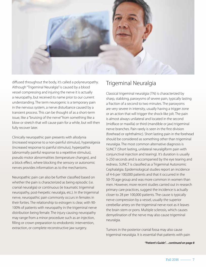
diffused throughout the body, it’s called a polyneuropathy. Although “Trigeminal Neuralgia” is caused by a blood vessel compressing and injuring the nerve it is actually a neuropathy, but received its name prior to our current understanding. The term neurogenic is a temporary pain in the nervous system, a nerve disturbance caused by a transient process. This can be thought of as a short-term issue, like a “bruising of the nerve” from something like a blow or stretch that will cause pain for a while, but will then fully recover later.
Clinically neuropathic pain presents with allodynia (increased response to a non-painful stimulus), hyperalgesia (increased response to painful stimulus), hyperpathia (abnormally painful response to a repetitive stimulus), pseudo motor abnormalities (temperature changes), and a block effect, where blocking the sensory or autonomic nerves provides information as to the mechanisms.
Neuropathic pain can also be further classified based on whether the pain is characterized as being episodic (i.e. cranial neuralgia) or continuous (ie traumatic trigeminal neuropathy, post-herpetic neuralgia, etc.). In the trigeminal nerve, neuropathic pain commonly occurs in females in their forties. The relationship to estrogen is clear, with 90-100% of patients with neuropathy in the trigeminal nerve distribution being female. The injury causing neuropathy may range from a minor procedure such as an injection, filling or crown preparation to endodontic intervention, extraction, or complete reconstructive jaw surgery.
Trigeminal Neuralgia
Classical trigeminal neuralgia (TN) is characterized by sharp, stabbing, paroxysms of severe pain, typically lasting a fraction of a second to two minutes. The paroxysms are very severe in intensity, usually having a trigger zone or an action that will trigger the shock-like jolt. The pain is almost always unilateral and located in the second (midface or maxilla) or third (mandible or jaw) trigeminal nerve branches. Pain rarely is seen in the first division (forehead or ophthalmic). Short lasting pain in the forehead should be considered as something other than trigeminal neuralgia. The most common alternative diagnosis is SUNCT (Short lasting, unilateral neuralgiform pain with conjunctival injection and tearing). It’s duration is usually 5-250 seconds and is accompanied by the eye tearing and redness. SUNCT is classified as a Trigeminal Autonomic Cephalalgia. Epidemiological studies report an incidence of 4-6 per 100,000 patients and that it occurred in the 50-70 age group and was more common in women than men. However, more recent studies carried out in research primary care practices, suggest the incidence is actually closer to 28 per 100,000 patients. The cause is typically nerve compression by a vessel, usually the superior cerebellar artery on the trigeminal nerve root as it leaves the brain stem or pons. Multiple sclerosis, which causes demyelination of the nerve may also cause trigeminal neuralgia.
Tumors in the posterior cranial fossa may also cause trigeminal neuralgia. It is essential that patients with pain
“Patient’s Guide”. . .continued on page 8

Fall 2016 -------------- 8
in the trigeminal nerve distribution obtain appropriate imaging to help define the diagnosis and etiology. This is usually an MRI of the brain with and without contrast with specific attention to the trigeminal nerve. Classical trigeminal neuralgia with purely paroxysmal pain is also marked by periods of complete pain-free remissions. However, there is another subtype of classical trigeminal neuralgia which has the same paroxysmal sharp pains, but with a concomitant continuous, persistent dull, aching
background pain that is mild to moderate in intensity. This subtype used to be called TN Type 2 or Atypical TN due to the “atypical” nature of its presentation.
The diagnostic classification of classical trigeminal neuralgia from the 3rd edition of The International Classification of Headache Disorders, devised by the International Headache Society, is as follows:
“Patient’s Guide”. . .continued from page 7
Table 3: Treatment Approach for Management of Trigeminal Neuralgia: Con�rm Diagnosis
Facial Pain:History, neurological exam, neurosensory testing
Facial Pain:Probable Trigeminal Neuralgia
Classical Trigeminal Neuralgia
Classical Trigeminal Neuralgia, purely paroxysmal
Classical Trigeminal Neuralgia,w/concomitant persistent facial pain
Painful Trigeminal Neuropathy,TN secondary to pathology
Atypical presentation, various characteristics w/occassional severe pain paroxysms
Sharp, shooting intermittent pain + Trigeminal sensory dysfunction & pain
MRI vascular compressionpain free remissions
MRI -continuous background pain
One Subclass: Post-Traumatic Trigeminal Neuropathy
Secondary pathology may be nerve damage
MRI + for underlying pathologyPersistent pain & dynamic mechanical allodynia test +

Fall 2016 -------------- 9
Classical trigeminal neuralgia
A. At least 3 attacks of unilateral facial pain fulfilling criteria B and C
B. In ≥1 divisions of trigemi-nal nerve, with no radiation beyond trigeminal distribu-tion
C. Pain has ≥3 of the follow-ing 4 characteristics:
1. recurring in paroxys-mal attacks lasting from a fraction of a second to 2 min2. severe intensity3. electric shock-like, shooting, stabbing or sharp in quality4. precipitated by innoc-uous stimuli to affected side of face
D. No clinically evident neu-rological deficit
E. Not better accounted for by another ICHD-3 diagnosis
There remains little doubt as to the efficacy of carbamazepine in the management of trigeminal neuralgia. The drug is highly effective and provides excellent pain relief within
a few days. The dose is between 100 mg and 1200 mg daily. Because there are potential side effects, base line blood tests should be obtained and repeated every few months to ensure there is no anemia or drop in sodium (hyponatremia). Oxcarbazepine, is a very useful alternative as it has similar efficacy but greatly improved tolerability and relative lack of interaction with other drugs. The dosing is between 150 mg and 1500 mg daily in two or three doses. As with carbamazepine blood tests to evaluate anemia and hyponatremia must be considered. Lamotrigine is a useful drug in patients who develop allergies to carbamazepine and oxcarbazepine but the need for slow escalation in order to reduce skin reactions means it is not useful for rapid relief. It is used in doses of 12.5 -300 mg daily. It may also be useful in those patients who have been mistakenly diagnosed as trigeminal neuralgia but in fact have SUNCT or SUNA. The second generation anticonvulsant drugs like gabapentin may be used as a third line drug. Although the side-effects of gabapentin are quoted as being low, sleepiness, weight gain
and edema seem to be frequently reported. Pregabalin appears to be a promising drug in that it can be escalated more rapidly and can be used on a twice daily dosage scheme and does appear to have some effect on reducing anxiety often present in these patients. In the type II trigeminal neuralgia where there is a continuous or longer lasting dull, burning, aching background pain, the addition of a tricyclic antidepressant such as nortriptyline, in doses around 50-100mg, at bedtime, may be helpful. Other anticonvulsants such as leviteracetam and zonisamide may be useful, but have not been studied in placebo controlled trials. Baclofen is a muscle relaxant that is very effective in trigeminal neuralgia in doses between 5 and 80 mg daily. Sedation is the most significant side effect. Phenytoin may be used as an alternative in doses of 100 -300 mg per day.
Surgical management for trigeminal neuralgia is very effective. The most effective surgery being microvascular decompression. Other options include stereotactic radiosurgery, balloon gangliolysis and radiofrequency rhizotomy.
Glossopharyngeal Neuralgia
Description: The International Association for the Study of Pain defines glossopharyngeal neuralgia as a sudden severe brief recurrent pain in the distribution of the glossopharyngeal nerve. This disorder may be due to symptomatic causes again due to compression of the nerve by tumors, malformations or vascular compression. Glossopharangeal
“Patient’s Guide”. . .continued on page 10
Fall 2016 -------------- 10
neuralgia is a severe, transient, stabbing, unilateral pain experienced in the ear, base of the tongue, tonsillar fossa and/or beneath the angle of the jaw. It is commonly provoked by swallowing, talking and/or coughing, and may remit and relapse in the fashion of classical trigeminal neuralgia. The pain can also be felt in the auricular and pharyngeal branches of the vagus nerve. The pain can be present for years and may have spontaneous remissions. The pain attacks last for no more than 2 minutes. Glossopharyngeal neuralgia is even rarer than trigeminal neuralgia, with an incidence rate of 0.7 per 100,000 patients. It occurs in the older age group and seems to predominate more in women. The drugs used in trigeminal neuralgia are also effective for glossopharangeal neuralgia.
Surgical management incudes nerve sectioning of glossopharangeal and pharyngeal branches of vagus. Microvascular decompression is also a possible treatment.
Burning mouth syndrome (BMS):
Description: An intraoral burning or dysesthetic sensation, recurring daily for more than 2 hours per day over more than 3 months, without clinically evident causative lesions. It is important to exclude other causes both local and systemic which can cause burning especially drugs such as the ACE inhibitors or infections such as candida or herpes simplex. It is particularly common in peri-menopausal or post- menopausal women. This is not a psychological disorder, rather there is evidence it is a neuropathic pain due to loss of the small pain fibers called c fibers.
Management of BMS is best treated with topically applied clonazepam. The patient is asked to suck a 0.5 mg tablet three times per day for three minutes and the tablet and saliva are spat out. This gives immediate relief. Continuing to do this for up to a month may be necessary to quiet the
Offering a full roster of advanced options for treatment, including:• Microvascular Decompression• Stereotactic Radiofrequency Lesion • Stereotactic Radiosurgery• Neurostimulation
Expert, integrated care for patients with trigeminal neuralgiaAddressing both your physical and emotional needs
The Facial Pain Program at Weill Cornell is directed by Dr. Philip E. Stieg, professor and chairman of the Department of Neurological surgery (left) and Dr. Michael Kaplitt, vice chairman, who specializes in advanced treatments for movement disorders and pain.
Advanced Treatment for Facial Pain
The Facial Pain Program at the Weill Cornell Brain and Spine Center is an innovative program that focuses on the diagnosis and treatment of trigeminal neuralgia, one of the most disabling causes of facial pain. Our team includes top specialists in vascular neurosurgery and pain disorders—internationally recognized experts in the field who have advanced training in the very latest minimally invasive procedures used to treat facial pain. Find out more at weillcornellbrainandspine.org or call 212-746-4684 to make an appointment.
Brain and Spine Center
TNA-Quarterly-Weill-Cornell-new branding.indd 1 11/12/2015 9:45:14 AM
“Patient’s Guide”. . .continued from page 9

Fall 2016 -------------- 11
burning. Alternative topical agents with peppermint, spice such as chili flakes, or capsaicin may also be useful. The addition of tricyclic antidepressants or antiepileptic agents may also be beneficial.
Post Traumatic Trigeminal Neuropathy:
Table 3: Persistent idiopathic facial pain (PIFP)Description: Persistent facial and/or oral pain, with varying presentations but recurring daily for more than 2 hours per day over more than 3 months, in the absence of clinical neurological deficit.
A. Facial and/or oral pain fulfilling criteria B and C
B. Recurring daily for >2 hours per day for >3 months
C. Pain has both of the following characteristics:
1. poorly localized, and not following the distribution of a peripheral nerve
2. dull, aching or nagging quality
D. Clinical neurological examination is normal
E. A dental cause has been excluded by appropriate investigations
Table 4. Painful Post-traumatic Trigeminal NeuropathyDescription: Head and/or facial pain in the distribution of one or more branches of the trigeminal nerve caused by another disorder and indicative of neural damage. The pain is highly variable in quality and intensity according to the cause.
A. Unilateral facial and/or oral pain fulfilling criterion “C”
B. History of an identifiable traumatic event to the trigeminal nerve, with clinically evident positive (hyperalgesia, allodynia) and/or negative (hypoaesthesia, hypoalgesia) signs of trigeminal nerve dysfunction
C. Evidence of causation demonstrated by both of the following:
1. pain is located in the distribution of the same trigeminal nerve
2. pain has developed within 3–6 months of the traumatic event
D. Not better accounted for by another ICHD-3 diagnosis
The most common cause of facial neuralgia is painful trigeminal neuropathy. This may be triggered by injury to any branch of the trigeminal nerve. Commonly this is secondary to a surgical or dental procedure, but can also occur with viral injury (post herpetic neuralgia). The injury may be a perfectly performed procedure that inadvertently sensitizes the nerve. An example would be following a root canal therapy. The purpose of this treatment is to remove an abscess or dead pulp in a tooth. This requires the removal of the nerve in the tooth. The nerve is a branch of the trigeminal nerve and when it is removed it can result in, nerve sensitization and persistent pain. This form of neuropathy occurs predominantly in females in their forties and is likely linked to a genetic predisposition and hormonally affected. The neuropathic pain following tissue or nerve injury in the trigeminal nerve distribution may be called a painful post traumatic trigeminal neuralgia (PPTN) – (Table 4) or trigeminal dysesthesia (TD). PPTN is defined as a continuous pain following complete or partial damage to a peripheral nerve or central nervous system structure. The pain is described as a continuous, burning numbness and often pulling pain.
There are a number of mechanisms described as causing traumatic induced neuropathy. They can be described as a) peripheral sensitization, b) ectopic activity due to sodium channel expression, c) central sensitization, d) A beta fiber reorganization, e) alteration in central inhibition systems, and f ) sympathetically maintained pain due to alpha receptor sprouting. More than one mechanism may be active to create individual clinical presentations.
a) Peripheral sensitization: Once stimulated or traumatized the peripheral nociceptors will release a variety of peptides including substance p, calcitonin gene related peptide (CGRP), and neurokinins. This results in a peripheral sensitivity that is characterized by an increased response to non-noxious (allodynia) and noxious stimuli (hyperalgesia).
b) Ectopic firing results after trauma, likely due to the
“Patient’s Guide”. . .continued on page 12

Fall 2016 -------------- 12
sprouting of sodium channels. The nerve is easily depolarized and spontaneous shooting pains result.
c) Central sensitization develops once the peripheral stimuli trigger second or third order neurons. Once again there is the release of peptides, though now in the dorsal horn (trigeminal nucleus) or thalamus, resulting in pain being generated without the presence of an ongoing peripheral stimulus. The role of glial cells and release of proinflammatory cytokines also plays a role in the central nervous system causing pain.
d) A beta fiber reorganization is another mechanism causing centrally mediated pain. It is described as occurring when the c fibers are destroyed, which usually have their second order neurons in lamina II, allowing for spontaneous growth of A beta fibers from lamina III into lamina II, making proprioceptive and temperature stimuli activate c fiber second order neurons. Hence non nociceptive activity causes pain.
e) Alterations in central inhibition, or disruption of normal pain inhibiting systems may also cause chronic pain. The brain has powerful opioid and non-opioid inhibitory systems. Neurochemicals such as serotonin, norepinephrine, and GABA all help the brain modulate nociception. If these inhibitory pathways are not efficient chronic pain may exist.
f ) Sympathetically maintained pain (SMP) may be a result of alpha receptor sprouting on peripheral nociceptors resulting in norepinephrine release peripherally causing pain. There is evidence that following neural injury, the sympathetic innervation in the dorsal root ganglia increase with age. It is not surprising that there is a higher incidence of neuropathic pain as we age. SMP is aggravated by non-noxious stimuli and interrupted temporarily by sympathetic block or alpha-adrenergic block with phentolamine.
The therapy for trigeminal dysesthesia is aimed at reducing peripheral nociceptive inputs and simultaneously enhancing central nervous system pain inhibitory systems. Management of neuropathy is achieved with integrating four treatment strategies: topicals, pharmacologic agents, nerve blocks/ nerve procedures, and behavioral strategies.
• Specializing in atypical facial pain
• Microvascular decompression
• Glycerol rhizotomy
• Comprehensive management of facial pain
Ramesh Babu MD,
NYU Medical Center530 First Ave.
Suite 7WNew York, NY 10016
212-263-7481
Associate Professor of Clinical Neurosurgery
“Patient’s Guide”. . .continued from page 11

Fall 2016 -------------- 13
Topical therapy allows a medication to be delivered to the source of pain via the skin or the mucosa in the mouth. An impression can be taken of the site of pain and the dentist can construct a plastic stent that fits over the pain site, called a “neurosensory shield”. The patient can take this in an out of their mouth and apply topical medications. The common medications include benzocaine, capsaicin, and compounded agents including tricyclic antidepressants, anti-inflammatories and antiepileptic agents. The use of clonazepam suck and spit as discussed in BMS may also be useful. Pharmacology often requires a combination of agents. Tricyclic antidepressants, such as nortriptyline in doses of 10 mg-100 mg is the first line therapy, followed by the use of antiepileptic agents such as pregabalin 50 mg-300 mg, or gabapentin 100mg-3000 mg. Nerve blocks help define if there is involvement of the sympathetic nervous system. Seeing the result of doing a sympathetic nerve block, either stellate ganglion block or sphenopalatine ganglion block will help the clinician choose a course of care. If there is a positive response to the procedure (greater than 60% reduction in pain) the patient may be a candidate for repeated blocks (up to 6 a week apart), or a radiofrequency (burning) procedure to the ganglion. Stimulation of the nerves may also be useful although are experimental. Onabotulinum toxin injected to the injured peripheral sensory nerve may desensitize it. Care should be taken not to create a weakness of the
face, as this agent also paralyzes the muscles. Behavioral therapy is very useful on its own or in combination with any pain management technique. This comes in many options. Use of cognitive behavioral techniques, stress management, biofeedback, hypnosis, and mindfulness are all very powerful tools that enhance the brain’s natural inhibitory systems. This can help the effectiveness of medications, procedures and natural healing.
Temporomandibular Disorders (TMD):
TMD is a collective term which may include a number of different clinical entities including muscle and joint components. Pain in the temporomandibular joint may occur in 10% of the population and TMD has been reported in 46.1% of the US population. In non-patient population studies, 75% have at least one joint dysfunction sign and about 33% have at least one symptom. Out of the 75% with a sign or symptom, fewer than 5% require
treatment. Inflammation within the joint accounts for TMD pain and the dysfunction is due to a disk/ condyle incoordination. Common suggested etiological factors for TMD include bruxism, trauma, bite abnormalities, and emotional stressors. Occlusal interferences (bite abnormalities) are by far the most controversial aspect of etiology and treatment in TMD. A literature meta-analysis does not support the theory that occlusion is a factor in TMD etiology. Trauma to the TMJ may result in acute capsulitis but this inflammatory process tends to resolve quickly without complication. Chronic joint disorders are more frequently associated with painful derangement of the TMJ. Articular disc displacement frequently underlies the mechanism of joint derangement but the etiology is unclear. The remarkable adaptive capacity of TMJ is well documented. Failure of this mechanism may lead to tissue destruction and disc displacement. This may be affected by age, stress, gender, systemic illness and previous trauma. However, acute and chronic
“Patient’s Guide”. . .continued on page 14

Fall 2016 -------------- 14
disc displacement is not always painful.
The TMJ is unique in its bilateral location with an upper and lower compartment separated by a fibrocartilaginous disc. This diarthrodial structure allows for both rotatory and translational movement of the mandible. Although the TMJ is subject to the same pathological disorders that affect other synovial joints, it is unique in certain anatomical aspects. Both joints move as a functional unit and are lined by a fibrous connective tissue, which is more resistant to degenerative change and has a greater capacity for repair. The masticatory system includes the articulation of the upper and lower dentition that may limit or support joint function and stability. A major component in TMD is joint noise and incoordination of the disc condyle relationship. This presents as noise in the joint with or without locking, or the inability to open with a normal range of motion. This is often referred to as an “internal derangement.” Articular disk displacement is the most common temporomandibular arthropathy and is characterized by an abnormal relationship or misalignment of the articular disk relative to the condyle. This is known as a disk derangement disorder and is classified as disk displacement with reduction and disk displacement without reduction. In disk displacement with reduction during mouth opening, the disk that begins in a misplaced position reduces or improves its structural relationship with the condyle. As it reduces, a sound often described as a clicking or popping is heard. When the mouth closes, a second sound called a “reciprocal click” may be audible as the disk moves off the condyle just before the teeth come together upon closing. Usually the closing noise is of less magnitude. Clicking sounds are not necessarily a sign of degeneration or an indication for treatment. Over one third of an asymptomatic sample can have moderate to severe derangement as seen on imaging and as many as 25% of clicking joints show normal or slightly displaced disks. Asymptomatic clicking does not require treatment. A disc displacement without reduction is described as an altered or misaligned disc-condyle structural relationship that is maintained during mandibular translation. This is also referred to as a “closed lock.”
Management of TMD is usually achieved with reducing stress on the joint through exercises and splint therapy,
coupled with medications such as anti-inflammatories and muscle relaxants. If locking occurs, an injection or flushing the joint may be necessary.
Myofascial Pain:
Pain associated with temporomandibular disorders (TMD) may often be muscular in origin. The most frequent diagnosis is myofascial pain. Myofascial pain is characterized by a regional muscle pain, described as dull or achy and associated with the presence of trigger points in muscles, tendons or fascia. Myofascial pain is a common cause of persistent regional pain involving the neck, shoulder head, and orofacial regions. The precise etiology of myofascial pain is unclear, but there are some developmental (e.g. stress or oral habits) and perpetuating (e.g. poor sleep, postural abnormalities, depression) factors that have been identified. The major characteristics of myofascial pain include trigger points in muscles and local and referred pain.
Management of myofascial pain is best achieved with a series of posture, stretching and strengthening exercises, coupled with physical medicine techniques such as spray and stretch and trigger point injections. Medications helpful in myofascial pain are muscle relaxants, tricyclic antidepressants and selective serotonin / norepinepherine reuptake blockers.
Conclusion:
Facial pain is a complex disorder with many causes and diagnoses. When suffering with facial pain the most important component in management is diagnosis. Too often the wastebasket term “Atypical Facial Pain” is used and if so the patient should challenge the clinician for a clearer diagnosis. It is clear that psychological factors may aggravate facial pain of any cause, but they are not to be blamed as the primary etiology. Once a diagnosis is established, persistence and perseverance in therapy will help the majority of those who suffer.
“Patient’s Guide”. . .continued from page 13

Fall 2016 -------------- 15
Can Medical Marijuana Conquer Pain?
Patients with trigeminal neuralgia and other facial pains often need to develop an armamentarium, a collection of resources and methods to carry out one’s purpose in order to reduce their pain. When the trigeminal nerve is involved, the pain can be unbearable, debilitating and result in hopelessness, loss of work, isolation and depression.
The pain usually cannot be controlled with analgesics or opioids and it can be difficult to find the right anticonvulsant, antidepressant or muscle relaxer. Over time, drug tolerance with these medications can occur, requiring dosage increases that may result in undesirable side-effects or the drug may no longer be effective in treating the pain.
Patients are inquiring about other options to control their pain and the use of marijuana is becoming a legitimate alternative.
Cannabis, or marijuana, was widely prescribed as early as the 1800s by physicians as a therapeutic agent until it became illegal in 1970 with the passage of the Controlled Substance Act. Public demand led to the legalization of marijuana for medical use in California 1996.
There are now 25 states, plus Washington DC, where the use of medical marijuana is legal (http://medicalmarijuana.procon.org). Marijuana possession is still considered a federal crime and possibly a felony (http://norml.org/laws/item/federal-penalties-2). Transportation into a state where it is illegal is a crime. Doctors may not “prescribe” cannabis
for medical use under federal law, though they can “recommend” its use under the First Amendment
Medical marijuana was brought to the forefront with the successful treatment of a young patient with seizures. Her name is Charlotte Figi and she started having seizures soon after birth. By age 3, she was having 300 a week, despite being on seven different medications. A special strain of medical marijuana, which was administered as a tincture and does not produce a “high” but acts to calm the brain, limited her seizures to 2 or 3 per month.
This special strain was named Charlotte’s Web and contained CBD, cannabidiol, which is a cannabis compound that has significant medical benefits but does not produce the high feeling that is associated with the psychoactive THC, tetrahydrocannabidiol.
In fact, CBD actually counteracts the psycho-activity of THC , making it an appealing option for patients for relief from inflammation, pain, seizures and spasms and other conditions without the disconcerting feelings of lethargy or dysphoria.
The amount of CBD in relation to THC is referred to as “CBD-rich” or “CBD-dominant”. “CBD-rich” is a cannabis strain that has equal amounts of CBD and THC or more CBD than THC. “CBD-dominant” is a strain that is CBD-rich but has very little THC content. CBD and THC work best for pain control when used together.
Editor’s note: This article is in response to the many patient inquiries we have received concerning medical marijuana. The FPA does not endorse the use of medical marijuana, or any specific treatment modality, our mission is to educate patients and aid them in advocating for their healthcare.
Written by Pam Neff, retired RN who is the patient support and services consultant for the Facial Pain Association. She can be contacted at (800) 923-3608 for more information.
For information on marijuana laws, go to http://www.safeaccessnow.org/federal_marijuana_law
“Marijuana”. . .continued on page 16
16 -------------- Quarterly: Journal of the Facial Pain Association
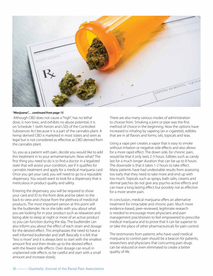
Although CBD does not cause a “high”, has no lethal dose, is non-toxic, and exhibits no abuse potential, it is on Schedule 1 (with heroin and LSD) of the Controlled Substances Act because it is a part of the cannabis plant. A hemp derived CBD is marketed in most states and seen as legal but is not considered as effective as CBD derived from the cannabis plant.
So, you as a patient with pain, decide you would like to add this treatment in to your armamentarium. Now what? The first thing you need to do is to find a doctor in a legalized state that will assess your condition, see if it qualifies for cannabis treatment and apply for a medical marijuana card. Once you get your card, you will need to go to a reputable dispensary. You would want to look for a dispensary that is meticulous in product quality and safety.
Entering the dispensary, you will be required to show your card and ID to the front desk and be taken to the back to view and choose from the plethora of medicinal products. The most important person at this point will be the budtender. He or she talks with you about what you are looking for in your product such as relaxation and being able to sleep at night or more of an active product so you can function during the day. The budtender can also inform you about the effect of each strain and dosage for the desired effect. This emphasizes the need to have a well informed budtender and staff. Basically it is felt that “less is more” and it is always best to start with the smallest amount first and then titrate up to the desired effect with the fewest side effects. Over dosage can result in unplanned side effects so be careful and start with a small amount and increase slowly.
There are also many various modes of administration to choose from. Smoking a joint or pipe was the first method of choice in the beginning. Now the options have increased to inhaling by vapeing (an e-cigarette), edibles that are in all flavors and forms, oils, topicals and teas.
Using a vape pen creates a vapor that is easy to smoke without irritation or negative side effects and also allows for a more rapid effect. The down side, for chronic pain, would be that it only lasts 2-3 hours. Edibles such as candy last for a much longer duration that can be up to 8 hours. The downside is that it takes 1-2 hours to take effect. Many patients have had undesirable results from assessing too early that they need to take more and end up with too much. Topicals such as sprays, bath salts, creams and dermal patches do not give any psycho-active effects and can have a long lasting effect but possibly not as effective for a more severe pain.
In conclusion, medical marijuana offers an alternative treatment for intractable and chronic pain. Much more evidence-based, peer-reviewed, legitimate research is needed to encourage more physicians and pain management practitioners to feel empowered to prescribe medical marijuana and to prove that it can be superior to or take the place of other pharmaceuticals for pain control.
The testimonies from patients who have used medical marijuana to control pain should be a beacon for patients, researchers and physicians that concurring pain drugs can be reduced or even eliminated to create a better quality of life.
“Marijuana”.. . .continued from page 15

Fall 2016 -------------- 17
Stress Exacerbated Systemic Illness
About the Author:Leesa Morrow, PhD is a clinical psychologist with extensive experience working with TN patients. Dr. Morrow is a frequent presenter at FPA conferences.
An example of courageOne patient with TN went to a psychotherapist after three failed microvascular decompression surgeries. At that time he was taking more than the highest recommended dose of carbamazepine, much to the consternation of his neurosurgery team. Before developing trigeminal neuralgia, this gentleman worked as an accountant for a local university. He enjoyed a leadership position in his church. He visited his adult
children frequently and he immensely enjoyed their companionship. He dearly loved his wife of many decades and he cherished their time together gardening, traveling, and socializing with friends. However, after years of TN pain, he had greatly reduced his social contact with members of his church and with his children. He and his wife did very little together other than sit at home reading or watching TV. Their conversations largely focused on the patient’s pain.
As he talked about his life, the therapist noticed that the patient tended to wince in pain when they began to discuss difficult topics. When he winced, she tended to instantly stop the discussion, even though the topic was crucially important to finish. The
“Stress. . .continued on page 18

Fall 2016 -------------- 18
same pattern occurred in the patient’s discussions with his wife. When the topic of discussion focused upon difficult content, the patient would wince and the discussion instantly stopped. The therapist had no sense that the patient was fabricating his experience of pain; it seemed very real. However, when he winced in pain, he did so in a manner that was
very dramatic and unusual among the trigeminal neuralgia patients she had treated. When in pain, his whole face contorted and he covered his face with trembling hands. The appearance of it was striking and it demanded silence.
Despite the authenticity of the patient’s pain, his pain-related behavior clearly worked to his benefit in that it allowed him to cut short conversations that dealt with topics he found personally difficult. While the patient could not avoid pain, he could avoid difficult conversations.
Before the onset of TN, this patient found difficult conversations especially unpleasant. He had always tended to be a little anxious and conflict avoidant. Nonetheless, he had routinely managed to participate in difficult conversations. His work required him to fire subordinates and restrict the spending of faculty. These work-related duties involved many difficult conversations, yet he was able to admirably perform in the workplace. The patient and his wife had three children and had elected to give up their home of many years in favor of a smaller residence at the time of retirement. Prior to developing TN, the patient was able to discuss matters of concern related to their children and the distribution of retirement assets without retreating from the discussion. The patient and his wife did not always agree on the issues, which was difficult for the patient, but he nonetheless managed to discuss issues of concern until they were resolved to both their satisfaction. After the onset of trigeminal neuralgia, the patient had increasingly avoided difficult conversations with significant others and increasingly focused more on discussions related to his pain.
Once the therapist was certain that the pattern of avoidance existed, she commented on it, and asked the patient whether he had noticed the impact that his wincing had upon conversations with his wife and children and in therapy. He thoughtfully considered the question, then responded that he had never thought about it before but, now that he had, he could see that the conversation entirely stopped when he winced. The therapist asked whether he was ever relieved to be done with the topic of conversation when that occurred. He responded, “Yes, sometimes.” The patient’s wife
was attending this session. The therapist asked her if she had noticed the pattern. She thought about it and responded that she often felt shut down by her husband’s pain. She explained that seeing him in pain made her feel that she must do something to help him but she had no idea what she might do to help. She said, “All I know to do is to hug him, but that doesn’t really help.” She asked what the therapist thought she might do to help him. The doctor said that it would help him just to understand how helpless she really felt in response to his pain, that she could do nothing more than be honest with him and do her best to take care of herself, so that he could use his energy to take care of himself. The patient very much loved his wife and was bothered by the thought of her feeling confused and helpless. He had never really understood the extent to which his wife felt intensely and uncomfortably inadequate in the face of his pain. She loved him too, yet she was clearly exhausted by her own self-imposed responsibility to ease his pain. She longed to be free of the responsibility for easing her husband’s pain but, as long as his pain was at the center of their lives, the only way for her to be free of this responsibility was to withdraw from him, which she was trying mightily not to do. They were able to discuss all these emotions, the patient’s fearful focus on his pain and his wife’s guilty obligation to ease his pain, to the point that each gained significant insight.
With time and growing awareness, the patient was able to minimize his wincing while talking with his wife and, more importantly, was able to continue discussions that were not easy for him. The patient was able to release his wife from any expectation that she would ease his pain. The patient openly expressed a sense
“Stress.” . .continued from page 17

Fall 2016 -------------- 19
of responsibility for doing what he could to master his fear of pain. He was able to return to social activities with the understanding that he would responsibly limit his interaction if his pain became overbearing. If he did not feel well enough to be social, he committed to assuming responsibility for communicating this and leaving the social interaction in order to care for himself. In these moments the patient was able to comfort himself through recognition that he would eventually feel better. Being freed from the largely self-imposed responsibility to ease her husband’s pain allowed the patient’s wife to hug him when she felt like being close, rather than out of fear or obligation. All of these changes helped to renew the connection between the patient and his wife.
Within four months of treatment, the patient and his wife were obviously happier, and the patient had cut down his use of carbamazepine to a safe and appropriate level. He accomplished this not because his pain was any less intense but because he had gained courage in the face of his pain, and because his awareness of the ways in which the pain had altered his behavior and identity had increased. All this was accomplished through the executive functions of the patient’s prefrontal cortex successfully inhibiting the fight-or-flight response, which allowed the patient to plan a new way of coping with ongoing pain that honored his self-concept as a loving husband and father. This is what it means to control your pain rather than it controlling you. But getting to the place where your prefrontal cortex is calling the shots takes effort and time.
Discussing courage in the face of excruciating, often uncontrollable pain, can strike a moral tone. The process of losing oneself to chronic facial pain does not involve moral failure. Fear in response to trigeminal neuralgic pain is natural and the brain’s response to fear is immediate and powerful. These neurophysiological changes are not a consequence of failure. However, it is important to recognize that the sense of self changes in response to the fearful experience of chronic facial pain—and this is rarely a good thing. The cycle of change is self-perpetuating as long as the cycle operates out of awareness. All need not be lost, however. Understanding that this process of changing identity commonly occurs in response to pain means that one can consciously and positively protect the sense of self, if one chooses to do so. Without exerting conscious effort, the personal and social forces of chronic facial pain will go unchecked and this typically leads to negative alterations in the sense of self.
If you decide that you would like to make changes in your response to chronic facial pain, be patient with yourself. Understand that you are not just making changes in behavior, beliefs, and attitudes: you are changing brain tissue. Through choice and self-reflection, you are altering the way neurons connect with one another to form the neural matrix that supports your identity and directs your responses to others. These changes take time. Be forgiving of yourself when things don’t go as you had hoped they would—and be very proud of yourself when they do.
The frightened brain and altered self-perceptionWe can define fear as a set of behavioral, autonomic, and hormonal events triggered by the brain in response to perceived physical danger. Most everyone will have heard the phrase “fight-or-flight response” and will know something about its value to survival. The fight-or-flight response involves many
“Stress”. . .continued on page 20

20 -------------- Quarterly: Journal of the Facial Pain Association
neural structures, some of which operate outside the brain in the periphery of the body and some of which operate in the very core of the brain. The amygdalae, two small almond-shaped structures situated deep in the temporal lobes of the brain on either side, are in charge of the fight-or-flight response.
Recent research interestingly indicates that the amygdalae also play a central role in the evaluation of faces. The role of the amygdalae in facial evaluation is to identify whether the person of interest is experiencing positive or negative emotion. This assessment is critical to determining whether the person presents a threat or not. Again, we see the primacy of the face to the human experience.
Research teaches us that the amygdalae learn from experience. If a threatening event occurs frequently enough, the amygdalae will come to regard the once neutral or even pleasant events that typically occur
before and after the threatening event as also dangerous. By the time this learning has occurred, many other brain structures have become part of the person’s generalized vigilance to danger. Thus, the fear response can generalize as a neural matrix that causes a person to irrationally identify benign life experiences as dangerous. Without intervention, this process can be very destructive to the sense of self.
The episodic, intensely painful nature of trigeminal neuralgia is perfect for provoking generalized fear. Imagine a grandmother who loves to hold and cuddle her infant grandchild. This seemingly small act of emotional bonding is critical to the grandmother’s sense of self. As she holds her grandchild, the grandmother recalls how she enjoyed cuddling her own infant children. Stored deep in her brain are memories of being lovingly held by her own mother. The simple unadorned connection of holding a much-loved child is consistent with the grandmother’s larger value system. Cuddling her infant grandchild is not a trivial thing: it is a small but lovely tile in the larger mosaic of her core beliefs about morality and interpersonal connection.
Imagine that as the grandmother and the grandchild gaze lovingly into one another’s eyes, the infant reaches up and touches her grandmother’s face. With this wholly innocent act of curiosity and love, the grandchild starts a rapid-fire series of fight-or-flight events in her grandmother’s brain. The child’s light touch triggers the pain of trigeminal neuralgia in her grandmother. With the painful stimulus of her grandchild’s touch, the grandmother’s amygdala instantly registers threat in the environment
and initiates the fight-or-flight response. If this event happens more than a few times, the grandmother will come to fear the touch of her grandchild. This fear will take on anticipatory dimensions that change the grandmother’s feeling about holding her grandchild. These changes are completely out of character for the grandmother: she is, after all, a loving person who deeply values her family; she is a person who enjoys the tenderness of loving touch. In this way, the fear of holding her grandchild threatens the grandmother’s sense of self. She must consciously adjust her thinking about these events if she is to preserve her core identity unaltered by chronic facial pain. If the grandmother does not actively problem-solve about these events, the fear they arouse will inevitably change her self-perception.
The self-preserving force of conscious thought cannot be overstated. Trigeminal neuralgia can initiate primal fear, which over time insidiously and deeply alters identity. This can happen without any fanfare or discussion through the simple interpersonal act of a child’s touch or a lover’s kiss. Still, it is not inevitable that these losses occur. Understanding the physical and psychological changes that chronic pain produces helps to create a sense of perceived control; this alone can help considerably.
It is important to recognize that the fight-or-flight response begins deep in the brain with the firing of the amygdalae, then extends up and out through most of the brain—all in a fraction of a second. Under the fierce provocation of fear, the amygdalae demand an increased synthesis of norepinephrine in the brain. High levels of norepinephrine are associated with increased
“Stress.” . .continued from page 19

Fall 2016 -------------- 21
vigilance to danger. This makes sense: hypervigilance to potential threat is adaptive within the context of what feels like life-threatening pain. The flight-or-fight circuitry activates the lateral hypothalamus and lateral medulla, causing increased blood pressure, constriction of the peripheral blood vessels, increased sweating, and loss of appetite. Part of the fight-or-flight response involves activation of the trigeminal nerve to create the automatic, involuntary facial expression of fright. By the time you have winced in response to the pain, the fight-or-flight response has already occurred. With repeated firing of these neural systems, the brain learns to associate pain with fear. With time, the distinction between fear and pain disappears. The frequent experience of fear pollutes relationships and self-perception with emotions of helplessness and defeat. As fear intensifies, pain intensifies, and so on. This cycle is obviously destructive, but it can be interrupted with knowledge, thought, and personal insight.
You may have heard the phrase, “what fires together, wires together.” Wiring by way of experience is the way the brain learns. The physical systems responsible for the fight-or-flight response learn to sense and avoid danger within the context of experience, albeit not without error. Repeated firings of the system change the brain so that it interprets ever more stimuli as threatening and so that it fires with less and less intense provocation. In other words, the fight-or-flight response can become enhanced through something like a practice effect. This is seen all the time in combat veterans. Those who fought in World War II, the Korean conflict, the Gulf War, Operation Desert Storm, Iraq, and Afghanistan
all say the same thing. When they first arrive in the war zone, they are terrified. However, the longer they stay, the more they adjust to the persistence of danger; they describe becoming robotic in their soldiering and going emotionally numb in a
seeming indifference to danger. It is not until they return home to the relative safety of their families and community environment that they recognize how much their brains have changed in response to persistent danger and multiple firings of their fight-or-flight system. In the relative safety of the home environment, they irrationally and automatically respond to everyday experiences with instantaneous fear. One veteran experienced a flashback when a small child popped a balloon in a pizza restaurant. Many veterans cannot tolerate shopping in a Target, Walmart, or similar large, open store; the space is too large to adequately scan for danger. There are sounds and people everywhere. From the perspective of their changed brains, any one of these sensory stimuli could signal a life-threatening risk. In response to
this generalized fear, these veterans do what is most natural: they avoid places and experiences that they cannot control. This means that they often become housebound and lose their most important relationships.
With persistent trigeminal neuralgic pain, a process can unfold not unlike the one the combat veteran experiences. Over time the patient with TN avoids more and more life experience as he or she attempts to avoid pain. This very human and understandable response to frequent, intense pain can result in the world becoming a very small place. The sense of self shrinks to accommodate a life reduced to nothing more than fearfully avoiding pain. In this way, bit by bit, trigeminal neuralgia alters and distorts identity. Fear and avoidance drive one to gradually let go of important relationships and activities. After trigeminal neuralgia, one may feel reduced to being mostly a patient, who remembers being a person. Having pain and avoiding pain thus become the defining features of one’s life.
These losses are not inevitable, however. Systems of the brain have evolved to modulate or check the automatic properties of the fight-or-flight response. We can learn not to fear what we know to be a non-life-threatening stimulus. While this won’t change the fact that TN pain is excruciating, it will limit the extent to which trigeminal neuralgia invades your life and alters who you are.
The essence of who we are is stored in the prefrontal cortex of the brain. That area is responsible for the executive functions that underlie purposeful behavior: it devotes concentration to cognition; it supports analytical problem-
“Stress”. . .continued on page 22
22 -------------- Quarterly: Journal of the Facial Pain Association
solving; it recognizes social cues and suppresses socially undesirable behavior; it imagines the future and plans complex action designed to achieve a particular goal; it makes choices possible. Specific areas in the prefrontal cortex support expressing and understanding language. Indeed, when a psychotherapist interacts with a patient, the therapist’s prefrontal cortex talks to the prefrontal cortex of the patient. Both learn much in the process of psychotherapy and both brains are changed by what they learn.
The prefrontal cortex can modulate activity in the amygdalae through descending inhibitory pathways. Generally speaking, the fight-or-flight response begins in the core basal structures of the brain, then rapidly fires outward through the brain. The inhibitory pathways of the prefrontal cortex begin on the outside structures of the brain and travel inward, where they can modify impulses. While the fight-or-flight system functions very fast, the pathways of the prefrontal cortex function relatively slowly and cover more tissue in the process. The fight-or-flight system of the brain functions automatically, while the systems of the prefrontal cortex function with purpose, on the heels of thought.
If you’ve ever heard people say that they’ve “learned to control their pain instead of the pain controlling them,” they are talking about utilizing the descending inhibitory pathways of the prefrontal cortex to control the tendency of the amygdalae to fire the fight-or-flight response in fearful anticipation of pain. When assisting patients to “control their pain,” the psychotherapist begins by emphasizing that the real lesson is in
learning to control their fear of pain. It is the fear of pain that shrinks the world and alters identity, not the pain itself.
The first step in the process of inhibiting fear is to learn what cardiologist and Harvard Professor of medicine Herbert Benson labeled the “quieting response” or “relaxation response.” Dr. Benson, a pioneer in mind-body medicine, thought of the quieting response as opposite to the fight-or-flight response. The quieting response can be learned in many ways, for example, through meditation, yoga, biofeedback, guided imagery, or hypnosis. Dr. Benson thought that prayer, too, could induce the quieting response.
Learning the relaxation response is only the first step in controlling pain-related fear, but it is an indispensable step. The person who wants to master pain-related fear must begin by learning the relaxation response through rehearsal and practice in a context free of pain. This will require a number of pain-free practice sessions. Patients should not attempt to use the relaxation response while experiencing pain during the initial
period of learning the response. Using it too early will result in the pain undoing what has been learned. This creates a one step forward, two steps back experience that diminishes hope and motivation. However, once patients can confidently and reliably produce the relaxation response, they can begin to use it immediately following the experience of TN pain. This helps to contain the fight-or-flight response so that it does not generalize throughout the brain. In other words, the relaxation response stops the fight-or-flight response in its tracks, before the rest of the brain can see it coming. It is essential for the patient to continue using the relaxation response during pain-free intervals so that the response remains robust.
The second step in learning to master pain-related fear is to do a cognitive appraisal of the fearful stimulus and the associated behavior it provokes. This appraisal may include recognizing and concentrating upon the fact that the pain will pass (or diminish) and that the pain will not kill you. With ongoing cognitive work, self-awareness typically increases through a process of discovery.
Fall 2016 -------------- 23
Honorary Tributes Our 60th Wedding Anniversary Eileen M. Gauger
Kelley Bergman Douglas Weeks
Linda Burnham Linda Burnham
Catherine Costello Janet L. O’Neil
Dr. Emad Eskandar, MD Donald Welcom
Jordan Grabel, MD Peter M. Fallon
Doris Haropulos Doris Haropulos
Kathy Hayes Helen J. Nicholson
Ally and Dan Kubik Melissa Anchan
Don Malley Mary A. Vanderwarf
Cecila Stachura Cecelia A. Stachura
Rob G. Parrish, MD Joe L Christian, Jr
Virginia Proffitt Kathleen Newcomb
Dr. Albert Rhoton, Jr Dorinda D. D’Agostino Dorothy Rainwater, RN
Liesl Sisson Judi van Resselaer
Dr. Christopher Slininger Ivy Lutzker
John Tew, MD Mona W. Marple
Mackenzie Winslow Floyd Winslow, Jr
Memorial TributeNattilee Bates Robert and Gail Russell
Danielle Beard Isaac R. Blair, Jr Wallace Putkowski
Frank and Muriel Borello Patricia Foggin
Gail Clark Skeeter Canevari
Les Cordonnier Roy S. McKay, Jr.
John DeMunter Catherine M. DeMunter
Gail Feiner Jerome Newman
Steve Goldstein Vasil and Lisa Vorsa
Chuck Grace Mary Ann Grace
Peter J. Jannetta, M.D. Iva Kay Conjelko Bronwyn G. Cowell
Nancy Mitchell Angelyn and David Furst
Effie S. Roth Helen I. Roth
Joyce Sweitzer Stanley Sweitzer
Andella Taylor Bonnie Pagano
Louise Tyson Carol Cornell
Rose Watts Barbara Noffke
FPA’s Memorial Tribute FundThere are special people in our lives we treasure. Increasingly, FPA supporters are making gifts in honor or in memory of such people. These thoughtful gifts are acknowledged with a special letter of thanks, are tax-deductible, and support FPA’s growing initiatives on behalf of TN patients and families. We are delighted to share recent Memorial Tribute gifts received as of June - August 2016:
24 -------------- Quarterly: Journal of the Facial Pain Association
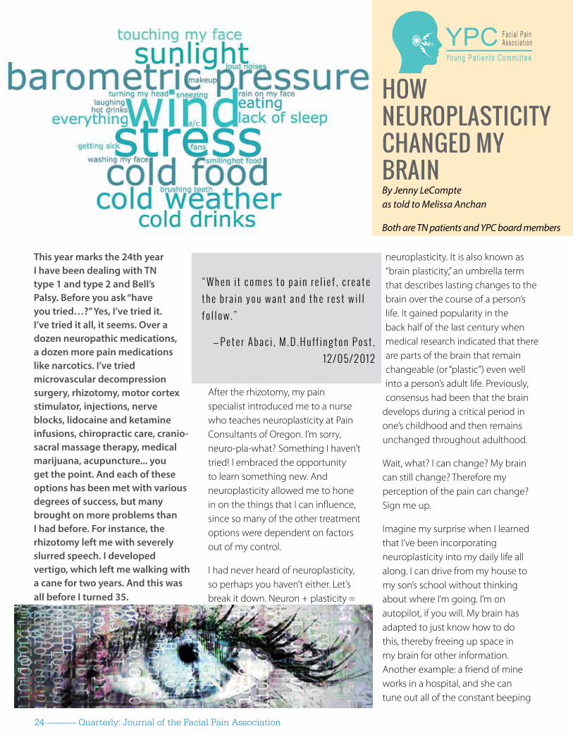
This year marks the 24th year I have been dealing with TN type 1 and type 2 and Bell’s Palsy. Before you ask “have you tried…?” Yes, I’ve tried it. I’ve tried it all, it seems. Over a dozen neuropathic medications, a dozen more pain medications like narcotics. I’ve tried microvascular decompression surgery, rhizotomy, motor cortex stimulator, injections, nerve blocks, lidocaine and ketamine infusions, chiropractic care, cranio-sacral massage therapy, medical marijuana, acupuncture... you get the point. And each of these options has been met with various degrees of success, but many brought on more problems than I had before. For instance, the rhizotomy left me with severely slurred speech. I developed vertigo, which left me walking with a cane for two years. And this was all before I turned 35.
After the rhizotomy, my pain specialist introduced me to a nurse who teaches neuroplasticity at Pain Consultants of Oregon. I’m sorry, neuro-pla-what? Something I haven’t tried! I embraced the opportunity to learn something new. And neuroplasticity allowed me to hone in on the things that I can influence, since so many of the other treatment options were dependent on factors out of my control.
I had never heard of neuroplasticity, so perhaps you haven’t either. Let’s break it down. Neuron + plasticity =
neuroplasticity. It is also known as “brain plasticity,” an umbrella term that describes lasting changes to the brain over the course of a person’s life. It gained popularity in the back half of the last century when medical research indicated that there are parts of the brain that remain changeable (or “plastic”) even well into a person’s adult life. Previously, consensus had been that the brain
develops during a critical period in one’s childhood and then remains unchanged throughout adulthood.
Wait, what? I can change? My brain can still change? Therefore my perception of the pain can change? Sign me up.
Imagine my surprise when I learned that I’ve been incorporating neuroplasticity into my daily life all along. I can drive from my house to my son’s school without thinking about where I’m going. I’m on autopilot, if you will. My brain has adapted to just know how to do this, thereby freeing up space in my brain for other information. Another example: a friend of mine works in a hospital, and she can tune out all of the constant beeping
YPC F a c i a l P a i nA s s o c i a t i o n
Young P a t i en t s Commi t t ee
HOW NEUROPLASTICITY CHANGED MY BRAINBy Jenny LeCompte as told to Melissa Anchan
Both are TN patients and YPC board members
“ W h e n i t c o m e s t o p a i n r e l i e f , c r e a t e t h e b r a i n y o u w a n t a n d t h e r e s t w i l l f o l l o w . ”
— P e t e r A b a c i , M . D . H u ff i n g t o n P o s t , 1 2 / 0 5 / 2 0 1 2

Fall 2016 -------------- 25
of the machines. Her brain learned how to do that over time. That’s neuroplasticity in its simplest form.
But this ability of our brains to change over time plays an interesting role when pain is factored in. One thing to note - there are big differences between acute pain and chronic pain. Acute pain is the result of tissue injury or bone damage, like a broken finger for instance. Chronic pain, on the other hand, is the disease. In the case of chronic pain, the brain makes maladpative changes to its wiring that make us feel lousy and change our behaviour. For people with persistent pain, the brain’s signals don’t allow us to tune out the pain, but rather they go into overdrive and cause us to
focus on it.
People with pain can experience a memory or “echo” of their original pain - such as amputees feeling “phantom pain” or MVD patients feeling “ghost pain” while healing. Our brains have been wired to hone in on the pain. But that can change. In the past few years, researchers have begun treating patients who suffer from chronic pain by teaching skills used in neuroplasticity, enabling them to rewire their brain to not feel (or at least decrease the sensation of ) pain. Think of these changes as the brain’s way of tuning itself to meet your needs. But how does that work?
One analogy to explain how the brain builds new neural pathways as new information or pain signals are introduced would be to think of the brain as an old radio (not the digital kind). If you were trying to listen to a particular station and could only somewhat make out the song but started to hear static, you would adjust the knob of the radio to get a clearer signal. Less static, more music. Building new neural pathways is the same thing - the more you focus and practice something, the better you become at learning that new skill or overcoming that obstacle or, in our case, tuning out the pain. By doing this, new neural connections are established in the brain as synapses that don’t usually fire at the same time start to sync up; this helps us sharpen our new skill.
Does it work? Science is starting to point to the notion that it does. Here’s a real-world example: researchers studied the brains of cab drivers and bus drivers in London and looked
at the size of their hippocampus, the part of the brain responsible for spatial recognition. It turns out that cab drivers have a larger average hippocampus size than their counterparts on the bus. This seems logical: bus drivers are creatures of habit, driving the same defined routes day in and day out. But cab drivers are constantly learning new roads, neighborhoods and taking different paths based on traffic, construction, etc. Their brains have had to grow and adapt to their new challenges.
It turns out you can teach an old dog (brain) some new tricks. Here are a few basic tools that could be considered neuroplastic techniques to help you use the positive aspects of neuroplasticity and rewire the nervous system connections not associated with pain
• New hobbies to help you use the positive aspects of neuroplasticity and rewire the nervous system connections not associated with pain
• Learn ways to manage stress and reduce the focus on your pain
• Exercise and movement done in a “paced” way can help reduce your hypersensitivity to pain
• Retrain the way in which your mind and body interpret and respond to pain
So how have I done it? Well, I’ll admit - I was a bit hesitant and it wasn’t easy. It still isn’t easy, even with a lot of practice. It’s a daily effort and it took me a good 12-18 months to really learn the process for me to be able to find the tools I keep handy in my
“Neuroplasticity”. . .continued on page 26

26 -------------- Quarterly: Journal of the Facial Pain Association
toolbox in order to live a pretty productive life. Learning to meditate has for sure been the most difficult part for me. As simple as it seems to learn breathing techniques, they take a lot of mental energy to keep going.
Neuroplasticity techniques, mindfulness and lots of self-care have helped me to not only regain my ability to speak, I’ve also gotten my vertigo issues under control to the point I can walk without a cane. My pain levels generally stay low so I can go on with my day. I am able to volunteer within the community and have lots of family time. My favorite byproduct of incorporating neuroplasticity in my daily routine has been finding all the fun and pleasures in life. My biggest joy is my son; I try and make sure we have some kind of adventure on a regular basis so I get to see the world through his eyes. Even on days I have increased pain, I find myself able to force the pain aside and enjoy the day instead.
It’s been important to find the main triggers that cause the pain and act in a way that intentionally fights them. Don’t get me wrong, I still avoid intense triggers, for instance hot days with the direct sunlight on my face, freezing cold weather or gusty winds. I try to go out in the overcast sunny days and enjoy my wind chimes on my patio. I go for walks with my son, even when it’s a little breezy outside. On days I choose to stay in, I love writing, reading, scrapbooking and baking. I keep a list handy of ways I can care for myself, either outside or inside. The better I feel, the more I find to add to this list - just more tools for the toolbox.
With enough practice, it’s said neuroplasticity could be seen as a “cure” or a way to heal what ails you. I think that’s a stretch. Of course someone who has lost a limb is not going to regrow it. The arteries that are wrapped around my trigeminal nerve are not going to magically unwrap themselves. I do believe, however, that neuroplasticity can get me to a state of remission. When I have an attack that’s more than I can handle, it’s a sure sign I haven’t been working on taking care of myself as well as I should have been. Of course there are some stressors and triggers in life that are beyond my control. What’s most important is I refuse to let the attacks get me down. I now know I’ll have more good days than bad, it takes work and persistence for it to become a way of life.
If I could give anyone advice on pursuing neuroplasticity as an option, it would be to not give up. Even with painful conditions like trigeminal neuralgia, neuroplasticity gets easier the longer you practice it. Attacks are not uncommon for me but the duration and intensity of the pain have been reduced significantly since I introduced neuroplasticity into my treatment.
Look, let’s be real here. I’m not a doctor - none of us on the YPC is. We’re all patients, just like you. And just because this has worked for me doesn’t mean it will work for you, or work in the same way. If there’s only one truth about facial pain patients it’s that each of us is different and has a unique story. The tools in my neuroplasticity toolbox will differ from yours. But if you’re like me and feel like you’ve tried everything, why not give it a shot?
For more information on neuroplasticity, here are a few resources we’ve found helpful:
Neuroplastic Transformation Workbook by Michael Moskowitz and Marla DePolo Golden
The Brain’s Way of Healing: Remarkable Discoveries and Recoveries from the Frontiers of Neuroplasticity by Norman Doidge
For more information about the YPC, visit ypc.tna-support.org OR /tnaypc
“Neuroplasticity”. . .continued from page 25

Fall 2016 -------------- 27
Break Free of Facial Pain Neurosurgeon Stephen E. Griffith, M.D., tailors surgical and nonsurgical treatments for trigeminal neuralgia. This includes:
• Microvascular decompression
• Internal neurolysis
• Radiofrequency gangliolysis
Trust Dr. Griffith and the experts at Saint Luke’s Marion Bloch Neuroscience Institute for world-class care to help you live life without pain.
Contact816-932-2700
4400 Broadway, Suite 510Kansas City, MO 64111
saintlukeskc.org/facialpain
Stephen E. Griffith, M.D.
Name: Lauren Hodkey
Current age: 17
Where do you live? Mentor, OH
How old were you when you first experienced facial pain? Age 14
How old were you when you were diagnosed? Age 14
What was your diagnosis? Type 1 Trigeminal Neuralgia on my right side
What do you do for fun? Theatre, Sing, Read, hang out with friends
What has TN taught you? It’s taught me to be grateful for everything and not take anything for granted, since there have been times where TN made everyday activities seem impossible. I appreciate the simplest things in life, like being able to make it through a school day, so much more now.
What treatments (non-surgical) have you tried? Everything from topical creams, and massage to
medications. Neurontin and Trileptal - both failed.
Have you had any procedures? Had 2 microvascular decompressions on my right side
How has your facial pain changed you? It’s made me a lot braver and stronger, I know now that I can get through anything since I’ve gotten through TN.
What tips do you have for other young patients? Don’t be afraid to say how you’re feeling. Stay positive, no matter how difficult things get. Better days are coming!
Young Patient Profile
28 -------------- Quarterly: Journal of the Facial Pain Association
FPA Membership The following individuals joined or renewed their FPA membership between June 1 and August 2016
JUNE
Maria Kash
JULY
Douglas Anderson
Jean Bauman
Helen Burrows
Evelyn Burry
Joyce Byram
Terry Czigan
Jay Donovan
Jean Gray
Richard Hersam
Robert Hirst
Darlene Kasten
Faith Laning
Wendy Lyka
Theresa Marston
Richard Martin
Maureen Muck
Donna Sullivan
Ginger Von Achen
Dennis Wenger
Candace Walkup
Dan Wegner
Amity Zepeda
Michael Zimmerman
AUGUST Jeremy Abbott
Charles Beaudoin
Pamela Biske
Kamla Brahmbhatt
Jonathan Cannon
Janet Ditslear
Marilyn Dixon
Stacey Ferguson
Darrell Fryer
Marvin Gorley
Monica Hart
Patricia Kolditz
Robert Liebler
Roger Martin
Betty McClelland
Mary McGinnis
Voncille Meyer
Miguel Pupiales
Patricia Riggins
Joseph Scheuchenzuber
Cora Schumann
Joyce Steadham
Brandi Sumpter
Shirley Thomas
Walter Toth
Cary Turner
William Willis
Without TN, life is full of possibilities.People with Trigeminal Neuralgia (TN) are often misdiagnosed and mistreated, referred from specialist to specialist without a solution. At UC Irvine Health, our experienced neurosurgeons are uniquely trained to diagnose and treat even the rarest cases of trigeminal neuralgia.
TN doesn’t have to be part of your daily life. Seek help from one of our specialists today.
Frank P.K. Hsu, MD, PhDChair, Dept. of Neurological Surgery Surgical Director of Functional, Epilepsy and Pain Neurosurgery
855-557-1531
Mark E. Linskey, MDWestern Regional Director Medical Advisory Board TNA–The Facial Pain Association
714-456-7002
ucirvinehealth.org

Fall 2016 -------------- 29
RELIEF AFTER A DECADE OF FACE PAIN
At first, Mary thought she just needed to see a dentist for her chronic face pain. But after being diagnosed with trigeminal neuralgia and over a decade of different high-dose medications, she had had enough. On December 15, 2015, Mary was treated with the Gamma Knife procedure. In less than an hour at The Valley Hospital Gamma Knife Center in Paramus, NJ, she went from managing her condition, to being free of it.
Start writing a new chapter to your story. Learn more about the procedure and our nurse navigator, who guides patients every step of the way. She will coordinate appointments, field questions and manage your follow-up care.
Schedule a consultation at The Valley Hospital Gamma Knife Center today by calling 201-634-5677 or visiting www.ValleyGammaKnife.com.
“The whole procedure took less than an hour—and it worked!”—Mary Hamilton, Warwick, NY

30 -------------- Quarterly: Journal of the Facial Pain Association
Alan Mechanic, M.D.
Jeffrey A. Brown, M.D.
Michael H. Brisman, M.D.
NSPC_Trigeminal NeuraVFeb18_TNA .indd 2 2/29/16 1:44 PM