Embed Size (px)
Citation preview
242 J La state Med soc vOL 159 September/October 2007
Journal of the Louisiana State Medical Society
cliNical caSe OF the mONth
a Paraplegic Man with altered Mental status and Fever
Credit
The LSMS Educational and Research Foundation designates this educational activity for a maximum of one (1) AMA PRA Category 1 CreditTM. Physicians should only claim credit commensurate with the extent of their participation in the activity.
disClosure
Drs. Ramirez, Martinez, Chaudhry, Hutchinson, O’Bryan, Heinrich, Anward, and Lowentritt have nothing to disclose. Ms. Tunuguntla and Mr. Rhodes have nothing to disclose.Dr. Lopez discloses that he is a member of the Journal Board of Trustees and the Journal Editorial Board.
originalreleasedateexpirationdate 9/30/2007 9/30/2008
cme iNFOrmatiONtargetaudienCe
The September/October Clinical Case of the Month is intended for medical students, general practitioners, medicine subspecialists, emergency medicine physicians, radiologists, urologists, pathologists and surgeons.
eduCationalobjeCtives
The Clinical Case of the Month is a regular educational feature presented by the Louisiana State University Department of Medicine. Medical students, residents, postdoctoral fellows, and faculty collaborate in the preparation of these discussions. After reading this article, physicians should be better able to identify and understand the pathophysiology, microbiology, epidemiology, clinical presentation, diagnosis, and treatment of emphysematous pyelonephritis. Estimated time to complete this activity is one (1) hour.
case PResenTaTion
A 48-year-old man was brought to the emergency department (ED) with persistent incomprehensible mumbling. His mother stated that four days prior to admission, after returning from a Mardi Gras parade, he went to bed and awoke the next morning vomiting and with no appetite. For the next three days he became progressively lethargic and complained of a vague headache. By the fourth day his mother was unable to understand his speech, and he had developed a subjective fever.
His past medical history was significant for chronic hepatitis C infection, diabetes mellitus type II, paraplegia secondary to a gunshot wound to the thoracic spine 22 years previously resulting in colostomy and chronic indwelling foley catheter, two prior sacral ulcers, and an episode of methicillin–resistant Staphylococcus aureus (MRSA)
Juan Ramirez, MD; Jorge Martinez, MD; Anila Chaudhry, MD; Jamie Hutchinson, MD; gerald O’Bryan, MD; Hari Tunuguntla, BS; Penny Heinrich, MD;
Mohammed Anward, MD; Brian Rhodes, BS; Joshua Lowentritt, MD; and Fred A. Lopez, MD (Section Editor)
bacteremia. He was hospitalized for treatment of a urinary tract infection secondary to E coli four months earlier.
Though adherence to his oral medication regimen was unknown, it included insulin, ciprofloxacin, clonidine, diazepam, hydrocodone-acetaminophen, isosorbide dinitrate, potassium chloride, lisinopril, quetiapine, and spironolactone. He had a documented urticarial reaction to iodine dye and ketorolac. Hospital medical records documented tobacco, marijuana, and inhalant (varnish) use. The patient lived with his mother.
Upon arrival to the ED, the patient’s vital signs were as follows: axillary temperature of 103.4oF, blood pressure of 101/52 mmHg, pulse of 141 beats per minute, respiratory rate of 40 breaths per minute, and oxygen saturation maintained at 98% with Venturi mask oxygen supplementation. On physical exam he was lethargic, responding only to painful stimuli, and the skin was warm
J La state Med soc vOL 159 September/October 2007 243
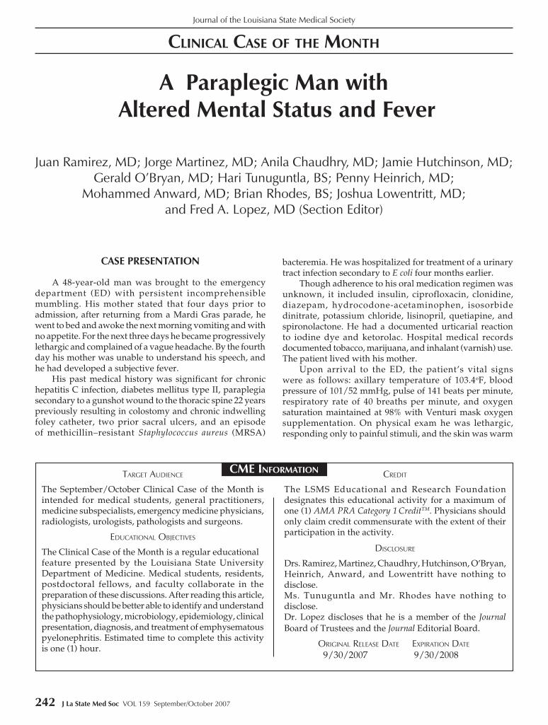
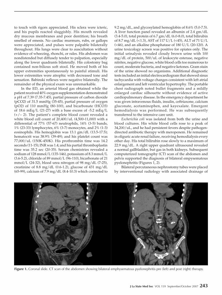
Figure 1. Coronal slide. CT scan of the abdomen showing bilateral emphysematous pyelonephritis pre (left) and post (right) therapy.
to touch with rigors appreciated. His sclera were icteric, and his pupils reacted sluggishly. His mouth revealed dry mucous membranes and poor dentition; his breath smelled of emesis. No cardiac murmurs, rubs, or gallops were appreciated, and pulses were palpable bilaterally throughout. His lungs were clear to auscultation without evidence of wheezing, rhonchi, or rales. His abdomen was nondistended but diffusely tender to palpation, especially along the lower quadrants bilaterally. His colostomy bag contained non-bilious soft stool. He moved both of his upper extremities spontaneously to painful stimuli. His lower extremities were atrophic with decreased tone and sensation. Babinski reflexes were negative bilaterally. The remainder of the physical exam was unremarkable.
In the ED, an arterial blood gas obtained while the patient received 40% oxygen supplementation demonstrated a pH of 7.39 (7.35-7.45), partial pressure of carbon dioxide (pCO2) of 31.5 mmHg (35-45), partial pressure of oxygen (pO2) of 110 mmHg (80-100), and bicarbonate (HCO3) of 18.6 mEq/L (21-27) with a base excess of -5.2 mEq/L (+/– 2). The patient’s complete blood count revealed a white blood cell count of 20,400/uL (4,500-11,000) with a differential of 77% (57-67) neutrophils, 14% (3-5) bands, 1% (23-33) lymphocytes, 6% (3-7) monocytes, and 2% (1-3) eosinophils. His hemoglobin was 13.1 gm/dL (13.5-17.5); hematocrit was 38.9% (39-49); and his platelet count was 77,000/uL (150K-450K). His prothrombin time was 14.2 seconds (11-15); INR was 1.4; and his partial thromboplastin time was 35.2 sec (20-35). Serum chemistries revealed a sodium of 128 mmol/L (135-146), potassium of 8.3 mmol/L (3.6-5.2), chloride of 89 mmol/L (96-110), bicarbonate of 21 mmol/L (24-32), blood urea nitrogen of 98 mg/dL (7-25), creatinine of 8.8 mg/dL (0.6-1.2), glucose of 431 mg/dL (65-99), calcium of 7.9 mg/dL (8.4-10.3) which corrected to
9.2 mg/dL, and glycosylated hemoglobin of 8.6% (5.0-7.5). A liver function panel revealed an albumin of 2.4 gm/dL (3.4-5.0), total protein of 6.7 gm/dL (6.0-8.0), total bilirubin of 8.7 mg/dL (<1.3), AST of 117 U/L (<45), ALT of 71 U/L (<46), and an alkaline phosphatase of 180 U/L (20-120). A urine toxicology screen was positive for opiates only. The initial urinalysis revealed cloudy brown urine with 100 mg/dL of protein, 500/uL of leukocyte esterase, negative nitrites, negative glucose, white blood cells too numerous to count, moderate bacteria, and negative ketones. Gram stain of the urine showed no organisms. Additional diagnostic tests included an initial electrocardiogram that showed sinus tachycardia with voltage changes consistent with left atrial enlargement and left ventricular hypertrophy. The portable chest radiograph noted bullet fragments and a mildly enlarged cardiac silhouette without evidence of active cardiopulmonary disease. In the emergency department he was given intravenous fluids, insulin, ceftriaxone, calcium gluconate, acetaminophen, and kayexalate. Emergent hemodialysis was performed. He was subsequently transferred to the intensive care unit.
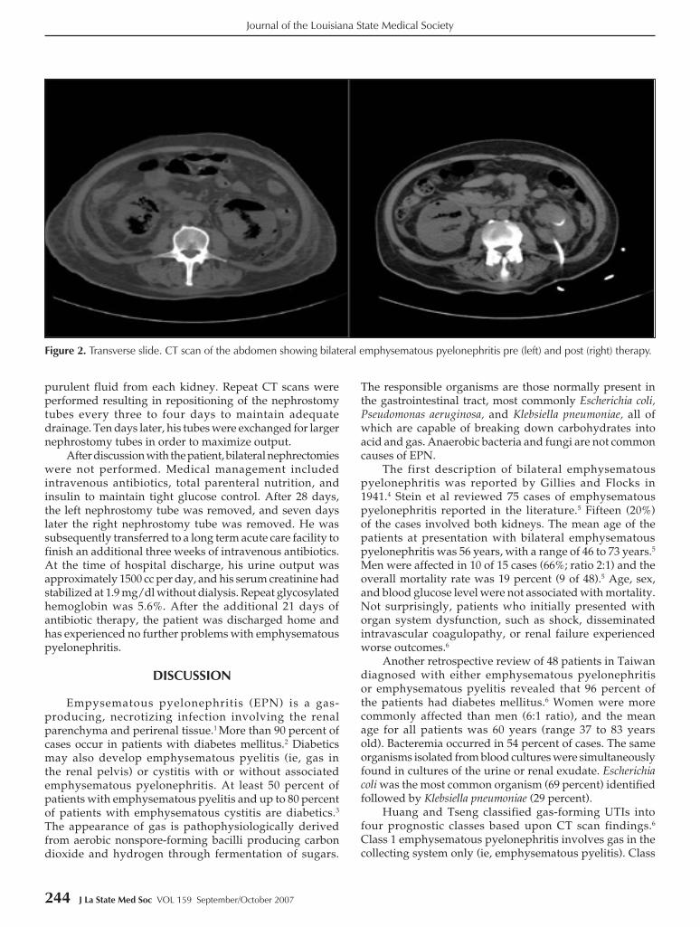
Escherichia coli was isolated from both the urine and blood cultures. His white blood cells rose to a peak of 34,200/uL, and he had persistent fevers despite pathogen-directed antibiotic therapy with meropenem. He remained in oliguric acute renal failure, receiving hemodialysis every other day. His total bilirubin rose slowly to a maximum of 22.9 mg/dL. A right upper quadrant ultrasound revealed a normal gallbladder, but gas in both kidneys. Subsequent computerized tomography (CT) scan of the abdomen and pelvis supported the diagnosis of bilateral empysematous pyelonephritis (Figures 1, 2).
Bilateral percutaneous nephrostomy tubes were placed by interventional radiology with associated drainage of
244 J La state Med soc vOL 159 September/October 2007
Journal of the Louisiana State Medical Society
Figure 2. Transverse slide. CT scan of the abdomen showing bilateral emphysematous pyelonephritis pre (left) and post (right) therapy.
purulent fluid from each kidney. Repeat CT scans were performed resulting in repositioning of the nephrostomy tubes every three to four days to maintain adequate drainage. Ten days later, his tubes were exchanged for larger nephrostomy tubes in order to maximize output.
After discussion with the patient, bilateral nephrectomies were not performed. Medical management included intravenous antibiotics, total parenteral nutrition, and insulin to maintain tight glucose control. After 28 days, the left nephrostomy tube was removed, and seven days later the right nephrostomy tube was removed. He was subsequently transferred to a long term acute care facility to finish an additional three weeks of intravenous antibiotics. At the time of hospital discharge, his urine output was approximately 1500 cc per day, and his serum creatinine had stabilized at 1.9 mg/dl without dialysis. Repeat glycosylated hemoglobin was 5.6%. After the additional 21 days of antibiotic therapy, the patient was discharged home and has experienced no further problems with emphysematous pyelonephritis.
Discussion
Empysematous pyelonephritis (EPN) is a gas-producing, necrotizing infection involving the renal parenchyma and perirenal tissue.1 More than 90 percent of cases occur in patients with diabetes mellitus.2 Diabetics may also develop emphysematous pyelitis (ie, gas in the renal pelvis) or cystitis with or without associated emphysematous pyelonephritis. At least 50 percent of patients with emphysematous pyelitis and up to 80 percent of patients with emphysematous cystitis are diabetics.3 The appearance of gas is pathophysiologically derived from aerobic nonspore-forming bacilli producing carbon dioxide and hydrogen through fermentation of sugars.
The responsible organisms are those normally present in the gastrointestinal tract, most commonly Escherichia coli, Pseudomonas aeruginosa, and Klebsiella pneumoniae, all of which are capable of breaking down carbohydrates into acid and gas. Anaerobic bacteria and fungi are not common causes of EPN.
The first description of bilateral emphysematous pyelonephritis was reported by Gillies and Flocks in 1941.4 Stein et al reviewed 75 cases of emphysematous pyelonephritis reported in the literature.5 Fifteen (20%) of the cases involved both kidneys. The mean age of the patients at presentation with bilateral emphysematous pyelonephritis was 56 years, with a range of 46 to 73 years.5 Men were affected in 10 of 15 cases (66%; ratio 2:1) and the overall mortality rate was 19 percent (9 of 48).5 Age, sex, and blood glucose level were not associated with mortality. Not surprisingly, patients who initially presented with organ system dysfunction, such as shock, disseminated intravascular coagulopathy, or renal failure experienced worse outcomes.6
Another retrospective review of 48 patients in Taiwan diagnosed with either emphysematous pyelonephritis or emphysematous pyelitis revealed that 96 percent of the patients had diabetes mellitus.6 Women were more commonly affected than men (6:1 ratio), and the mean age for all patients was 60 years (range 37 to 83 years old). Bacteremia occurred in 54 percent of cases. The same organisms isolated from blood cultures were simultaneously found in cultures of the urine or renal exudate. Escherichia coli was the most common organism (69 percent) identified followed by Klebsiella pneumoniae (29 percent).
Huang and Tseng classified gas-forming UTIs into four prognostic classes based upon CT scan findings.6 Class 1 emphysematous pyelonephritis involves gas in the collecting system only (ie, emphysematous pyelitis). Class

J La state Med soc vOL 159 September/October 2007 245
2 emphysematous pyelonephritis includes gas in the renal parenchyma without extension to the extrarenal space. Class 3 emphysematous pyelonephritis is divided into two subclasses. Class 3A involves the extension of gas or abscess to the perinephric space, while Class 3B involves the extension of gas or abscess into the pararenal space. Class 4 includes bilateral emphysematous pyelonephritis or a solitary kidney with emphysematous pyelonephritis.
Clinical features of emphysematous pyelonephritis and emphysematous pyelitis are usually indistinguishable from those seen in severe acute pyelonephritis. Onset may be abrupt or evolve slowly over two to three weeks. Most patients complain of fever, chills, flank or abdominal pain, nausea, and vomiting. Physical examination may reveal an abdominal mass.7 A careful distinction should be made between gas confined to the collecting system, gas confined to the parenchyma, and gas outside the parenchyma. In advanced stages gas may surround the kidney as a crescent formation within the renal capsule, may accumulate in the perirenal space within Gerota’s fascia, and/or may perforate and enter the pararenal and retroperitoneal spaces. Crepitation over the flank or thigh of a patient with diabetes mellitus suggests a gas-forming renal infection with retroperitoneal extension.8 Patients with emphysematous cystitis typically present with frequency and urgency, dysuria, abdominal pain, and pneumaturia.7
The diagnosis of a emphysematous pyelonephritis can be made by plain films of the abdomen, which will reveal gas in the renal parenchyma or surrounding tissue in one- third of cases. If gas is present on plain films, abdominal CT scanning is mandatory to visualize the extent of gas in the genitourinary system.7 Laboratory testing usually reveals hyperglycemia, leukocytosis, elevated blood urea nitrogen and creatinine, and pyuria. The initial treatment of bilateral emphysematous pyelonephritis consists of vigorous hydration and intravenous antibiotics, along with aggressive control of hyperglycemia in the diabetic patient. Treatment of obstruction of the kidney or ureters and drainage of the kidneys are essential.7,9
In Class 1 or Class 2 emphysematous pyelonephritis, antibiotics and percutaneous catheter placement are usually sufficient. In Class 3A or 3B emphysematous pyelonephritis without systemic organ dysfunction, antibiotics plus percutaneous catheter placement is recommended. However, immediate nephrectomy is necessary if patients with Class 3A or 3B emphysematous pyelonephritis develop systemic organ dysfunction. Immediate bilateral percutaneous drainage is mandatory for all patients with Class 4 bilateral emphysematous pyelonephritis. In addition, nephrectomy is recommended for all patients when the clinical situation does not respond to percutaneous drainage.
ReFeRences
1. Kumar A, Turney JH, Brownjohn AM, et al. Unusual bacterial infections of the urinary tract in diabetic patients—rare but
frequently lethal. Nephrol Dial Transplant 2001; 16:1062-1065.2. Evanoff GV, Thompson CS, Foley R, et al. Spectrum of gas within
the kidney. Emphysematous pyelonephritis and emphysematous pyelitis. Am J Med 1987; 83:149-154.
3. Gropper M, Kravtsov A, Potasman I. Emphysematous cystitis: Illustrative case report and review of the literature. Medicine 2007; 86:47-53.
4. Gilles CL, Flocks R. Spontaneous renal and perirenal enphysema: report of a case in a diabetic from Escherichia coli infection. Am J Roentgenol 1941; 46: 173-174.
5. Stein JP, Spitz A, Elmajian DA, et al. Bilateral emphysematous pyelonephritis: A case report and review of the literature. Urology 1996; 47:129-134.
6. Huang JJ, Tseng CC. Emphysematous pyelonephritis: clinicoradiological classification, management, prognosis, and pathogenesis. Arch Intern Med 2000; 160:797-805.
7. Joshi N, Caputo GM, Weitekamp MR, et al. Infections in patients with diabetes mellitus. N Engl J Med 1999; 1906-1912.
8. Ellenbogen PH, Talner LB. Uroradiology of diabetes mellitus. Urology 1976; 8:413-419.
9. Chen MT, Huang CN, Chou YH, et al. Percutaneous drainage in the treatment of emphysematous pyelonephritis: 10-year experience. J Urol 1997; 157:1569-1573.
Dr. Ramirez and Dr. o’Bryan are house officers in the Department of Medicine-Emergency Medicine training program at Louisiana State university Health Sciences Center in New Orleans, Lousiana (LSuHSC-NO). Dr. heinrich is a house officer in the Department of Medicine at LSuHSC-NO. Dr. hutchinson and Dr. anward are a house officers in the Department of Psychiatry at LSuHSC-NO. Ms. Tunuguntla and Mr. Rhodes are fourth year medical students at LSuHSC-NO.
246 J La state Med soc vOL 159 September/October 2007
Journal of the Louisiana State Medical Society
Lead authors: earn CMe Credit For PubLishing artiCLes
Physicians may claim AMA PRA Category 1 Credit™ directly from the AMA for learning that occurs as a result of publishing articles. Credit can be awarded only for articles published within the last three years. Applicants should keep a copy of the application and supporting documentation submitted.
Credit may be claimed only for meeting the following criteria:
Publishing an article in a journal as a lead author.
The journal must be included in the MEDLINE bibliographic database.
The AMA will award 10 credits per article. Go to www.ama-assn.org/ama/pub/category/16244.html to print a Direct Credit Application and to receive more details.
1.
2.
Dr. chaudhry was a chief resident in Medicine at LSuHSC-NO. Dr. Martinez is the program director for both Internal Medicine and the combined program Internal Medicine / Emergency Medicine at LSuHSC-NO. Dr. Lowentritt is a nephrologist at Touro Infirmary in New Orleans, Louisiana. Dr. Lopez is associate professor and vice chair in the Department of Medicine at LSuHSC in New Orleans, LA.
cMe QuesTions
Read the preceding CME article and complete the registration, evaluation, and answer form on page 285 to earn CME credit. Mail or fax the registration, evaluation, and answer form to the LSMS Educational and Research Foundation. Answers must be postmarked or faxed prior to September 30, 2008. Participants must attain a minimum score of 75% to receive credit. LSMS members may also go online at http://www.lsms.org. Click on the Journal logo and then click on the Journal CME link. Complete the interactive answer sheet for each CME article.
Choose the one answer that is most correct for each question.
1. All of the following are true concerning emphysematous pyelonephritis except:a. Emphysematous pyelonephritis is rare in patients
with diabetes.b. Onset may be abrupt or evolve slowly over two to
three weeks.
c. The responsible organisms are those normally present in the gastrointestinal tract, most commonly Escherichia coli, Pseudomonas aeruginosa, and Klebsiella pneumoniae
d. More than 90 percent of cases occur in patients with diabetes.
2. True or False: Emphysematous pyelonephritis (EPN) is a non-gas-producing, necrotizing infection involving the renal parenchyma and perirenal tissue.
3. Class 2 emphysematous pyelonephritis includes:a. Gas in the collecting system only.b. Gas in the renal parenchyma without extension to
the extrarenal space.c. The extension of gas or abscess to the pararenal
space.d. Bilateral emphysematous pyelonephritis or solitary
kidney with emphysematous pyelonephritis.
4. True or False: Clinical features of emphysematous pyelonephritis and emphysematous pyelitis are usually indistinguishable from those seen in severe, acute pyelonephritis.