Embed Size (px)
Citation preview
AnAtomy of Phyllodina Persica (BivAlviA: tellinidAe), And its first occurrence in southeAstern BrAziliAn wAters
rodrigo cesAr mArques1,2
luiz ricArdo l. simone2
AbstrAct
This study presents a detailed anatomy of a rare Western Atlantic tellin, Phyllodina persica, under a comparative scenario. Some characters are shared with other tellinids such as the large hemipalps compared to gills; gills with outer demibranch with a single lamella absent from the pericardial region; the type‑V stomach associated with the style sac conjoined with the proxi‑mal intestine, and distal intestine presenting a dorsal and ventral group of loops, separated by the transverse muscle. The stomach presents a laterally enlarged typhlosole, although shallow, without flange in the margins. This feature is not found in other tellinid species. Another noteworthy feature in the stomach is the aperture of both caeca, which are larger than the left pouch aperture, and as wide as the style sac aperture. Furthermore, there is an interesting small process in the anterior hinge, and a pair of oblique protractor muscles placed posteriorly to the anterior foot retractor muscle, being a new type of intrinsic muscle described in bivalves. In addition to anatomy, this study presents the southernmost record of P. persica, expanding its distribution to the southeastern region of Brazil.
Key-Words: Tellinidae; Anatomy; Morphology; Phyllodina; Western Atlantic.
IntroductIon
Phyllodina Dall 1900 is a tellinid taxon of rare occurrence, characterized by an elongated oval out-line, oblique and detached pallial sinus connected to the pallial line by a linear scar and a foliated concen-tric sculpture (Boss, 1969). It has traditionally been considered as a subgenus of Tellina Linné, 1758. Due to this distinctive characteristics, Phyllodina possibly constitutes a natural group (Boss, 1966, 1969). Re-cently this taxon, like of most subgenera belonging to Tellina, have been shifted to the genus status (e.g., Turgeon et al., 2009). Four species of Phyllodina are
present in the American continents: Phyllodina fluctig‑era (Dall, 1908) and P. pristiphora (Dall, 1900) from Mexican to Costa Rica Pacific coasts (Keen, 1971), P. persica (Dall & Simpson, 1901) and P. squamifera (Deshayes, 1855) in the Western Atlantic, ranging from North Carolina, USA (Boss, 1969), through Ca-ribbean, and Brazilian Northeastern Coast (Tenório, 1984; Cauquoin, 1967). P. persica, the focus of the present paper, is characterized by a more ellipsoid out-line and a sculpture less foliated than P. squamifera and P. pristophora, resembling P. fluctigera of the Eastern Pacific, a possible closely allied species (Boss, 1969). P. persica presents a very rare record, characterized so
1. Programa de Pós-Graduação em Zoologia, Instituto de Biociências da Universidade de São Paulo. E-mail: [email protected]. Museu de Zoologia, Universidade de São Paulo. Caixa Postal 42.494, 04218-970, São Paulo, SP, Brasil. E-mail: [email protected]
Volume 53(9):115‑127, 2013
far by few occurrences from Cuba through the Lesser Antilles, and the Colombian Caribean. The present reported occurrence in northern Rio de Janeiro State expands the geographic distribution of the species. All the registers are in depths below 25 m up to 400 m. All of these past registers are constituted only by valves, without soft parts preserved.
Recently, in a marine research study carried out by Petrobras S/A, called Habitats Project, a well-pre-served specimen was dredged from northern Rio de Janeiro State. Due to paucity of the Phylodina records, as well as the first occurrence of a sample with soft parts, the assessment of the anatomy of P. persica im-proves considerably the systematics of the taxon, this being a concern of the present paper in a comparative scenario.
MAterIAl And Methods
The specimen is fixed in Alcohol 70% and dis-sected by standard techniques, immersed in alcohol. Pictures were taken with a digital camera mounted on stereomicroscope and later edited in Corel Photo-Paint®. Drawings were done with the aid of a camera lucida and later edited in Corel Draw®.
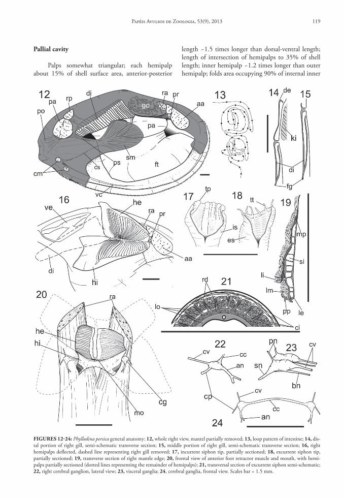
Abbreviations in figures and text: aa, anterior adduc-tor muscle; ab, aortic bulb; am, anterior transverse muscle above stomach; ap, dorsal-posterior appendix; bm, branchial muscle; bn, branchial nerve; an, an-terior nerve; at, anterior lateral tooth; au, auricle; bt, bifid cardinal tooth; ci, circular siphonal mus-cle; cc, cerebral commissure; cg, cerebral ganglion; cm, cruciform muscle; cp, cerebro-pedal connective; cs, cruciform to siphonal muscle; ct, ctenidial axis; cv, cerebro-visceral connective; dd, diverticulum; de, outer demibranch; dh, dorsal hood; di, inner demibranch; es, excurrent siphon; fl, flap over style sac aperture; fn, pedal nerve; ft, foot; gd, gonoduct; gf, gastric flap; go, gonad; gp, gonopore; gs, gastric shield; he, outer hemipalp; hi, inner hemipalp; ig, in-testinal food groove im, intestinal transverse muscle; is, incurrent siphon; ki, kidney; lg, ligament; la, lat-eral extension of typhlosole; lc, left caecum; le, ex-ternal mantle lamella; li, internal mantle lamella; lm, middle mantle lamella; lo, longitudinal siphonal muscle; lp, left pouch; lt, laminate cardinal tooth; mp, pallial muscle; mo, mouth; ne, nephropore; ny, nymph; oe, esophagus; om, oblique protactor muscle; pa, posterior adductor muscle; pg, pedal gan-glion; pl, palp; po, posterior pallial muscle; pp, pa-pilla; pn, posterior nerve; pm, posterior transverse
muscle; pr, foot protractor muscle; ps, septal siphonal muscle; pt, posterior lateral tooth; r, dorsal stomach ridge; r1, minor dorsal stomach ridge; ra, anterior foot retractor muscle; rc, right caecum; rd, radial si-phonal muscle re, rectum; rg, ring around style sac aperture; rm, esophageal rim; rp, posterior foot retrac-tor muscle; rs1, superior branch of siphonal muscle; rs2, inferior branch of siphonal muscle; sa, selection area; si, blood sinus; sm, siphonal muscle; sn, sipho-nal nerve; sp, intestine-style sac septum; ss, style sac; st, stomach; tp, projections around distal siphon tip; tm, middle transverse muscle; tt, siphonal ten-tacle; ty, typhlosole; vc, ventral channel; ve, ventricle; vg, visceral ganglion; vi, visceral sac; vs, vestibule.
descrIptIon
Phyllodina persica dall and simpson 1901 (Figs. 1‑40)
Tellina (Arcopagia; Phyllodina) persica: Dall & Simp-son 1901: 479 (pl. 55, fig. 1).
Tellina (Arcopagia) persica: McLean, 1951: 94.Tellina (Eurytellina) persica: Warmke & Abbott,
1961: 195 (pl. 29, fig. g).Tellina persica: Cauquoin, 1967: 227.Tellina (Phyllodina) persica: Boss, 1969: 255 (pl. 136,
fig. 2); Rios, 1975: 241 (pl. 77, fig. 1157); 1994: 271 (pl. 93, fig. 1328); Abbott, 1974 497 (fig. 5646); Díaz & Puyana, 1994: 88 (fig. 219).
Type locality: Mayaguez, Puerto Rico.
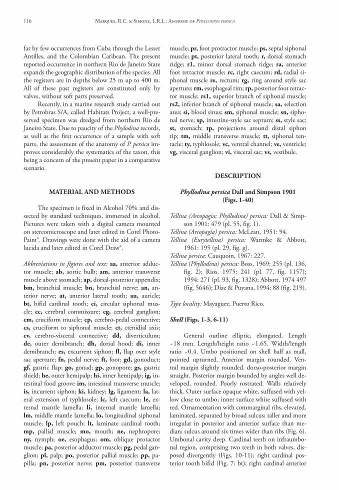
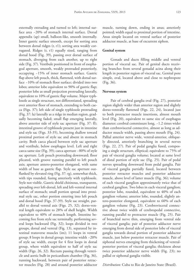
Shell (Figs. 1‑3, 6‑11)
General outline elliptic, elongated. Length ~18 mm. Length/height ratio ~1.65. Width/length ratio ~0.4. Umbo positioned on shell half as mall, pointed upturned. Anterior margin rounded. Ven-tral margin slightly rounded. dorso-posterior margin straight. Posterior margin bounded by angles well de-veloped, rounded. Poorly rostrated. Walls relatively thick. Outer surface opaque white, suffused with yel-low close to umbo; inner surface white suffused with red. Ornamentation with commarginal ribs, elevated, laminated, separated by broad sulcus; taller and more irregular in posterior and anterior surface than me-dian; sulcus around six times wider than ribs (Fig. 6). Umbonal cavity deep. Cardinal teeth on infraumbo-nal region, comprising two teeth in both valves, dis-posed divergently (Figs. 10-11); right cardinal pos-terior tooth bifid (Fig. 7: bt); right cardinal anterior
Marques, R.C. & Simone, L.R.L.: Anatomy of Phyllodina Persica116
tooth laminate (Fig. 7); left cardinal posterior tooth laminate (Fig. 8: lt); left cardinal anterior tooth bi-fid (Fig. 8: bt). Cardinal laminate teeth not reaching cardinal plate margin (Fig. 9). Anterior and posterior lateral teeth present in both valves, sockets present in right valve only; anterior lateral teeth distal, ranging
from posterior third to posterior half of dorso-an-terior margin, with taller elevation in half, turned dorsally upward (Fig. 10: at); posterior lateral teeth distal, located above posterior adductor scar level, in third posterior of dorso-posterior margin, equivalent to 30% of anterior lateral teeth length (Fig. 10: pt).
FIgures 1‑11: Shell and anatomic aspects of Phyllodina persica (MZSP 96780): 1, left valve, external view; 2, right valve, internal view; 3, dorsal view; 4, right view, valves and part of mantle removed; 5, detail of right mantle edge, internal view (gray arrow = thin muscles; white arrow = thick muscles); 6, detail of commarginal ornamentation on posterior surface of left valve; 7, right cardinal hinge tooth, inferior view; 8, left cardinal hinge tooth, inferior view; 9, right cardinal tooth and process anterior hinge (white arrow); 10, right hinge; 11, left hinge. Scales bar = 1.5 mm.
Papéis Avulsos de Zoologia, 53(9), 2013 117
Small process on third posterior between cardinal teeth and anterior lateral teeth (Fig. 9). Ligament seat-ed on nymph, brown, ranging from posterior side of umbo to half of dorso-posterior margin (Figs. 10-11). Nymph wall shorter than lateral teeth, parallel to mar-gin. Muscle scars well visible; both adductor muscle scars of equivalent size, each scar occupying ~10% of valve inner surface. Anterior adductor muscle scars el-liptical, dorso-ventrally elongated. Posterior adductor muscle scars rounded. Pallial line parallel and at 1/10 of shell height distant from margin. Pallial sinus an-tero-posteriorly elongated, detached from pallial line, extending to half of valve; ventral margin straight, raised 45° from pallial line; posterior margins slightly pointed, dorsal margin slightly curved (Fig. 2). Cruci-form muscle scar inconspicuous.
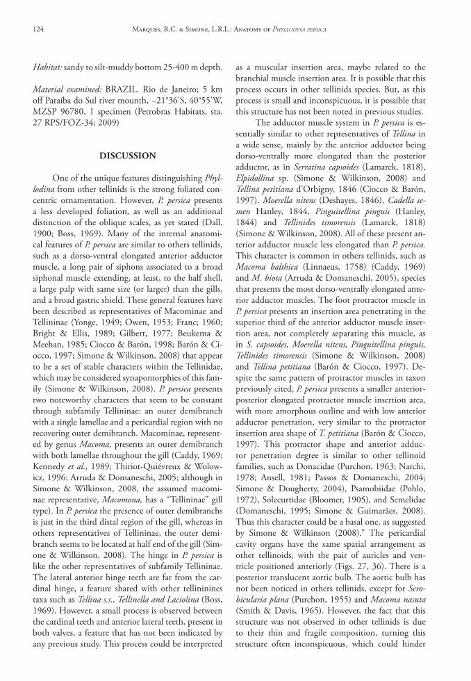
Muscle system
Anterior adductor muscle insertion area about 7% of shell surface area, dorsoventrally elongated, 3 times dorso-ventrally longer than antero-posterior length, divided by protractor muscle insertion into two regions (Figs. 4, 11); ventral region equivalent to 2/3 of anterior adductor insertion area; dorsal region equivalent to 1/3 of anterior adductor insertion area. Posterior adductor muscle rounded in cross section, located more ventrally than anterior adductor muscle; area equivalent to 5% of shell surface (Figs. 4, 11). Pair of anterior pedal retractor muscles covering an-terior edge of foot (Fig. 25), bifurcating just below mouth (Fig. 20), extending to insertion area, just above anterior adductor muscle; insertion area wider anteriorly than posteriorly, ~1/3 of anterior adduc-tor insertion area. Pair of posterior pedal retractor muscles running with each other from posterior edge of foot, bifurcating in superior third of shell height (Fig. 25); bifurcation angle ~50° (Fig. 28); insertion area ~40% of posterior adductor insertion area. Pair of pedal protractor muscles originating from posterior region, close to pericardial region, extending to an-terior adductor insertion area; distal muscle bundles thin, becoming thicker proximally (Fig. 25); insertion area triangular, separating dorsal and ventral regions of adductor anterior, tapering anteriorly, extending to posterior third of anterior adductor muscle (Fig. 12). Umbonal muscles unidentifiable. Branchial muscle thin, ranging from below pericardial chamber region to infraumbonal cavity, just parallel to ctenidial axis (Fig. 36). Pallial muscles consisting of individual muscle bundles laterally arranged, ranging from base of three mantle folds to insertion area ~10% of shell height (Figs. 5, 19); two types of muscle bundles, thin
bundles, equally spaced (Fig. 5: gray arrow) and thick bundles, about three times thickness of thin bundles, ventrally branched, spaced at 4 thin bundles (Fig. 5: white arrow). Siphonal muscle ranging from proximal aperture of siphons (Fig. 29) to lateral median region of shell, spreading in numerous fibers (Fig. 12); in-sertion area fan-like, antero-posteriorly elongated (Fig. 30); origin divided in two bundles of same thick-ness (Fig. 29: rs1), connected to proximal excurrent aperture, and rs2, below rs1, connected to proximal incurrent aperture. Siphonal septal muscle originat-ing from each side of posterior adductor insertion area, inserting in proximal opening of excurrent si-phon, parallel to siphonal septum (Figs. 4, 12: ps); length ~1/6 of shell size; area of origin spreading in ventral-anterior side of adductor muscle insertion. Siphonal muscle ranging from posterior branch of cruciform muscle to bottom of inhalant siphon (Figs. 4, 12: cs), consisting of few fibers, inconspicu-ous, of same length and thinner than siphonal sep-tal muscle. Posterior pallial muscles (Fig. 12: po) on posterior side, originating below posterior adductor muscle, inserting in ventral-posterior margin, length ~15% of shell height; origin ~1/10 of posterior ad-ductor muscle insertion area. Pair of oblique protrac-tor muscles (Fig. 25: om) ranging from dorsal margin, above and between insertion area of anterior retractor muscles, descending obliquely, backward, each pair inserting laterally on anterior quarter of protractor muscles, bifurcating; thickness ~2/3 of esophagus di-ameter. Median transverse muscles present in visceral sac (Fig. 25), surrounding style sac (Fig. 32); insertion area same size of esophageal diameter. Anterior trans-verse muscle thinner than median transverse muscle, trespassing digestive diverticula, above right and left caeca (Fig. 32: am). Intestinal transverse muscles composed of flattened fibers (Fig. 32: im), separat-ing intestinal loops in two groups. Posterior trans-verse muscles compound of indivisible set of muscle fibers aligned conjoining with connective tissue; set of muscle fibers, as wall, separating visceral cavity from pericardial region (Figs. 25, 32: pm); ventral portion thicker than dorsal portion; average thickness ~2/3 of median transverse muscle. Cruciform muscles to-tal length equivalent to posterior adductor muscle (Fig. 31); anterior and posterior branches same size, located posterior to horizontal level of posterior ad-ductor muscle; cruciform organ not grooved, closed; insertion area of posterior branch of cruciform mus-cles rounded, area ~¼ of posterior area of retractor insertion; anterior cruciform muscles insertion area subquadrate, same size as posterior cruciform inser-tion area (Fig. 12).
Marques, R.C. & Simone, L.R.L.: Anatomy of Phyllodina Persica118
pallial cavity
Palps somewhat triangular; each hemipalp about 15% of shell surface area, anterior-posterior
length ~1.5 times longer than dorsal-ventral length; length of intersection of hemipalps to 35% of shell length; inner hemipalp ~1.2 times longer than outer hemipalp; folds area occupying 90% of internal inner
FIgures 12‑24: Phyllodina persica general anatomy: 12, whole right view, mantel partially removed; 13, loop pattern of intestine; 14, dis-tal portion of right gill, semi-schematic transverse section; 15, middle portion of right gill, semi-schematic transverse section; 16, right hemipalps deflected, dashed line representing right gill removed; 17, incurrent siphon tip, partially sectioned; 18, excurrent siphon tip, partially sectioned; 19, transverse section of right mantle edge; 20, frontal view of anterior foot retractor muscle and mouth, with hemi-palps partially sectioned (dotted lines representing the remainder of hemipalps); 21, transversal section of excurrent siphon semi-schematic; 22, right cerebral ganglion, lateral view; 23, visceral ganglia; 24, cerebral ganglia, frontal view. Scales bar = 1.5 mm.
Papéis Avulsos de Zoologia, 53(9), 2013 119
hemipalp surface and 75% of internal outer surface areas; palp smooth area occupying dorsal edge and posterior area; palp folds originating from hemi-palp intersection spreading toward palp dorsal edge (Fig. 16); folds becoming relatively wider distally. Each fold width ~1/30 of palp length on average (Figs. 16, 20). Gill ~80% times inner hemipalp area (Fig. 20); anterior-posterior axis of gill three times longer than dorso-ventral axis; outer demibranch with one lamella (Fig. 14), present only in posterior quar-ter of gill, lacking in pericardial region (Figs. 15, 26); inner demibranch with two lamella; inner lamella same size as outer lamella (Figs. 14, 15). Food groove running along ventral edge of inner demibranch (Fig. 15). Association gills-palp of Type III of Stasek; region of under-lapped gill extend to 2/5 of hemipalp intersection (Fig. 16). Mantle edge four times thicker than remaining mantle; thickened mantle edge occu-pying ~1/10 of shell height in middle region and up to 1/8 in anterior region (Fig. 4). Mantle edge three-fold; depth of sulcus between middle and internal fold equivalent to 25% of height of thickened mantle edge; sulcus between external and middle fold equivalent to 1/7 of sulcus depth between middle and internal fold, middle fold with series of small, narrow, same sized, uniformly distributed papillae; papillae height equivalent to 6% of height of thickened mantle edge (Fig. 19); each papilla cylindrical, with tip rounded, separated from others by small space, almost touch-ing each other. Ventral channel (vc) on both internal sides of mantle (Figs. 4, 12), ranging from anterior side and same level of anterior branch of cruciform muscles (Fig. 31) up to middle region (Fig. 4), distant from margin equivalent to 1/8 of shell height; ventral channel wall blunt and curved posteriorly, shorten-ing forward. Foot flattened, ~40% of shell volume when retracted; distal tip pointed (Figs. 4, 25). In-current and excurrent siphons totally separate from each other, except for their base. Retracted siphons length equivalent to 1/3 of shell length. Siphons outer surface lacking pigment; inner surface of incur-rent siphon smooth (Fig. 17); inner surface of excur-rent siphon corrugated (Fig. 18). Incurrent siphonal tip with six little elevations (tp), separated by space equivalent to twice its width (Fig. 17); excurrent tip with six tentacles (tt); each tentacle three times mantle papillae (Fig. 18). Siphonal septum, thin, translucent, membrane-like, ranging from siphon base, between incurrent and excurrent proximal aperture, up to gill distal tip; gill connected to siphonal membrane by cilia. Cruciform membrane connecting dorsal region of cruciform muscles to base of incurrent siphon, thin, transparent. Proximal incurrent siphon diameter
equivalent to 3/4 that of adductor posterior mus-cle, becoming gradually narrower distally, diameter equivalent to 1/6 that of adductor posterior muscle. Proximal excurrent siphon diameter ~1.2 times larger than excurrent siphon, becoming gradually narrower distally, with same diameter as distal incurrent siphon (Fig. 29); incurrent siphonal wall thickness ~22% of incurrent siphon total diameter; excurrent siphonal wall thickness ~20% of excurrent siphon total diam-eter. Radial, longitudinal and circular muscles lying in both siphonal walls; 28 radial muscles in siphon wall, spaced by interval 3 times their width. Three sets of longitudinal and three sets of circular siphonal wall muscles intercalate each one (Fig. 21); outer circular siphonal muscles ~30% of siphonal wall thickness on both siphons; outer longitudinal siphonal muscle ~30% on both siphons; middle and inner circular si-phonal muscles thinner, ~10% of outer circular siph-onal muscle; middle longitudinal muscle equivalent to 15% of siphonal wall thickness; inner longitudinal with same thickness of middle longitudinal muscle layer. Six longitudinal nerves present in inner longitu-dinal muscle layer, equally spaced (Fig. 21).
circulatory and excretory systems
Pericardial structures occupying 25% of total visceral volume (Fig. 25). Ventricle thick, surround-ing rectum, occupying 1/6 of pericardial chamber. Pair of auricles connected to middle third of gill. Au-ricles inserted in middle-anterior region of ventricle. Aortic bulb translucent, surrounding rectum, as intu-mescence (Figs. 26, 36), equivalent to 1/10 of ventri-cle size, located in third posterior of pericardial cham-ber length (Fig. 36). Kidney yellowish, solid, with fusiform lobes on both sides of pericardial chamber, just behind outer demibranch (Figs. 25, 36). Each lobe equivalent to 1/8 of pericardial chamber; ante-rior-posterior length two times dorso-ventral length. Nephropores as slits, 1/10 of kidney antero-posterior length, located just below ctenidial axis (Fig. 36).
digestive system
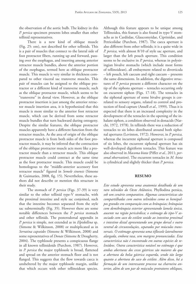
Palps as described above: Mouth conic, with relatively thick lips, lacking folds (Fig. 20). Esophagus some-what cylindrical, length ~75% of stomach length; thickness of ~25% of style sac thickness (Fig. 32). Inner surface folded only in region connected with stomach (esophageal rim – Fig. 37); remaining in-ner surface smooth. Stomach type V of Purchon, with style sac and intestine conjoined. Stomach oval, somewhat flattened ventrally, weakly positioned to
Marques, R.C. & Simone, L.R.L.: Anatomy of Phyllodina Persica120
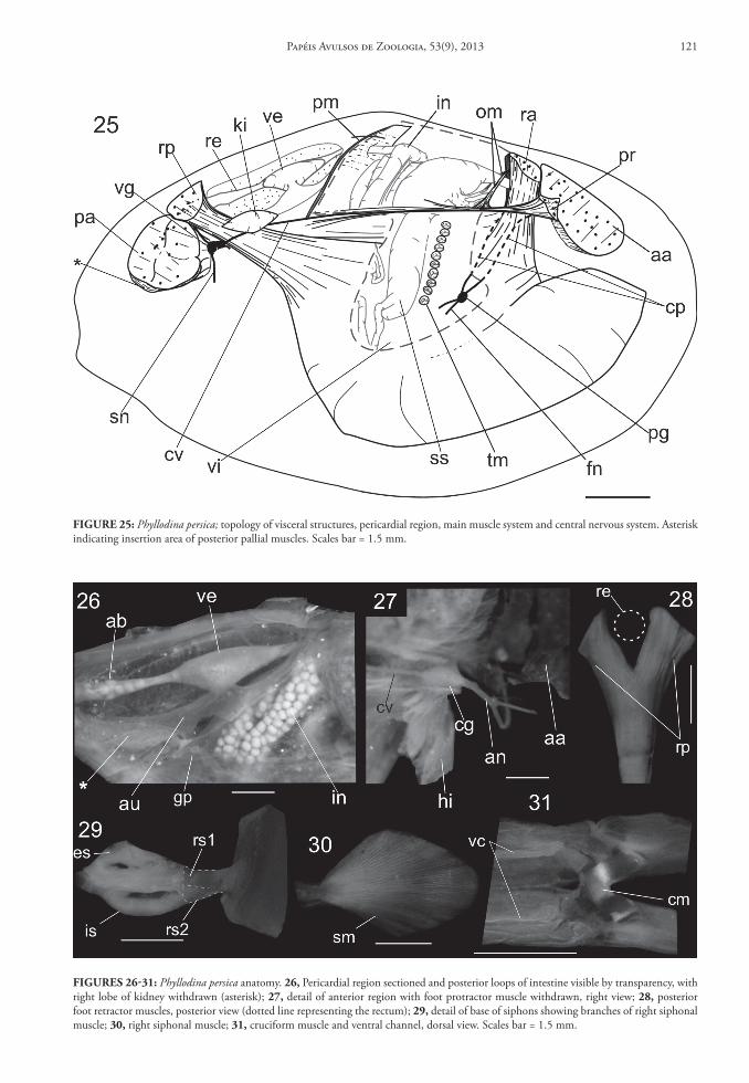
FIgure 25: Phyllodina persica; topology of visceral structures, pericardial region, main muscle system and central nervous system. Asterisk indicating insertion area of posterior pallial muscles. Scales bar = 1.5 mm.
FIgures 26‑31: Phyllodina persica anatomy. 26, Pericardial region sectioned and posterior loops of intestine visible by transparency, with right lobe of kidney withdrawn (asterisk); 27, detail of anterior region with foot protractor muscle withdrawn, right view; 28, posterior foot retractor muscles, posterior view (dotted line representing the rectum); 29, detail of base of siphons showing branches of right siphonal muscle; 30, right siphonal muscle; 31, cruciform muscle and ventral channel, dorsal view. Scales bar = 1.5 mm.
Papéis Avulsos de Zoologia, 53(9), 2013 121
right, few wider ventrally than dorsally; length 1.2 times of height. Ducts to digestive diverticula running from right cecum, spreading ventrally and anteriorly;
digestive diverticula from left cecum spreading ven-trally (Fig. 32). Digestive diverticula occupying mid-dle and dorsal portions of visceral sac. Dorsal hood
FIgures 32‑39: Phyllodina persica anatomy.32, digestive system and associated muscles, right view; 33, section of intestine, median region; 34, section of style sac, distal portion; 35, section of style sac, proximal portion; 36, pericardial region and adjacent structures; 37, stomach sectioned on right side, dorsolateral view; 38, detail of dorsal hood aperture; 39, detail of right and left caeca and left pouch, with gastric fold deflected.
Marques, R.C. & Simone, L.R.L.: Anatomy of Phyllodina Persica122
externally extruding and turned to left; internal sur-face area ~20% of stomach internal surface. Dorsal appendix (ap) small, balloon-like, smooth internally. Inner gastric surface smooth, except for sorting area between dorsal ridges (r, r1); sorting area weakly cor-rugated. Ridges (r, r1) equally sized, ranging from dorsal hood (Fig. 39), passing over dorsal surface of stomach, diverging from each another, up to right side (Fig. 37). Vestibule positioned in front of esopha-geal aperture, smooth, weakly protruded posteriorly, occupying ~15% of inner stomach surface. Gastric flap above left pouch, thick, flattened, with dorsal sur-face ~10% of stomach floor surface; divided into two lobes; anterior lobe equivalent to 90% of gastric flap; posterior lobe as small projection protruding laterally, equivalent to 10% of gastric flap volume. Major typh-losole as single structure, not differentiated, spreading over anterior floor of stomach, extending to both cae-ca (Figs. 37); left side of single typhlosole protruding (Fig. 37: la) laterally as a ridge in median region, grad-ually becoming faded; small flap emerging laterally, above anterior side of style sac aperture (Fig. 37: fl); intestinal groove of typhlosole present just in intestine and style sac (Figs. 33-35), becoming shallow toward proximal portion of style sac and absent in stomach cavity. Both caeca placed between style sac aperture and vestibule, below esophagus level. Left and right caeca same size (Fig. 39); caeca aperture ~80% of style sac aperture. Left pouch protruding downward; floor plicated, with groove running parallel to left pouch axis; aperture antero-posterior elongated, with same width of base as gastric flap. Style sac aperture out-flanked by elevated ring (Fig. 37: rg), somewhat thick, with tips rounded, fusing anteriorly with typhlosole. Style not visible. Gastric shield chitinous, transparent, spreading over left-dorsal, left and left-ventral internal surface of stomach; small portion spread into proxi-mal style sac, other portion entering into left pouch and dorsal hood (Figs. 37-39). Style sac straight, par-allel to dorsal ventral axis (Figs. 25, 32); dorso-ven-tral length equivalent to 28% of shell height. Width equivalent to 40% of stomach length. Intestine be-coming free from style sac terminally, performing sev-eral loops backward (Fig. 32); loops divided in two groups, dorsal and ventral (Fig. 13), separated by in-testinal transverse muscles (im); 11 loops in ventral group; 8 loops in dorsal group; intestine width ~20% of style sac width, except for 4 first loops in dorsal group, where width equivalent to half of style sac width (Figs. 26, 32). Rectum passing through ventri-cle and aortic bulb in pericardium chamber (Fig. 36), running backward, between pair of posterior retrac-tor muscles (Fig. 28) and around posterior adductor
muscle, turning down, ending in anus; anteriorly pointed; width equal to proximal portion of intestine. Anus simple located on ventral surface of posterior adductor muscle, at base of excurrent siphon.
genital system
Gonads and ducts filling middle and ventral portion of visceral sac. Pair of genital ducts receiv-ing branches from several gonadal acina along their length in posterior region of visceral sac. Genital pore simple, oval, located above and close to nephropore (Fig. 36).
nervous system
Pair of cerebral ganglia oval (Fig. 27), posterior region slightly wider than anterior region and slightly dorso-ventrally flattened (Figs. 22, 24), located just to both protractor muscle insertions, almost mouth level (Fig. 20), equivalent to same size of esophagus diameter; cerebral commissure long, ~1.2 times wider than cerebrovisceral connective, almost as long as ad-ductor muscle width, passing above mouth (Fig. 24). Pallial anterior nerve wide, ventral-anterior, oblique-ly directed, anteriorly branching in several nerves (Figs. 22, 27). Pair of pedal ganglia fused, compos-ing a single structure, elliptical; equivalent to 75% of pair of visceral ganglia volume; located at same level of distal portion of style sac (Fig. 25). Pair of pedal nerves spreading downward from pedal ganglia. Pair of visceral ganglia partially fused, located between posterior retractor muscles and posterior adductor muscle, above level of latter muscle (Fig. 36); volume of each visceral ganglion approximately twice that of cerebral ganglion. Two lobes in each visceral ganglion; posterior lobe, rounded, equivalent to 40% of each ganglion volume; anterior lobe, rounded, slightly an-tero-posterior elongated, equivalent to 60% of each ganglion volume (Fig. 23). Cerebrovisceral connec-tive about twice width of cerebropedal connective, running parallel to protractor muscle (Fig. 25). Pair of branchial nerve thin, emerging from ventral side of visceral ganglia; pair of posterior adductor nerve emerging from dorsal side of posterior lobe of visceral ganglia towards dorsal portion of posterior adductor muscle, just below posterior retractor muscle. Pair of siphonal nerves emerging from thickening of ventral-posterior portion of visceral ganglia; thickness about twice posterior adductor nerve width (Fig. 23); no pallial or siphonal ganglia visible.
Distribution: Cuba to Rio de Janeiro State (Brazil).
Papéis Avulsos de Zoologia, 53(9), 2013 123
Habitat: sandy to silt-muddy bottom 25-400 m depth.
Material examined: BRAZIL. Rio de Janeiro; 5 km off Paraíba do Sul river mounth, ~21°36’S, 40°55’W, MZSP 96780, 1 specimen (Petrobras Habitats, sta. 27 RPS/FOZ-34; 2009)
dIscussIon
One of the unique features distinguishing Phyl‑lodina from other tellinids is the strong foliated con-centric ornamentation. However, P. persica presents a less developed foliation, as well as an additional distinction of the oblique scales, as yet stated (Dall, 1900; Boss, 1969). Many of the internal anatomi-cal features of P. persica are similar to others tellinids, such as a dorso-ventral elongated anterior adductor muscle, a long pair of siphons associated to a broad siphonal muscle extending, at least, to the half shell, a large palp with same size (or larger) than the gills, and a broad gastric shield. These general features have been described as representatives of Macominae and Tellininae (Yonge, 1949; Owen, 1953; Franc; 1960; Bright & Ellis, 1989; Gilbert, 1977; Beukema & Meehan, 1985; Ciocco & Barón, 1998; Barón & Ci-occo, 1997; Simone & Wilkinson, 2008) that appear to be a set of stable characters within the Tellinidae, which may be considered synapomorphies of this fam-ily (Simone & Wilkinson, 2008). P. persica presents two noteworthy characters that seem to be constant through subfamily Tellininae: an outer demibranch with a single lamellae and a pericardial region with no recovering outer demibranch. Macominae, represent-ed by genus Macoma, presents an outer demibranch with both lamellae throughout the gill (Caddy, 1969; Kennedy et al., 1989; Thiriot-Quiévreux & Wolow-icz, 1996; Arruda & Domaneschi, 2005; although in Simone & Wilkinson, 2008, the assumed macomi-nae representative, Macomona, has a “Tellininae” gill type). In P. persica the presence of outer demibranchs is just in the third distal region of the gill, whereas in others representatives of Tellininae, the outer demi-branch seems to be located at half end of the gill (Sim-one & Wilkinson, 2008). The hinge in P. persica is like the other representatives of subfamily Tellininae. The lateral anterior hinge teeth are far from the car-dinal hinge, a feature shared with other tellininines taxa such as Tellina s.s., Tellinella and Laciolina (Boss, 1969). However, a small process is observed between the cardinal teeth and anterior lateral teeth, present in both valves, a feature that has not been indicated by any previous study. This process could be interpreted
as a muscular insertion area, maybe related to the branchial muscle insertion area. It is possible that this process occurs in other tellinids species. But, as this process is small and inconspicuous, it is possible that this structure has not been noted in previous studies.
The adductor muscle system in P. persica is es-sentially similar to other representatives of Tellina in a wide sense, mainly by the anterior adductor being dorso-ventrally more elongated than the posterior adductor, as in Serratina capsoides (Lamarck, 1818), Elpidollina sp. (Simone & Wilkinson, 2008) and Tellina petitiana d’Orbigny, 1846 (Ciocco & Barón, 1997). Moerella nitens (Deshayes, 1846), Cadella se‑men Hanley, 1844, Pinguitellina pinguis (Hanley, 1844) and Tellinides timorensis (Lamarck, 1818) (Simone & Wilkinson, 2008). All of these present an-terior adductor muscle less elongated than P. persica. This character is common in others tellinids, such as Macoma balthica (Linnaeus, 1758) (Caddy, 1969) and M. biota (Arruda & Domaneschi, 2005), species that presents the most dorso-ventrally elongated ante-rior adductor muscles. The foot protractor muscle in P. persica presents an insertion area penetrating in the superior third of the anterior adductor muscle inser-tion area, not completely separating this muscle, as in S. capsoides, Moerella nitens, Pinguitellina pinguis, Tellinides timorensis (Simone & Wilkinson, 2008) and Tellina petitiana (Barón & Ciocco, 1997). De-spite the same pattern of protractor muscles in taxon previously cited, P. persica presents a smaller anterior-posterior elongated protractor muscle insertion area, with more amorphous outline and with low anterior adductor penetration, very similar to the protractor insertion area shape of T. petitiana (Barón & Ciocco, 1997). This protractor shape and anterior adduc-tor penetration degree is similar to other tellinoid families, such as Donacidae (Purchon, 1963; Narchi, 1978; Ansell, 1981; Passos & Domaneschi, 2004; Simone & Dougherty, 2004), Psamobiidae (Pohlo, 1972), Solecurtidae (Bloomer, 1905), and Semelidae (Domaneschi, 1995; Simone & Guimarães, 2008). Thus this character could be a basal one, as suggested by Simone & Wilkinson (2008).” The pericardial cavity organs have the same spatial arrangement as other tellinoids, with the pair of auricles and ven-tricle positioned anteriorly (Figs. 27, 36). There is a posterior translucent aortic bulb. The aortic bulb has not been noticed in others tellinids, except for Scro‑bicularia plana (Purchon, 1955) and Macoma nasuta (Smith & Davis, 1965). However, the fact that this structure was not observed in other tellinids is due to their thin and fragile composition, turning this structure often inconspicuous, which could hinder
Marques, R.C. & Simone, L.R.L.: Anatomy of Phyllodina Persica124
the observation of the aortic bulb. The kidney in this P. persica specimen presents lobes smaller than other tellinid representatives.
There is a new kind of oblique muscle (Fig. 25: om), not described for other tellinids. This is a pair of muscles that connect to the lateral side of foot protractor fibers, running up and forward, pass-ing over the esophagus, and inserting among anterior retractor muscle bundles, above the anterior portion of the esophagus, termed here as oblique protractor muscle. This muscle is very similar in thickness com-pared to other visceral sac transverse muscles. This pair of muscles can be assigned to the oblique pro-tractor or a different kind of transverse muscle, such as the oblique protractor muscle, which seems to be “transverse” in dorsal view. However, as the oblique protractor insertion is just among the anterior retrac-tor muscle insertion area, it is hypothesized that this muscle is more similar to the anterior foot retractor muscle, which can be derived from some retractor muscle bundles that turn backward during ontogeny. Despite the similar features, the oblique protractor muscles apparently have a different function from the retractor muscles. As the area of origin of the oblique protractor muscle is from both sides of the foot pro-tractor muscle, it may be inferred that the contraction of the oblique protractor muscle acts more like a pro-tractor muscle than a retractor muscle. This oblique protractor muscle could contract at the same time as the foot protractor muscle. This muscle could be homologous to the “middle-anterior accessory foot retractor muscle” figured in Semele sinensis (Simone & Guimarães, 2008, fig. 15). Nevertheless, these au-thors did not describe or mention this structure in their study.
The stomach of P. persica (Figs. 37-39) is very similar to the other tellinid type-V stomachs, with the proximal intestine and style sac conjoined, such that the intestine becomes separated from the style sac subterminally (Fig. 35). However there are some notable differences between the P. persica stomach and other tellinids. The posterodorsal appendix in P. persica is simple, not extended as in Elpidollina sp. (Simone & Wilkinson, 2008) or multiplicated as in Serratina capsoides (Simone & Wilkinson, 2008) and some representatives of Donax (Simone & Dougherty, 2004). The typhlosole presents a conspicuous flange in all known tellinidoids (Purchon, 1987). However, in P. persica the major typhlosole is broad, flattened and spread on the anterior stomach floor and is not flanged. This suggests that the flow towards caeca is unsheltered by the major typhlosole margin, unlike that which occurs with other tellinoidean species.
Although this feature appears to be unique among Tellinoidea, this feature is also found in type-V stom-achs as in Cardiidae, Glaucomyidae, Ciprinidae, and Petricolidae (Purchon, 1987). The caeca aperture is also different from other tellinids; it is a quite wide in P. persica, with almost 8/10 of style sac aperture, and larger than the left pouch aperture. This condition seems to be exclusive in P. persica, whereas in polysir-ingian bivalve stomachs (which include most forms of tellinoidean), all embayment related to diverticula – left pouch, left caecum and right caecum – presents the same dimensions. In addition, the digestive struc-tures of P. persica present a different character on the tip of the siphons aperture – tentacles occurring only on excurrent siphon (Figs. 17-18). The tentacles in the siphonal aperture of some bivalve groups is cor-related to sensory organs, related to control and pro-tection of food capture (Ansell et al., 1999). Thus it is expected that there is an increased complexity in the development of the tentacles in the opening of the in-halant siphon, a condition observed in donacids (Nar-chi, 1972, 1978). In tellinids there was a reduction of tentacles to six lobes distributed around both sipho-nal apertures (Levinton, 1972). However, in P. persica, whereas the incurrent siphonal aperture is composed of six lobes, the excurrent siphonal aperture has six well-developed digitiform tentacles. This feature was also observed in Macoma biota (Piffer et al., 2011; per‑sonal observation). The excurrent tentacles in M. biota is cylindrical and slightly thicker than P. persica.
resuMo
Este estudo apresenta uma anatomia detalhada de um raro telinídeo do Oeste Atlântico, Phyllodina persica, sob um cenário comparativo. Algumas características são compartilhadas com outros telinídeos como os hemipal‑pos grandes em comparação com as brânquias; brânquias com demibrânquia exterior apresentando lamela única, ausente na região pericárdica; o estômago do tipo‑V as‑sociado com saco do estilete unido ao intestino proximal e intestino distal apresentando um grupo dorsal e outro ventral de circunvoluções, separados por músculos trans‑versais. O estômago apresenta uma tiflossole lateralmente alargada, embora rasa, sem margens pronunciadas. Esta característica não é encontrado em outras espécies de te‑linídeos. Outra característica notável no estômago é que ambas aberturas dos cecos gástricos são maiores do que a abertura da bolsa gástrica esquerda, sendo tão larga quanto a abertura do saco do estilete. Além disso, há a formação de um interessante processo na charneira an‑terior, além de um par de músculos protratores oblíquos,
Papéis Avulsos de Zoologia, 53(9), 2013 125
Domaneschi, O. 1995. A comparative study of the functional morphology of Semele purpurascens (Gmelin, 1791) and Semele proficua (Pulteney, 1799) (Bivalvia: Semelidae). Veliger, 38(4):323-342.
Franc, A. 1960. Classe de Bivalvia. In: Grassé, P.P. (ed.) Traite de zoologie; anatomie, systematique, biologie. Paris, Masson et Co. p. 1605-1802.
Gilbert, M.A. 1977. The behavior and functional morphology of deposit feeding in Macoma balthica (Linné, 1758) in New England. Journal of Molluscan Studies, 43(1):18-27.
Keen, A.M. 1971. Sea shells of tropical West America: marine mollusks from Baja California to Peru. 2. ed. Stanford, Ed. Stanford. 1064 p.
Kennedy, V.S.; Lutz, R.A. & Fuller, S.C. 1989. Larval and early postlarval development of Macoma mitchelli Dall (Bivalvia, Tellinidae). Veliger, 32(1):29-38.
Levinton, J.S. 1972. Stability and trophic structure in deposit-feeding and suspension feeding communities. American Naturalist, 106(4):472-486.
McLean, R.A. 1951. The Pelecypoda or bivalve mollusks of Porto Rico and the Virgin Islands. Scientific Survey of Porto Rico and the Virgin Islands, 17(1):1-183.
Narchi, W. 1972. On the biology of Iphigenia brasiliensis Lamarck, 1818 (Bivalvia, Donacidae). Proccedings of the Malacological Society of London, 40(2):79-91.
Narchi, W. 1978. Functional anatomy of Donax hanleyanus Philippi 1874 (Donacidae-Bivalvia). Boletim de Zoologia da Universidade de São Paulo, 3(1):121-142.
Owen, G. 1953. The shell in the Lamellibranchia. Quarterly Journal of Microscopical Science, 94(1):57-70.
Passos, F.D. & Domaneschi, O. 2004. Biologia e anatomia funcional de Donax gemmula Morrison (Bivalvia, Donacidae) do litoral de Sao Paulo, Brasil. Revista Brasileira de Zoologia, 21(4):1017-1032.
Piffer, P.R.; Arruda, E.P. & Passos, F.D. 2011. The biology and functional morphology of Macoma biota (Bivalvia: Tellinidae: Macominae). Zoologia, 28(3):321-333.
Pohlo, R.H. 1972. Feeding and associated functional morphology of Sanguinolaria nuttallii (Bivalvia: Tellinacea). Veliger, 14(3):298-301.
Purchon, R.D. 1955. The functional morphology of the rock boring lamellibranch, Petricola pholadiformis (Lam.). Journal of Marine Biological Association, 34(2):257-278.
Purchon, R.D. 1963. A note on the biology of Egeria radiata Lam. (Bivalvia: Donacidae). Proceedings of the Malacological Society of London, 35(2):251-271.
Purchon, R.D. 1987. The stomach in the Bivalvia. Philosophical Transactions of the Royal Society of London, B 316:183-276.
Rios, E.C. 1975. Brazilian marine mollusks iconography. Rio Grande, Fundação Universidade do Rio Grande. 331 p.
Rios, E.C. 1994. Seashells of Brazil. 2. ed. Rio Grande, Fundação Universidade do Rio Grande. 368 p.
Simone, L.R.L. & Dougherty, J.R. 2004. Anatomy and systematics of northwestern Atlantic Donax (Bivalvia, Veneroidea, Donacidae). Malacologia, 46(2):459-472.
Simone, L.R.L. & Guimarães, C.H. 2008. Comparative anatomical study of two species of Semele from Thailand (Bivalvia: Tellinoidea). The Raffles Bulletin of Zoology, 18(Suppl.): 137-149.
Simone, L.R.L. & Wilkinson, S. 2008. Comparative morphological study of some Tellinidae from Thailand (Bivalvia: Tellinoidea) The Raffles Bulletin of Zoology, 18(Suppl.): 151-190.
Smith, L.S. & Davis, J.C. 1965. Haemodynamics in Tresus nuttallii and certain other bivalves. Journal of Experimental Biology, 43(1):171-180.
Tenório, D.O. 1984. O gênero Tellina Linnaeus, 1758 (Mollusca, Bivalvia) na plataforma continental brasileira.
localizados posteriormente ao músculo retrator anterior do pé, sendo um novo tipo de musculatura íntrinseca ob‑servada em bivalves. Além da anatomia, este estudo apre‑senta um registro mais austral para P. persica, expandin‑do sua distribuição para a região sudeste do Brasil.
Key-Words: Tellinidae; Anatomy; Morphology; Phyllodina; Western Atlantic.
reFerences
Abbott, R.T. 1974. American Seashells. 2. ed. New York, Van Nostrand. 663 p.
Ansell, A.D. 1981. Functional morphology and feeding of Donax serra Röding and Donax sordidus Hanley (Bivalvia: Donacidae). Journal of Molluscan Studies, 47(1):59-72.
Ansell, A.D.; Harvey, R. & Gunther, C.P. 1999. Recovery from siphon damage in Donax vittatus (da Costa) (Bivalvia: Donacidae). Journal of Molluscan Studies, 65(2):223-232.
Arruda, E.P. & Domaneschi, O. 2005. New species of Macoma (Bivalvia: Tellinoidea: Tellinidae) from southeastern Brazil, and with description of its gross anatomy. Zootaxa, 1012:13-22.
Barón, P.J. & Ciocco, N.F. 1997. Anatomía de la almeja Tellina petitiana d'Orbigny, 1846. I. Organización general, valvas, manto, sifones, aductores, pie y branquias (Bivalvia, Tellinidae). Revista de Biología Marina y Oceanografía, 32(2):95-110.
Barón, P.J. & Ciocco, N.F. 1998. Anatomía de la almeja Tellina petitiana d'Orbigny, 1846. III. Sistema nervioso y gónada (Bivalvia, Tellinidae). Revista de Biología Marina y Oceanografía, 33(1):139-154.
Beukema, J.J. & Meehan, B.W. 1985. Latitudinal variation in linear growth and other shell characteristics of Macoma balthica. Marine Biology, 90(1):27-33.
Bloomer, H.H. 1905. On the anatomy of certain species of Solenidae. Journal of Malacology, 12(1):78-85.
Boss, K.J. 1966. Evolutionary sequence in Phyllodina (Bivalvia: Tellinidae). Report of American Malacological Union, Pacific Division, 1:21-23.
Boss, K.J. 1969. The subfamily Tellininae in the Western Atlantic (Part I). Johnsonia, 4(45-47):217-364.
Bright, D.A. & Ellis, D.V. 1989. Aspects of histology in Macoma carlottensis (Bivalvia: Tellinidae) and in situ histopathology related to mine-tailings discharge. Marine Biological Association of U.K, 69(2):447-464.
Caddy, J.F. 1969. Development of mantle organs, feeding, and locomotion in postlarval Macoma balthica (L.) (Lamellibranchiata). Canadian Journal Zoology, 47(4):609-617.
Cauquoin, M. 1967. Lamellibranchiate molluscs – Tellinidae, Scrobiculariidae and Donacidae. Annales de l’Institut Oceanographique, 45(2):227-223.
Ciocco, N.F. & Barón, P.J. 1998. Anatomía de la almeja Tellina petitiana d'Orbigny, 1846. II. Sistema digestivo, corazón, riñones, cavidad y glándulas pericárdicas (Bivalvia, Tellinidae). Revista de Biología Marina y Oceanografía, 33(1):73-87.
Dall, W.H. 1900. Synopsis of the family Tellinidae and of the North American species. Proceedings of the United States National Museum, 23(1210):285-326.
Dall, W.H. & Simpson, C.T. 1901. The Mollusca of Porto Rico. Bulletin of the United States Fish Commission, 20(1):351-52.
Díaz, J.M. & Puyana, M. 1994. Moluscos del Caribe Colombiano. Un catálogo Ilustrado. Bogotá, Colciencias, Fundación Natura and Invemar. 367 p.
Marques, R.C. & Simone, L.R.L.: Anatomy of Phyllodina Persica126
Trabalhos Oceanográficos, Universidade Federal de Pernambuco, 8(1):7-138.
Thiriot-Quiévreux, C. & Wolowicz, M. 1996. Etude caryologique d'une néoplasie branchiale chez Macoma balthica (Mollusca, Bivalvia). Comptes rendus de l’Académie des sciences. Série 3, Sciences de la vie, 319(10):887-89.
Turgeon, D.D.; Lyons W.G.; Mikkelsen P.; Rosenberg G. & Moretzsohn, F. 2009. Bivalvia (Mollusca) of the Gulf of Mexico. In: Felder, D.L. and D.K. Camp (eds.), Gulf of
Mexico‑Origins, Waters, and Biota. Biodiversity. Texas A&M Press, College Sattion. p. 711-744.
Warmke, G.L. & Abbott, R.T. 1961. New York, Caribean Seashells, Dover Publications. 348 p.
Yonge, C.M. 1949. On the structure and adaptation of the Tellinacea, deposit-feeding Eulamellibranchia. Philosophical Transactions of the Royal Society of London, Series B, Biological Sciences, 234(609):29-76.
Aceito em: 22/03/2013 Impresso em: 30/06/2013
Papéis Avulsos de Zoologia, 53(9), 2013 127

![Cell Wall Changes in Nectarines (Prunus persica)'Nectarine fruit (Prunus persica L. Batsch var nectarina [Ait] maxim) ... Inc.). The columnwasmaintainedat 1000Cfor 2minandincreasedto](https://img.pdfslide.us/doc/110x75/5f12baf6290ffe2baf0f91a2/cell-wall-changes-in-nectarines-prunus-persica-nectarine-fruit-prunus-persica.jpg)