Embed Size (px)
Citation preview
A ONE-WAY-VALVE CHEST WOUND DRESSING: EVALUATION IN A CANINE MODEL OF OPEN CHEST WOUNDS
Ernest Ruiz, M.D., F.A.C.E.P. Assistant Professor of Surgery
University of Minnesota Department of Emergency Medicine
Hennepin County Medical Center Jonathan Lueders, M.D.
Emergency Medicine Resident Hennepin County Medical Center
Christian Petersen Medical Student
University of Minnesota
From Department of Emergency Medicine
Hennepin County Medical Center 701 Park Avenue
Minneapolis, Minnesota 54415 and
Minneapolis Medical Research Foundation 914 South 8th Street
Minneapolis, Minnesota 55404
Supported in part with a grant from: Brunswick Biomedical Technologies, Inc.
6 Thacher lane Wareham, Massachusetts 02571
ABSTRACT
Objective: To test a chest wound dressing incorporating a low profile one-
way valve dressing (OWVD) designed for use in prehospital treatment of
pneumothorax in penetrating chest trauma by comparing it to conventional
petrolatum impregnated gauze dressings (PGD). Design: six dogs were used
to develop an anesthetized breathing model using ketamine (40 mg/kg),
midazolam (200 ug/kg), and fentanyl (10 ug/kg) maintained with an infusion of
600 ug/kg/min, 5 ug/kg/min, and .5 ug/kg/min respectively. Four dogs served
as a control group evaluation this model on and off the ventilator. Eight dogs
with bilateral standardized chest wounds were randomized into two groups
in a crossover design study. One group tested the PGD first and then the
OWVD, both with and without positive pressure ventilation (PPV). The
second group tested the OWVD then the PGD. Dogs were stabilized between
tests of each device. Respiratory rate, heart rate, arterial blood gases and
hemoglobin oxygen saturation (qualitative) were monitored. Results: The
control group showed stable vital signs throughout testing. Animals on PPV
maintained stable vital signs regardless of the dressing applied. Dogs
without PPV were unable to survive a 15 minute period with the PGD,
whereas dogs with the OWVD were able to adequately maintain vital signs.
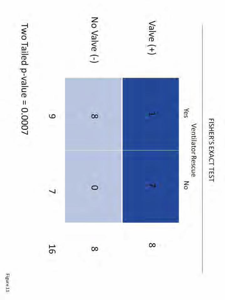
The OWVD prevented collapse in 7 of 8 tests while the PGD prevented
collapse in none of 8 tests in dogs without PPV. A probability of p = 0.0007
was found when Fisher’s exact test was applied to this combined data.
Conclusion: The OWVD out-performed the conventional PGD in preventing
severe decompensation in dogs with bilateral open chest wounds without
PPV.
INTRODUCTION
The purpose of our study is to test a chest wound dressing incorporating a
one-way valve and compare it to the traditional occlusive petrolatum gauze
dressing used in pre-hospital care of penetrating chest wounds. A simple,
disposable chest wound dressing incorporating a light weight, low profile,
and low resistance silicone-leaflet one-way valve has recently become
available.
Penetrating chest wounds are increasing at an astronomical rate1. These
patients pose difficult pre-hospital problems. The patient is usually unstable
and in need of immediate transport. The traditional sterile occlusive
dressing taped to the skin on three sides is difficult and time consuming to
apply under field conditions. Frequently there are multiple wounds to
manage, as in through and through gunshot wounds.
The open, “sucking” chest wound allows air to enter the chest and separate
the visceral and parietal pleural surfaces, breaking the thin fluid layer
between the lung and chest wall. The elastic tissues of the lung normally
produce a physiologic negative pressure differential between the potential
intrapleural space and the outside of 4 – 12 cm water2. The elastic recoil of
the lung causes it to pull away from the chest wall and air is drawn into the
chest through the wound. Unassisted ventilation becomes impossible when
respiratory movements of the chest wall and diaphragms result only in air
movement through the wound instead of through oropharynx. If the wound is
small, the patient can compensate with exaggerated chest wall and
diaphragmatic movement because the wound offers more resistance to air
flow than the bronchial tree and airway. It has been estimated that wound
two-thirds the diameter of the trachea offers less resistance than the normal
airway3. When positive pressures is used, the lung can expand if air moves
out of the wound or if the pressure of ventilation is high enough to compress
the air within the pleural space. Tension pneumothorax can develop if there
is a lung injury with an air leak into the pleural space and the wound is
occluded or functions as a one-way valve allowing air into but not out of the
chest. Tension pneumothorax can develop with or without positive pressure
ventilation. A small pneumothorax can quickly become a tension
pneumothorax when positive pressure ventilation is added to an occluded
wound with underlying lung injury and air leak.
An ideal dressing should possess certain qualities. It should be simple,
quick, easily applied, and non-invasive. It should have a low profile and be
sturdy enough to be used on the posterior thorax of a supine patient. Most
importantly it should prevent influx of air on spontaneous inspiration through
the wound and allow escape of air from the potential pleural space during
any escape of air from the potential pleural space during any phase of
respiration or assisted ventilation to avoid a tension pneumothorax.
A disposable chest wound dressing incorporating a light weight, low profile
and low resistance silicone leaf one-way valve has become available. This
chest wound dressing appears to satisfy the qualities discussed above.
From experience at our institution, conventional occlusive petrolatum-gauze
dressings are frequently unreliable in preventing influx and allowing egress
of air from a chest wound. It is virtually impossible for providers to place
these dressings while wearing latex gloves and to get tape to stick to a
bloody surface. They are time consuming to place at a time when other
procedures also need to be performed. This study was designed to
determine if this device in a laboratory model of open chest wounds.
MATERIALS AND METHODS
Approval of the institutional animal use review committee was obtained for
this study. A total of 18 mature mongrel dogs weight 10 to 25 kg were used.
Six dogs were used to develop an anesthesia regimen that reliably resulted
in a fully anesthetized but breathing model. The regimen included an IV
anesthetizing dose of midazolam HCL, 200 ug/kg, alfentanil citrate, 10 ug/kg,
and ketamine HCL, 40 mg/kg. Anesthesia was maintained with an infusion
pump delivering a solution of midazolam HCL, 5 ug/kg/min, alfentanyl citrate
0.4 ug/kg/min, and ketamine HCL, 600 ug/kg/min. If a dog made spontaneous
non-ventilatory movements, additional IV boluses of midazolam HCL, 100
ug/kg were given during the experimental period.
All dogs were monitored as follows: Hemoglobin O2 saturation (SaO2) using a
Oxisensor adult digit oxygen transducer wrapped around the tongue; Heart
rate (HR) using ECG lead II; Central venous pressure (CVP) and mean arterial
pressure (MAP) monitored via catheters placed percutaneously into an
external jugular vein and femoral artery; Core temperature (CT) using an
esophageal probe. The SaO2, HR, MAP and CT were displayed and recorded
using a Hewlett-Packard Merlin monitoring system and pressure transducer.
CVP was measured using a saline manometer in mmH2O = 9.807 Pa).
Arterial blood gases were measured using a Corning Model 168 blood gas
analyzer.
All dogs had their chests, neck, and groins shaved after induction of
anesthesia. All dogs were orotracheally intubated using a cuffed 8.5 mm I.D.
tube. Positive pressure ventilation with room air with a Bennett MA-1
respirator at ten breaths per minute with a tidal volume taken from the
Kleinman ventilation graph plus 50 cc to compensate for dead-space in the
apparatus. Peak flow was set at 30 L/min. Sigh ventilations of 1000 cc were
administered as needed to return the animal to stable state between
experimental periods. An infra red heating lamp was placed over the torso to
maintain body temperature at 37.0 to 38.5 C.
Dogs that were to receive open chest wounds had plastic sleeves with an
internal diameter of 6 mm placed bilaterally in the 6th intercostal space at
the anterior axillary line using a guidewire technique. The sleeves were
curved to conform to the chest wall and were 7 cm in length with multiple
side holes. They were sutured flush to the skin. This ended the period of
preparation.
Twelve dogs were randomly assigned to one of three groups. Group 3
consisted of four dogs and served as in anesthesia control group. They were
monitored at five minute internals for four 20 minute periods, the first and
third in which they were breathing spontaneously while the second and
fourth were respirator dependant. Groups 1 and 2 were the experimental
groups. These dogs had bilateral chest holes placed followed by the
application of chest suction to the sleeves at 200 mm of H2O vacuum while
on the respirator allowing them to recover from the brief collapse of the
lungs cased by the procedure. After a fifteen minute period of stabilization
the lungs were allowed to collapse by removing the respirator and chest wall
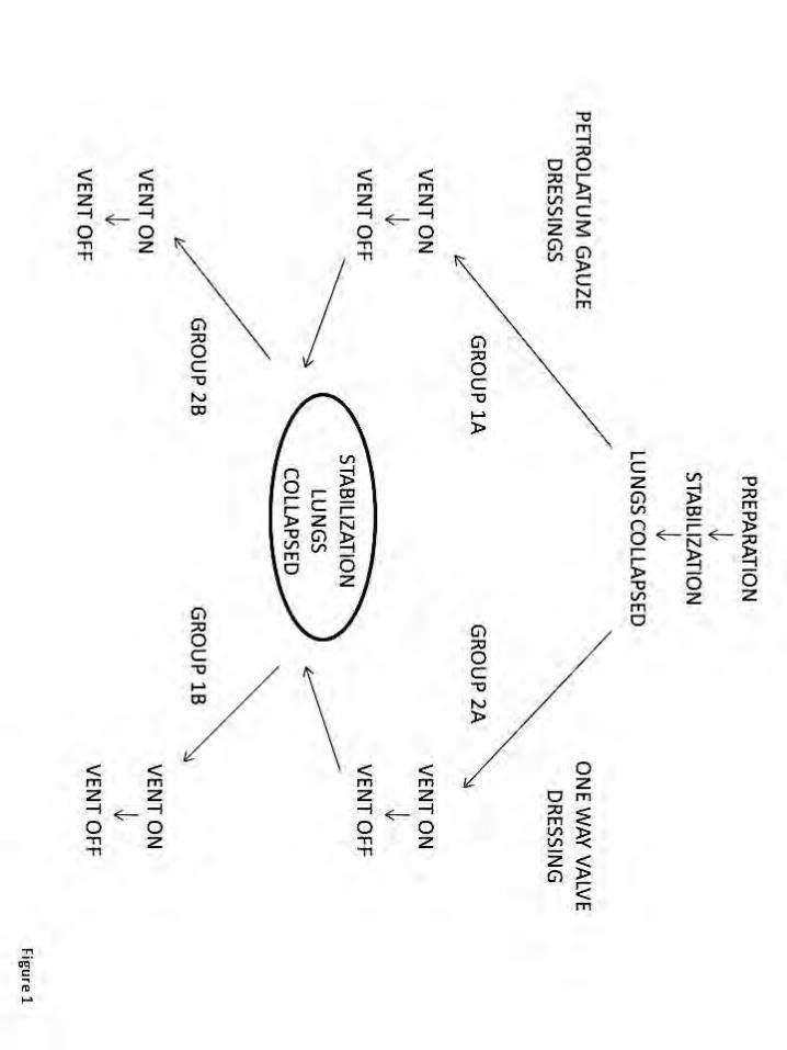
suction for one minute. Either the conventional gauze dressing of the
experimental one-way valve dressing was then applied as shown in our
experimental algorhytm (Figure 1). During this time the dressings were
tested with fifteen minute trials on and off the respirator respectively. The
dressing was then removed and the dogs were placed back on the respirator
and chest wall suction for a fifteen minute period of restabilization. Lungs
were collapsed again by removal of the respirator and chest wall suction for
one minute. The opposite dressing as used before was then applied and
tested with fifteen minute trials on and then off the respirator. Following this
the dogs were euthanized with an IV KCL bolus.
The petroleum gauze (PG) dressings were placed over the chest holes,
molded to the chest and taped using two inch strips of waterproof adhesive
tape. The one-way valve (OWV) dressings were placed over the chest holed
and pasted to the skin with silicone sealant. If a dog became agonal with a
SaO2 of less than 50 percent while off the respirator, the respirator was re-
applied and sigh ventilations were used to re-expand the lungs. When a dog
was unable to sustain itself during the fifteen minute trial without respirator
support, multiple attempts were made to re-stabilize the dog and try again
until the fifteen minute trial had elapsed.
One dog randomly assigned to Group 2 was found to have evidence of
pneumonia with copious purulent drainage from the trachea and severe
hypotension / hypoxia throughout. Another dog was subsequently added to
Group 2 and the dog with pneumonia eliminated from the study. One other
dog had black pigmentation of the tongue which interfered with SaO2
measurements. In this instance, the dog’s vital signs and ECG were used to
confirm an agonal condition.
RESULTS
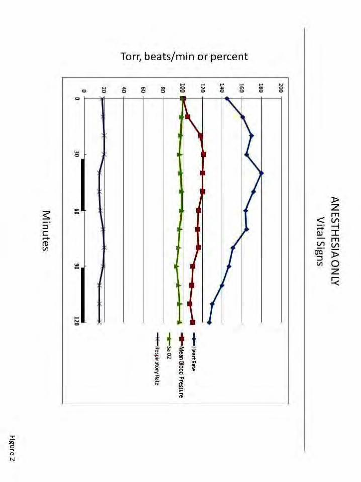
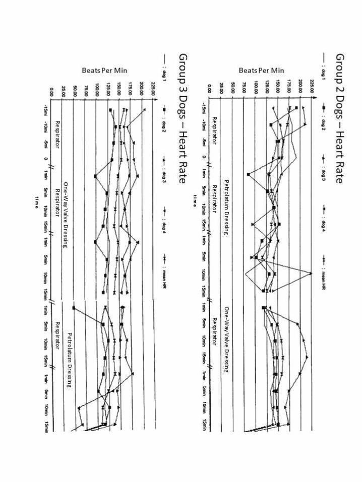
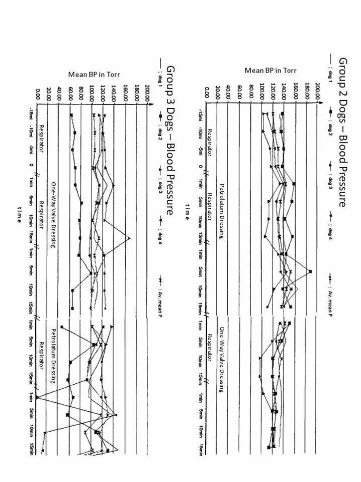
Anesthesia control Group 3 revealed no significant deterioration of vital
signs or blood gases over the experimental period. See Figure 2.
We then compared data from Groups 1 and 2 which showed that vital signs of
dogs testing the gauze and one-way valve dressings were very stable and
similar while the dogs were being ventilated.
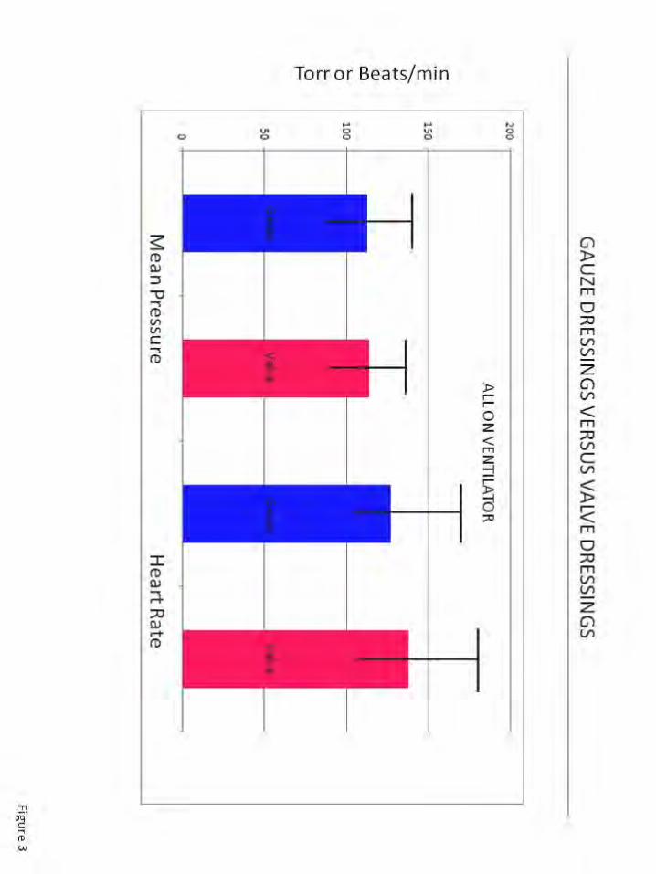
Data from Group 1A and 2B which represents gauze dressing results was
compared with data from Groups 2A and 1B which represents the valve
dressing results. Figure 3 graphs the gauze results compared to valve
results. Both dressings appeared perform similar based on vital signs. We
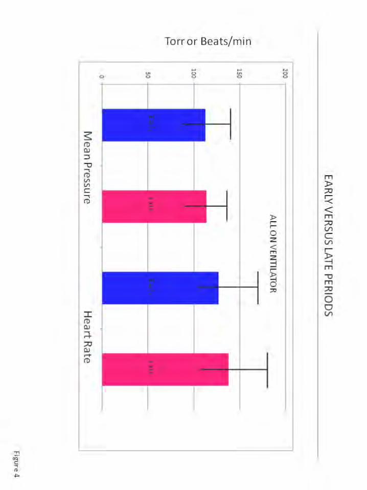
then looked at whether it made any difference if the dressing was tested
early (Groups 1A and 2A) prior to testing the opposite dressing, or late
(groups 2A and 2B) after previously testing the opposite dressing. Figure 4
shows these vitals also little variation.
This finding allowed the combining of data from groups 1 and 2 to determine
the significance of differences in response to either of the two variable
dressings. Data showed that both dressings performed equal when dogs
were supported by the respirator.
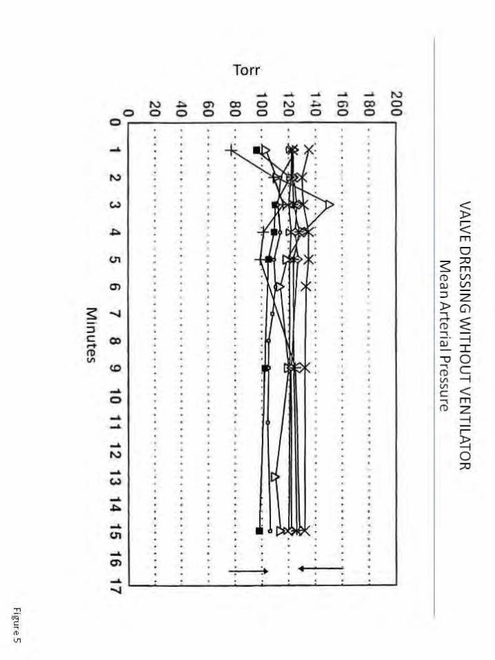
There was a very significant difference between the two dressings when the
dogs were not supported with a respirator. The one-way valve dressing
protected the dogs from severe collapse 7 out of 8 times. Figure 5 shows
mean arterial blood pressure of the dogs testing the valve dressing after
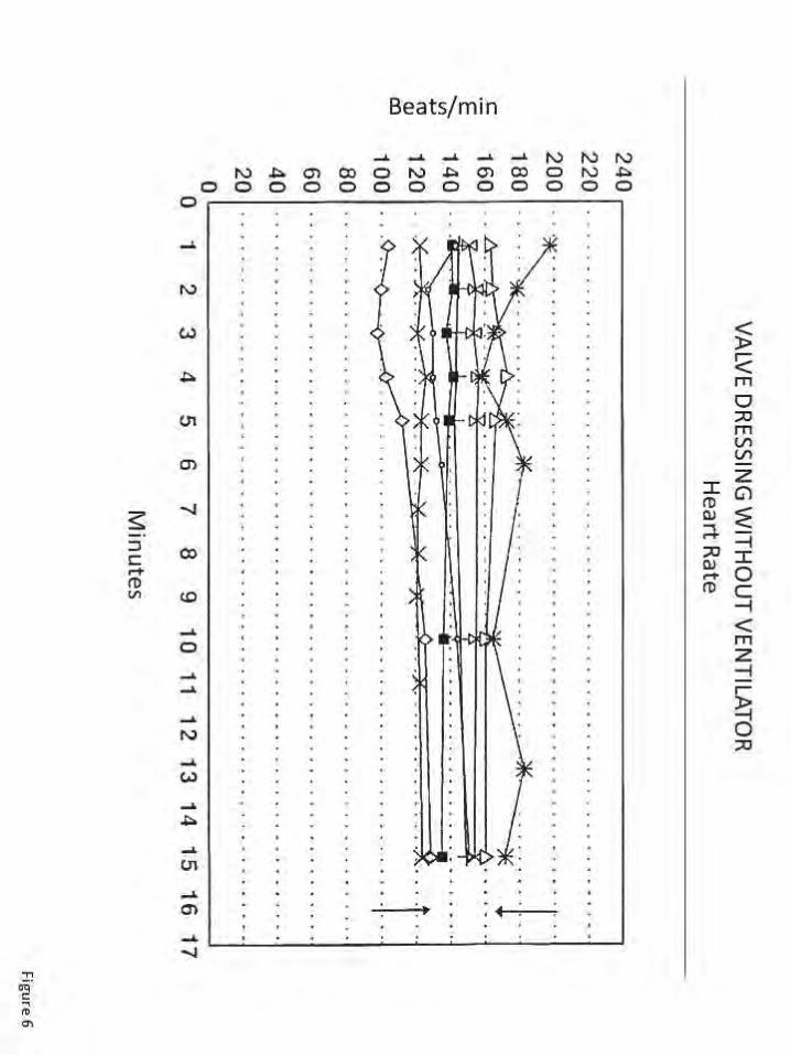
respirator support has been removed. Mean pressure remains stable. Figure
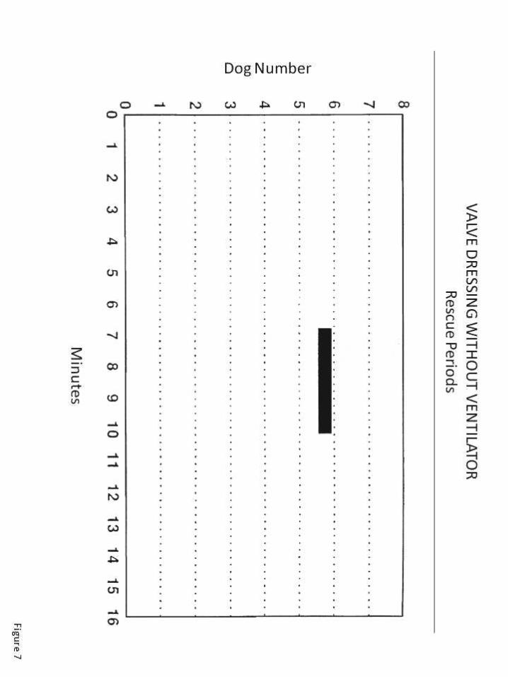
6 show the heart rate of this same group which also remains stable. Only
one dog required rescue support by being placed back on the respirator for
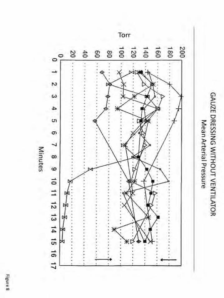
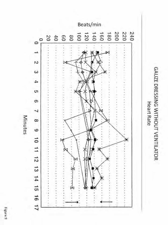
three minutes as shown in Figure 7. In contrast, dogs testing the gauze
dressings were very unstable and none of them survived the 15 minute trial
without rescue and ventilatory support. These vital signs were very
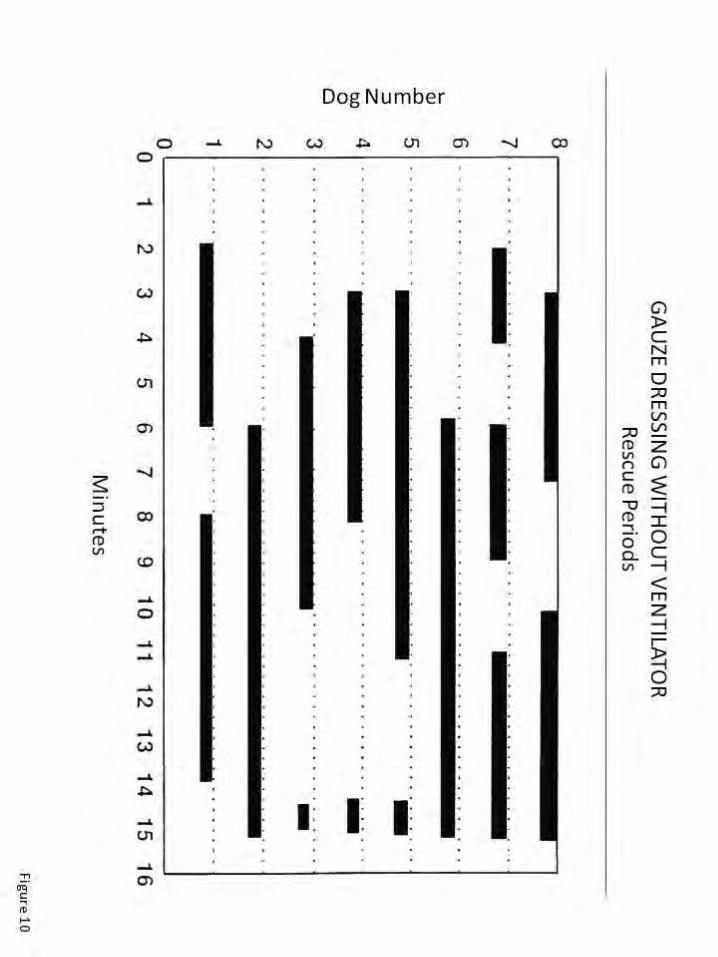
unstable, and much of the time actually represents time on a respirator
during rescue periods. Figure 10 shows these rescue periods. Figure 10
shows these rescue periods which all eight dogs testing the gauze dressing
required. Multiple attempts for rescue were performed in most dogs.
ANALYSIS
Descriptive statistical techniques were used to determine the stability of the
anesthesia control group. Descriptive statistical techniques were also used
to determine the effect of the timing of experimental variable periods in
group 1 and 2. Fisher’s Exact Test of probability was used to determine if
there was a significant difference in performance of the two dressings with
and without respirator support giving a p – value of 0.0007. See Figure 11.
DISCUSSION
Bench research of pneumothorax and tension pneumothorax is severely
limited by the anatomic characteristics of most laboratory animals. The dog,
pig, cat, rabbit, and rat all have an incomplete mediastinum4. Sheep and
goats do have a complete mediastinum 5 but are relatively unavailable in the
laboratory setting. Since the dog has an oxyhemoglobin dissociation curve
almost identical to that of humans, very similar blood viscosity properties
and red blood cell size and configuration, we elected to develop our model
using dogs6. By placing a hole on both sides of the chest we created a
severe, constant model of open chest wound simulating bilateral chest
wounds in humans.
The conventional method of pre-hospital treatment of penetrating chest
wounds is to apply an occlusive dressing. Although they make intuitive
sense, there is no recent experimental data supporting the use of occlusive
dressings taped to the chest. Gauze dressings used in our studies consisted
of 4 x 4 inch petrolatum impregnated gauze covered with 4 x 4 inch gauze
bandages which were taped to the skin on all four edges. This is similar to a
wound dressing on the posterior thorax of a supine patient. Dressings
applied were not air-tight.
LIMITATIONS
Limitations include relatively small numbers used and the assumption that
our crossover design was valid to make the most of our small numbers. The
incomplete mediastinum of a dog was also used.
CONCLUSION
An open chest wound dressing with a one-way valve improves pulmonary
functioning in the absence of positive pressure ventilation, and functions as
well as the standard occlusive gauze dressing when positive pressure
ventilation is applied.
REFERENCES:
1.
2. Sabiston: Textbook of Surgery, 14th Edition, p 1719.
3.
4.
5.
6.

Ernest Ruiz, MD FACEP
Professor Emeritus of Emergency MedicineUniversity of Minnesota
Chief of Emergency Medicine (Ret)Surgery Faculty (Ret)
Hennepin County Medical Center, Minneapolis
Author
Clinical Studies Team Leader
US ArmyKorean Conflict
Note: Dr. Ernest Ruiz, MD, has not received financial reward for performing this laboratory study, nor will he,at his own request, receive any financial reward for the commercial sale of this product.
Copyright® 1992 Dr. Ernest Ruiz. All Rights Reserved. Unauthorized use without written permission is prohibited.