Embed Size (px)
Citation preview

THE JOURNAL OF BIOLOGICAL CHEMISTRY 0 1992 by The American Soeiety for Biochemistry and Molecular Biology, Inc.
VOl. 267, No. 20, Issue of July 15, PP. 14443-14450,1992 Printed in U.S.A.
A Novel Site-directed Mutant of Myoglobin with an Unusually High O2 Affinity and Low Autooxidation Rate*
(Received for publication, January 27,1992)
Theodore E. Carver$, Robert E. Brantley, Jr.5, Eileen W. Singleton, Robert M. Arduini, Michael L. Quillinn, George N. Phillips, Jr.11, and John S. Olson** From the W. M. Keck Center for Computational Bwlogy and the Department of Biochemistry and Cell Biology, Rice University, Houston, Texas 77251
Mutants of sperm whale myoglobin were constructed at position 29 (SI0 in helix notation) to examine the effects of distal pocket size on the rates of ligand binding and autooxidation. Leuze was replaced with Ala, Val, and Phe using the synthetic gene and Esche- richia coli expression system of Springer and Sligar (Springer, B. A., and Sligar, S. G. (1987) Proc. Natl. Acad. Sci. U. S. A. 84, 8961-8965). Structures of the ferric forms of Val” and Phe”, and the oxy form of Pheae myoglobin were determined to 1.7 A by x-ray crystallography. The ferric mutant proteins are re- markably isomorphous with the wild type protein ex- cept in the immediate vicinity of residue 29. Thus, the protein structure in the distal pocket of myoglobin can accommodate either a large “hole” (i.e. Ala or Val) or a large side chain (Le. Phe) at position 29 without perturbation of tertiary structure. PheZe oxymyoglobin is also identical to the native oxy protein in terms of overall structure and interactions between the bound Oa and Hise4, Vales, Phe43, and Ile107. The distance between the nearest side chain atom of residue 29 and the second atom of the boundpxygen molecule is 3.2 A in the PheZm protein and 4.9 A in native myoglobin.
The equilibrium constants for Oz binding to Alaze, Valae, and Leuae (native) myoglobin are the same, -1.0 X lo6 M” at 20 OC, whereas that for the PheZe protein is markedly greater, 16 X loe M-’. This increase in affinity is due primarily to a 10-fold decrease in the O2 dissociation rate constant for the Pheae mutant and appears to be the result of stabilizing interactions be- tween the negative portion of the bound O2 dipole and the partially positive edge of the phenyl ring. Increas- ing the size of residue 29 causes large decreases in the rate of autooxidation of myoglobin: ko, = 0.24, 0.23, 0.066, and 0.006 h“ for Ala”, Valze, Leuze (native), and Phe” myoglobin, respectively, in air at 37 “C. Thus, the LeuZe + Phe mutation produces a reduced protein that is remarkably stable and is expressed in
* The costs of publication of this article were defrayed in part by the payment of page charges. This article must therefore be hereby marked “advertisement” in accordance with 18 U.S.C. Section 1734 solely to indicate this fact.
of Health Training Grant GM-07933. $ Recipient of a graduate fellowship from the National Institutes
of Health Training Grant GM-08280. Recipient of a graduate fellowship from the National Institutes
ll Recipient of a National Science Foundation predoctoral fellow- ship.
11 Supported by United States Public Health Service Grant AR40252, Grant C-1142 from the Robert A. Welch Foundation, and the W. M. Keck Foundation.
** Supported by United States Public Health Service Grants GM- 35649 and HL-47020, Grant C-612 from the Robert A. Welch Foun- dation, and the W. M. Keck Foundation.
E. coli as 100% MbOz. The selective pressure to con- serve Leuze at the B10 position probably represents a compromise between reducing the rate of autooxida- tion and maintaining a large enough O2 dissociation rate constant to allow rapid oxygen release during respiration.
Over the past 3 years, site-directed mutagenesis techniques have been used to examine the functional roles of key amino acid residues in the active sites of human hemoglobin and several mammalian myoglobins. Most work has focused on ligand binding studies with distal histidine (E7) and valine ( E l l ) mutants (i.e. Nagai et al., 1987; Mathews et al., 1989; Springer et al., 1989; Egeberg et al., 1990). In the course of these studies, it became apparent that the overall polarity and physical size of the distal pocket are also key factors in regulating the rate and equilibrium constants for ligand bind- ing. Smerdon et al. (1991) used the isosteric Val6* + Thr substitution to show that increasing the polarity of the distal pocket in pig myoglobin causes marked decreases in 02 and CO affinity. Carver et al. (1990) have shown that conventional and geminate recombination rate constants are markedly affected by ligand size and distal pocket volume. The structure of myoglobin and molecular dynamics simulations suggest that the distal cavity surrounded by residues His64 (E7), Valrn (El l ) , Ile”’ (G12), Ilelo7 (G8), Leu3’ (B13), Leuz9 (BlO), and Phe43 (CD1) can readily accommodate thermally or photo- chemically dissociated diatomic gases (i.e. Sassaroli and Rous- seau, 1986; Kottalam and Case, 1988; Carver et al., 1990; Elber and Karplus, 1991).
In view of these previous studies, we decided to vary the packing of distal side chains around the bound ligand by replacing Leuz9 with Ala, Val, and Phe in sperm whale myo- globin using the synthetic gene and expression system of Springer and Sligar (1987). Leuz9 is the 10th residue in the B helix and forms part of the top of the distal pocket (see Fig. 1). The isobutyl side chain is not in Van der Waals contact with either bound diatomic ligands or the heme group. Thus, we hoped that functional changes produced by substitutions at this position would arise from changes in the contours of the distal pocket rather than alterations in the geometry of the Fe-heme complex or the tertiary structure of the mutant protein. These assumptions were evaluated directly by deter- mining the x-ray crystallographic structures of Valz9 and PheZ9 aquometmyoglobin and of PheZ9 oxymyoglobin. The overall association and dissociation rate constants for 02, CO, methyl, and ethyl isocyanide binding to the mutant proteins were then compared to those of the native protein. Finally, the rates of autooxidation of all four myoglobins were measured at 37 “C.
Even though its side chain does not interact directly with
14443

14444 L29F Sperm Whale Myoglobin
either bound oxygen or the porphyrin ring, Leuz9 is highly conserved in mammalian myoglobins and hemoglobins. The only known exception is Valz9 in the a subunits of rabbit hemoglobin.’ In reptile myoglobins, Ile often replaces Leuz9, and even greater variance is observed among invertebrate oxygen carrying heme proteins and plant leghemoglobins where Phe and Tyr are often found at this position.’ Thus, another goal of this study was to examine the nature of the selective pressure in mammals which has preserved Leuz9.
METHODS AND MATERIALS
Mutagenesis of the Myoglobin Gene-The PstI-KpnI fragment of pMb413a (synthetic sperm whale myoglobin (Springer and Sligar, 1987)) was subcloned into pEMBL19 for mutagenesis, sequencing, and expression. The product of these manipulations was designated pEMbS-1. DNA manipulations were made as described by Sambrook et al. (1989). For the Leuz9 + Phe and Leuz9 + Val substitutions, the Kunkel method for oligonucleotide-directed mutagenesis was used (Kunkel, 1985). In this method, uracil-containing single-stranded pEMbS-1 DNA was prepared using the Escherichia coli strain BW313. The codon for position 29 was altered using 18 or 21 bpz mutagenic oligonucleotides as primers for extension on the uracil template DNA. Selection against the nonmutant strand was achieved by transfor- mation of the product of the extension reaction into E. coli strain 71/18. For the Leuz9 -+ Ala substitution, oligonucleotide-directed mutagenesis was performed according to the method of Taylor et al. (1985). Single-stranded wild type pEMbS-1 DNA was prepared from E. coli 71/18. All subsequent manipulations were performed using the Amersham oligonucleotide-directed in uitro mutagenesis system. The final extension product was transformed into E. coli strain TG-1. Mutants produced using either method were screened and confirmed by sequencing single-stranded DNA from several of the transformants using Sequenase kits purchased from United States Biochemical Corporation. Enzymes used in producing mutant constructions were purchased from Boehringer Mannheim, Promega, Sigma, and Amer- sham.
The single-stranded form of pEMbS was sequenced in the region of the mutation to confirm the substitution and then transformed into E. coli strain TB-1, and the Mb gene was expressed as described previously (Springer and Sligar, 1987; Egeberg et al., 1990). In our procedure, the 100L fermentation facility a t Rice was used to obtain roughly 400-500 g of packed cells containing recombinant myoglobin. Half of these cells (-200 g) were used in each protein preparation procedure. The remaining cells were stored at -70 or -20 “C. The purification scheme was similar to that described by Springer and Sligar (1987) and Egeberg et al. (1990) and involved 1) lysis by freezing and thawing and digestion with lysozyme; 2) 50 and 95% ammonium sulfate cuts; 3) anion-exchange chromatography with DEAE cellulose (Whatman DE-52); and 4) a cation-exchange column (Whatman CM-52). An additional HPLC step with a weak cation exchanger was used before crystal preparation. The final purified protein samples were concentrated to about 1 mM in heme, frozen, and stored under liquid nitrogen.
X-ray Crystallography-Crystals for the recombinant myoglobins were grown in ammonium sulfate as described by Phillips et al. (1990). The ferric forms of the Valz9 and PheZ9 myoglobins and the oxy form of PheZ9 myoglobin produced large crystals (0.5 X 0.3 X 0.15 mm) which belonged to the space group P6, with one molecule per asym- metric unit and cell constants close to those for wild-type metmy- oglobin. Diffraction data for each mutant were collected on a Siemens X-1000 area detector with a rotating-anode source and processed using XDS (Kabsch, 1988). Relevant statistics for data collection are presented in Table I.
The starting models for the refinement of mutant structures were generated from the wild-type recombinant myoglobin coordinates (Protein Data Bank entry lMBW, Brookhaven National Laboratory).
Listings of known myoglobin and human hemoglobin a and P sequences were obtained using the EuGene & SAM software package developed by the Molecular Biology Information Resource, Depart- ment of Cell Biology, Baylor College of Medicine, Houston, TX and the Protein Sequence Data Bank from the National Biomedical Research Foundation, Wash., DC.
The abbreviations used are: bp, base pair(s); HPLC, high-per- formance liquid chromatography.
TABLE I Summary of crystallogrwhic data for position 29 mutants
Val” Leu” aauomet aauomet“ aauomet Phe” oxy
Reflections Measured 68,019 50,003 68,499 38,051 Unique 20,549 12,308 22,278 18,399
Highest resolution (A) 1.7 1.9 1.7 1.7
Completeness (%) 84.9 71.0 92.0 76.0
R-factor (%)* 15.5 14.8 14.9 16.7 * From G. N. Phillips et al. (1990).
R-factor = ZIF, - KI ZFO
for all data from 10 A to the given
resolution.
Initial difference Fourier maps were calculated with PROTEIN (Rem- ington et al., 1982). The mutated residue was built into the observed density using the molecular modeling program CHAIN (Sack, 1988). Constrained least-squares refinement of the met and oxy PheZ9 myo- globin structures was accomplished with the program PROFFT (Fin- sel, 1987). All refinement and map calculations for the mutant Valz9 were performed with XPLOR using the PROLSQ parameter set (Brunger et al., 1989). After several cycles of refinement, manual refitting, and solvent placement, the crystallographic R-factors con- verged to the values shown in Table I.
Kinetic Measurements-Association and dissociation rate con- stants were measured using stopped-flow rapid mixing and conven- tional flash photolysis techniques as described in detail by Rohlfs et al. (1990). For Leuz9 (native), Valz9, and Alam myoglobins, oxygenated samples were usually prepared by reducing about 30 pl of concentrated ferric protein with 3-4 grains of sodium dithionite and then quickly passing the sample through a small Sephadex G-25 column equili- brated with oxygenated buffer to remove residual reducing agent and its byproducts. PheZ9 myoglobin was isolated from E. coli completely in the oxy form and did not require reduction. Small fractions of reduced, oxygenated Alaz9 and Valz9 myoglobins were also obtained. Rate constants measured for these fractions were found to be identical to those measured for oxy complexes obtained by dithionite reduction. Association rate constants for OZ binding were determined by com- pletely photolyzing the MbOz complexes with a 300-ns dye laser and following recombination on microsecond time scales at three different oxygen concentrations: 0.263 mM (air); 0.625 mM (?h a h ) ; 1.25 mM (1 atm). Association rate constants for CO, methyl, and ethyl iso- cyanide binding were obtained by mixing the deoxy forms of the proteins with various concentrations of ligand or by photolyzing the fully liganded complexes and following recombination after the flash. Identical results were obtained in the two types of experiments. Dissociation rate constants were obtained by analyzing replacement reactions in which Oz was displaced by CO, CO was displaced by NO, and the isocyanides were displaced by CO(Roh1fs et al., 1990). Asso- ciation equilibrium constants were calculated as the ratio of the rate constants. Wild-type sperm whale myoglobin expressed in E. coli has previously been shown to be identical in tertiary structure and func- tion to native sperm whale myoglobin (Rohlfs et al., 1990; Phillips et al., 1990) so that the values given for Leuz9 myoglobin represent both native and recombinant wild type proteins.
For some autooxidation experiments, the purified oxy fractions of recombinant Ala”, Valz9, Leuz9 (wild type), and PheZ9 were used without dithionite reduction. Otherwise, mutants purified in the oxidized form, and native sperm whale myoglobin obtained from Sigma in the oxidized form, were reduced with a few grains of sodium dithionite and passed through a G-25 column containing air-equili- brated buffer. The resultant protein was diluted to -20 p M heme in 3 ml of 0.1 M phosphate, 0.001 M EDTA, pH 7.0, and catalase and superoxide dismutase were each added to 3 mmol/mol heme. The absorbance changes due to autooxidation were followed in the range 500-700 nm or at 581 nm alone for multicell experiments. All of these experiments were performed using a Shimadzu UV-visible PC2100 spectrophotometer equipped with a multicell chamber and a ther- mostat. The autooxidation of native and wild-type sperm whale myoglobin was followed to near completion to ensure definition of the endpoint. Alternatively, final absorbance values were obtained by adding sufficient potassium ferricyanide to oxidize all protein. Time courses were computed as aA uersus time where AA is defined as the
L29F Sperm Whale Myoglobin 14445
absorbance at any time minus that at infinite time. The resultant traces were then fitted to a single exponential expression with an offset using an iterative, nonlinear least squares algorithm to obtain the observed first order autooxidation rate constants, kx (see Table VI). Since the reaction for Phez9 myoglobin is 10 times slower than that for native myoglobin, b. was estimated from the half-life. For Alaz9 and Valz9 myoglobins, precipitation of oxidized protein became significant after about 1-2 half-lives, and this effect often precluded fitting the time courses to an exponential expression. Thus, for these proteins, k,, was often determined from the measured half-time of the reaction which was determined using the expected total absorb- ance change.
RESULTS
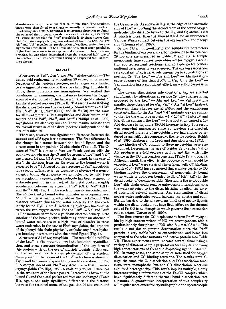
Structures of Valzg, Leuz9, and Phe2’ Metmyoglobins-The amino acid replacements a t position 29 caused no large per- turbation of the protein structure, and changes were limited to the immediate vicinity of the side chain (Fig. 1, Table 11). Thus, these mutations are isomorphous. We verified this conclusion by examining the distances between the oxygen atom of water bound to the heme iron and specific atoms of key distal pocket residues (Table 11). The results were striking: the distances between the covalently bound water and His64 (E7), Vala (El l ) , Phe43 (CDl), and Ile1O7 (G8) are the same for all three proteins. The amplitudes and distribution of B- factors of the Valz9, PheZ9, and Leuz9 (Phillips et al., 1990) myoglobins are also very similar. These results indicate that the overall structure of the distal pocket is independent of the size of residue 29.
There are, however, two significant differences between the mutant and wild type ferric structures. The first difference is the change in distance between the bound ligand and the closest atom in the position 29 side chain (Table 11). The C{ atom of PheZ9 is almost in Van der Waals contact with the bound water, whereas the C6 and Cy atoms of Leuz9 and Valz9 are located 5.4 and 6.3 A away from the ligand. In the case of Alaz9, the distance from the C@ atom to the bound water is expected to be 7.4 A based on the structure of Valz9 myoglobin. The second difference is the presence or absence of a nonco- valently bound distal pocket water molecule. In wild type metmyoglobin, a second water molecule has been assigned to a discrete position in the back of the distal pocket, roughly equidistant between the edges of Phe43 (CDl), Vala (El l ) , and Ile’07 (G8) (Fig. 1). The electron density associated with this noncovalently bound water indicates an occupancy level of 38% which is significantly above the background. The distance between this second water molecule and the cova- lently bound HzO is 2.7 A, indicating hydrogen bonding be- tween the two oxygen atoms. For the Leuz9 + Val and Leuz9 + Phe mutants, there is no significant electron density in the interior of the heme pocket, indicating either an absence of bound water molecules or a high level of disorder in these water molecules. In the case of PheZ9 myoglobin, the large size of the phenyl side chain physically excludes any direct hydro- gen bonding interactions with the bound ligand (Fig. 1).
Structure of P h Z 9 Oxymyoglobin-The remarkable stability of the Leuz9 + Phe mutant allowed the isolation, crystalliza- tion, and x-ray structure determination of the oxy form of this protein without the use of multiple crystals, a flow cell, or low temperatures. A stereo photograph of the electron density map in the region of the PheZ9 side chain is shown in Fig. 2 and two views of space filling models are shown in Fig. 3. A comparison of our PheZ9 oxy structure to that of native oxymyoglobin (Phillips, 1980) reveals only minor differences in the structure of the heme pocket. Interactions between the bound O2 and the distal pocket residues are unchanged (Table 111). Again, the only significant difference is the distance between the terminal atoms of the position 29 side chain and
the Oz molecule. As shown in Fig. 3, the edge of the aromatic ring of PheZ9 is touching the second atom of the bound oxygen molecule. The distance between the 0(2, and C{ atoms is 3.2 A, which is closer than the allowed 3.6 A for an unhindered Van der Waals contact between the oxygen atom and phenyl ring (Thomas et al., 1982).
0, and CO Binding-Kinetic and equilibrium parameters for the binding of oxygen and carbon monoxide to the position 29 mutants are presented in Table IV and Fig. 4. Simple monophasic time courses were observed for oxygen associa- tion and replacement reactions, and no evidence for confor- mational heterogeneity was observed. The oxygen association rate constant, k‘ o,, is relatively insensitive to substitutions at position 29. The Leuz9 + Phe and Leuz9 + Ala mutations cause changes of less than &30% in k’o,. Only the Leuz9 + Val mutation has a significant effect, an -2-fold decrease in k’o,.
The oxygen dissociation rate constants, b,, are affected significantly by alterations at residue 29. The changes in b, produced by the Leuz9 + Ala and Leuz9 + Val mutations parallel those observed for k’o,: Valz9 < Alaz9 5 Leuz9 (native). However, these changes are 5 &50%, and the equilibrium constants, K O , , for the Alaz9 and Valz9 mutants are very similar to that for the wild type protein, -1 X lo6 M” (Table IV and Fig. 4). In contrast, the Leuz9 4 Phe mutation caused a 10- fold decrease in ko, and a 15-fold increase in K O , . This result was somewhat unexpected since all previous site-directed, distal pocket mutants of myoglobin have had similar or re- duced oxygen affinities compared to the native protein (Rohlfs et al., 1990; Egeberg et al., 1990; and Smerdon et al., 1991).
The kinetics of CO binding to these myoglobins were also examined. Decreasing the size of residue 29 to either Val or Ala produces a 2-fold decrease in k’co and Kc0 with little change in the CO dissociation constant (Table IV and Fig. 4). Although small, this effect is the opposite of what would be expected if Leuz9 were sterically interfering with CO binding. Rohlfs et al. (1991) have suggested that a major barrier to CO binding involves the displacement of noncovalently bound water which is hydrogen bonded to N, of His64 (ET) in the distal pocket of deoxymyoglobin. Shortening the hydrophobic Leuz9 side chain could remove unfavorable interactions with the water attached to the distal histidine or allow the entry of additional solvent molecules. Any stabilization of distal pocket water molecules would increase the kinetic and equi- librium barriers to the noncovalent binding of apolar ligands within the distal pocket, but have little effect on the thermal rate of Fe-CO bond disruption which governs the dissociation rate constant (Carver et al., 1990).
The time courses for CO displacement from Phez9 myoglo- bin by high concentrations of NO are heterogeneous with a predominantly slow phase (-70% with kc0 = 0.003 s-’). This result is not due to protein denaturation since the Phez9 protein is very stable both to autooxidation and heme loss compared to the other mutants and native protein (see Table VI). These experiments were repeated several times using a variety of different sample preparation techniques and using high concentrations of O2 as the displacing ligand instead of NO. In many cases, the same samples were used for oxygen dissociation and CO binding reactions. The results were al- ways the same: the 02 dissociation and CO association reac- tions were monophasic, but the CO dissociation reactions exhibited heterogeneity. This result implies multiple, slowly interconverting conformations of the Fe . CO complex which have significantly different thermal bond dissociation rate constants. A quantitative interpretation of this complexity will require more extensive crystallographic and spectroscopic

14446 L29F Sperm Whale Myoglobin
FIG. 1. Stereo pair of the superposition of distal pocket residues Ior several position 29 mutants. The structures are of Valz9 (blue), LeuB native (red), and PheB (yellow) myoglobins in the aquomet form. In this view, the heme plane is horizontal, and the solvent interface is located to the left. Only minor shifts in position are seen in residues surrounding the mutations.
FIG. 2. Stereo pair of a I 2F. - F, I electron density map for the Leuz8 -., Phe mutant in the oxy form. This view is taken from the interior of the protein, looking toward the solvent. Density surrounding the bound oxygen is clearly visible just above the heme group.
investigations. Similar problems were observed for the Leu" + Ala mutant. For the present, we have estimated single values for k o , Kco, and M (Kco/Ko,) for the Phem and Alaz9 proteins to allow simple comparisons with the oxygen binding data and the results for native and Valz9 myoglobin. As shown in Table IV, the Leuz9 Phe replacement caused little change in CO affinity but did decrease both rate constants -2-fold.
Curiously, all three position 29 mutations caused greater discrimination against CO binding in favor of Oz binding. This property is usually evaluated in terms of the ratio of Kco/Ko, which defines the constant M. Sterically unhindered model heme compounds have M values equal to -4,000, whereas native mammalian hemoglobins and myoglobins show values equal to -200 and -20, respectively. Nagai, Olson, Sligar, and coworkers have shown that this discrimi- nation is due partly to stabilization of bound Oz by hydrogen
bonding to His (E7) and partly to destabilization of bound CO by steric hindrance with His (E7) and Val (El l ) (see Springer et al., 1989; Mathews et al., 1989; Egeberg et al., 1990). The 2-fold decrease in M produced by the Leuz9 4 Ala and Leuz9 + Val substitutions is the result of a decrease in Kc0 relative to KO,. In the case of the Leu2' + Phe mutation, the 10-fold reduction in M is due exclusively to preferential stabilization of bound oxygen by the Phe" side chain since Kc0 increases slightly (Table IV).
Binding of Larger Ligands-The rate constants for methyl and ethyl isocyanide binding to the position 29 mutants were measured to assess the size of the distal pockets in these proteins. As shown in Table V and Fig. 4, isocyanide binding is inhibited by increasing the size of residue 29. The associa- tion rate constants for the larger ligands decrease 5-10-fold as residue 29 is increased from Ala to Val to Leu. When Leu"

L29F Sperm Whale Myoglobin 14447
TABLE I1 Ligand-side chain distances in aqmmet sperm whale myoglobin
structures of msition 29 mutants
Atom pair
HisM (Ne)-OHz 2.6 HiP (Cc)-OHz 3.5 Valrn (Cyz)-OHz 3.2 Phe" (CS)-OHZ 3.9 neaw (C6)-OH2 5.6 Val" (Crz)-OHz 6.3 Leu" (Cdz)-OH2 Phe" (C{)-OH2
,,From G. N. Phillips et al. (1990).
A 2.5 2.7 3.5 3.5 3.1 3.2 3.9 3.9 5.7 5.8
5.4 4.0
FIG. 3. Space-fiing representations of the distal pocket of the oxy form of the Leurn + Phe mutant. The orientation is the same as in Fig. 2 in the top pane l and is rotated 90' around the vertical axis in the bottom panel . Yellow, Phe"; red, bound oxygen; purple, heme group; light blue, other polar residues; and green, other nonpolar residues. The close proximity of the phenyl ring of Phe" to the oxygen ligand is apparent in these images.
is replaced by Phe, there is little change in the association rate constants for methyl and ethyl isocyanide binding, but the dissociation rate constants for these ligands increase
TABLE I11 Contacts between convalently bound oxygen and the side chains of
HisM, Val", and residue 29
Atom pair Distance
Leu" (native)" Phe" A
His@' (Ne)-O(l,b 3.0 2.7 His@' (Nt)-O(z, 2.8 2.8 Valas (Crz)-o(l) 3.2 3.2 Valrn (cYz)-O(z) 3.5 3.6 Phe" (CS)-O(Z) 3.5 3.5 Ile" (C6)-O(2) 5.2 4.9 Leu" (cbz)-o~2, 4.9 Phe" (CS)-O(Z) 3.2
From S. E. V. Phillips et al. (1981).
and O(Z) to the oxygen atom bound to O(]).
almost 15-fold. In the crystal structure of ethyl isocyanide myoglobin, the
ligand adopts a bent geometry (Johnson et d, 1989). The ethyl carbon atoms point toward the back of the distal pocket and are located in a cavity between IlelO' (G8), Phe@ (CDl), and Leu" (B13). The bottom of the cavity is the heme ring, and the top is Leum, which appears to restrict the ligand side chain from adopting a more upright conformation. Thus, increasing the size of residue 29 would be expected to cause significant decreases in the affinity Constants for isocyanide binding. As shown in Table V and Fig. 4, replacing Leum with Val or Ala produces a myoglobin which exhibits an affinity for ethyl isocyanide which is identical to that for Oz. In contrast to the gases, the isocyanide affinity constanta for the Phem mutant decrease dramatically compared to those of the native protein. This result is readily interpreted from the structural resulta in Figs. 1-3; there is simply no room for large ligands in the distal pocket of the Phem mutant.
Autooxidution-When the wild type sperm whale myoglobin gene is expressed in E. coli in our 100 L fermentation facility, the protein is isolated as a mixture of ferric and ferrous forms and the cell paste has a reddish-brown color. S i m i i results are obtained with the Leum + Ala and Leum + Val mutants. However, cell paste obtained with plasmids containing the Leuzg + Phe mutation is bright red, and the resultant Phez9 protein remains 100% in the reduced, oxy form throughout the purifkation procedure and after repeated freezing and thawing. The effects of mutating residue 29 on the stability of the ferrous oxy complex were determined quantitatively by measuring rates of autooxidation of mutant, wild type, and native sperm whale proteins at pH 7.0, 37 "C in air-equili- brated buffer. The apparent first order autooxidation rate constants, kX, are given in Table VI along with the association equilibrium constants for oxygen binding at 37 'C. As shown, there is a 4-fold decrease in the rate of autooxidation of myoglobin when residue 29 is increased in size from Ala or Val to Leu even though these substitutions do not affect oxygen affinity. When Leum is replaced by Phe, there is an additional 10-fold reduction in the rate of autooxidation, and the reaction is so slow that it is very difficult to obtain a complete time course (ie. tS 6 days nt 37 OC).
Two mechanisms have been proposed for the autooxidation of oxymyoglobin: 1) direct fmt order dissociation of the neutral superoxide radical and 2) indirect second order reac- tion of O2 with deoxymyoglobin containing a weakly bound ferric ligand, normally HzO (Weiss, 1964, Shikama, 1983, Wallace et d., 1982). We have recently confirmed that the
* O(I) refers to the oxygen atom bound directly to the iron atom

14448 L29F Sperm Whale Myoglobin
TABLE IV Rate and equilibrium parameters for oxygen and carbon monoxide binding to position 29 (BIO) mutants
of sperm whale myoglobin at 20 "C, pH 7.0 The errors for both native and mutant myoglobins were calculated as the standard deviation from the mean of at least three independent
experiments at different ligand concentrations. M is equal to Kco/KO,. Protein k 'a2 ko2 K O 2 k'co k0 Kc0 M
q"' s-1 s-1 fi" q"1 s" s-1 m" Ala" 14rt 1 18f 1 0.8 f 0.1 0.26 f 0.02 0.019" 14' 17'
Leuz9 (native) 16 f 2 14 f 2 1.1 f 0.2 0.55 f 0.02 0.019 f 0.001 29 f 2 26 f 6 PheZ9
~ ~ 1 2 9 8.8 f 0.3 8.3 f 1 1.1 f 0.1 0.18 f 0.01 0.016 f 0.002 11 f 1 10*2
21 f 2 1.4 f 0.4 15 rf: 4 0.22 f 0.01 0.0060" 37' 2.5' These rate parameters were calculated from a weighted average of fitted parameters because the time courses for CO dissociation were
heterogeneous, and a sum of two exponentials provided better fits to the data. For a two-exponential fit, the amplitudes ( A ) and rate constants ( k ) were for Ala? A , = 0.81, k , = 0.022 s-', Az = 0.19, kp = 0.0074 s-'; and for Phe? A , = 0.29, kl = 0.012 s-'; A2 = 0.71, k, = 0.0030 s-'. ' These equilibrium parameters were calculated using the weighted averages for kc0 presented in column 6 (see footnote a ) .
6
8 s M
5
4
3
ENC \
I I I
Ala Val Leu Phe
Position 29 Residue FIG. 4. Dependence of ligand affinity on the size of residue
29 (B10) at pH 7,20 OC. Logarithms of the association equilibrium constants, K, for 02, CO, methyl (MNC), and ethyl (ENC) isocyanide binding are plotted uersus the size of the residue at position 29. Values of the constants are given in Tables IV and V.
superoxide dissociation mechanism predominates at high [02] and that the bimolecular reaction predominates at low [OZ] where deoxymyoglobin is the major form of the protein (Brantley et al., 1992a, 1992b). The former condition applies in our experiments since the myoglobin molecules were com- pletely saturated with ligand at the oxygen concentration used (i.e. [Oz] = 40-700 X Kd or P ~ o ) . The simplest interpre- tation of the results in Table VI is that decreasing the size of residue 29 from Leu to Ala or Val increases the equilibrium occupancy of disordered waters near the bound ligand. Such a phenomenon would increase the polarity of the ligand environment, facilitate protonation of the Fe . O2 complex, enhance HOB dissociation, and promote autooxidation with- out causing significant changes in oxygen affinity. In the case of PheZ9 myoglobin, the 10-fold decrease in the rate of au- tooxidation is accompanied by a large increase in KO,. Al- though the side chain of PheZ9 does decrease the polarity of the ligand environment, it is likely that the dramatic decrease in k,, is due primarily to specific interactions of the phenyl side chain which stabilize the bound oxygen and prevent its protonation.
DISCUSSION
Dipolar Interactions with PheZ9-The 15-fold increase in oxygen affinity produced by the Leuz9 + Phe mutation is readily explained in terms of favorable electrostatic interac- tions between the polar Fe.OZ complex and the multipole of the benzene ring of the PheZ9 side chain. Thomas et al. (1982) examined the spatial distribution of oxygen atoms around the side chains of 170 phenylalanines in 28 different protein crystal structures. The dominant orientation was one in which the oxygen atoms were located at the edge of the aromatic rings. An empirical estimate of the free energy of this inter- action was obtained by computing the ratio of the number of oxygen atoms located near the edge of phenyl rings to the number located at the top of the ring face for the complete data set. The results suggested that the edge orientation was favored by "0.8 kcal/mol for all oxygen atoms and could be as large as -1.5 kcal/mol for carbonyl oxygen atoms and Phe side chains in helical environments.
Thomas et al. (1982) also carried out ab initio quantum mechanical calculations to examine the energy change for bringing formamide in close proximity to benzene. A E was computed as a function of: 1) the distance between the nearest ring carbon atom and the carbonyl oxygen and 2) the relative angle between the carbonyl dipole and the ring plane. The computed values of hE are negative for angles less than 45" and distances between 3 and 6 A with a minimum at about 3.6 A which is the Van der Waals limit. At this distance and an angle of O", the computed value of AE is -2.0 kcal/mol. At the same interatomic distance but with the carbonyl dipole oriented perpendicular to and directly above the center of the aromatic ring, the computed value of AE is - +2-3 kcal/mol.
The Fe.0, complex in heme compounds is highly polar with a partial negative charge on the second oxygen atom and a partial positive charge on the iron atom (Weiss, 1964). In native myoglobin, the N, atom of HisM (E7) acts as a proton donor to form a hydrogen bond with the second bound oxygen atom. This interaction stabilizes the oxy complex by -1 to -2 kcal/mol based on the effects of replacing HisM with Gly, Val, Leu, and Phe (Springer et al., 1989) and decreases the rate of autooxidation by inhibiting both dissociation and protonation of the Fe.02 complex (Springer et d., 1989; Brantley et al., 1992a, 1992b). In the Phem mutant, the hydrogen bond with the distal histidine is preserved, and the second bound oxygen atom is also located 3.2 8, away from the edge of the B10 phenyl group at an angle of -15" with respect to the plane of the aromatic ring. According to Thomas et al. (1982), this orientation should stabilize the bound oxy- gen enthalpically by an additional -1 to -2 kcal/mol if the Fe. O2 complex has a dipole moment similar to the carbonyl group in formamide. As shown in Table IV, replacing Leuz9

L29F Sperm Whale Myoglobin 14449
with Phe decreased the free energy change for oxygen binding by -1.5 kcal/mol, an amount compatible with the calculations of Thomas et al. (1982).
The evidence in favor of this interpretation of the high 02 affinity of PheZ9 myoglobin is 3-fold. First, the distance be- tween the O(,) oxygen atom and Cf of the phenyl ring is 0.4 A shorter than the 3.6 A expected for a simple Van der Waals contact (Table 111, Thomas et al., 1982). In the absence of favorable interactions with the aromatic ring, this distance would imply steric hindrance and a decreased oxygen affinity. In contrast, a 15-fold increase in affinity is observed. Second, this increase in O2 affinity is due almost exclusively to a decreased dissociation rate constant. Carver et al. (1990) have shown the overall dissociation rate constant of oxymyoglobin is governed primarily by the rate of thermal bond disruption. Therefore, a decrease in b, without a change in k'o, implies (but does not prove) stabilization of the ligand complex rather than an increase in reactivity of the unliganded iron atom. Third, except for the position 29 side chains, both the ferric aquo and ferrous oxy forms of the mutant are isomorphous with native or wild type myoglobin. Consequently, secondary conformational changes or alterations in the iron-heme co- ordination geometry cannot be invoked to explain the increase in KO,. Thus, it seems clear that PheZ9 is stabilizing bound oxygen in sperm whale myoglobin through polar interactions, and the marked increase in Oz affinity produced by the Leuz9 + Phe mutation provides an independent experimental veri- fication of the importance of aromatic multipole interactions in proteins (see Thomas et al., 1982; Burley and Petsko, 1985).
Importance of Phe (BlO) in Leghemoglobins-Phe and Tyr are found at the B10 position in the distal pockets of leghe- moglobins, and these proteins, like PheZ9 sperm whale myo- globin, exhibit a high affinity for 0, (KO, 2 10 pL"' at 20 "C; Wittenberg et al., 1986).' In lupine leghemoglobin, Phe resi- dues are present at the B9 and B10 positions, but in the aquomet structure of this protein, the edges of both aromatic rings are located -6 A from the water bound to the iron atom (Vainshtein et al., 1978).' This conformation syggests that the phenyl rings would be located at least 5.0 A away from the second atom of the bound ligand in oxyleghemoglobin if there are no large structural changes roduced by reduction of the iron atom. This distance is 1.8 x greater than the C { - 0 ,~ ) distance observed in sperm whale PheZ9 oxymyoglobin and is beyond the optimal range for polar interactions with the aromatic rings (Thomas et al., 1982). Thus, the phenyl- alanines at B9 and B10 in leghemoglobin do not appear to be close enough to stabilize the O2 complex. This conclusion is supported by the observation that the rate constant for oxygen dissociation from lupine leghemoglobin is 25 s-' at 20 "C, which is 80% greater than ko, for native sperm whale myoglo- bin and 18 times greater than &, for PheZ9 myoglobin (Wit- tenberg et al., 1986; Table IV). Soybean leghemoglobin has a lower oxygen dissociation rate constant, 2-5 s-l (Wittenberg
et al., 1986; Gibson et al., 1989). In this protein, the B10 residue is a tyrosine,' and it is possible that the -OH of the Tyr(B10) side chain forms a hydrogen bond with the oxygen ligand, supplementing the hydrogen bond with His (E7). However, the abnormally high oxygen affinities of both these leghemoglobins are due primarily to large association rate constants, V O 2 = 1-2 x 10' M" s-', rather than small disso- ciation rate constants, in contrast to what is observed for PheZ9 sperm whale myoglobin (Wittenberg et al., 1986, and Table IV).
Function of Leuz9 in Myogbbin-The crystallographic re- sults in Figs. 1 and 2 show that Leuz9 is not mandatory for the structural integrity of the distal pocket. Oxygen binding is little affected by decreasing the size of the B10 residue from Leu to Val to Ala as judged both by the equilibrium constants in Tables IV and VI and by the ligand-side chain contacts listed in Tables I1 and 111. However, the stability of oxymy- oglobin to autooxidation is affected markedly by substitutions at position 29. As shown in Table VI, decreasing the size of residue 29 from Leu to either Ala or Val decreases the half- life of oxymyoglobin from -13 to 3 h at 37 "C. This increase in autooxidation rate is most readily explained in terms of an increase in accessibility of the Fe . Oz complex to water mole- cules when the length of the side chain at position 29 is reduced. This interpretation is supported by results for Hb St. Louis in which Gln replaces Leu at the B10 position in p subunits of human hemoglobin (Fermi and Perutz, 1982). The crystal structure of the mutant subunit shows an expanded distal pocket due to the binding of a water molecule between Gln (B10) and His (E7), and this increase in polarity readily accounts for the high rate of autooxidation of Hb St. Louis ,!l chains. The Leuz9 + Ala and Leuz9 + Val mutations also appear to enhance the rate of denaturation of metmyoglobin at 37 'C as judged by the extent of precipitation in our autooxidation experiments.
In contrast, increasing the size of residue 29 from Leu to Phe inhibits autooxidation markedly and produces an exceed- ingly stable oxymyoglobin with a half-life of roughly 6 days at 37 "C. This remarkable stability is probably due to the general exclusion of water from the distal pocket as a result of the large size of the phenyl group and a more specific interaction between the positive edge of the aromatic multi- pole and the negative dipole of bound oxygen. The latter polar interaction in the Leuz9 * Phe mutant also causes a dramatic decrease in the rate of oxygen dissociation which is likely to be detrimental to the physiological function of myoglobin. Since intracellular diffusion of oxygen is very rapid and the reaction of cytochrome oxidase with oxygen is almost diffu- sion-controlled (-lo8 "'s-'; Greenwood and Gibson, 1965), the speed of 0, delivery from myoglobin to mitochondria is determined mainly by the rate constant for O2 dissociation during periods of rapid respiration. The value of ko, is -15 s-' at 20 "C and -40 s-' at 37 "C for all mammalian myoglo-
TABLE V Rate and equilibrium parameters for methyl and ethyl isocyanide binding to position 29 (BIO) mutants
of sperm whale myoglobin at 20 "C, pH 7.0 Errors for the native myoglobin parameters have been estimated to be +16% for rate constants and *20% for equilibrium constants based
on previous measurements with native and wild type myoglobin and are assumed to apply also to the mutant parameters (Rohlfs et al., 1990). Protein k'hmcO khmc KMNC k ' m c k m c KENC
PM' s" S-1 P" p" s" S" P"
1.1 1.3 0.23
~ 1 ~ 2 9
va1z9 0.58 7.7 0.075 0.24 4.7 0.21 0.16
0.63 0.051
Leuz9 (native) 0.12 4.3 0.028 PheZ9 0.19
0.30 0.0033 0.090 4.2 0.021
0.70
0.069 57.
The isocyanide abbreviations are: MNC, methyl isocyanide; ENC, ethyl isocyanide.
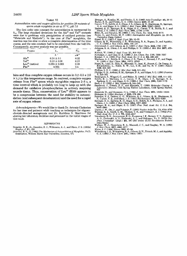
14450 L29F Sperm Whale Myoglobin TABLE VI
Autooxidation rates and oxygen affinities for position 29 mutants of sperm whale myoglobin in air at 37 “C, pH 7.0
The first order rate constant for autooxidation is represented by k,,,. The large standard deviations for the Alaz9 and Valz9 mutants were due to problems with precipitation of oxidized proteins (see “Materials and Methods”). In the case of PheZg myoglobin, the reaction was too slow to allow a complete determination of the time course, and the rate constant had to be calculated from the half-life. Consequently, an error analysis was not possible.
Protein k,. K O ,
h- ’ N W 1
0.24 +- 0.1 0.22 0.23 f 0.05 0.23
Leuz9 (native) 0.055 f 0.005 0.28 Phe2’ 0.005 3.4
~ 1 ~ 2 9
va1*9
bins and thus complete oxygen release occurs in 0.2-0.5 s (10 X tlh) in this temperature range. In contrast, complete oxygen release from PheZ9 sperm whale myoglobin requires 2-5 s, a time interval which is probably too long to keep up with the demand for oxidative phosphorylation in actively respiring muscle tissue. Thus, conservation of Leuz9 (B10) appears to be a compromise between the need for stability to autooxi- dation (and subsequent denaturation) and the need for a rapid rate of oxygen release.
Acknowledgments-We would like to thank Dr. Artemis Chakerian for her time and patience while teaching us techniques for oligonu- cleotide-directed mutagenesis and Dr. Kathleen S. Matthews for sharing her laboratory facilities and personnel in the initial stages of this work.
REFERENCES Hrantley, R. E., Jr., Smerdon, S. J., Wilkinson, A. J., and Olson, J. S. (1992a)
Brantley, R. E., Jr. (1992) The Mechanism ofAutoosidation ofMyoglobin. Ph.D. Biophys. J . 6 1 , 298
dissertation, William Marsh Rice University, Houston, TX
Brunger, A., Karplus, M., and Petsko, G. A. (1989) Acta Crystallogr. 46,50-61 Burley, S. K., and Petsko, G. A. (1985) Science 229,23-28 Carver, T. E., Rohlfs, R. J., Olson, J. S., Gibson, Q. H., Blackmore, R., Springer,
Egeberg, K. D., Springer, B. A., Sligar, S. G., Carver, T. E., Rohlfs, R. J., and
Elber, R., and Karplus, M. (1990) J. Am. Chem. SOC. 112,9161-9175 Fermi, G., and Perutz, M. F. (1981) Haemoglobin and Myoglobin, pp. 26-29,
Finsel, B. C. (1987) J. Appl. Cryst. 20,53-55 Gibson, Q. H., Wittenberg, J. B., Wittenberg, B. A., Bogusz, D., and Appleby,
C. A. (1989) J. Biol. Chem. 264,100-107
Johnson, K. A,, Olson, J. S., and Phillips, G. N. (1989) J. Mol. Biol. 207 , 459- Greenwood, C., and Gibson, Q. H. (1967) J. Biol. Chem. 242,1782-1787
Kabsch, W. (1988) J. Appl. Cryst. 21,916-924 Kottalam, J., and Case, D. A. (1988) J. Am. Chem. SOC. 1 1 0 , 7690-7697 Kunkel, T. A. (1985) Proc. Natl. Acad. Sci. U. S. A. 82,488-493 Mathews, A. J., Rohlfs, R. J., Olson, J. S., Tame, J., Renaud, J. P., and Nagai,
K. (1989) J. Biol. Chem. 264,16573-16583 Nagai, K., Luisi, B., Shih, K., Miyazaki, G., Imai, K., Poyart, C., De Young, A.,
Kwiatkowsky, L., Noble, R. W., Lin, S.-H., and Yu, N. T. (1987) Nature 329,858-860
B. A,, and Sligar, S. G. (1990) J. Biol. Chem. 2 6 6 , 20007-20020
Olson, J. S. (1990) J. Biol. Chem. 266,11788-11795
Clarendon Press, Oxford
463
Phillips, S. E. V. (1980) J. Mol. Biol. 142 , 531-554 Phillips, G. N., Arduini, R. M., Springer, B. A., and Sligar, S. G. (1990) Proteins
7.358-365 Reminson, S. , Wiegard, G., and Huber, R. (1982) J. Mol. Biol. 168, 111-152 Rohlfs, R. J., Mathews, A. J., Carver, T. E., Olson, J. S., Springer, B. A,,
Sambrook, J., Fritsch, E. F., and Maniatis, T. (1989) Moleculnr Cloning: A Sack, J. S. (1988) J. Mol. Graphics 6 , 244-245
Laboratory Manual, Cold Spring Harbor Laboratory, Cold Spring Harbor, NY
Egeberg, K. D., and Sligar, S. G. (1990) J. Biol. Chem. 266,3168-3176
Sassaroli, M., and Rousseau, D. L. (1986) J. Biol. Chem. 261 , 16292-16294 Shikama, K. (1984) Biochem. J. 223,279-280 Smerdon, S. J., Dodson, G. G., Wikinson, A. J., Gibson, Q. H., Blackmore, R.
Springer, B. A., Egeberg, K. D., Sligar, S. G., Rohlfs, R. J., Mathews, A. J., and
Springer, B. A,, and Sligar, S. G. (1987) Proc. Natl. Acad. Sci. U. S. A. 8 4 ,
Taylor, J. W., Ott, J., and Eckstein, F. (1985) Nucleic Acids Res. 13,8764-8785 Thomas, K. A., Smith, G. M., Thomas, T. B., and Feldmann, R. J. (1982) Proc.
Natl. Acad. Sci. U. S. A. 79,4843-4847 Vainshtein, B. K., Arutyunyan, E. G., Kuranova, I. P., Borisov, V. V., Sosfenov,
A. G., Pavlovskii, A. G., Grebenko, A. I., and Nekrasov, Yu. V. (1978) Sou. Phys. Crystallogr. (Engl.) 2 3 , 287-293 (entry 2LH2 Brookhaven Protein Data Bank)
Wallace, W. J., Houtchens, R. A,, Maxwell, J. C., and Caughey, W. S. (1982) J. Biol. Chem. 267,4966-4977
Weiss, J. J. (1964) Nature 202 , 83-84 Wittenberg, J. B., Wittenberg, B. A,, Gibson, Q. H., Trinick, M. J., and Appleby,
S., Carver, T. E., and Olson, J. S. (1991) Biochemistry 30,6252-6260
Olson, J. S. (1989) J. Biol. Chem. 264,3057-3060
8961-8965
C. A. (1986) J. Biol. Chem. 261,13624-13631