Embed Size (px)
Citation preview
Proceedings of Machine Learning Research 102:370–379, 2019 MIDL 2019 – Full paper track
A novel segmentation framework for uveal melanoma in magneticresonance imaging based on class activation maps
Huu-Giao Nguyen1,2,3 [email protected] Proton Therapy Center, Paul Scherrer Institut, ETH Domain, Villigen, Switzerland2 Ophthalmic Technology Lab., ARTORG Center, University of Bern, Switzerland3 Radiology Department, Lausanne University Hospital (CHUV), Switzerland
Alessia Pica1 and Jan Hrbacek1 and Damien C. Weber1,4
[email protected] Radiation Oncology Department, Inselspital, University of Bern, SwitzerlandFrancesco La Rosa5 [email protected] Signal Processing Lab., Ecole Polytechnique Federale de Lausanne, Switzerland
Ann Schalenbourg6 [email protected] Adult Ocular Oncology Unit, Jules-Gonin Eye hospital, Lausanne, Switzerland
Raphael Sznitman2 [email protected]
Meritxell Bach Cuadra3,5,7 [email protected] Medical Image Analysis Laboratory, CIBM, University of Lausanne, Switzerland
AbstractAn automatic and accurate eye tumor segmentation from Magnetic Resonance images (MRI) couldhave a great clinical contribution for the purpose of diagnosis and treatment planning of intra-ocularcancer. For instance, the characterization of uveal melanoma (UM) tumors would allow the inte-gration of 3D information for the radiotherapy and would also support further radiomics studies.In this work, we tackle two major challenges of UM segmentation: 1) the high heterogeneity oftumor characterization in respect to location, size and appearance and, 2) the difficulty in obtainingground-truth delineations of medical experts for training. We propose a thorough segmentationpipeline consisting of a combination of two Convolutional Neural Networks (CNN). First, we con-sider the class activation maps (CAM) output from a Resnet classification model and the combina-tion of Dense Conditional Random Field (CRF) with a prior information of sclera and lens from anActive Shape Model (ASM) to automatically extract the tumor location for all MRIs. Then, theseimmediate results will be inputted into a 2D-Unet CNN whereby using four encoder and decoderlayers to produce the tumor segmentation. A clinical data set of 1.5T T1-w and T2-w images of 28healthy eyes and 24 UM patients is used for validation. We show experimentally in two differentMRI sequences that our weakly 2D-Unet approach outperforms previous state-of-the-art methodsfor tumor segmentation and that it achieves equivalent accuracy as when manual labels are used fortraining. These results are promising for further large-scale analysis and for introducing 3D oculartumor information in the therapy planning.Keywords: Activation map, CAM, Unet, tumor segmentation, Uveal melanoma
1. Introduction
UM is the most common primary intraocular malignancy in the white adult population, makingup 79-88% of primary intraocular cancers (Singh et al., 2014; Lemke et al., 1999). Several 2-
c© 2019 H.-G. Nguyen, A. Pica, J. Hrbacek, D. Weber, F.L. Rosa, A. Schalenbourg, R. Sznitman & M. Bach Cuadra.
SEGMENTATION OF UVEAL MELANOMA IN MRI
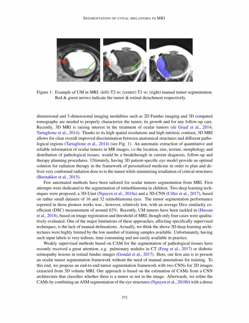
Figure 1: Example of UM in MRI: (left) T2-w; (center) T1-w; (right) manual tumor segmentation.Red & green arrows indicate the tumor & retinal detachment respectively.
dimensional and 3-dimensional imaging modalities such as 2D Fundus imaging and 3D computedtomography are needed to properly characterize the tumor, its growth and for any follow-up care.Recently, 3D MRI is raising interest in the treatment of ocular tumors (de Graaf et al., 2014;Tartaglione et al., 2014). Thanks to its high spatial resolutions and high intrinsic contrast, 3D MRIallows for clear overall improved discrimination between anatomical structures and different patho-logical regions (Tartaglione et al., 2014) (see Fig. 1). An automatic extraction of quantitative andreliable information of ocular tumors in MR images, i.e the location, size, texture, morphology anddistribution of pathological tissues, would be a breakthrough in current diagnosis, follow-up andtherapy planning procedures. Ultimately, having 3D patient-specific eye model provide an optimalsolution for radiation therapy in the framework of personalized medicine in order to plan and de-liver very conformal radiation dose to to the tumor while minimizing irradiation of critical structures(Beenakker et al., 2015).
Few automated methods have been tailored for ocular tumors segmentation from MRI. Firstattempts were dedicated to the segmentation of retinoblastoma in children. Two deep learning tech-niques were proposed, a 3D-Unet (Nguyen et al., 2018a) and a 3D-CNN (Ciller et al., 2017), basedon rather small datasets of 16 and 32 retinoblastoma eyes. The tumor segmentation performancereported in those pioneer works was , however, relatively low, with an average Dice similarity co-efficient (DSC) measurement of around 62%. Recently, UM tumors have been tackled in (Hassanet al., 2018), based on image registration and threshold of MRI, though only four cases were qualita-tively evaluated. One of the major limitations of these approaches, affecting specifically supervisedtechniques, is the lack of manual delineations. Actually, we think the above 3D deep learning archi-tectures were highly limited by the low number of training samples available. Unfortunately, havingsuch input labels is very tedious, time consuming and not easily available in practice.
Weakly supervised methods based on CAM for the segmentation of pathological tissues haverecently received a great attention, e.g. pulmonary nodules in CT (Feng et al., 2017) or diabeticretinopathy lesions in retinal fundus images (Gondal et al., 2017). Here, our first aim is to presentan ocular tumor segmentation framework without the need of manual annotations for training. Tothis end, we propose an end-to-end tumor segmentation framework with two CNNs for 2D imagesextracted from 3D volume MRI. Our approach is based on the estimation of CAMs from a CNNarchitecture that classifies whether there is a tumor or not in the image. Afterwards, we refine theCAMs by combining an ASM segmentation of the eye structures (Nguyen et al., 2018b) with a dense
371
SEGMENTATION OF UVEAL MELANOMA IN MRI
Table 1: MR imaging acquisition parameters at 1.5T with a surface coil.Repetitiontime(ms)
Echo time(ms)
Flip AngleVoxel size
(mm3)FOV
(Voxels)Healthy UM
T1-VIBE 6.55 2.39 12◦ 0.5x0.5x0.5 256x256x80 28 eyes 24 eyes
T2-SPACE 1400 185 150◦0.5x0.5x0.5 and0.82x0.82x0.8
256x256x80 25 eyes 22 eyes
CRF to maximize label agreement between similar pixels in images. Finally, we use these refinedCAMs as input training data for a 2D-Unet segmentation (Ronneberger et al., 2015). The proposedframework is cheaper in training data (only sclera segmentations are needed for the ASM) andoutperforms in segmentation compared existing deep learning approaches (Nguyen et al., 2018a;Rosa et al., 2018).
A second major contribution of this work is the quantitative evaluation of several 2D and 3Darchitectures for the UM segmentation. To the best of our knowledge this is the first study reportingautomated segmentation accuracy for such ocular tumor. Our proposed segmentation technique willbe compared with previous related work: 1) our previous work tailored for retinoblastoma tumorsin children and based on a 3D-Unet (Nguyen et al., 2018a), with a 2D-Unet using manual labels astraining from an expert, and 2) a cascade of two 3D patch-wise CNNs used for lesion segmentationin Multiple Sclerosis (Rosa et al., 2018).
2. Dataset
MR acquisitions were performed by a 1.5T Siemens scanner with surface coil for both T1w andT2w contrasts at the Paul Scherrer Institute. A set of 16 healthy volunteers (mean age 29± 5.4y.o., range [23− 46] years) and 24 UM patients (mean age 63± 14 y.o., range [36− 74] years)was considered. The cohort median eye size was 24.4mm of diameter (range, 22.1-26.5). Tab. 1shows the different parameters used for the MRI acquisition protocol. The study was approved bythe Ethics Committee of the involved institutions and all subjects (anonymized and de-identified)provided written informed consent prior to participation.
Images were pre-processed as follows. First, an anisotropic diffusion filtering (Perona and Ma-lik, 1990) was applied to reduce noise without removing significant image content. Second, weapplied the N4 algorithm (Tustison et al., 2010) to correct for bias field variations and performedhistogram-based intensity normalization (Nyul et al., 2000) for an intensity normalisation. Finally,in order to improve the performance in segmentation and computation time, the whole MRI wascropped using a volume of interest of 64x64x64 voxels centered in the eye.
Manual delineations were done by radiation oncologist expert for 16 UM patients and all healthyeyes using Velocity software(Varian Medical System, Palo Alto, CA). First, segmentation for sclera,lens and tumor was done individually through intensity threshold. Second, manual editing wasperformed to refine borders and remove outlier regions.
3. Proposed segmentation framework
The proposed framework is over-viewed in Fig. 2. It mainly consists of the concatenation of a 2DResNet model (He et al., 2016) to classify MRI slices (with or without tumor) that combined with
372
SEGMENTATION OF UVEAL MELANOMA IN MRI
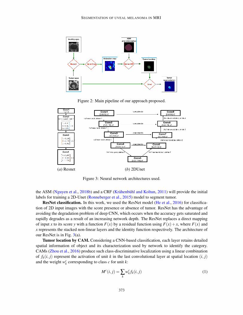
Figure 2: Main pipeline of our approach proposed.
(a) Resnet (b) 2DUnet
Figure 3: Neural network architectures used.
the ASM (Nguyen et al., 2018b) and a CRF (Krahenbuhl and Koltun, 2011) will provide the initiallabels for training a 2D-Unet (Ronneberger et al., 2015) model to segment tumor.
ResNet classification. In this work, we used the ResNet model (He et al., 2016) for classifica-tion of 2D input images with the score presence or absence of tumor. ResNet has the advantage ofavoiding the degradation problem of deep CNN, which occurs when the accuracy gets saturated andrapidly degrades as a result of an increasing network depth. The ResNet replaces a direct mappingof input x to its score y with a function F(x) by a residual function using F(x)+ x, where F(x) andx represents the stacked non-linear layers and the identity function respectively. The architecture ofour ResNet is in Fig. 3(a).
Tumor location by CAM. Considering a CNN-based classification, each layer retains detailedspatial information of object and its characterization used by network to identify the category.CAMs (Zhou et al., 2016) produce such class-discriminative localization using a linear combinationof fk(i, j) represent the activation of unit k in the last convolutional layer at spatial location (i, j)and the weight wc
k corresponding to class c for unit k:
Mc(i, j) = ∑k
wck fk(i, j) (1)
373
SEGMENTATION OF UVEAL MELANOMA IN MRI
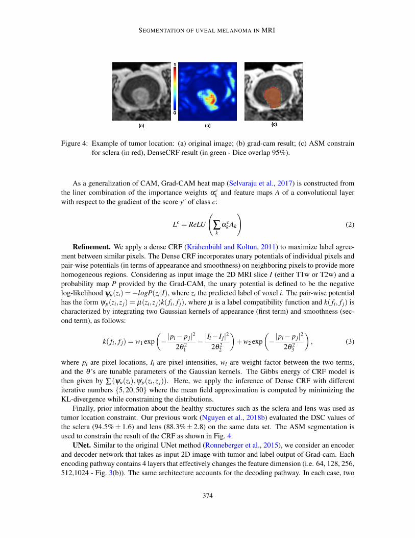
Figure 4: Example of tumor location: (a) original image; (b) grad-cam result; (c) ASM constrainfor sclera (in red), DenseCRF result (in green - Dice overlap 95%).
As a generalization of CAM, Grad-CAM heat map (Selvaraju et al., 2017) is constructed fromthe liner combination of the importance weights αc
k and feature maps A of a convolutional layerwith respect to the gradient of the score yc of class c:
Lc = ReLU
(∑k
αck Ak
)(2)
Refinement. We apply a dense CRF (Krahenbuhl and Koltun, 2011) to maximize label agree-ment between similar pixels. The Dense CRF incorporates unary potentials of individual pixels andpair-wise potentials (in terms of appearance and smoothness) on neighboring pixels to provide morehomogeneous regions. Considering as input image the 2D MRI slice I (either T1w or T2w) and aprobability map P provided by the Grad-CAM, the unary potential is defined to be the negativelog-likelihood ψu(zi) =−logP(zi|I), where zi the predicted label of voxel i. The pair-wise potentialhas the form ψp(zi,z j) = µ(zi,z j)k( fi, f j), where µ is a label compatibility function and k( fi, f j) ischaracterized by integrating two Gaussian kernels of appearance (first term) and smoothness (sec-ond term), as follows:
k( fi, f j) = w1 exp(−|pi− p j|2
2θ 21−|Ii− I j|2
2θ 22
)+w2 exp
(−|pi− p j|2
2θ 23
), (3)
where pi are pixel locations, Ii are pixel intensities, wl are weight factor between the two terms,and the θ ’s are tunable parameters of the Gaussian kernels. The Gibbs energy of CRF model isthen given by ∑(ψu(zi),ψp(zi,z j)). Here, we apply the inference of Dense CRF with differentiterative numbers {5,20,50} where the mean field approximation is computed by minimizing theKL-divergence while constraining the distributions.
Finally, prior information about the healthy structures such as the sclera and lens was used astumor location constraint. Our previous work (Nguyen et al., 2018b) evaluated the DSC values ofthe sclera (94.5%± 1.6) and lens (88.3%± 2.8) on the same data set. The ASM segmentation isused to constrain the result of the CRF as shown in Fig. 4.
UNet. Similar to the original UNet method (Ronneberger et al., 2015), we consider an encoderand decoder network that takes as input 2D image with tumor and label output of Grad-cam. Eachencoding pathway contains 4 layers that effectively changes the feature dimension (i.e. 64, 128, 256,512,1024 - Fig. 3(b)). The same architecture accounts for the decoding pathway. In each case, two
374
SEGMENTATION OF UVEAL MELANOMA IN MRI
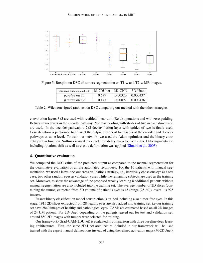
Figure 5: Boxplot on DSC of tumors segmentation on T1-w and T2-w MR images.
Wilcoxon test compared with M-2DUnet 3D-CNN 3D-Unetp value on T1 0.679 0.00320 0.000437p value on T2 0.147 0.00097 0.000436
Table 2: Wilcoxon signed rank test on DSC comparing our method with the other strategies.
convolution layers 3x3 are used with rectified linear unit (Relu) operations and with zero padding.Between two layers in the encoder pathway, 2x2 max pooling with strides of two in each dimensionare used. In the decoder pathway, a 2x2 deconvolution layer with strides of two is firstly used.Concatenation is performed to connect the output tensors of two layers of the encoder and decoderpathways at same level. To train our network, we used the Adam optimizer and the binary crossentropy loss function. Softmax is used to extract probability maps for each class. Data augmentationincluding rotation, shift as well as elastic deformation was applied (Simard et al., 2003).
4. Quantitative evaluation
We computed the DSC value of the predicted output as compared to the manual segmentation forthe quantitative evaluation of all the automated techniques. For the 16 patients with manual seg-mentation, we used a leave-one-out cross-validations strategy, i.e., iteratively chose one eye as a testcase, two other random eyes as validation cases while the remaining subjects are used as the trainingset. Moreover, to show the advantage of the proposed weakly learning 8 additional patients withoutmanual segmentation are also included into the training set. The average number of 2D slices (con-taining the tumor) extracted from 3D volume of patient’s eyes is 45 (range [25-60]), overall is 925images.
Resnet binary classification model construction is trained including also tumor-free eyes. In thisstage, 1915 2D slices extracted from 28 healthy eyes are also added into training set, i.e our trainingset have 2840 images of healthy and pathological eyes. CAMs are estimated based on all 2D imagesof 24 UM patient. For 2D-Unet, depending on the patients leaved out for test and validation set,around 850 2D images with tumors were selected for training.
Our framework (Grad-CAM-2DUnet) is evaluated in comparison with three baseline deep learn-ing architectures. First, the same 2D-Unet architecture included in our framework will be usedtrained with the expert manual delineations instead of using the refined activation maps (M-2DUnet).
375
SEGMENTATION OF UVEAL MELANOMA IN MRI
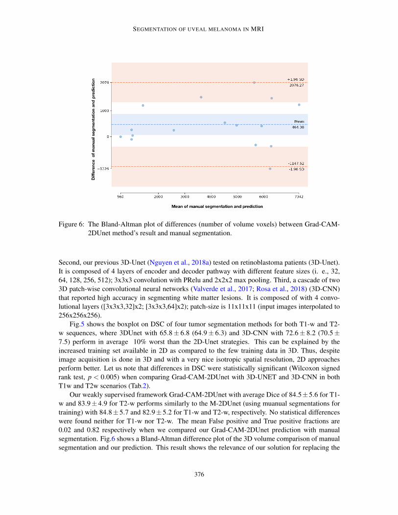
Figure 6: The Bland-Altman plot of differences (number of volume voxels) between Grad-CAM-2DUnet method’s result and manual segmentation.
Second, our previous 3D-Unet (Nguyen et al., 2018a) tested on retinoblastoma patients (3D-Unet).It is composed of 4 layers of encoder and decoder pathway with different feature sizes (i. e., 32,64, 128, 256, 512); 3x3x3 convolution with PRelu and 2x2x2 max pooling. Third, a cascade of two3D patch-wise convolutional neural networks (Valverde et al., 2017; Rosa et al., 2018) (3D-CNN)that reported high accuracy in segmenting white matter lesions. It is composed of with 4 convo-lutional layers ([3x3x3,32]x2; [3x3x3,64]x2); patch-size is 11x11x11 (input images interpolated to256x256x256).
Fig.5 shows the boxplot on DSC of four tumor segmentation methods for both T1-w and T2-w sequences, where 3DUnet with 65.8± 6.8 (64.9± 6.3) and 3D-CNN with 72.6± 8.2 (70.5±7.5) perform in average 10% worst than the 2D-Unet strategies. This can be explained by theincreased training set available in 2D as compared to the few training data in 3D. Thus, despiteimage acquisition is done in 3D and with a very nice isotropic spatial resolution, 2D approachesperform better. Let us note that differences in DSC were statistically significant (Wilcoxon signedrank test, p < 0.005) when comparing Grad-CAM-2DUnet with 3D-UNET and 3D-CNN in bothT1w and T2w scenarios (Tab.2).
Our weakly supervised framework Grad-CAM-2DUnet with average Dice of 84.5±5.6 for T1-w and 83.9±4.9 for T2-w performs similarly to the M-2DUnet (using muanual segmentations fortraining) with 84.8±5.7 and 82.9±5.2 for T1-w and T2-w, respectively. No statistical differenceswere found neither for T1-w nor T2-w. The mean False positive and True positive fractions are0.02 and 0.82 respectively when we compared our Grad-CAM-2DUnet prediction with manualsegmentation. Fig.6 shows a Bland-Altman difference plot of the 3D volume comparison of manualsegmentation and our prediction. This result shows the relevance of our solution for replacing the
376
SEGMENTATION OF UVEAL MELANOMA IN MRI
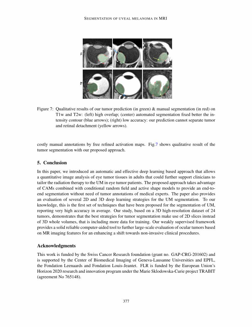
Figure 7: Qualitative results of our tumor prediction (in green) & manual segmentation (in red) onT1w and T2w: (left) high overlap; (center) automated segmentation fixed better the in-tensity contour (blue arrows); (right) low accuracy: our prediction cannot separate tumorand retinal detachment (yellow arrows).
costly manual annotations by free refined activation maps. Fig.7 shows qualitative result of thetumor segmentation with our proposed approach.
5. Conclusion
In this paper, we introduced an automatic and effective deep learning based approach that allowsa quantitative image analysis of eye tumor tissues in adults that could further support clinicians totailor the radiation therapy to the UM in eye tumor patients. The proposed approach takes advantageof CAMs combined with conditional random field and active shape models to provide an end-to-end segmentation without need of tumor annotations of medical experts. The paper also providesan evaluation of several 2D and 3D deep learning strategies for the UM segmentation. To ourknowledge, this is the first set of techniques that have been proposed for the segmentation of UM,reporting very high accuracy in average. Our study, based on a 3D high-resolution dataset of 24tumors, demonstrates that the best strategies for tumor segmentation make use of 2D slices insteadof 3D whole volumes, that is including more data for training. Our weakly supervised frameworkprovides a solid reliable computer-aided tool to further large-scale evaluation of ocular tumors basedon MR imaging features for an enhancing a shift towards non-invasive clinical procedures.
Acknowledgments
This work is funded by the Swiss Cancer Research foundation (grant no. GAP-CRG-201602) andis supported by the Center of Biomedical Imaging of Geneva-Lausanne Universities and EPFL,the Fondation Leenaards and Fondation Louis-Jeantet. FLR is funded by the European Union’sHorizon 2020 research and innovation program under the Marie Sklodowska-Curie project TRABIT(agreement No 765148).
377
SEGMENTATION OF UVEAL MELANOMA IN MRI
References
J. Beenakker, D. Shamonin, A. Webb, G. Luyten, and B. Stoel. Automated retinal topographicmaps measured with magnetic resonance imaging. Investig. Ophthalmol. Vis. Sci., 56:1033–1039,2015.
C. Ciller, S. De Zanet, K. Kamnitsas, P. Maeder, B. Glocker, F. Munier, D. Rueckert, J-P. Thiran,M. Bach Cuadra, and R. Sznitman. Multi-channel mri segmentation of eye structures and tumorsusing patient-specific features. PLoS ONE, 12(3), 2017.
P. de Graaf, S. Goricke, F. Rodjan, P. Galluzzi, P. Maeder, J. Castelijns, and H. Brisse. Guidelinesfor imaging retinoblastoma: imaging principles and mri standardization. Pediatric radiology,pages 2–14, 2014.
X. Feng, J. Yang, A. Laine, and E. Angelini. Discriminative localization in cnns for weakly-supervised segmen- tation of pulmonary nodules. MICCAI, page 568–576, 2017.
W. Gondal, J. Kohler, R. Grzeszick, G. Fink, and M. Hirsch. Weakly-supervised localization ofdiabetic retinopathy lesions in retinal fundus images. arXiv preprint arXiv:1706.09634, 2017.
M. Hassan, D. Shamonin, R. Shahzad, A. Webb, B. Stoel, and J-W. Beenakker. Automated analysisof eye tumor mr-images for an improved treatment determination. ISMRM, 2018.
K. He, X. Zhang, S. Ren, and J. Sun. Deep residual learning for image recognition. CVPR, pages770–778, 2016.
P. Krahenbuhl and V. Koltun. Efficient inference in fully connected crfs with gaussian edge poten-tials. Adv. in Neural Information Processing Systems, pages 109–117, 2011.
A. Lemke, N. Hosten, N. Bornfeld, N. Bechrakis, A. Schuler, M. Richter, C. Stroszczynski, andR. Felix. Uveal melanoma: correlation of histopathologic and radiologic findings by using thin-section mr imaging with a surface coil. Radiology, 210(3):775–783, 1999.
H-G. Nguyen, A. Pica, P. Maeder, A. Schalenbourg, M. Peroni, J. Hrbacek, DC. Weber, M. BachCuadra, and R. Sznitman. Ocular structures segmentation from multi-sequences mri using 3dunet with fully connected crfs. Comput. Path. & Opht. Med. Image Analysis, pages 167–175,2018a.
H-G. Nguyen, R. Sznitman, P. Maeder, A. Schalenbourg, M. Peroni, J. Hrbacek, DC. Weber,A. Pica, and M. Bach Cuadra. Personalized anatomic eye model from t1-weighted vibe mrimaging of patients with uveal melanoma. Journal of Radiation Oncology, Biology, Physics,2018b.
L.G. Nyul, J.K. Udupa, and Xuan Zhang. New variants of a method of mri scale standardization.IEEE Transactions on Medical Imaging, 19(2):143–50, 2000.
P. Perona and J. Malik. Scale-space and edge detection using anisotropic diffusion. IEEE Transac-tions on Pattern Analysis Machine Intelligence, 12(7):629–639, 1990.
O. Ronneberger, P. Fischer, and T. Brox. U-net: Convolutional networks for biomedical imagesegmentation. MICCAI, 9351:234–241, 2015.
378
SEGMENTATION OF UVEAL MELANOMA IN MRI
F. La Rosa, M. Fartaria, T. Kober, J. Richiardi, C. Granziera, J-P. Thiran, and M. Bach Cuadra.Shallow vs deep learning architectures for white matter lesion segmentation in the early stages ofmultiple sclerosis. MICCAI workshop, 2018.
R. Selvaraju, M. Cogswell, A. Das, R. Vedantam, D. Parikh, and D. Batra. Grad-cam: Visualexplanations from deep networks via gradient-based localization. In arX- iv:1610.02391v3, 2017.
P.Y. Simard, D. Steinkraus, and J.C. Platt. Best practices for convolutional neural networks appliedto visual document analysis. ICDAR, pages 958–963, 2003.
A. Singh, L. Bergman, and S. Seregard. Uveal melanoma: epidemiologic aspects. Clini. Opht.Onc., page 75–87, 2014.
T. Tartaglione, M. Pagliara, M. Sciandra, C. Caputo, R. Calandrelli, G. Fabrizi, S. Gaudino,M. Blasi, and C. Colosimo. Uveal melanoma: evaluation of extrascleral extension using thin-section mr of the eye with surface coils. Radiol Med., 119(10):775–783, 2014.
N. Tustison, B. Avants, P. Cook, Y. Zheng, A. Egan, P. Yushkevich, and J. Gee. N4itk: Improved n3bias correction. IEEE Transactions on Medical Imaging, 29(6):1310–1320, 2010.
S. Valverde, M. Cabezas, E. Roura, S. Gonzalez-Villa, D. Pareto, J-C. Vilanova, L. Ramio-Torrenta,A. Rovira, A. Oliver, and X. Llado. Improving automated multiple sclerosis lesion segmentationwith a cascaded 3d convolutional neural network approach. NeuroImage, 2017.
B. Zhou, A. Khosla, A. Lapedriza, A. Oliva, and A. Torralba. Learning deep features for discrimi-native localization. CVPR, pages 2921–2929, 2016.
379


![VOCA: Cell Nuclei Detection In Histopathology Images By ...proceedings.mlr.press/v102/xie19a/xie19a.pdf · 0;v0]) (3) where 1 jj(u0 0i;v j)jj 2](https://img.pdfslide.us/doc/110x75/5fd35a1ff5347b4904567d2f/voca-cell-nuclei-detection-in-histopathology-images-by-0v0-3-where-1-jju0.jpg)