Embed Size (px)
Citation preview
A Novel Octreotide Modified Lipid Vesicle Improved theAnticancer Efficacy of Doxorubicin in Somatostatin
Receptor 2 Positive Tumor Models
Junlin Zhang,† Wu Jin,† Xueqing Wang, Jiancheng Wang, Xuan Zhang, andQiang Zhang*
State Key Laboratory of Natural and Biomimetic Drugs, School of PharmaceuticalSciences, Peking UniVersity, Beijing, 100191, P. R. China
Received February 1, 2010; Revised Manuscript Received April 27, 2010; Accepted June 4,2010
Abstract: Octreotide (Oct) is a potential ligand due to its high affinity to somatostatin receptors(SSTRs), especially subtype 2 (SSTR2), as many tumor cells specifically overexpress SSTR2.In this study, we conjugated Oct to the PEG end of DSPE-PEG and prepared a novel doxorubicin(DOX)-loaded and Oct-modified sterically stabilized liposomes (Oct-SSL-DOX), in order tofacilitate intracellular delivery of chemotherapeutic agent to the related tumor cells through activetargeting and finally improve its antitumor activity. Three cells were proved to be different inexpression level of SSTR2 and were used as model or control. It was demonstrated byfluorescence spectrophotometry, confocal laser scanning microscopy and flow cytometry thatactive sterically stabilized liposomes (SSL) increased intracellular delivery of DOX in SSTR2-positive cells, through a mechanism of receptor-mediated endocytosis. Compared to SSL, Octmodification on SSL exhibited little effect on the physicochemical properties of SSL. However,it reduced the circulation time of loaded-DOX to some extent in rats, increased cytotoxicity inSSTR2-positive tumor cells, enhanced drug accumulation in tumor tissue and improvedanticancer efficacy in SSTR2-overexpressing tumor model. The correlation was found amongintracellular uptake, cytotoxicity, drug distribution in tumor and pharmacodynamics of Oct-SSL-DOX, but not the pharmacokinetics based on plasma drug concentration. In summary, octreotide-modified SSL might be a promising system for the treatment of SSTR2-overexpressing cancers.
Keywords: Octreotide; somatostatin receptors; sterically stabilized liposomes; doxorubicin;antitumor effect
IntroductionAmong various drug delivery systems, liposomes represent
an advanced technology to provide a biocompatible carrierand favorable pharmacokinetic properties. The developmentof PEGylated liposomes, also called sterically stabilizedliposomes (SSL), which are characterized by long half-lifein plasma compartment, has inaugurated a new era inliposomal drug delivery.1,2 Several chemotherapeutic agents,
including doxorubicin (DOX), have been successfully as-sociated to liposomes or SSL3,4 which are commercially
* Corresponding author. Mailing address: Peking University,Pharmaceutical Sciences Department, 38 Xueyuan Road,Haidian District, Beijing, 100191, China. E-mail: [email protected]. Tel/fax: +86-10-82802791.
† These authors contributed equally to this work.
(1) Chonn, A.; Cullis, P. R. Recent advances in liposome technologiesand their applications for systemic gene delivery. AdV. DrugDeliVery ReV. 1998, 30, 73–83.
(2) Minko, T.; Pakunlu, R. I.; Wang, Y.; Khandare, J. J.; Saad, M.New generation of liposomal drugs for cancer. Anti-Cancer AgentsMed. Chem. 2006, 6, 537–552.
(3) Xiong, X. B.; et al. Enhanced intracellular delivery and improvedantitumor efficacy of doxorubicin by sterically stabilized liposomesmodified with a synthetic RGD mimetic. J. Controlled Release2005, 107 (2), 262–275.
(4) Murphy, E. A.; et al. Nanoparticle-mediated drug delivery to tumorvasculature suppresses metastasis. Proc. Natl. Acad. Sci. U.S.A.2008, 105 (27), 9343–9348.
articles
10.1021/mp1000235 2010 American Chemical Society VOL. 7, NO. 4, 1159–1168 MOLECULAR PHARMACEUTICS 1159Published on Web 06/04/2010
available now. DOX, an anthracycline analogue, is one ofthe most frequently prescribed antineoplastic agents forcancer chemotherapy. However, its cardiotoxicity, whichcould lead to congestive heart failure or death, compromisesits clinical application. DOX encapsulated in PEGylatedliposomes, known as Doxil, has reduced cardiotoxicitycompared to free DOX.5,6
As we know, PEGylated liposomes could significantlyimprove drug (e.g., DOX) distribution in ViVo.7,8 Butprevious reports, including ours,3 demonstrated that drug-loaded PEGylated liposomes were very similar to free drugin terms of therapeutic effect. It was quite possible thatPEGylated liposomes could only increase drug concentrationin tumor tissue, but not the intracellular drug level. On thecontrary, it was found that the increased intracellular drugconcentration usually led to improved antitumor efficacy.Therefore, the significance of enhancing drug delivery intotumor cells caught the attention of scientists. In fact, variousefforts have been made, for instance, modifying PEGylatedliposomes with monoclonal antibodies or unique ligands.9,10
Main ligands reported included folate,11-13 transferrin,14
EGF,15,16 albumin17 and so on.
Somatostatin (SST) is a neuropeptide that demonstrates apowerful inhibitory action against several endocrine systems,including brain, pituitary, gut, exocrine and endocrinepancreas, adrenals, thyroid, and kidneys. The biologicaleffects of SST are mediated through high-affinity plasmamembrane receptors, named SSTRs, which have beendemonstrated on a variety of human tumors by classicalbiochemical binding techniques. Extremely high levels ofSSTR expression are generally detected in neuroendocrinetumors,18 including pituitary adenomas, endocrine pancreatictumors, gastrointestinal and lung carcinoids, paragangliomas,pheochromocytomas, small cell carcinomas, Merkel cellcarcinomas, neuroblastomas and medullary thyroid carcino-mas, with a preferential expression of SSTR subtypes 2, 3and 5.19
The value of SST as ligand in targeting delivery systemshas been already demonstrated by the clinical use of [111]In-DTPA-octreotide (OctreoScan, Pentetreotide), a radiodiag-nostic agent with high binding affinity to SSTRs, especiallysubtype 2. Pentetreotide was approved for clinical use bythe FDA in scintigraphically identifying various neuroen-docrine tumors.20 In addition, somatostatin analogues con-jugated with antitumor agents, such as DOX,21-23
paclitaxel24,25 and camptothecin,26,27 have also been assessedand proved potentially in anticancer drug delivery. Allcommentaries focused on SST and its analogues in tumordiagnosis and therapy convinced us to consider Oct as apotential candidate in ligand-receptor mediated targetingDDS.
It is supposed that coupling Oct to PEGylated liposomesmight be favorable for enhancing their binding to SSTR
(5) O’Brien, M. E. R.; et al. Reduced cardiotoxicity and comparableefficacy in a phase III trial of pegylated liposomal doxorubicinHCl (CAELYXTM/Doxil) versus conventional doxorubicin forfirst-line treatment of metastatic breast cancer. Ann. Oncol. 2004,15 (3), 440–449.
(6) Harris, L.; et al. Liposome-Encapsulated Doxorubicin Comparedwith Conventional Doxorubicin in a Randomized Multicenter Trialas First-Line Therapy of Metastatic Breast Carcinoma. Cancer2002, 94 (1), 25–36.
(7) Uster, P. S.; Working, P. K.; Vaage, J. Pegylated liposomaldoxorubicin (DOXIL, CAELYX) distribution in tumourmodels observed with confocal laser scanning microscopy. Int.J. Pharm. 1998, 162 (1-2), 77–86.
(8) Vail, D. M.; et al. Pegylated liposomal doxorubicin: Proof ofprinciple using preclinical animal models and pharmacokineticstudies. Semin. Oncol. 2004, 31 (Suppl. 13), 16–35.
(9) Forssena, E.; Willisb, M. Ligand-targeted liposomes. AdV. DrugDeliVery ReV. 1998, 29, 249–271.
(10) Eliaz, R. E.; Francis, C.; Szoka, J. Liposome-encapsulatedDoxorubicin Targeted to CD44: A Strategy to Kill CD44-overexpressing Tumor Cells. Cancer Res. 2001, 61, 2592–2601.
(11) Lu, Y.; et al. Role of Formulation Composition in Folate Receptor-Targeted Liposomal Doxorubicin Delivery to Acute MyelogenousLeukemia Cells. Mol. Pharmaceutics 2007, 4 (5), 707–712.
(12) Gabizon, A.; et al. In Vivo Fate of Folate-Targeted Polyethylene-Glycol Liposomes in Tumor-Bearing Mice. Clin. Cancer Res.2003, 9, 6551–6559.
(13) Leamon, C. P.; et al. Preclinical Antitumor Activity of a NovelFolate-Targeted Dual Drug Conjugate. Mol. Pharmaceutics 2007,4 (5), 659–667.
(14) Li, X.; Ding, L.; Xu, Y.; Wang, Y.; Ping, Q. Targeted deliveryof doxorubicin using stealth liposomes modified with transferrin.Int. J. Pharm. 2009, 373, 116–123.
(15) Song, S.; et al. Peptide ligand-mediated liposome distribution andtargeting to EGFR expressing tumor in vivo. Int. J. Pharm. 2008,363 (1-2), 155–161.
(16) Alves, J. B.; et al. Local delivery of EGF-liposome mediated bonemodeling in orthodontic tooth movement by increasing RANKLexpression. Life Sci., in press.
(17) Yokoe, J.-i. Albumin-conjugated PEG liposome enhances tumordistribution of liposomal doxorubicin in rats. Int. J. Pharm. 2008,353, 28–34.
(18) Reubi, J. C.; Kvols, L.; Krenning, E.; Lamberts, S. W. J.Distribution of somatostantin receptors in normal and tumor tissue.Metabolism 1990, 39 (Suppl. 2), 78–81.
(19) Volante, M.; et al. Somatostatin, cortistatin and their receptors intumours. Mol. Cell. Endocrinol. 2008, 286, 219–229.
(20) Olsen, J. O.; et al. Somatostatin receptor imaging of neuroendo-crine tumors with indium-111 pentetreotide (Octreoscan). Semin.Nucl. Med. 1995, 25 (3), 251–261.
(21) Engel, J. B.; Schally, A. V.; Dietl, J.; Rieger, L.; Honig, A.Targeted Therapy of Breast and Gynecological Cancers withCytotoxic Analogues of Peptide Hormones. Mol. Pharmaceutics2007, 4 (5), 652–658.
(22) Nagy, A.; et al. Synthesis and biological evaluation of cytotoxicanalogs of somatostatin containing doxorubicin or its intenselypotent derivative, 2-pyrrolinodoxorubicin. Proc. Natl. Acad. Sci.U.S.A. 1998, 95, 1794–1799.
(23) Kiaris, H.; et al. A targeted cytotoxic somatostatin (SST) analogue,AN-238, inhibits the growth of H-69 small-cell lung carcinoma(SCLC) and H-157 non-SCLC in nude mice. Eur. J. Cancer 2001,37, 620–628.
(24) Shen, H.; et al. Paclitaxel-octreotide conjugates in tumor growthinhibition of A549 human non-small cell lung cancer xenograftedinto nude mice. Eur. J. Pharmacol. 2008, 60 (1-3), 23–29.
(25) Huang, C.-M.; Wu, Y.-T.; Chen, S.-T. Targeting delivery ofpaclitaxel into tumor cells via somatostatin receptor endocytosis.Chem. Biol. 2000, 7 (7), 453–461.
articles Zhang et al.
1160 MOLECULAR PHARMACEUTICS VOL. 7, NO. 4
positive tumor cells and increasing intracellular delivery ofantitumor drugs such as DOX. With this hypothesis, weconstructed a targeting delivery system of liposomal DOXthrough combination of passive (PEGylation) and active (Octmodification) delivery strategies. Namely, DOX was encap-sulated into Oct modified SSL (Oct-SSL). Three cells withdifferent expression level of SSTR2 were used as model orcontrol. In Vitro and in ViVo characteristics of the targetingDDS were investigated in different cell models as well astumor bearing mice models.
Materials and MethodsMaterials. Octreotide acetate (Mw 1019.26) was custom
synthesized (purity 98%) by Zaichuang Biotechnology Co.,Ltd. (Shanghai, China). DSPE-PEG and DSPE-PEG-NHS(PEG Mw 2000) were purchased from NOF Co. (Tokyo,Japan). Cholesterol and Sephadex G50 were obtained fromPharmacia Biotech (Piscataway, NJ), and EPC was obtainedfrom Lipoid GmbH (Ludwigshafen, Germany). Doxorubicinhydrochloride was kindly provided as a gift by HaizhengPharmaceutical Co., Ltd. (Zhejiang, China). ICG was pur-chased from Acros Organics (Geel, Belgium). Trypsin waspurchased from AMRESCO Inc. (Solon, OH).
SSTR2 goat polyclonal IgG (sc11606), donkey anti-goatIgG-FITC (sc2024), donkey anti-goat IgG-HRP (sc2020),GAPDH rabbit polyclonal IgG (sc25778) and Westernblotting luminol reagent (sc-2048) were sourced from SantaCruz Biotechnology, Inc. (Santa Cruz, CA). Goat anti-rabbitIgG-HRP was from Zhongshan Golden-Bridge Biotechnol-ogy Co., Ltd. (Beijing, China). RIPA lysis buffer (P1053),protease inhibitor (cocktail, P1265), BCA protein assay kits(P1511), 5× SDS-PAGE loading buffer (B1012), X-rayfilms and cassette were obtained from Applygen Technolo-gies Inc. (Beijing, China). PVDF membrane was obtainedfrom Millipore China (Beijing, China). Prestained proteinmolecular weight marker (#SM0441) was purchased fromFermentas China (Beijing, China). All other chemicals wereof analytical grade purity.
Cell Culture and Animals. Human small cell lung cancercell line NCI-H446, human breast cancer cell line MCF-7and Chinese hamster ovary cell line CHO were obtained fromInstitute of Basic Medical Science, Chinese Academy ofMedical Science (Beijing, China). Cells were cultured inRPMI-1640, DMEM and Ham’s F12 medium (M&C GeneTechnology, Beijing, China) respectively, supplemented with10% FBS at 37 °C in 5% CO2 atmosphere.
Male SD rats (180-200 g) and BALB/c nude mice(18-22 g) provided by Vital River Laboratory Animal
Center (Beijing, China) were acclimated at 25 °C and 55%humidity under natural light/dark conditions for 3 days beforestudies, with free access to standard lab food (Vital RiverLaboratory Animal Center, Beijing, China) and water duringexperiments. All care and handling of animals were per-formed with the approval of the Institutional Animal Careand Use Committee at Peking University Health ScienceCenter.
DSPE-PEG-Oct Conjugation. Oct was conjugated toDSPE-PEG through the activated NHS group. DSPE-PEG-NHS was incubated in DMF with Oct at 2:1 molar ratio,adjusting pH to 10.0 with triethylamine. Reaction wasperformed for 4 days at room temperature under moderatestirring and traced by PR-HPLC (Shimadzu, LC-10AT,Japan). The mobile phase consisted of 30% acetonitrile and70% PBS (pH 7.4), and UV absorbance was monitored at220 nm. Finally, the reaction mixture was dialyzed (molec-ular mass cutoff 3500) against deionized water for 24 h toremove the unconjugated peptide, followed by lyophilization,and stored at -20 °C until used. The structure of DSPE-PEG-Oct was characterized by MALDI-TOF MS. Toindicate the coupling site between DSPE-PEG-NHS and Oct,a proteolytic digestion was carried out. Briefly, DSPE-PEG-Oct was treated with dithiothreitol at a final concentra-tion of 5 mM for 4 h to reduce the disulfide bond, followedby treating with trypsin (final concentration 1 mg/mL) in0.1 M Tris-HCl buffer (pH 8.0) for 1 h at 37 °C.28-30 Thedigests were directly analyzed by MALDI-TOF MS. Theconjugating sites were identified from the molecular weightof the DSPE-PEG-Oct fragments.
Preparation of Liposomes. Lipid compositions of EPC/cholesterol/DSPE-PEG (15.9:4.1:6.0, w/w) and EPC/cho-lesterol/DSPE-PEG/DSPE-PEG-Oct (15.9:4.1:5.7:0.3, w/w)were used for SSL-DOX and Oct-SSL-DOX, respectively.Liposomes were prepared by thin lipid film hydrationmethod. Briefly, lipids were dissolved in chloroform in apear-shaped flask and evaporated at 37 °C on a rotaryevaporator until dry. The obtained dried lipid films werehydrated in 123 mM ammonium sulfate, followed bysonication for 10 min. External buffer was exchanged byeluting through a Sephadex G50 column equilibrated withPBS (pH 7.4). DOX was loaded by ammonium sulfategradient method. In brief, DOX was added into emptyliposomes at a lipid-drug ratio of 20:1 (w/w) and incubatedfor 10 min at 60 °C with gentle shaking. Encapsulated DOXwas separated from free DOX through a Sephadex G50column eluted with PBS (pH 7.4). For liposomes loading
(26) Moody, T. W.; et al. Camptothecin-somatostatin conjugates inhibitthe growth of small cell lung cancer cells. Peptides 2005, 26 (9),1560–1566.
(27) Sun, L.-C.; Luo, J.; Mackey, L. V.; Fuselier, J. A.; Coy, D. H. Aconjugate of camptothecin and a somatostatin analog againstprostate cancer cell invasion via a possible signaling pathwayinvolving PI3K/Akt, RV�3/RV�5 and MMP-2/-9. Cancer Lett.2007, 246 (1-2), 157–166.
(28) Na, D. H.; et al. Identification of the Modifying Sites of Mono-Pegylated Salmon Calcitonins by Capillary Electrophoresis andMALDI-TOF Mass Spectrometry. J. Chromatogr., B: Biomed.Sci. Appl. 2001, 754 (1), 259–263.
(29) Na, D. H.; Lee, K. C.; DeLuca, P. P. PEGylation of Octreotide:II. Effect of N-terminal Mono-PEGylation on Biological Activityand Pharmacokinetics. Pharm. Res. 2005, 22, 5, 743–749.
(30) Na, D. H.; DeLuca, P. P. PEGylation of Octreotide_ I. Separationof Positional Isomers and Stability Against Acylation by Poly(D,L-lactide-co-glycolide). Pharm. Res. 2005, 22, 5, 736–742.
ImproVed DOX Anticancer Efficacy articles
VOL. 7, NO. 4 MOLECULAR PHARMACEUTICS 1161
ICG, obtained dried lipid films were hydrated with ICGsolution (0.25 mg/mL in water), followed by sonication. Allliposomes were stored at 4 °C and used within one week.
Average particle size, PDI and zeta potential of liposomeswere determined by dynamic light scattering (DLS) usingMalvern Zetasizer Nano ZS (Malvern, U.K.) at 25 °C.Concentration of DOX and ICG was determined by UVspectrophotometry at 485 and 784 nm, respectively.
Drug Release in Vitro. To determine the release kineticsof DOX from liposomes, 0.5 mL of liposomes (SSL-DOXor Oct-SSL-DOX) was mixed with 0.5 mL of FBS andplaced in a dialysis bag (molecular mass cutoff 3500). Bagswere incubated in 50 mL of PBS (pH 7.4) at 37 °C withgentle shaking (100 rpm). Aliquots of 0.5 mL of incubationmedium were removed at predetermined time points (0.5,1, 2, 4, 8, 12, 24, 36, 48 h) and replaced with an equal volumeof fresh medium. Released DOX was quantified by fluoro-photometer (470/585 nm).
Western Blot Analysis. NCI-H446, MCF-7 and CHOcells were seeded in 6-well plates for one day. Cells werewashed with cold PBS three times and then lysed in RIPAbuffer for 5 min on ice. Lysate was centrifuged for 10 minat 12000 rpm at 4 °C, and the supernatants were maintainedat -20 °C. Protein assay was performed using the BCAmethod with BSA as standard.
Western blot analysis was carried out by the standardmethods. Briefly, samples were solubilized in SDS-PAGEloading buffer for 10 min at 100 °C and resolved on 12%SDS-PAGE gels. Fractionated proteins were then transferredonto PVDF membranes. Membranes were blocked with 5%nonfat milk and incubated with primary antibodies, followedby the corresponding HRP-conjugated secondary antibodies.Signal was visualized by the ECL method using luminolreagent according to the manufacturer’s protocol.
Confocal Microscopy Studies. Following the culturingof NCI-H446, MCF-7 and CHO cells for 24 h on 14 mm2
glass coverslips that were placed in culture dishes, variousformulations (free DOX, SSL-DOX or Oct-SSL-DOX) at aDOX concentration of 10 µM were added to each dish andincubated for another 3 h at 37 °C. The medium wasremoved, and cells were washed with cold PBS followedby fixing with 4% paraformaldehyde in PBS for 10 min.Nuclear staining was performed by Hoechst 33258 for 10min, and the fluorescent images of cells were analyzed usinga laser scanning confocal microscope (LSCM, leica, TCSSP2, Germany).31
For SSTR2 expression study, three different cells werecultured on coverslips 24 h prior to experiments. Mediumwas removed, and cells were washed and fixed, followedby incubation with 100 µL of primary antibody (SSTR2 goatpolyclonal IgG, 1:75) overnight at 4 °C. Negative controls(PBS added) were included. Cells were then washed threetimes with cold PBS and incubated again with 100 µL of
secondary antibody (donkey anti-goat IgG-FITC, 1:100) for1 h at room temperature. Then nuclear staining wasconducted and a laser scanning confocal microscope wasused as described above.
Fluorescent Detection of Cellular Uptake. To study thekinetics of cellular uptake, NCI-H446 cells (1 × 10⁵ cellsper well in a 24-well plate) were incubated with variousformulations of DOX-loaded liposomes at 37 °C. The finalconcentration of DOX was diluted by serum free mediumto 10 µM.32 After incubation, medium was removed and cellswere washed with cold PBS three times. An aliquot of 1mL of DMSO was added to lyse cells. Fluorescence intensityof the DOX in DMSO was determined by fluorophotometer(470/585 nm). Values were then normalized with respect tototal cellular protein content, which was quantified by theBCA method. Percentage of uptake was calculated asnormalized concentration of uptaked DOX to the concentra-tion of encapsulated DOX being added.33
Competition Experiments. Approximately 5 × 10⁵ NCI-H446 cells per well were seeded in a 6-well plate 24 h priorto study, and cells were preincubated with 5 mg/mL excessfree Oct or primary antibody (SSTR2 goat polyclonal IgG,1:75) for 0.5 h at 37 °C to saturate receptors.34 Then SSL-DOX or Oct-SSL-DOX was added to designated wells witha concentration of DOX as 10 µM. After the incubationperiod of 3 h, cells were trypsinized and pelleted bycentrifugation, then washed three times with cold PBS andexamined by flow cytometry using the FACScan (BectonDickinson, San Jose, CA). Cells associated DOX wereexcited with an argon laser (488 nm), and fluorescence wasdetected at 560 nm. Files were analyzed with the FACStationsoftware program.
In Vitro Cytotoxicity Study. Cytotoxicity in Vitro wasassessed in NCI-H446, MCF-7 and CHO cell lines. Cellswere plated at a density of 5 × 10³ cells per well in 200 µLof medium in 96-well plates and grown for 24 h, and thenthey were exposed to a series of concentrations of free DOX,SSL-DOX or Oct-SSL-DOX for 6 h, followed by washingwith PBS and replacing with fresh medium. Next, cells wereadditionally incubated for another 42 h (NCI-H446 andMCF-7) or 66 h (CHO). The viability of cells was measuredusing the MTT method.35 Briefly, 180 µL of medium with20 µL of MTT solution (final concentration 0.5 mg/mL) wasadded to each well and plates were incubated for 4 h at 37 °C.After that, 200 µL of DMSO was added to each well for 10
(31) Tang, N.; et al. Improving Penetration in Tumors With Nanoas-semblies of Phospholipids and Doxorubicin. J. Natl. Cancer Inst.2007, 99, 13.
(32) Kobayashi, T.; et al. Effect of transferrin receptor-targetedliposomal doxorubicin in P-glycoprotein-mediated drug resistanttumor cells. Int. J. Pharm. 2007, 329, 94–102.
(33) Hu, F.-Q.; Wu, X.-l.; Du, Y.-Z.; You, J.; Yuan, H. Cellular uptakeand cytotoxicity of shell crosslinked stearic acid-grafted chitosanoligosaccharide micelles encapsulating doxorubicin. Eur. J. Pharm.Biopharm. 2008, 69, 117–125.
(34) Garga, A.; Tisdalea, A. W.; Haidarib, E.; Kokkoli, E. Targetingcolon cancer cells using PEGylated liposomes modified with afibronectin-mimetic peptide. Int. J. Pharm. 2009, 366, 201–210.
(35) Blumenthal, R. D. In Vitro Assays; Humana Press Inc.: Totowa,NJ, 2005; Vol. 1, p 69-78.
articles Zhang et al.
1162 MOLECULAR PHARMACEUTICS VOL. 7, NO. 4

min at room temperature. Absorbance was measured at 540nm using a 96-well plate reader (Biorad, 680, America).
Pharmacokinetic Experiments in ViWo. Male SD rats(180-200 g) were randomly divided into 3 groups (6 ratsper group) and injected intravenously through the tail veinwith free DOX, SSL-DOX or Oct-SSL-DOX (2.5 mg/kgDOX each). At scheduled time points (0.5, 1, 2, 4, 6, 8, 12,24, 36, 48 h) after injection, blood samples were seriallydrawn from orbits, and centrifuged for 10 min at 14000 rpmat 4 °C immediately to isolate plasma and stored at -20 °C.Plasma extracts were prepared by mixing plasma with fourtimes volume of methanol. Precipitated proteins wereremoved by centrifugation for 10 min at 14000 rpm at4 °C.36 The clear supernatants were detected by fluoropho-tometer (470/585 nm). Main PK parameters were calculatedby WinNonlin V5.2.
Living Imaging Studies. Approximately 5 × 10⁶ NCI-H446 cells were inoculated subcutaneously in the flank regionof nude mice. On the 15th day after tumor inoculation (whenthe tumor volume reached 1000-1500 mm3), mice wereinjected with ICG-loaded SSL or Oct-SSL (5 mg/kg) via thetail vein and then anesthetized using intraperitoneal injectionof pentobarbital (60 mg/kg). NIRF imaging experiments wereperformed at 3, 6, and 9 h postinjection using a Kodakmultimodal-imaging system IS2000MM (Kodak, USA) equippedwith an excitation bandpass filter at 760 nm and an emission at830 nm. Exposure time was 60 s per image. Images wasanalyzed using the imaging station IS2000MM software (KodakID Image Analysis Software; Kodak).37
After living imaging, mice were sacrificed. Tumors andorgans were excised and analyzed again with the samesystem as described above.
In ViWo Antitumor Activity. Approximately 5 × 10⁶ NCI-H446 cells in 200 µL of serum free medium were subcutane-ously incubated into the right flank of mice. When averagetumor volume reached about 50 mm3, mice were randomlyassigned to 4 groups (n ) 6): briefly, group 1 for salinesolution as control,38 group 2 for DOX PBS-solution, group3 for SSL-DOX and group 4 for Oct-SSL-DOX. Variousformulations were intravenously administered with a doseof 2 mg/kg. DOX was given every other day for 5 times,and tumor volumes were measured with a Vernier caliper,and calculated using the following equation:
volume (mm3) ) longer diameter × (shorter one)2 × 0.5
Tumors were also excised from sacrificed mice after 15 days’observation.
Statistical Analysis. All the experiments were repeatedat least three times. All data are shown as means ( SD unlessparticularly outlined. Student’s t test or one-way analysesof variance (ANOVA) were performed in statistical evalu-ation. A value of p less than 0.05 was considered to besignificant.
Results and DiscussionDSPE-PEG-Oct Conjugation. The conjugation reaction
was monitored by RP-HPLC, and the final product wasidentified by MALDI-TOF MS as shown in Figure 1. After4 days, the reaction between peptide and polymer reachedequilibrium, with 33.1 ( 3.1% uncoupled Oct. As Oct hastwo functional amino groups (at the N-terminus and the sidechain of Lys), both reactive, we may get mono- or disub-stituted derivatives (Figure 1A). But in our test condition,the appearance of peaks in the m/z range around 4000 andno peak around 5000 in the MALDI-TOF MS (Figure 1D,E)indicated that DSPE-PEG and Oct conjugated at a molar ratioof 1:1. In order to further confirm the coupling site, a Lys-Cdigestion test was performed. Trypsin cleaves peptide chainsmainly at the carboxyl side of lysine or arginine, except wheneither is followed by proline. Oct has only one Lys residue.If PEGylation occurred at the Lys residue, there would beno change in the mass spectrum after trypsinization; other-wise the modification occurring at the N-terminus wouldexhibit a reduced mass fragment.30 As shown in Figure 1F,DSPE-PEG-Oct digests produced mass peaks around m/z3700. These results indicated that the conjugation occurredat the N-terminus of Oct.
Liposome Preparation. All liposomes prepared were∼100 nm (PDI < 0.30) with negative charge on the surface.The loading efficiency of DOX was consistently greater than95% (Table 1). The concentration of DOX was about 300µg/mL, and the amount of lipid (liposomes) was about 6mg/mL. Stored at 4 °C, no significant leakage of DOX wasfound within one week. All these parameters were notsignificantly affected by the Oct modification.
In Vitro Release of DOX. The profiles of DOX releaseversus time in Vitro are presented in Figure 2. To bettersimulate conditions in ViVo, we preferred to mix liposomeswith FBS directly. After 48 h about 40% of DOX releasedfrom liposomes, suggesting a favorable drug releasingproperty at this condition. The relatively slow leakage ofDOX from lipid vesicles was likely due to its encapsulationmechanism. However, there were no significant differencesbetween SSL-DOX and Oct-SSL-DOX in drug release ateach time point, indicating little effect by the peptidemodification.
Expression Level of SSTR2 in Different Cell Lines.Obviously, the receptor-mediated endocytosis is very muchdependent on the expressing level of receptors. It wasreported that Oct showed high affinity to SSTRs, especially
(36) Fundaro, A.; et al. Nonstealth and stealth solid lipid nanoparticlesSLN carrying doxorubicin pharmacokinetics and tissue distributionafter iv administration to rats. Pharmacol. Res. 2000, 42 (4), 337–343.
(37) Peng, L.; et al. Combinatorial chemistry identifies high-affinitypeptidomimetics against a4b1 integrin for in vivo tumor imaging.Nat. Chem. Biol. 2006, 2, 7.
(38) Ogawara, K.-i.; Un, K.; Tanaka, K.-i.; Higaki, K.; Kimura, T. Invivo anti-tumor effect of PEG liposomal doxorubicin (DOX) inDOX-resistant tumor-bearing mice: Involvement of cytotoxiceffect on vascular endothelial cells. J. Controlled Release 2009,133, 4–10.
ImproVed DOX Anticancer Efficacy articles
VOL. 7, NO. 4 MOLECULAR PHARMACEUTICS 1163

subtype 2,39 and both NCI-H446 and MCF-7 cell linesexpressed SSTR2.40,41 Therefore, it is necessary to detectthe expression level of SSTR2 on these kinds of cancer celllines. The CHO cell line was used as negative control.25 The
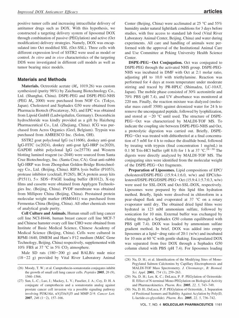
results (Figure 3) showed obvious expression of SSTR2 onthe surface of NCI-H446 and MCF-7, while there was noSSTR2 detected on CHO. Furthermore, NCI-H446 exhibited
(39) Siehler, S.; Seuwen, K.; Hoyer, D. [125I][Tyr3]octreotide labelshuman somatostatin sst2 and sst5 receptors. Eur. J. Pharmacol.1998, 348, 2–3.
(40) Xu, B.; et al. Expression of somatostatin receptor subtype 2 in12 tumor cell lines. Guangdong Med. J. 2006, 27, 3.
(41) Watt, H. L.; Kumar, U. Colocalization of somatostatin receptorsand epidermal growth factor receptors in breast cancer cells.Cancer Cell Int. 2006, 6, 5–23.
Figure 1. Conjugation of Oct to DSPE-PEG-NHS. (A) Amino acid sequence of Oct which has two amino groups (atthe N-terminus and the side chain of Lys). (B) HPLC determination of unconjugated peptide: (a) free Oct in mobilephase and (b) DSPE-PEG-NHS and Oct with a molar ratio of 2:1 in mobile phase after reaction for 4 days. (C) Massspectrum of DSPE-PEG-NHS with a peak at the m/z range around 3000. (D, E) Mass spectrum of DSPE-PEG-Octwith a peak at the m/z range around 4000 and there was no peak around 5000. (F) Mass spectrum ofDSPE-PEG-Oct digested by trypsin with a peak at the m/z range around 3700. MALDI-TOF MS results indicated thatDSPE-PEG-NHS and Oct conjugated at the N-terminus of Oct with a molar ratio of 1:1.
Table 1. Characteristics of Different Formulations ofSterically Stabilized Liposomes (n ) 3)
formulation size (nm) PDI
zetapotential
(mV)
encapsulationefficiency
(%) of DOX
SSL 86.03 ( 0.88 0.23 ( 0.03 -4.86 ( 1.17
SSL-DOX 92.02 ( 2.68 0.22 ( 0.03 -5.27 ( 1.29 97.3 ( 2.03
Oct-SSL 87.56 ( 2.93 0.23 ( 0.03 -4.97 ( 1.24
Oct-SSL-DOX 93.95 ( 1.55 0.24 ( 0.01 -4.70 ( 0.89 96.3 ( 1.08
Figure 2. In vitro release of DOX from liposomes. 0.5mL of liposomes and 0.5 mL of FBS were mixed in adialysis bag (molecular mass cutoff 3500) andincubated at 37 °C in 50 mL of PBS. At predeterminedtime points, 0.5 mL of medium was replaced, and theamount of DOX was quantified as described inMaterials and Methods (n ) 3).
articles Zhang et al.
1164 MOLECULAR PHARMACEUTICS VOL. 7, NO. 4
more than MCF-7. Therefore, the NCI-H446 cell line waspreferred throughout the whole study unless particularlyoutlined.
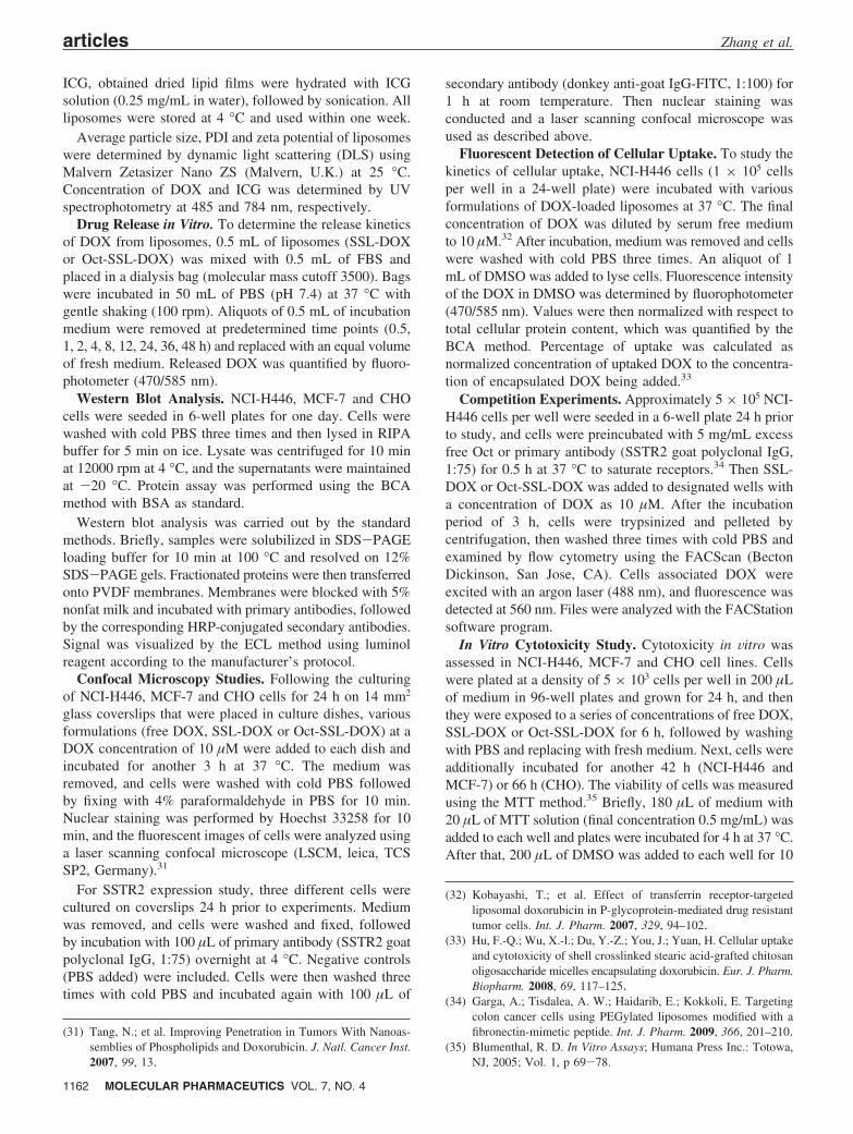
Confocal Microscopy Studies. Figure 4 showed theconfocal microscopy images of NCI-H446, MCF-7 and CHOcells after 3 h incubation with SSL-DOX, Oct-SSL-DOX orfree DOX at 37 °C. Without the release process, free DOXdirectly penetrated into cells through the membrane diffusion,leading to a greater amount of intracellular accumulation,which was taken as positive control (C1, C2 and C3). ForNCI-H446, the images of the Oct-SSL-DOX group (B1)displayed more red fluorescence of DOX than that of theSSL-DOX one (A1), suggesting the favorable effect of Octon the cellular uptake of liposomes. A similar result wasobserved on MCF-7 (A2, B2), while the difference betweenthe two liposomal groups was minor compared with NCI-H446, likely due to their difference in SSTR2 expressionlevel. In terms of CHO (A3, B3), there was no red
fluorescence in both passive and active targeting liposomegroups, proving no expression of SSTR2 on the cells andno targeting effect of Oct to this cell line.
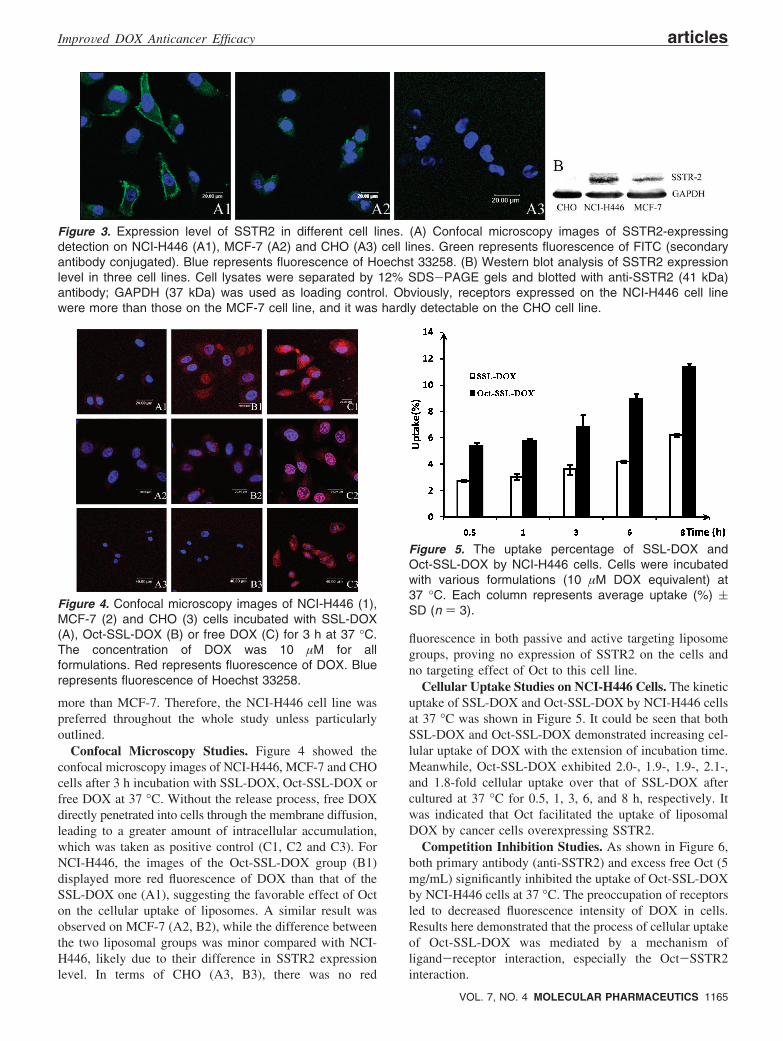
Cellular Uptake Studies on NCI-H446 Cells. The kineticuptake of SSL-DOX and Oct-SSL-DOX by NCI-H446 cellsat 37 °C was shown in Figure 5. It could be seen that bothSSL-DOX and Oct-SSL-DOX demonstrated increasing cel-lular uptake of DOX with the extension of incubation time.Meanwhile, Oct-SSL-DOX exhibited 2.0-, 1.9-, 1.9-, 2.1-,and 1.8-fold cellular uptake over that of SSL-DOX aftercultured at 37 °C for 0.5, 1, 3, 6, and 8 h, respectively. Itwas indicated that Oct facilitated the uptake of liposomalDOX by cancer cells overexpressing SSTR2.
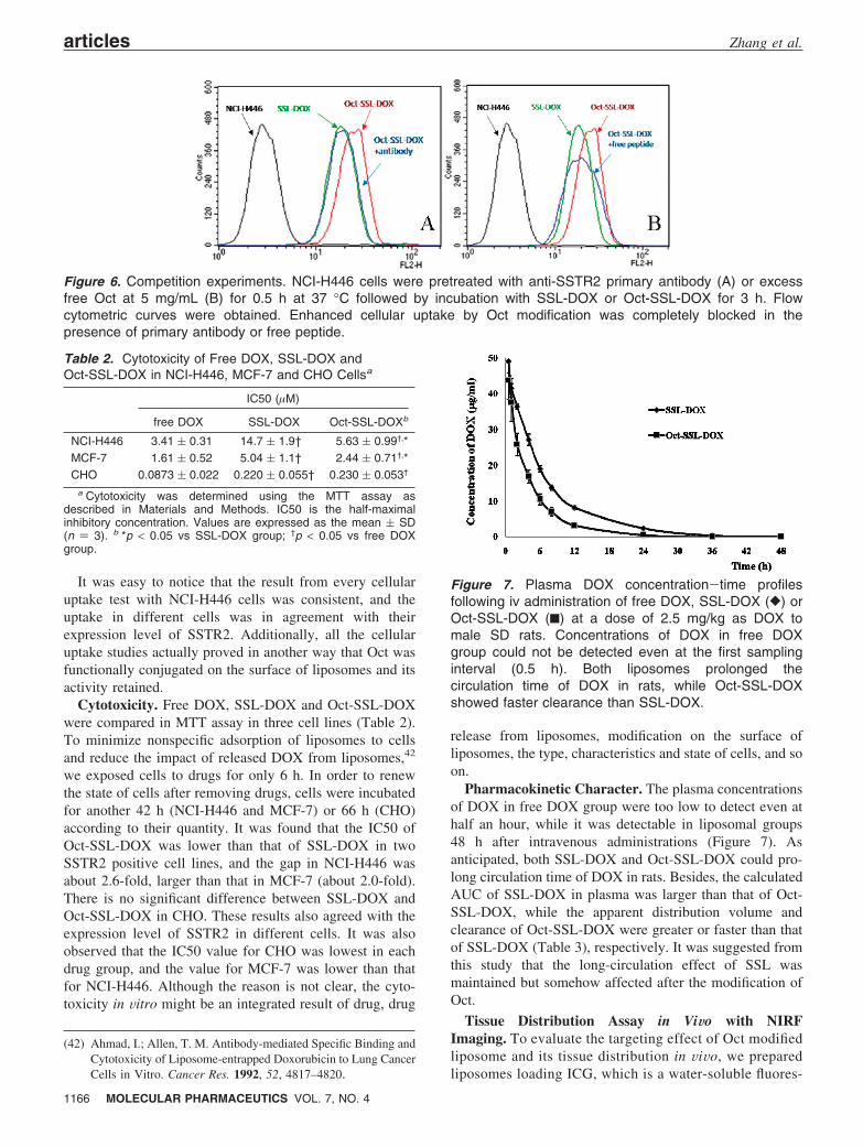
Competition Inhibition Studies. As shown in Figure 6,both primary antibody (anti-SSTR2) and excess free Oct (5mg/mL) significantly inhibited the uptake of Oct-SSL-DOXby NCI-H446 cells at 37 °C. The preoccupation of receptorsled to decreased fluorescence intensity of DOX in cells.Results here demonstrated that the process of cellular uptakeof Oct-SSL-DOX was mediated by a mechanism ofligand-receptor interaction, especially the Oct-SSTR2interaction.
Figure 3. Expression level of SSTR2 in different cell lines. (A) Confocal microscopy images of SSTR2-expressingdetection on NCI-H446 (A1), MCF-7 (A2) and CHO (A3) cell lines. Green represents fluorescence of FITC (secondaryantibody conjugated). Blue represents fluorescence of Hoechst 33258. (B) Western blot analysis of SSTR2 expressionlevel in three cell lines. Cell lysates were separated by 12% SDS-PAGE gels and blotted with anti-SSTR2 (41 kDa)antibody; GAPDH (37 kDa) was used as loading control. Obviously, receptors expressed on the NCI-H446 cell linewere more than those on the MCF-7 cell line, and it was hardly detectable on the CHO cell line.
Figure 4. Confocal microscopy images of NCI-H446 (1),MCF-7 (2) and CHO (3) cells incubated with SSL-DOX(A), Oct-SSL-DOX (B) or free DOX (C) for 3 h at 37 °C.The concentration of DOX was 10 µM for allformulations. Red represents fluorescence of DOX. Bluerepresents fluorescence of Hoechst 33258.
Figure 5. The uptake percentage of SSL-DOX andOct-SSL-DOX by NCI-H446 cells. Cells were incubatedwith various formulations (10 µM DOX equivalent) at37 °C. Each column represents average uptake (%) (SD (n ) 3).
ImproVed DOX Anticancer Efficacy articles
VOL. 7, NO. 4 MOLECULAR PHARMACEUTICS 1165
It was easy to notice that the result from every cellularuptake test with NCI-H446 cells was consistent, and theuptake in different cells was in agreement with theirexpression level of SSTR2. Additionally, all the cellularuptake studies actually proved in another way that Oct wasfunctionally conjugated on the surface of liposomes and itsactivity retained.
Cytotoxicity. Free DOX, SSL-DOX and Oct-SSL-DOXwere compared in MTT assay in three cell lines (Table 2).To minimize nonspecific adsorption of liposomes to cellsand reduce the impact of released DOX from liposomes,42
we exposed cells to drugs for only 6 h. In order to renewthe state of cells after removing drugs, cells were incubatedfor another 42 h (NCI-H446 and MCF-7) or 66 h (CHO)according to their quantity. It was found that the IC50 ofOct-SSL-DOX was lower than that of SSL-DOX in twoSSTR2 positive cell lines, and the gap in NCI-H446 wasabout 2.6-fold, larger than that in MCF-7 (about 2.0-fold).There is no significant difference between SSL-DOX andOct-SSL-DOX in CHO. These results also agreed with theexpression level of SSTR2 in different cells. It was alsoobserved that the IC50 value for CHO was lowest in eachdrug group, and the value for MCF-7 was lower than thatfor NCI-H446. Although the reason is not clear, the cyto-toxicity in Vitro might be an integrated result of drug, drug
release from liposomes, modification on the surface ofliposomes, the type, characteristics and state of cells, and soon.
Pharmacokinetic Character. The plasma concentrationsof DOX in free DOX group were too low to detect even athalf an hour, while it was detectable in liposomal groups48 h after intravenous administrations (Figure 7). Asanticipated, both SSL-DOX and Oct-SSL-DOX could pro-long circulation time of DOX in rats. Besides, the calculatedAUC of SSL-DOX in plasma was larger than that of Oct-SSL-DOX, while the apparent distribution volume andclearance of Oct-SSL-DOX were greater or faster than thatof SSL-DOX (Table 3), respectively. It was suggested fromthis study that the long-circulation effect of SSL wasmaintained but somehow affected after the modification ofOct.
Tissue Distribution Assay in ViWo with NIRFImaging. To evaluate the targeting effect of Oct modifiedliposome and its tissue distribution in ViVo, we preparedliposomes loading ICG, which is a water-soluble fluores-
(42) Ahmad, I.; Allen, T. M. Antibody-mediated Specific Binding andCytotoxicity of Liposome-entrapped Doxorubicin to Lung CancerCells in Vitro. Cancer Res. 1992, 52, 4817–4820.
Figure 6. Competition experiments. NCI-H446 cells were pretreated with anti-SSTR2 primary antibody (A) or excessfree Oct at 5 mg/mL (B) for 0.5 h at 37 °C followed by incubation with SSL-DOX or Oct-SSL-DOX for 3 h. Flowcytometric curves were obtained. Enhanced cellular uptake by Oct modification was completely blocked in thepresence of primary antibody or free peptide.
Table 2. Cytotoxicity of Free DOX, SSL-DOX andOct-SSL-DOX in NCI-H446, MCF-7 and CHO Cellsa
IC50 (µM)
free DOX SSL-DOX Oct-SSL-DOXb
NCI-H446 3.41 ( 0.31 14.7 ( 1.9† 5.63 ( 0.99†,*MCF-7 1.61 ( 0.52 5.04 ( 1.1† 2.44 ( 0.71†,*CHO 0.0873 ( 0.022 0.220 ( 0.055† 0.230 ( 0.053†
a Cytotoxicity was determined using the MTT assay asdescribed in Materials and Methods. IC50 is the half-maximalinhibitory concentration. Values are expressed as the mean ( SD(n ) 3). b *p < 0.05 vs SSL-DOX group; †p < 0.05 vs free DOXgroup.
Figure 7. Plasma DOX concentration-time profilesfollowing iv administration of free DOX, SSL-DOX ([) orOct-SSL-DOX (9) at a dose of 2.5 mg/kg as DOX tomale SD rats. Concentrations of DOX in free DOXgroup could not be detected even at the first samplinginterval (0.5 h). Both liposomes prolonged thecirculation time of DOX in rats, while Oct-SSL-DOXshowed faster clearance than SSL-DOX.
articles Zhang et al.
1166 MOLECULAR PHARMACEUTICS VOL. 7, NO. 4

cence dye with EX 795 nm and EM 835 nm (DOX wasnot considered as optional dye here for its EX 488 nmwas not suitable for NIRF imaging). And the ICGliposomes were first tested here in the NIRF imagingstudy. The use of ICG as fluorescence dye but not lipid-soluble ones like Cy5 series is based on the existence ofOct. Activated lipid-soluble dyes could label liposomesvia covalent bond with activated NH2 group of DSPE-PEG, but such a group also exists in the active site ofOct. Therefore, water-soluble dyes like ICG were pre-ferred, which was commonly used clinically in angiog-raphy, and reported as label reagent in inhalant powder43
and nanoparticles.44,45 The ICG liposomes prepared herewere 100-110 nm and negatively charged on the surfacewith a loading efficiency of ICG greater than 95%.
During the living imaging test, the fluorescence signalsin whole bodies of mice were relatively weak (data notshow). However, further experiment with excised organs(Figure 8) clearly demonstrated that most of the ICGaccumulated in liver 3 h after iv administration of variousICG formulations. Both liposomal formulations increasedthe fluorescence in liver, spleen, kidneys and lung, andthe strongest fluorescence was found in the SSL-ICGgroup. Similar results can be seen in Figure 8B. Consider-ing the liver-specific distribution of ICG itself and RESuptake of particulates in circulation, these data seemedreasonable and were in accordance with the pharmacoki-netics assay.
For the tumor accumulation, it was found that activetargeting liposomes enhanced the ICG distribution intumor compared to passive vesicles as shown in Figure8A, although the signal in tumor was relatively weak.More clear images could be seen in Figure 8C. The ICGfluorescence in the Oct-SSL-ICG group was the strongesteven 9 h after dosing, stronger than that of SSL-ICG ateach time point, while there was already no signal detected
at 9 h in the free ICG group. In this way, it was provedin ViVo that Oct-SSL enhanced targeting delivery of DOXto related tumor tissue.
Antitumor Effect in ViWo. Figure 9 shows the PD ofvarious DOX formulations on nude mice bearing NCI-H446cancer xenografts. It was demonstrated that the tumor growthinhibition efficacy of Oct-SSL-DOX was better than that ofSSL-DOX (P < 0.05). The strongest antitumor effect of Oct-SSL-DOX revealed its highest targeting efficiency. FreeDOX showed nearly the same efficacy with the SSL-DOXgroup, similar to the previous reports,3 suggesting nocorrelation between PK and PD, and also the necessity ofactive targeting. Besides, it was indicated that the antitumorefficacy in ViVo for Oct-SSL-DOX and SSL-DOX wasconsistent with the drug distribution in tumor and cellexperiments in Vitro.
ConclusionsA novel DOX-loaded and Oct-modified SSL was prepared
and studied here. Various investigations in cell level achievedsimilar results, namely, the active SSL exhibited higher celluptake than that of passive SSL in SSTR2-positive cells. Themechanism was proved to be SSTR2 mediated endocytosis.Besides, the Oct modification on the SSL showed littleimpact on the in Vitro properties of SSL, however it reducedthe circulation time of loaded-DOX to some extent in rats,increased cytotoxicity in SSTR2-positive tumor cells, en-
(43) Mizuno, T.; Mohri, K.; Nasu, S.; Danjo, K.; Okamoto, H. Dualimaging of pulmonary delivery and gene expression of dry powderinhalant by fluorescence and bioluminescence. J. ControlledRelease 2009, 134, 149–154.
(44) Kirchherr, A.-K.; Briel, A.; Mader, K. Stabilization of IndocyanineGreen by Encapsulation within Micellar Systems. Mol. Pharma-ceutics 2009, 6, 2, 480–491.
(45) Ohashi, K.; Kabasawa, T.; Ozeki, T.; Okada, H. One-steppreparation of rifampicin/poly(lactic-co-glycolic acid) nanopar-ticle-containing mannitol microspheres using a four-fluid nozzlespray drier for inhalation therapy of tuberculosis. J. ControlledRelease 2009, 135, 19–24.
Table 3. Main Pharmacokinetic Parameters in Plasma after Intravenous Administration of SSL-DOX or Oct-SSL-DOX toRatsa
formulation AUC ((µg/mL)h) k (1/h) T1/2 (h) Vd (mL) Cl (mL/h)
SSL-DOX 384 ( 13 0.11 ( 0.016 6.3 ( 0.96 8.30 ( 1.5 0.91 ( 0.032Oct-SSL-DOX 219 ( 34** 0.15 ( 0.010* 4.7 ( 0.31** 11.4 ( 3.5** 1.7 ( 0.56**
a Each preparation was dosed at 2.5 mg/kg as DOX. AUC, area under the plasma concentration-time curve; k, elimination rate constant;T1/2, plasma half-life; Vd, apparent distribution volume; and Cl, clearance, were calculated based on one-compartment model. Results areexpressed as the mean ( SD. *p < 0.01 and **p < 0.05 vs SSL-DOX group.
Figure 8. NIRF imaging of main organs at 3 h (A) afteriv injection of ICG formulations; and at various timepoints (B), including excised tumors. (C) The color onimages represents fluorescence from ICG.
ImproVed DOX Anticancer Efficacy articles
VOL. 7, NO. 4 MOLECULAR PHARMACEUTICS 1167
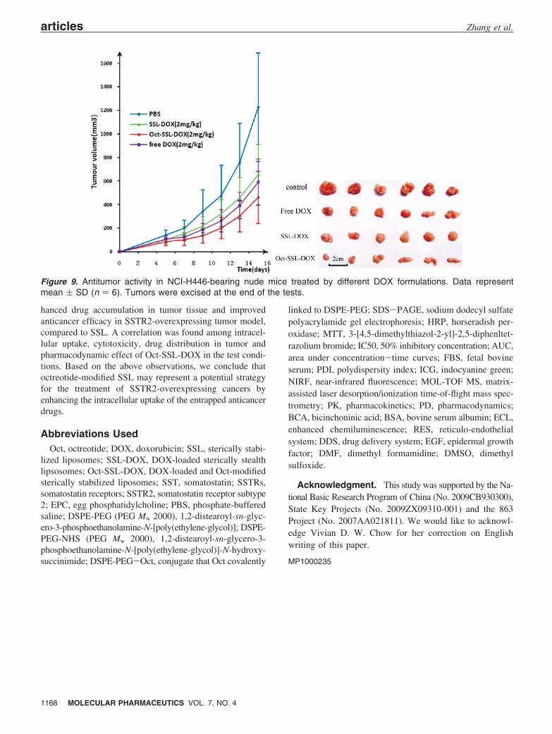
hanced drug accumulation in tumor tissue and improvedanticancer efficacy in SSTR2-overexpressing tumor model,compared to SSL. A correlation was found among intracel-lular uptake, cytotoxicity, drug distribution in tumor andpharmacodynamic effect of Oct-SSL-DOX in the test condi-tions. Based on the above observations, we conclude thatoctreotide-modified SSL may represent a potential strategyfor the treatment of SSTR2-overexpressing cancers byenhancing the intracellular uptake of the entrapped anticancerdrugs.
Abbreviations UsedOct, octreotide; DOX, doxorubicin; SSL, sterically stabi-
lized liposomes; SSL-DOX, DOX-loaded sterically stealthlipsosomes; Oct-SSL-DOX, DOX-loaded and Oct-modifiedsterically stabilized liposomes; SST, somatostatin; SSTRs,somatostatin receptors; SSTR2, somatostatin receptor subtype2; EPC, egg phosphatidylcholine; PBS, phosphate-bufferedsaline; DSPE-PEG (PEG Mw 2000), 1,2-distearoyl-sn-glyc-ero-3-phosphoethanolamine-N-[poly(ethylene-glycol)]; DSPE-PEG-NHS (PEG Mw 2000), 1,2-distearoyl-sn-glycero-3-phosphoethanolamine-N-[poly(ethylene-glycol)]-N-hydroxy-succinimide; DSPE-PEG-Oct, conjugate that Oct covalently
linked to DSPE-PEG; SDS-PAGE, sodium dodecyl sulfatepolyacrylamide gel electrophoresis; HRP, horseradish per-oxidase; MTT, 3-[4,5-dimethylthiazol-2-yl]-2,5-diphenltet-razolium bromide; IC50, 50% inhibitory concentration; AUC,area under concentration-time curves; FBS, fetal bovineserum; PDI, polydispersity index; ICG, indocyanine green;NIRF, near-infrared fluorescence; MOL-TOF MS, matrix-assisted laser desorption/ionization time-of-flight mass spec-trometry; PK, pharmacokinetics; PD, pharmacodynamics;BCA, bicinchoninic acid; BSA, bovine serum albumin; ECL,enhanced chemiluminescence; RES, reticulo-endothelialsystem; DDS, drug delivery system; EGF, epidermal growthfactor; DMF, dimethyl formamidine; DMSO, dimethylsulfoxide.
Acknowledgment. This study was supported by the Na-tional Basic Research Program of China (No. 2009CB930300),State Key Projects (No. 2009ZX09310-001) and the 863Project (No. 2007AA021811). We would like to acknowl-edge Vivian D. W. Chow for her correction on Englishwriting of this paper.
MP1000235
Figure 9. Antitumor activity in NCI-H446-bearing nude mice treated by different DOX formulations. Data representmean ( SD (n ) 6). Tumors were excised at the end of the tests.
articles Zhang et al.
1168 MOLECULAR PHARMACEUTICS VOL. 7, NO. 4


![Pharmacokinetics Octreotide Hypertension; Relationship ...quently, octreotide has a muchlonger circulat-ing half-life than somatostatin in healthy volunteers [4, 5]. In normal healthy](https://img.pdfslide.us/doc/110x75/60e40bd6a8bffe3dd6583b84/pharmacokinetics-octreotide-hypertension-relationship-quently-octreotide-has.jpg)


![Non-peptide ligands in the characterization of peptide ... · has been shown for the somatostatin analogue octreotide [38]. However, this application route appears not suitable for](https://img.pdfslide.us/doc/110x75/5e6ab21500d45501f147a942/non-peptide-ligands-in-the-characterization-of-peptide-has-been-shown-for-the.jpg)