Embed Size (px)
Citation preview
Scientia Iranica F (2018) 25(3), 1815{1823
Sharif University of TechnologyScientia Iranica
Transactions F: Nanotechnologyhttp://scientiairanica.sharif.edu
A novel nano-composite sca�old for cartilage tissueengineering
M. Mehdikhania, E. Tavakolia, A. Zargar-Kharazib;�,and B. Hashemibenic
a. Department of Biomedical Engineering, Faculty of Engineering, University of Isfahan, Isfahan, P.O. Box 8174613441, Iran.b. Department of Biomaterials, Nanaotechnology and Tissue Engineering, School of Advanced Technology in Medicine, Isfahan
University of Medical Sciences, Isfahan, P.O. Box 8174673461, Iran.c. Department of Anatomical Sciences, School of Medicine, Isfahan University of Medical Sciences, Isfahan, P.O. Box 8174673461,
Iran.
Received 12 December 2016; received in revised form 16 August 2017; accepted 16 October 2017
KEYWORDSOsteoarthritis (OA);Human Adipose-Derived MesenchymalStem Cells(hADMSCs);PLGA;Tissue engineering;Hybrid sca�olds.
Abstract. In this study, hybrid Poly (lactic-co-glycolic acid) (PLGA)/Hyaluronic Acid(Ha)/Fibrin/45S Bioactive Glass (45SBG) nanocomposite sca�olds seeded with humanAdipose-Derived Mesenchymal Stem Cells (hADMSCs) were investigated as a construct forosteoarthritis (OA), Articular Cartilage (AC), and subchondral bone defects therapies. Thebioactivity and biodegradation of the nanocomposite sca�olds were assessed in SimulatedBody Fluid (SBF) and Phosphate Bu�er Saline (PBS) solution, respectively. Furthermore,MTT analysis was performed in order to determine attachment and viability of hADMSCs.Ultimately, results indicate the increase of bioactivity in nanocomposite sca�olds, ascompared to the pure PLGA sca�old. In addition, biodegradation assay exhibits thatthe addition of Ha, �brin, and 45SBG nanoparticles could modify the degradation rate ofPLGA. The nanocomposite sca�olds did not exhibit any cytotoxicity, and the hADMSCswere attached to the sca�olds , which proliferate properly. According to our investigation,it was concluded that using natural and synthetic polymers along with BG nanoparticlesmay provide a suitable construct and could show a bene�cial role in AC tissue engineeringand OA therapy.© 2018 Sharif University of Technology. All rights reserved.
1. Introduction
Osteoarthritis (OA) is a rampant disease in industrialsocieties imposing more than $30 billion a year totheir therapeutic budget [1]. Articular Cartilage (AC)lesions have a con�ned capacity to reform following in-jury, trauma, and pathological diseases [2]. Numeroustreatment strategies have been utilized to repair trau-
*. Corresponding authors. Tel.: 03137923857E-mail address: a [email protected] (A.Zargar-Kharazi)
doi: 10.24200/sci.2017.4595
matic cartilage defects; however, in these techniques,�brocartilage formation, immune rejection, pathogentransmission, donor site morbidity, and slower remod-elling is not pending away [1-3]. Tissue Engineering(TE) and regenerative medicine are new approachesthat can promote cartilage regeneration and transcendthe previous methods limitations [4]. TE with threebasic elements, including cells, biodegradable sca�olds,and growth factors, strives to compensate the per-formance of the damaged tissues and organs [5,6].The sca�olds have a pivotal role in regeneration oftissues and provide simulated environment similar tothe Extra Cellular Matrix (ECM) for cell migration,adhesion, and proliferation. In order to design a
1816 M. Mehdikhani et al./Scientia Iranica, Transactions F: Nanotechnology 25 (2018) 1815{1823
successful structure for regeneration of cartilage tissuedisorders, signi�cant parameters include biocompati-bility, biodegradability, interconnected porous struc-ture, su�cient mechanical properties, adequate abilityto cell attachment, proliferation, di�erentiation, non-cytotoxicity, and non-antigenicity must be consid-ered [7]. Native biological materials (e.g., hyaluronicacid (Ha), �brin, etc.) and synthetic polymeric mate-rials are the two biggest groups of material suitable forforming sca�olds [8]. Poly (Lactic-co-Glycolic Acid)(PLGA) is an approved synthetic polymer materialby the US Food and Drug Administration (FDA)due to its biocompatibility, biodegradability, facilebiodegradation, and good mechanical properties. Withits plausible biocompatibility property, PLGA is widelyused in bone and cartilage TE applications. However,limitation on cell adhesion and high price are twodrawbacks of PLGA as a biomaterial [6-9]. Despitelow cell attachment a�nity of PLGA, using othermaterials, such as bioceramics (e.g., Bioactive Glasses(BGs) nanoparticle) as �llers/reinforcement compo-nents in PLGA matrix, could provide a distinguishedsurface in order to improve cellular interaction andintensify its application in hard tissue regeneration [10-12]. Hyaluronic acid (Ha) is a kind of native biologicalmaterials that can be found in synovial uid with ex-cellent ability to support chondrocytes and Mesenchy-mal Stem Cells (MSCs), noticeable biocompatibility,biodegradability, di�erentiated cell phenotype, well cellexpansion, and little toxicity that can be used in softand hard tissues [6,13]. Researchers have exhibitedthat the hybrid sca�olds, containing Ha and the othernatural materials (e.g., collagen and �brin), can restoreAC performance and are an e�ective way for AC reha-bilitation [14,15]. Fibrin is a blood derivative that waswidely used in clinical operations as �brin glue. From acell delivery behaviour's point of view, �brin has beenutilized with Mesenchymal Stem Cells (MSCs) for softand hard tissues (e.g., ligament, bone and cartilage).Meanwhile, because it su�ers from weak mechanicalproperties and short biodegradation time, TE applica-tions have been limited [6,16,17]. Fibrin applicationfor immobilizing cells and providing homogenous cellsdistribution in PLGA sca�olds have displayed chon-drocytes' proliferation, high level of cartilage-speci�cproteins secretion and promotion, and the cartilaginoustissue formation [18]. In recent years, researchershave fabricated hybrid nanocomposite sca�olds fromsynthetically and naturally derived polymeric materialsdue to high regeneration potential in the context ofregenerative medicine [10,19]. Nanocomposite sca�oldsare a nano-scale version of a conventional compos-ite, where nanoparticles are dispersed in a polymermatrix [20]. Recently, synthesized Bioactive Glass(BG) nanoparticles, such as 45S, with high biologicalactivity, have been widely used as a �ller or coating
with polymers in order to regenerate tissues such asbone, lungs, cartilage, and other organs [21]. Previousresearches have demonstrated the eminent role of BGsin subchondral and cartilage regeneration [22-25]. Untilnow, several techniques have been used to confectporous sca�olds, such as Thermal-Induced Phase Sep-aration (TIPS), electrospinning, gas foaming, solventcasting and particulate leaching (SC/PL), and somecombined or modi�ed processes of the above [10,26].The objective of this study, accordingly, is to preparenanocomposite sca�olds containing PLGA, Ha, �brin,and 45SBG nanoparticles with SC/ PL techniques andassessment of biological behaviour of hADMSCs on thesurface of these sca�olds as a candidate for cartilage TEapplications. Likewise, a correlation among Ha, �brin,and 45SBG nanoparticles' contents will be ascertained.
2. Materials and methods
2.1. Nanocomposite porous sca�oldsfabrication
Nanocomposite sca�olds with di�erent amounts ofHa, �brin, and 45SBG nanoparticles were made viaSC/PL techniques, according to our previous workelsewhere [10]. Brie y, PLGA powder (RESOMER®
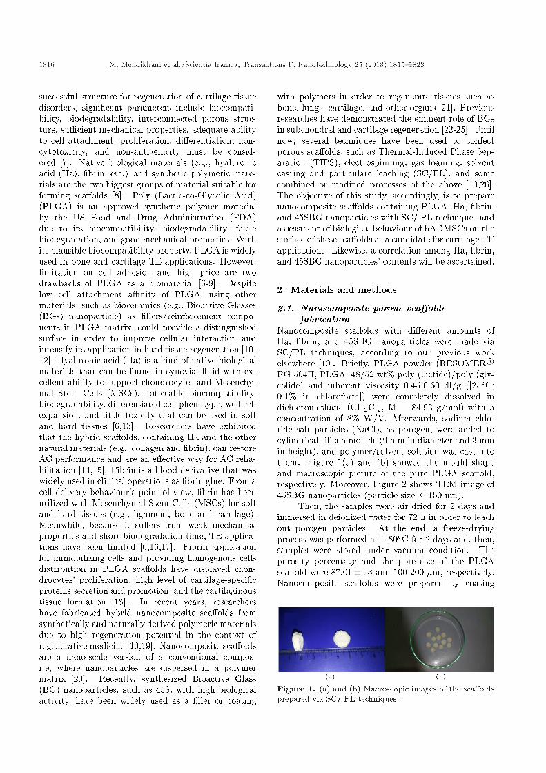
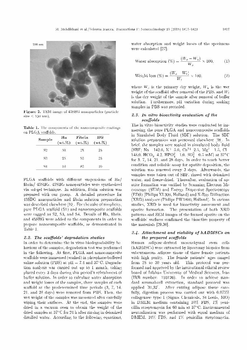
RG 504H, PLGA; 48/52 wt% poly (lactide)/poly (gly-colide) and inherent viscosity 0.45-0.60 dl/g ([25�C;0.1% in chloroform]) were completely dissolved indichloromethane (CH2Cl2, M = 84.93 g/mol) with aconcentration of 8% W/V. Afterwards, sodium chlo-ride salt particles (NaCl), as porogen, were added tocylindrical silicon moulds (9 mm in diameter and 3 mmin height), and polymer/solvent solution was cast intothem. Figure 1(a) and (b) showed the mould shapeand macroscopic picture of the pure PLGA sca�old,respectively. Moreover, Figure 2 shows TEM image of45SBG nanoparticles (particle size � 150 nm).
Then, the samples were air-dried for 2 days andimmersed in deionized water for 72 h in order to leachout porogen particles. At the end, a freeze-dryingprocess was performed at �80�C for 2 days and, then,samples were stored under vacuum condition. Theporosity percentage and the pore size of the PLGAsca�old were 87:01� 03 and 100-200 �m, respectively.Nanocomposite sca�olds were prepared by coating
Figure 1. (a) and (b) Macroscopic images of the sca�oldsprepared via SC/ PL techniques.
M. Mehdikhani et al./Scientia Iranica, Transactions F: Nanotechnology 25 (2018) 1815{1823 1817
Figure 2. TEM image of 45SBG nanoparticles (particlesize � 150 nm).
Table 1. The components of the nanocomposite coatingson PLGA sca�olds.
Sample Ha(wt.%)
Fibrin(wt.%)
BG(wt.%)
S2 50 25 25
S3 25 50 25
S4 10 80 10
PLGA sca�olds with di�erent suspensions of Ha/�brin/ 45SBG. 45SBG nanoparticles were synthesizedvia sol-gel technique. In addition, �brin solution wasprepared with our group. A detailed procedure for45SBG nanoparticles and �brin solution preparationwas described elsewhere [10]. For the sake of simplicity,pure PLGA sca�old (S1) and nanocomposite sca�oldswere tagged as S2, S3, and S4. Details of Ha, �brin,and 45SBG were added to the components in order toprepare nanocomposite sca�olds, as demonstrated inTable 1.
2.2. The sca�olds' degradation studiesIn order to determine the in vitro biodegradability be-haviour of the samples, degradation test was performedin the following. At �rst, PLGA and nanocompositesca�olds were immersed (soaked) in phosphate-bu�eredsaline solution (PBS) at pH = 7.4 and 37�C. Degrada-tion analysis was carried out up to 1 month, takingplaced every 3 days during this period's refreshment ofbu�er solution. In order to calculate water absorptionand weight losses of the samples, three samples of eachsca�old at the predetermined time periods (3, 7, 14,21, and 28 days) were removed from PBS. Then, thewet weight of the samples was measured after carefullywiping their surfaces. At the end, the samples weredried in a vacuum oven to obtain the weight of thedried samples at 37�C for 24 h after rinsing in deionizeddistilled water. According to the following equations,
water absorption and weight losses of the specimenswere calculated [27]:
Water absorption (%) =(Wo �Wa)
Wo� 100; (1)
Weight loss (%) =(Wo �Wt)
Wt� 100; (2)
where Wo is the primary dry weight, Wa is the wetweight of the sca�old after removal of the PBS, and Wtis the dry weight of the sample after removal of bu�ersolution. Furthermore, pH variation during soakingsamples in PBS was recorded.
2.3. In vitro bioactivity evaluation of thesca�olds
The in vitro bioactivity studies were conducted by im-mersing the pure PLGA and nanocomposite sca�oldsin Simulated Body Fluid (SBF) solution. The SBFsolution preparation was presented elsewhere [28]. Inbrief, the samples were soaked in simulated body uid(SBF; Na+ 142.0, K+ 5.0, Ca2+ 2.5, Mg2+ 1.5, Cl�148.0, HCO�3 4.2, HPO2�
4 1.0, SO2�4 0.5 mM) at 37�C
for 3, 7, 14, 21, and 28 days. In order to reach bettercondition and reliable assay for apatite deposition, thesolution was renewed every 3 days. Afterwards, thesamples were taken out of SBF, rinsed with deionizedwater, and freeze-dried. Thereafter, evaluation of ap-atite formation was veri�ed by Scanning Electron Mi-croscopy (SEM) and Energy Dispersive Spectroscopy(EDS) (Philips XL300, Holland) and X-Ray Di�raction(XRD) analyzer (Philips PW1800, Holland). In variousstudies, XRD is used for bioactivity assessment andapatite formation. The presentation of both XRDpatterns and SEM images of the formed apatite on thesca�olds' surfaces con�rmed the bioactive property ofthe materials [29,30].
2.4. Attachment and viability of hADMSCs onthe prepared sca�olds
Human adipose-derived mesenchymal stem cells(hADMSCs) were extracted by lipectomy biopsies fromsubcutaneous adipose tissue of three female patientswith high purity. The female patients' ages rangedfrom 28 to 30 years old. This protocol was per-formed and approved by the institutional ethical reviewboard of Isfahan University of Medical Sciences, Iran(IRB number: 193126). In order to achieve stan-dard stromal-cell extraction, standard protocol wasapplied [31,32]. After cutting adipose tissue care-fully, digestion process was carried out with 0.075%collagenase type I (Sigma Chemicals, St-Louis, MO)in DMEM medium containing 10% FBS, 1% peni-cillin streptomycin for 60 min at 37�C. Environmentalneutralization was performed with equal medium ofDMEM, 10% FBS, and 1% penicillin streptomycin.
1818 M. Mehdikhani et al./Scientia Iranica, Transactions F: Nanotechnology 25 (2018) 1815{1823
Afterwards, the medium was cast in 50 cc falcon tube,and centrifugation process was performed for 10 minwith 1400 rpm. Then, oating adipocytes were scrapedand added DMEM, 10% FBS to the cells depositedsolution. The cells' suspension was transformed intothe culture asks and kept at 37�C, 5% CO2 conditions.To achieve high con uence of the cells and apply it thein vitro study, suspension was passaged three times andthe culture medium exchange was done every 2 days.In order to calculate cells' attachment and viability,samples were sterilized with UV radiation for 2 h andsoaked in 70% ethanol overnight. A de�nite numberof the cells were seeded onto the sterile samples. Forcells' attachment and viability determination, sterilesamples were transformed into six multi-wall platesand, then, seeded at a cell density of 1� 104 cells/cm2
in a complete culture medium. Cell culture plateswere utilized as negative control. For each sample,100-microliter cell suspension was added to each welland allowed to be attached �rmly on the surfacesof the samples. Then, 3 ml of culture medium wasadded to every samples. Finally, after determiningincubation times (3 days), a rinsing process was carriedout in order to omit non-adherent cells from eachwell. In order to determine viable cells, MTT (3-(4, 5-dimethylthiazol-2-yl)-2, 5-diphenyl tetrazoliumbromide) assay was performed. A 2 mg/ml stocksolution of MTT in PBS was prepared and sterilizedby Millipore �ltration and kept in a dark conditionbefore any tests. Afterwards, a certain amount ofMTT solution was cast into each well, and the plateswere transformed into the incubator for 3 h. Then,supernatant liquid on the surface of wells was removed.In order to dissolve the formazan crystals, dimethylsulfoxide (DMSO) was added to each well under slightshaking. At the end, solution was transformed in96 well plates and optical density was calculated byELISA plate reader (Hyperion, Florida, USA) at 540nm. The amount of absorption indicates the numberof viable cells attached to each sample. Our resultswere recorded and reported as optical density values.Morphology of the cells incubated for 3 days wasobserved using SEM. Cells adhering to the substrateswere washed with PBS. Subsequently, the cells were�xed with 2.5% glutaraldehyde in PBS for 1 h at 4�C.The specimens, after being thoroughly washed withPBS, were dehydrated using graded ethanol changes,gold splattered in vacuum, and examined using SEM.
3. Results and discussion
3.1. In vitro biodegradation properties of thesca�olds
The in vitro degradation of pure PLGA and nanocom-posite sca�olds was studied in PBS at 37�C. Degrada-tion was carried out by measuring water absorption,
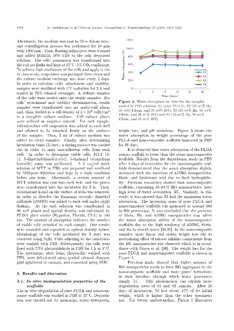
Figure 3. Water absorption vs. time for the samplessoaked in PBS solution: S1 (pure PLGA), S2 (50 wt% Ha,25 wt% Fibrin, and 25 wt% BG), S3 (25 wt% Ha, 50 wt%Fibrin, and 25 wt% BG) and S4 (10 wt% Ha, 80 wt%Fibrin, and 10 wt% BG).
weight loss, and pH variations. Figure 3 shows thewater absorption in weight percentage of the purePLGA and nanocomposite sca�olds immersed in PBSfor 28 days.
It is observed that water absorption of the PLGAporous sca�old is lower than the other nanocompositesca�olds. Results from the degradation study in PBSafter 3 days of immersion for the nanocomposite scaf-folds demonstrated that the water absorption slightlyincreased with the insertion of 45SBG nanoparticles,�brin, and hyaluronic acid due to their hydrophilic-ity. Previous researches showed that nanocompositesca�olds, containing 10 wt.% BG nanoparticles, havehigh level of water absorption [33]. Similarly, in thisstudy, it was showed that S3 had the maximum waterabsorption. The increasing mass of pure PLGA andnanocomposite sca�olds was measured at around 600to 900 percentage. It was concluded that the presenceof �brin, Ha, and 45SBG nanoparticles may a�ectthe water absorption ability of the nanocompositessca�olds due to the high tendency of 45SBG, �brin,and Ha to absorb water [34,35]. In the nanocompositesamples, more linear and slower weight loss due toneutralizing e�ect of release alkaline components fromthe BG nanoparticles was observed which is in accor-dance with Orava et al. [36]. The weight loss for thepure PLGA and nanocomposite sca�olds is shown inFigure 4.
Previous study showed that higher amount ofBG nanoparticles tends to form BG aggregates in thenanocomposite sca�olds and more pores' formationsin their interface through which water penetratessimply [11]. This phenomenon can explain moredegradation rates of S2 and S3 samples. After 28days of incubation, S3 lost about 12% of its initialweight, which is higher than the other specimensare. For better understanding, Figure 5 illustrates
M. Mehdikhani et al./Scientia Iranica, Transactions F: Nanotechnology 25 (2018) 1815{1823 1819
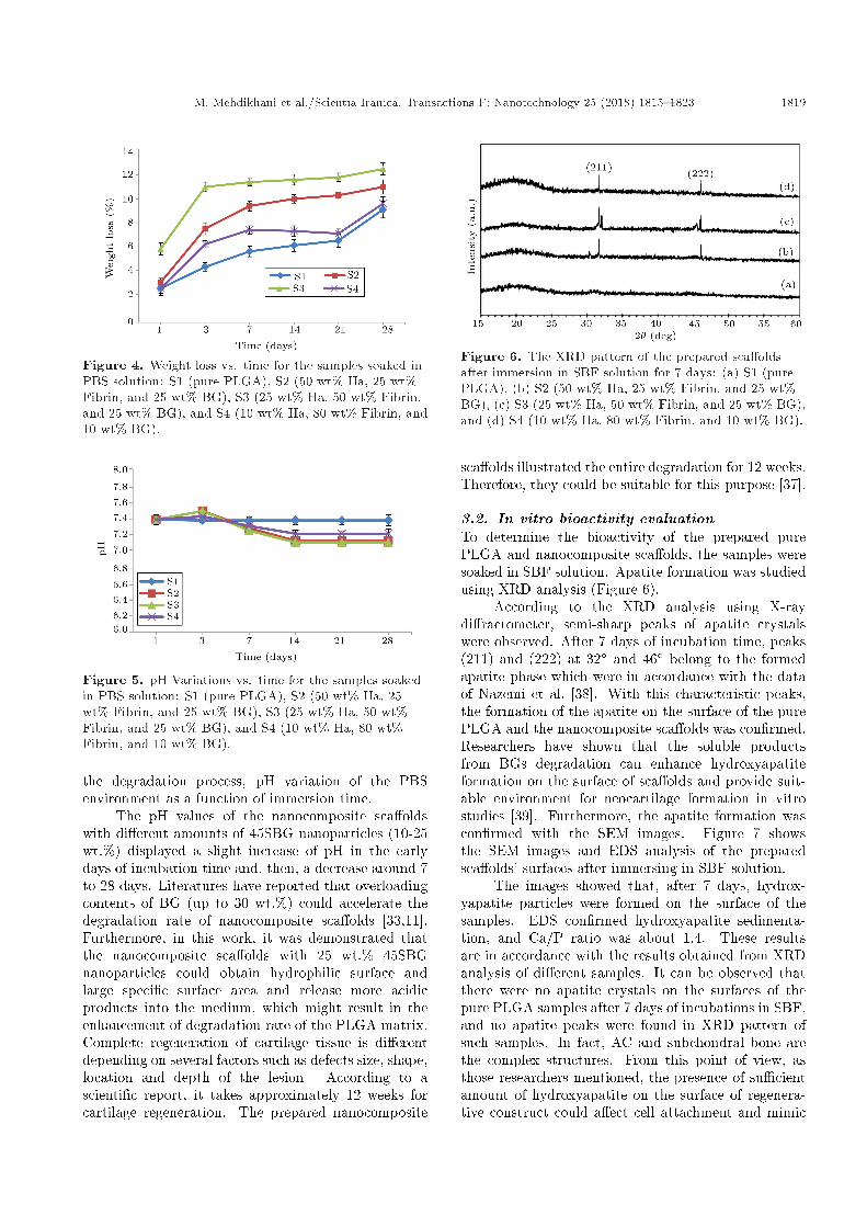
Figure 4. Weight loss vs. time for the samples soaked inPBS solution: S1 (pure PLGA), S2 (50 wt% Ha, 25 wt%Fibrin, and 25 wt% BG), S3 (25 wt% Ha, 50 wt% Fibrin,and 25 wt% BG), and S4 (10 wt% Ha, 80 wt% Fibrin, and10 wt% BG).
Figure 5. pH Variations vs. time for the samples soakedin PBS solution: S1 (pure PLGA), S2 (50 wt% Ha, 25wt% Fibrin, and 25 wt% BG), S3 (25 wt% Ha, 50 wt%Fibrin, and 25 wt% BG), and S4 (10 wt% Ha, 80 wt%Fibrin, and 10 wt% BG).
the degradation process, pH variation of the PBSenvironment as a function of immersion time.
The pH values of the nanocomposite sca�oldswith di�erent amounts of 45SBG nanoparticles (10-25wt.%) displayed a slight increase of pH in the earlydays of incubation time and, then, a decrease around 7to 28 days. Literatures have reported that overloadingcontents of BG (up to 30 wt.%) could accelerate thedegradation rate of nanocomposite sca�olds [33,11].Furthermore, in this work, it was demonstrated thatthe nanocomposite sca�olds with 25 wt.% 45SBGnanoparticles could obtain hydrophilic surface andlarge speci�c surface area and release more acidicproducts into the medium, which might result in theenhancement of degradation rate of the PLGA matrix.Complete regeneration of cartilage tissue is di�erentdepending on several factors such as defects size, shape,location and depth of the lesion. According to ascienti�c report, it takes approximately 12 weeks forcartilage regeneration. The prepared nanocomposite
Figure 6. The XRD pattern of the prepared sca�oldsafter immersion in SBF solution for 7 days: (a) S1 (purePLGA), (b) S2 (50 wt% Ha, 25 wt% Fibrin, and 25 wt%BG), (c) S3 (25 wt% Ha, 50 wt% Fibrin, and 25 wt% BG),and (d) S4 (10 wt% Ha, 80 wt% Fibrin, and 10 wt% BG).
sca�olds illustrated the entire degradation for 12 weeks.Therefore, they could be suitable for this purpose [37].
3.2. In vitro bioactivity evaluationTo determine the bioactivity of the prepared purePLGA and nanocomposite sca�olds, the samples weresoaked in SBF solution. Apatite formation was studiedusing XRD analysis (Figure 6).
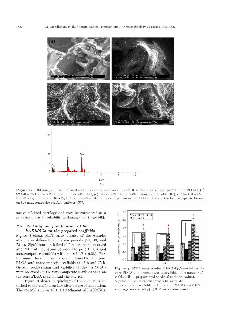
According to the XRD analysis using X-raydi�ractometer, semi-sharp peaks of apatite crystalswere observed. After 7 days of incubation time, peaks(211) and (222) at 32� and 46� belong to the formedapatite phase which were in accordance with the dataof Nazemi et al. [38]. With this characteristic peaks,the formation of the apatite on the surface of the purePLGA and the nanocomposite sca�olds was con�rmed.Researchers have shown that the soluble productsfrom BGs degradation can enhance hydroxyapatiteformation on the surface of sca�olds and provide suit-able environment for neocartilage formation in vitrostudies [39]. Furthermore, the apatite formation wascon�rmed with the SEM images. Figure 7 showsthe SEM images and EDS analysis of the preparedsca�olds' surfaces after immersing in SBF solution.
The images showed that, after 7 days, hydrox-yapatite particles were formed on the surface of thesamples. EDS con�rmed hydroxyapatite sedimenta-tion, and Ca/P ratio was about 1.4. These resultsare in accordance with the results obtained from XRDanalysis of di�erent samples. It can be observed thatthere were no apatite crystals on the surfaces of thepure PLGA samples after 7 days of incubations in SBF,and no apatite peaks were found in XRD pattern ofsuch samples. In fact, AC and subchondral bone arethe complex structures. From this point of view, asthose researchers mentioned, the presence of su�cientamount of hydroxyapatite on the surface of regenera-tive construct could a�ect cell attachment and mimic
1820 M. Mehdikhani et al./Scientia Iranica, Transactions F: Nanotechnology 25 (2018) 1815{1823
Figure 7. SEM images of the prepared sca�olds surface after soaking in SBF solution for 7 days: (a) S1 (pure PLGA), (b)S2 (50 wt% Ha, 25 wt% Fibrin, and 25 wt% BG), (c) S3 (25 wt% Ha, 50 wt% Fibrin, and 25 wt% BG), (d) S4 (10 wt%Ha, 80 wt% Fibrin, and 10 wt% BG) and Sca�old structures and porosities, (e) EDS analysis of the hydroxyapatite formedon the nanocomposite sca�old surfaces (S3).
native calci�ed cartilage and may be considered as aprominent way to rehabilitate damaged cartilage [40].
3.3. Viability and proliferation of thehADMSCs on the prepared sca�olds
Figure 8 shows MTT assay results of the samplesafter three di�erent incubation periods (24, 48, and72 h). Signi�cant statistical di�erences were observedafter 24 h of incubation between the pure PLGA andnanocomposite sca�olds with control (P < 0:05). Fur-thermore, the same results were obtained for the purePLGA and nanocomposite sca�olds at 48 h and 72 h.Greater proliferation and viability of the hADMSCswere observed on the nanocomposite sca�olds than onthe pure PLGA sca�old and the control.
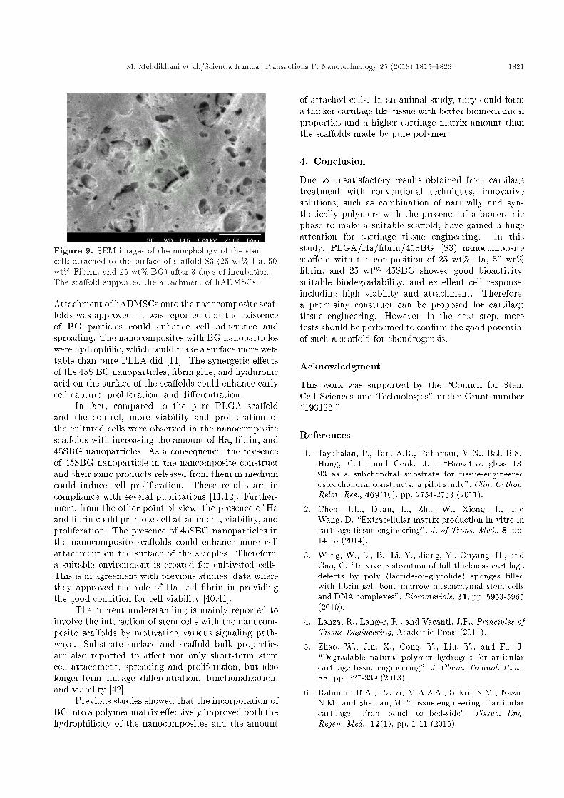
Figure 9 shows morphology of the stem cells at-tached to the sca�old surface after 3 days of incubation.The sca�old supported the attachment of hADMSCs.
Figure 8. MTT assay results of hADMSCs seeded on thepure PLGA and nanocomposite sca�olds. The number ofviable cells is proportional to the absorbance values.Signi�cant statistical di�erences between thenanocomposite sca�olds and S1 (pure PLGA) +p < 0:05,and negative control py < 0:05 were determined.
M. Mehdikhani et al./Scientia Iranica, Transactions F: Nanotechnology 25 (2018) 1815{1823 1821
Figure 9. SEM images of the morphology of the stemcells attached to the surface of sca�old S3 (25 wt% Ha, 50wt% Fibrin, and 25 wt% BG) after 3 days of incubation.The sca�old supported the attachment of hADMSCs.
Attachment of hADMSCs onto the nanocomposite scaf-folds was approved. It was reported that the existenceof BG particles could enhance cell adherence andspreading. The nanocomposites with BG nanoparticleswere hydrophilic, which could make a surface more wet-table than pure PLLA did [11]. The synergetic e�ectsof the 45S BG nanoparticles, �brin glue, and hyaluronicacid on the surface of the sca�olds could enhance earlycell capture, proliferation, and di�erentiation.
In fact, compared to the pure PLGA sca�oldand the control, more viability and proliferation ofthe cultured cells were observed in the nanocompositesca�olds with increasing the amount of Ha, �brin, and45SBG nanoparticles. As a consequence, the presenceof 45SBG nanoparticle in the nancomposite constructand their ionic products released from them in mediumcould induce cell proliferation. These results are incompliance with several publications [11,12]. Further-more, from the other point of view, the presence of Haand �brin could promote cell attachment, viability, andproliferation. The presence of 45SBG nanoparticles inthe nanocomposite sca�olds could enhance more cellattachment on the surface of the samples. Therefore,a suitable environment is created for cultivated cells.This is in agreement with previous studies' data wherethey approved the role of Ha and �brin in providingthe good condition for cell viability [40,41].
The current understanding is mainly reported toinvolve the interaction of stem cells with the nanocom-posite sca�olds by motivating various signaling path-ways. Substrate surface and sca�old bulk propertiesare also reported to a�ect not only short-term stemcell attachment, spreading and proliferation, but alsolonger-term lineage di�erentiation, functionalization,and viability [42].
Previous studies showed that the incorporation ofBG into a polymer matrix e�ectively improved both thehydrophilicity of the nanocomposites and the amount
of attached cells. In an animal study, they could forma thicker cartilage-like tissue with better biomechanicalproperties and a higher cartilage matrix amount thanthe sca�olds made by pure polymer.
4. Conclusion
Due to unsatisfactory results obtained from cartilagetreatment with conventional techniques, innovativesolutions, such as combination of naturally and syn-thetically polymers with the presence of a bioceramicphase to make a suitable sca�old, have gained a hugeattention for cartilage tissue engineering. In thisstudy, PLGA/Ha/�brin/45SBG (S3) nanocompositesca�old with the composition of 25 wt% Ha, 50 wt%�brin, and 25 wt% 45SBG showed good bioactivity,suitable biodegradability, and excellent cell response,including high viability and attachment. Therefore,a promising construct can be proposed for cartilagetissue engineering. However, in the next step, moretests should be performed to con�rm the good potentialof such a sca�old for chondrogensis.
Acknowledgment
This work was supported by the \Council for StemCell Sciences and Technologies" under Grant number\193126."
References
1. Jayabalan, P., Tan, A.R., Rahaman, M.N., Bal, B.S.,Hung, C.T., and Cook, J.L. \Bioactive glass 13-93 as a subchondral substrate for tissue-engineeredosteochondral constructs: a pilot study", Clin. Orthop.Relat. Res., 469(10), pp. 2754-2763 (2011).
2. Chen, J.L., Duan, L., Zhu, W., Xiong, J., andWang, D. \Extracellular matrix production in vitro incartilage tissue engineering", J. of Trans. Med., 8, pp.14-15 (2014).
3. Wang, W., Li, B., Li, Y., Jiang, Y., Ouyang, H., andGao, C. \In vivo restoration of full-thickness cartilagedefects by poly (lactide-co-glycolide) sponges �lledwith �brin gel, bone marrow mesenchymal stem cellsand DNA complexes", Biomaterials, 31, pp. 5953-5965(2010).
4. Lanza, R., Langer, R., and Vacanti, J.P., Principles ofTissue Engineering, Academic Press (2011).
5. Zhao, W., Jin, X., Cong, Y., Liu, Y., and Fu, J.\Degradable natural polymer hydrogels for articularcartilage tissue engineering", J. Chem. Technol. Biot.,88, pp. 327-339 (2013).
6. Rahman, R.A., Radzi, M.A.Z.A., Sukri, N.M., Nazir,N.M., and Sha'ban, M. \Tissue engineering of articularcartilage: From bench to bed-side", Tissue. Eng.Regen. Med., 12(1), pp. 1-11 (2015).
1822 M. Mehdikhani et al./Scientia Iranica, Transactions F: Nanotechnology 25 (2018) 1815{1823
7. Garg, T., Singh, O., Arora, S., and Murthy R.\Sca�old: a novel carrier for cell and drug delivery",Crit. Revi. inThera. Drug. Carrier. Sys., p. 29 (2012).
8. Dumitriu, S., Polymeric Biomaterials, revised andexpanded: CRC Press (2001).
9. Gentile, P., Chiono, V., Carmagnola, I., and Hatton,P.V. \An overview of poly (lactic-co-glycolic) acid(PLGA)-based biomaterials for bone tissue engineer-ing", Int. J. Mol. Sci., 15, pp. 3640-3659 (2014).
10. Tavakoli, E., Mehdikhani-Nahrkhalaji, M., Hashemi-Beni, B., Zargar-Kharazi, A., and Kharaziha, M.\Preparation, characterization, and mechanical as-sessment of poly (Lactide-co-Glycolide)/hyaluronicacid/ �brin/bioactive glass nano-composite sca�oldsfor cartilage tissue engineering applications", Procedia.Mater. Sci., 11, pp. 124-130 (2015).
11. Mehdikhani-Nahrkhalaji, M., Fathi, M.H., Mortazavi,V., Mousavi, S.B., Hashemi-Beni, B., and Razavi,S.M. \Novel nanocomposite coating for dental implantapplications in vitro and in vivo evaluation", J. Mater.Sci. Mater. Med., 23, pp. 485-495 (2012).
12. Mehdikhani-Nahrkhalaji, M., Fathi, M.H., Mortazavi,V., Mousavi, S.B., Hashemi-Beni, B., Razavi, S.B.,Akhavan, A., and Haghighat, A. \In vivo and in vitroevaluation of poly (lactide-co-glycolide)/bioactive glassnanocomposite coating", Adv. Mater. Res., 829, pp.309-313 (2014).
13. Song, J.E., Kim, M.J., Yoon, H., Yoo, H., Lee,Y.J., Kim, H.N., Lee, D., Yuk, S.H., and Khang,G. \E�ect of hyaluronic acid (HA) in a HA/PLGAsca�old on annulus �brosus regeneration: In vivotests", Macromol. Res., 21, pp. 1075-1082 (2013).
14. Jang, J.-D., Moon, Y.-S., Kim, Y.-S., Choi, N.-Y.,Mok, H.-S., Kim, Y.-J., Shetty, A.A., and Kim, S.-J.\Novel repair technique for articular cartilage defectusing a �brin and hyaluronic acid mixture", Tissue.Eng. Regen. Med., 10(1), pp. 1-9 (2013).
15. Kim, H.J., Kim, K.K., Park, I.K., Choi, B.S., Kim,J.H., and Kim, M.S. \Erratum to: Hybrid sca�oldscomposed of hyaluronic acid and collagen for carti-lage", Tissue. Eng. Regen. Med., 9(4), pp. 231-231(2012).
16. Sha0ban, M., Kim, S.H., Idrus, R., and Khang, G.\The use of �brin and poly (lactic-co-glycolic acid) hy-brid sca�old for articular cartilage tissue engineering:an in vivo analysis", Eur. Cell. Mat., 15, pp. 41-52(2008).
17. Hofmann, S. and Garcia-Fuentes, M. \Bioactive scaf-folds for the controlled formation of complex skeletaltissues", INTECH. Open. Access. Publ., 15, pp. 41-52(2008).
18. Sha0Ban, M., Kim, S.H., Idrus, R.B., and Khang,G. \Fibrin and poly (lactic-co-glycolic acid) hybridsca�old promotes early chondrogenesis of articularchondrocytes: an in vitro study", J. Orthop. Surg.Res., 3(1), pp. 1-10 (2008).
19. Subia, B., Kundu, J., and Kundu, S. \Biomaterialsca�old fabrication techniques for potential tissue engi-neering applications", Intech. Open. Access. Publisher.(2010).
20. Hench, L.L. and Wilson, J., An Introduction to Bioce-ramics, 2nd Ed. Imperial College Press., p. 478 (2013).
21. Rahaman, M.N., Day, D.E., Bal, B.S., Fu, Q., Jung,S.B., Bonewald, L.F., and Tomsia, A.P. \Bioactiveglass in tissue engineering", Acta. Biomater., 7, pp.2355-2373 (2011).
22. Kim, S.H. and Min, B-H. \A new era of cartilage repairusing cell therapy and tissue engineering: turningcurrent clinical limitations into new ideas", Tissue.Eng. Regen. Med., 9(5), pp. 240-248 (2012).
23. Niederauer, G.G., Slivka, M.A., Leatherbury, N.C.,Korvick, D.L., Harro� Jr, H.H., Ehler, W.C., Dunn,C.J., and Kieswetter, K. \Evaluation of multiphaseimplants for repair of focal osteochondral defects ingoats", Biomaterials, 21(24), pp. 2561-2574 (2000).
24. Suominen, E., Aho, A.J., Vedel, E., Kangasniemi, I.,Uusipaikka, E., and Yli-Urpo, A. \Subchondral boneand cartilage repair with bioactive glasses, hydroxyap-atite, and hydroxyapatite-glass composite", J. Biomed.Mater. Res., 32(4), pp. 543-551 (1996).
25. Rahaman, M.N., Day, D.E., Bal, B.S., Fu, Q., Jung,S.B., Bonewald, L.F., and Tomsia, A.P. \Bioactiveglass in tissue engineering", Acta. Biomater, 7(6), pp.2355-2373 (2011).
26. Ma, P.X. and Elissee�, J., Sca�olding in Tissue Engi-neering, CRC press (2005).
27. Diba, M., Kharaziha, M., Fathi, M.H., Gholipour-malekabadi, M., and Samadikuchaksaraei, A. \Prepa-ration and characterization of polycaprolactone/forsteritenanocomposite porous sca�olds designed forbone tissue regeneration", Compos. Sci. Technol., 72,pp. 716-723 (2012).
28. Kokubo, T. and Takadama, H. \How useful is SBFin predicting in vivo bone bioactivity?", Biomaterials,27, pp. 2907-2915 (2006).
29. Saadaldin, S.A., Dixon, S.J., Costa, D.O., andRizkalla, A.S. \Synthesis of bioactive and machin-ablemiserite glass-ceramics for dental implant applica-tions", Dent. Mater., 29, pp. 645-655 (2013).
30. Magyari, K., Stefan, R., Vulpoi, A., and Baia, L.\Bioactivity evolution of calcium-free borophosphateglass with addition of titanium dioxide", J. Non-Cryst.Solids, 410, pp. 112-117 (2015).
31. Mardani, M., Kabiri, A., Esfandiari, E., Esmaeili,A., Pourazar, A., Ansar, M., and Hashemibeni, B.\The e�ect of platelet rich plasma on chondrogenicdi�erentiation of human adipose derived stem cells intranswell culture", Iran J. Basic Med. Sci., 16(11), pp.1163-1169 (2013).
32. Hauner, H., Entenmann, G., Wabitsch, M., Gaillard,D., Ailhaud, G., Negrel, R., and Pfei�er, E. \Pro-moting e�ect of glucocorticoids on the di�erentiation
M. Mehdikhani et al./Scientia Iranica, Transactions F: Nanotechnology 25 (2018) 1815{1823 1823
of human adipocyte precursor cells cultured in achemically de�ned medium", J. Clin. Invest., 84, pp.1663-1670 (1989).
33. Hong, Z., Reis, R.L., and Mano, J.F. \Preparationand in vitro characterization of sca�olds of poly (L-lactic acid) containing bioactive glass ceramic nanopar-ticles", Acta. Biomater., 4, pp. 1297-1306 (2008).
34. Day, R.M., Maquet, V., Boccaccini, A.R., J�erome,R., and Forbes, A. \In vitro and in vivo analysisof macroporous biodegradable poly (D, L-lactide-co-glycolide) sca�olds containing bioactive glass", J.Biomed. Mater. Res. A., 75, pp. 778-787 (2005).
35. Ho�man, A.S. \Hydrogels for biomedical applica-tions", Adv. Drug. Delivery. Rev., 64, pp. 18-23 (2012).
36. Orava, E., Korventausta, J., Rosenberg, M., Jokinen,M., and Rosling, A. \In vitro degradation of porouspoly (DL-lactide-co-glycolide)(PLGA)/bioactive glasscomposite foams with a polar structure", Polym.Degrad. Stab., 92, pp. 14-23 (2007).
37. Jackson, D.W., Lalor, P.A., Aberman, H.M., andSimon, T.M. \Spontaneous repair of full-thicknessdefects of articular cartilage in a goat model", J. Bone.Joint. Surg. Am., 83(1), pp. 53-53 (2001).
38. Nazemi, K., Moztarzadeh, F., Jalali, N., Asgari,S., and Mozafari, M. \Synthesis and characterizationof poly (lactic-co-glycolic) acid nanoparticles-loadedchitosan/bioactive glass sca�olds as a localized deliverysystem in the bone defects", Biomed. Res. Int., pp. 1-9(2014).
39. Spoliti, M., Iudicone, P., Leone, R., De Rosa, A.,Rossetti, F.R., and Pierelli, L. \In vitro release andexpansion of mesenchymal stem cells by a hyaluronicacid sca�old used in combination with bone marrow",Muscles. Ligaments. Tendons. J., 2(4), pp. 289-294(2012).
40. Wang, Y., Meng, H., Yuan, X., Peng, J., Guo, Q.,Lu, S., and Wang, A. \Fabrication and in vitro eval-uation of an articular cartilage extracellular matrix-hydroxyapatite bilayered sca�old with low permeabil-ity for interface tissue engineering", Biomed. Eng.Online, 13(1), p. 80 (2014).
41. Snyder, T.N., Madhavan, K., Intrator, M., Dregalla,R.C., and Park, D. \A �brin/hyaluronic acid hydrogelfor the delivery of mesenchymal stem cells and poten-tial for articular cartilage repair", J. Biol. Eng., 8, pp.1-11 (2014).
42. Osathanon, T., Chuenjitkuntaworn, B., Nowwarote,N., Supaphol, P., Sastravaha, P., Subbalekha, K.,and Pavasant, P. \The responses of human adipose-derived mesenchymal stem cells on polycaprolactone-based sca�olds: an in vitro study", Tissue Eng. Regen.Med., 11(3), pp. 239-246 (2014).
Biographies
Mehdi Mehdikhani (PhD of Biomaterials) is now anAssistant Professor of Biomaterials at the Departmentof Biomedical Engineering, Faculty of Engineering,University of Isfahan, Isfahan, Iran. He receivedhis BSc in Materials Engineering, MSc in CorrosionEngineering, and PhD in Biomaterials at Isfahan Uni-versity of Technology. Currently, Dr. Mehdikhani-Nahrkhalaji's researches are focused on designing andfabricating of sca�olds and hydrogels containing de-sired drugs loaded nanoparticles for cartilage, bone,cardiovascular, and skin tissue engineering.
Ebrahim Tavakoli received his BSc degree in Ma-terials Engineering (area of concentration: IndustrialMetallurgy), from Azad University of Najafabad, Na-jafabad, Iran in 2010; he also received his MSc degreein Biomedical Engineering (area of concentration: Bio-materials) in 2015 from University of Isfahan, Isfahan,Iran. His MSc dissertation on articular cartilage regen-eration was carried out in collaboration with IsfahanUniversity of Medical Sciences and Council for StemCell Sciences and Technologies, Tehran, Iran, underGrant number \193126". His main research interestsare tissue engineering, nanobiomaterials, drug deliverysystems design, and stem cell therapy.
Anousheh Zargar-Kharazi (PhD: Biomaterials) isan Assistant Professor of Biomaterials Science andEngineering at the Department of Biomaterials andTissue Engineering, School of Advanced Technologyin Medicine, Isfahan University of Medical Sciences,Isfahan, Iran. She received her Bachelor's degreein Mechanical Engineering from Isfahan University ofTechnology and Master's degree in Biomechanics fromthe Iran University of Science and Technology. Shecompleted her PhD in Biomaterials from the IsfahanUniversity of Technology, Iran. Her research interestsare �nite-element modelling, application of compositesin biomaterials, fabrication and characterisation ofcomposite biomaterials and vascular tissue engineering.
Batool Hashemibeni, PhD, is an Associate Professorof Isfahan University of Medical Sciences. She receivedher BSc degree in Biology from Isfahan Universityof Medical Science, Isfahan, Iran, her MSc degree inHistology from Tarbiat Modarres University, Tehran,Iran, and her PhD degree in Anatomical Sciences fromIsfahan University of Medical Sciences, Isfahan, Iran.Her research background lies in stem cells �eld andtissue engineering.


![Comparative study of chitosan and chitosan–gelatin scaffold ......International Nano Letters (2017) 7:285–290 2871 volume.Thedensityofchitosanandchitosan–gelatinscaf-foldswascalculatedbytheformula[16],](https://img.pdfslide.us/doc/110x75/60ce433a5b5f1b495614599f/comparative-study-of-chitosan-and-chitosanagelatin-scaffold-international.jpg)