Embed Size (px)
Citation preview

Accepted Manuscript
A novel MYH7 founder mutation causing Laing distal myopathy inSouthern Spain
P. Carbonell-Corvillo MD , E. Tristan-Clavijo PhD ,M. Cabrera-Serrano MD , E. Servian-Morilla PhD ,G. Garcıa-Martın MD , L. Villarreal-Perez MD ,E. Rivas-Infante MD , E. Area-Gomez PhD ,M.I. Chamorro-Munoz MD , A. Gil-Galvez BSc ,A. Miranda-Vizuete PhD , A. Martinez-Mir PhD , N. Laing PhD ,C. Paradas MD
PII: S0960-8966(18)30445-0DOI: 10.1016/j.nmd.2018.07.006Reference: NMD 3585
To appear in: Neuromuscular Disorders
Received date: 29 May 2018Revised date: 12 July 2018Accepted date: 19 July 2018
Please cite this article as: P. Carbonell-Corvillo MD , E. Tristan-Clavijo PhD ,M. Cabrera-Serrano MD , E. Servian-Morilla PhD , G. Garcıa-Martın MD , L. Villarreal-Perez MD ,E. Rivas-Infante MD , E. Area-Gomez PhD , M.I. Chamorro-Munoz MD , A. Gil-Galvez BSc ,A. Miranda-Vizuete PhD , A. Martinez-Mir PhD , N. Laing PhD , C. Paradas MD , A novel MYH7founder mutation causing Laing distal myopathy in Southern Spain, Neuromuscular Disorders (2018),doi: 10.1016/j.nmd.2018.07.006
This is a PDF file of an unedited manuscript that has been accepted for publication. As a serviceto our customers we are providing this early version of the manuscript. The manuscript will undergocopyediting, typesetting, and review of the resulting proof before it is published in its final form. Pleasenote that during the production process errors may be discovered which could affect the content, andall legal disclaimers that apply to the journal pertain.

ACCEPTED MANUSCRIPT
ACCEPTED MANUSCRIP
T
1
Highlights
We report the novel variant p.R1560P in MYH7 gene as a founder mutation in
southern Spain
Most patients display the classic Laing distal myopathy phenotype
Interestingly, neck flexor weakness is revealed as early sign in some cases
MRI shows early involvement of the sartorius muscle, even in asymptomatic
carriers

ACCEPTED MANUSCRIPT
ACCEPTED MANUSCRIP
T
2
TITLE: A novel MYH7 founder mutation causing Laing distal myopathy in Southern
Spain
Carbonell-Corvillo, P.1
(MD); Tristán-Clavijo, E.2 (PhD); Cabrera-Serrano, M.
1,3 (MD);
Servián-Morilla E.1 (PhD); García-Martín, G.
4 (MD); Villarreal-Pérez, L.
1 (MD); Rivas-
Infante, E.5
(MD); Area-Gómez, E.6 (PhD); Chamorro-Muñoz, M.I.
4 (MD); Gil-Gálvez A.
7
(BSc); Miranda-Vizuete A.7
(PhD); Martinez-Mir, A.2 (PhD); Laing, N.
3 (PhD); Paradas,
C.1
(MD)
1 Neuromuscular Unit, Department of Neurology. Hospital U. Virgen del Rocío/
Instituto de Biomedicina de Sevilla (IBiS), Sevilla, Spain.
2 Instituto de Biomedicina de Sevilla (IBIS)/Hospital Virgen del
Rocío/CSIC/Universidad de Sevilla. Spain.
3 Centre for Medical Research. University of Western Australia, Harry Perkins
Institute of Medical Research. Perth, Australia
4 Department of Neurology. Hospital Universitario Virgen de la Victoria, Málaga,
Spain.
5 Department of Neuropathology. Hospital U. Virgen del Rocío/ Instituto de
Biomedicina de Sevilla (IBiS), Sevilla. Spain
6 Department of Neurology, Columbia University Medical Center, New York, NY,
USA
7 Redox Homeostasis Group. Instituto de Biomedicina de Sevilla (IBIS)/Hospital
Virgen del Rocío/CSIC/Universidad de Sevilla. Spain.

ACCEPTED MANUSCRIPT
ACCEPTED MANUSCRIP
T
3
Corresponding author: Carmen Paradas. Phone: +34687748886. Fax: +34955013536. e-
mail: [email protected]
ABSTRACT
MYH7 gene mutations are associated with wide clinical and genetic heterogeneity. We
report a novel founder mutation in MYH7 in Southern Spain (Andalucía). We studied two
index patients and 24 family members from two apparently independent families by
physical examination, serum creatine-kinase, muscle MRI, sequencing studies and genetic
linkage analysis. Sixteen individuals were heterozygous for a (p.R1560P) variant in the
MYH7 gene. Haplotype was consistent with a common ancestor for the two families. The
patients displayed the classic Laing distal myopathy phenotype, with hanging first toe as
the initial presentation, even in mildly affected patients who declared themselves
asymptomatic, although neck flexor weakness was revealed as an early sign in some cases.
MRI showed that the sartorius was the first muscle involved, even in two out of three
asymptomatic carriers. Our findings support the novel variant p.R1560P in MYH7 as a
founder mutation in Andalucía. The early involvement of the sartorius muscle in MRI may
be useful as an indicator of affection status.
Keywords: MYH7; founder mutation; distal myopathy; muscle MRI; genetic linkage
analysis
INTRODUCTION
Laing distal myopathy is an autosomal dominant disease, commonly characterized by
slowly progressive distal weakness, involving the anterior compartment of the legs, thus
causing the typical “hanging big toe” sign [1]. Nevertheless, highly variable clinical and
histological phenotypes have been widely reported in Laing distal myopathy [2-4], and the

ACCEPTED MANUSCRIPT
ACCEPTED MANUSCRIP
T
4
same mutation in the MYH7 gene can cause a different clinical picture regarding age of
onset, weakness pattern or severity and variable pathologic profiles, even within the same
family [3].
Concerning muscle MRI in the Laing distal myopathy, the tibialis anterior is the earliest
and more severely affected muscle in all the reported cases. To date, several case reports
[3, 5, 6] and one study with detailed analysis on radiological pattern in a cohort of Laing
distal myopathy patients [7] have been published, reporting quite variable and not always
consistent conclusions regarding the muscle imaging findings other than the tibialis
anterior early involvement.
The largest series of distal myopathy patients with mutations in MYH7 reported a high rate
of de novo mutations. This high frequency of sporadic mutations suggest that there should
be a wide geographical distribution [7-9]. Exceptionally, a founder mutation in the MYH7
gene, p.K1729del has been found to cause the majority of Laing distal myopathy cases in
the Safor region, Spain [10]. Most mutations are associated with the classic phenotype of
Laing distal myopathy. However, in the series reported by Fiorillo et al [7] some atypical
clinical features were reported which delayed the diagnosis.
Lamont et al reported eight out of 15 families with missense mutations introducing a
proline in MYH7 associated with distal myopathy [9] . These mutations involve amino-
acid residues at all positions of the heptad repeat of the protein which would induce
significant modifications in the protein structure, which suggests that proline substitutions
at any of these positions has a pathogenic effect [11, 12].
Here, we report a novel mutation in the MYH7 gene with a founder effect in Andalucía
(South of Spain). The description of founder mutations and the analysis of a potential
genotype-phenotype correlation are important from a diagnostic point of view, especially
when variable phenotypical expression is associated with the disease.

ACCEPTED MANUSCRIPT
ACCEPTED MANUSCRIP
T
5
METHODS
Patients and study protocol
We initially identified two apparently unrelated patients with a distal anterior myopathy,
both from Andalucía (Fig. 1). Patient II.6/Family A reported several affected relatives
including his father and at least four siblings. Patient II.3/Family B was a sporadic case.
Affected and unaffected relatives from both families were contacted and offered the
possibility of participating in the study. The participants were examined using a
standardized protocol which included interview, physical exam, muscle MRI, serum CK
and blood sample to obtain DNA. The first step of the clinical protocol was the interview
of the recruited family members to collect the clinical data and classify them as
symptomatic or asymptomatic, followed by a complete general and neurological
examination, including manual muscle testing, and the rest of the tests. This study received
approval from the HUVR local Ethics Committee and all participants provided informed
consent.
Muscle biopsy
Muscle samples were obtained by open biopsy. We processed the muscle following the
standard procedures. Routine stains included hematoxylin & eosin (HE), modified Gomori
trichrome, Oil Red O, periodic acid-Schiff (PAS), nicotinamide adenosine dinucleotide
dehydrogenase (NADH) nicotinamide adenine dinucleotide (NADH), succinate
dehydrogenase (SDH), cytochrome oxidase (COX), and combined COX/SDH, and
myofibrillar adenosine triphosphatase (ATPase) after preincubation at pH 9.4, 4.5 and 4.3.
Immunohistochemistry for fast and slow myosin was performed in one case.

ACCEPTED MANUSCRIPT
ACCEPTED MANUSCRIP
T
6
Muscle MRI
Muscle MRI was performed at the level of the thighs and lower legs. We used a 1.5 Tesla
scanner (MRI Intera, Philips Medical Systems). Axial MRI sections were obtained
including a T1-weighted spin-echo sequence (TR 600-700 ms, TE 30 ms) and a short-time
inversion recovery (STIR) sequence (TR 2500-3500 ms, TE 60 ms, TI 150 ms), in 10 mm
slices. Fatty degeneration of muscles was identified according to the increased signal in T1
sequences and quantified according to a subjective scale [13], and STIR sequences were
used to evaluate the presence of edema.
MYH7 genetic study
Genomic DNA was extracted as per standard protocols from peripheral blood (Chemagic
DNA Blood 5K Kit, Perkin Elmer) or saliva samples (Oragene DNA Self-Collection Kit,
DNA Genotek) from patients and the rest of family members. In the index patient
II.6/Family A, the coding regions of MYH7 gene were Sanger-sequenced at the
Neuromuscular Unit, Hospital U. Virgen del Rocío, Sevilla (NMU-HUVR). DNA from
index patient II.3/Family B was sent to the Lotterywest State Biomedical Facility
Genomics (Perth, Australia) for neurogenetic sub-exomic supercapture and next generation
sequencing (Proton™, Life Technologies) of 336 genes, which included 254 disease genes
listed in the Neuromuscular Disorders Gene Table (www.musclegenetable.fr). DNA
samples from all recruited relatives were PCR-amplified using specific primers (5´-
CCATGATTAGTGAGCAGGCC-3´, 5´-TCAGAGGTCCCTGCAGTAAC-3´) and
directly sequenced using BigDye Terminator v3.1 Cycle Sequencing kit (Thermo Fisher
Scientific).
ACCEPTED MANUSCRIPT
ACCEPTED MANUSCRIP
T
7
Haplotype analysis
Microsatellite and SNP markers covering the MYH7 gene were selected based on their
genomic location and heterozygosity (UCSC Genome Browser, Human GRCh37/hg19;
http://genome.ucsc.edu/) [14]. They spanned a genomic region of 2,92 Mb. Microsatellite
markers were amplified by PCR using fluorescently-labelled primers and resolved in an
ABI 3500 Genetic Analyzer (Applied Biosystems). SNP loci were PCR-amplified and
genotypes were determined by visual inspection of the sequence chromatograms.
Haplotypes were built assuming the minimum number of recombinations.
RESULTS
Clinical phenotype
We performed the interview and physical examination in 23 individuals from the two
families, nine women and 14 men. The age at the time of the examination ranged from
eight to 79 years. The symptoms appeared between four and 30 years of age. Most carriers
recalled abnormal gait and falls during infancy but did not complain of weakness until
adulthood. Twelve carriers of the p.R1560P mutation (age range 8-79 years) showed a
pathological physical exam. Three of them (age range 21-42 years) declared themselves to
be asymptomatic and weakness restricted to big toe extension was the only sign found in
the exam (III.4/Family A, II.5/Family B, III.2/Family B). Two of them presented transient
cervical weakness at eight years old but not later (III-8/Family A, III-15/Family A). Three
out of the 16 carriers were asymptomatic with normal physical exam (age range 11-38
years). One carrier was not examined (I.2/Family B). No patient referred respiratory or
cardiac symptoms, and none of them referred a familial history of sudden death,

ACCEPTED MANUSCRIPT
ACCEPTED MANUSCRIP
T
8
arrhythmia or myocardiopathy. Echocardiography and respiratory functional studies were
performed in some affected patients and all the studies were normal. CK was in normal
range in all the carriers. Clinical findings are summarized in Table 1 and Figure 2.
Muscle histology
Muscle biopsies from four patients (II.5/Family A, II.6/Family A, II.14/Family A,
II.3/Family B), two tibialis anterior and two gastrocnemius muscles, were available for
review. Myopathic pattern with increased variability in fiber size and internal nuclei were
present in all biopsies. More severe changes with prominent atrophy and increased
endomysial connective tissue were observed in both tibialis anterior muscles (Fig 3A and
3B). Small group of angulated fibers and nuclear clumps suggesting neurogenic features
were seen in three of the four samples (Fig 3A and 3F). Oxidative enzyme stains (NADH,
SDH and COX) revealed prominent internal sarcoplasmic disorganization with core, core
with poor defined bordersand minicore lesions in all biopsies (Fig 3C and 3G). We
observed type I fiber predominance in three cases (Fig 3D) and scattered rimmed vacuoles
in one case. No hypotrophy of type I fibers was observed.
Imaging study
Muscle MRI was performed in 11 patients (Family A: II.5, II.6, II.14, III.2, III.4, III.14,
III.15; Family B: II.3, II.5, III.1, III.2). All the affected carriers with moderate or severe
phenotype (mean age 54.1±12.2 years old [range 39 to 79 years], disease duration
44.6±17.2 years [range 9 to 66 years]) showed fatty degeneration of lower limb muscles.
The tibialis anterior, gluteus major and sartorius muscles were involved in all of them,
although with different degrees of degeneration. Degeneration was severe in the tibialis
anterior and mild in the gluteus and sartorius muscles. The other affected muscles in the
MRI were the gracilis (75%), the quadriceps (50%) and the gastrocnemius (25%). Two
slightly affected patients (III.4/ Family A, II.5/Family B) and two asymptomatic carriers
ACCEPTED MANUSCRIPT
ACCEPTED MANUSCRIP
T
9
(III.2/Family A, III.1/Family B) presented mild fatty infiltration only in the sartorius
muscle. Two mildly affected patients (III.15/Family A, III.2/Family B) and one
asymptomatic carrier (III.14/Family A) showed normal MRI. The sartorius muscle was the
only one involved in 50% of slightly affected patients and in 66’6% of asymptomatic
carriers. MRI findings are summarized in Table 2 and Figure 4.
MYH7 mutation screening
The novel heterozygous mutation c.4679G>C (NG_007884.1) p.R1560P in exon 34 of
MYH7 was identified in both index patients (Fig. 1). The variant is absent from databases
of controls (http://gnomad.broadinstitute.org/; last accessed in November 2017) and is
predicted to introduce a proline in the slow myosin tail. Next generation sequencing in the
index patient II.3/B excluded pathogenic mutations in other genes related to neuromuscular
disorders. The novel variant was subsequently analyzed by Sanger sequencing in all the
recruited family members. In total, 26 subjects from the two families were recruited (17
from Family A and nine from Family B), comprising twelve clinically affected patients and
14 asymptomatic individuals. Segregation analysis showed that 16 individuals were
heterozygous for variant p.R1560P in the MYH7 gene, including twelve symptomatic
individuals.
Haplotype analysis
Given the identification of the same novel mutation in two families from different cities in
Andalucía, Southern Spain, we studied the possibility of them being related. Haplotype
analysis revealed a shared genomic region of 306 Kb in all carriers of p.R1560P mutation,
restricted to four SNP markers (Fig 5).
DISCUSSION

ACCEPTED MANUSCRIPT
ACCEPTED MANUSCRIP
T
10
We report a novel founder mutation in MYH7 causing Laing distal myopathy in Andalucía.
The shared haplotype identified in our study spans a short genomic region (306 Kb)
defined by SNP markers, however, the data are in accordance with the findings by Muelas
et al. [10], that show a common SNP-defined genomic region of 41,2-71,2 Kb. In our
Neuromuscular Unit at HUVR, which is a national reference unit, only patients with
ancestors from Southern Spain carry the novel c.4679G>C (p.R1560P) mutation in MYH7,
while in patients from other geographic regions of Spain the disease is due to different
mutations, all of them already described. Thus, this mutation should be the first mutation to
study in patients with this disease phenotype from Southern Spain. Proline residues are
widely known to have a pathogenic effect on the assembly of the protein [15, 16].
Moreover, most mutations in MYH7 associated with distal myopathy are located in the
mid-rod region of MYH7 protein encoded by exons 32 to 36 [17, 18]. All together, these
findings support the pathogenic role of this novel mutation. The high rate of de novo
mutations demonstrated in MYH7 together with the long lifespan of patients with Laing
distal myopathy could explain the high prevalence of cases, regardless of geographic or
ethnic distribution [7, 9]. Hence, the identification of founder mutations in specific
geographic areas facilitates the molecular diagnosis of patients.
A noticeable clinical and genetic heterogeneity is associated with the MYH7-related
myopathies [3, 4, 9]. Until now, only one founder mutation in MYH7 has been reported,
and it was also associated with a wide range of clinical and morphologic features among
the carriers [3, 10]. In this study, we identified the clinical variability previously reported
in other series, finding patients severely or mildly affected at a similar age, in the same
family. Four previously reported large series of patients showed that age at onset varied
between 0 and 50 years [3, 7, 9, 19] and in our series the onset was between four and 30
years of age (none of the patients with congenital onset), and the asymptomatic ones
ACCEPTED MANUSCRIPT
ACCEPTED MANUSCRIP
T
11
ranged from 11 to 38 years old. Fiorillo et al. described a series of 21 patients identifying
12 heterozygous mutations in MYH7 affecting the C-terminal rod domain, and the patients
with congenital or early onset presented predominant distal weakness, while patients with
later onset presented prevalent proximal weakness [7]. In contrast, in our series with a
novel mutation affecting the rod domain we found no differences in the distribution of the
weakness related to the age of onset. In our cohort, we found three asymptomatic carriers
(age ranged 11-38 years old) and five slightly affected patients (age ranged 8-42 years old).
Tasca et al. reported one family carrying a novel mutation in the myosin tail, and the age of
onset of all three affected members was not prior the fifth decade and shared mild
weakness limited to long toe and foot extensors [4]. Feinstein-Linial et al., who reported
two novel heterozygous proline substitutions located in exon 31 of MYH7, described also
one asymptomatic carrier aged 40 [5]. This confirms that more benign forms than the
classic phenotype exist, probably associated with better prognosis regarding the
progression of the disease.
We uncovered interesting manifestations after a thorough anamnesis and clinical exam.
The majority of carriers of the mutation reported abnormal gait with increasing tendency to
stumble and fall in infancy or childhood, although most of them did not complain of
motility or muscle problems until the third or fourth decade, a finding also described by
Muelas et al [3]. A review of 34 patients with Laing distal myopathy by Lamont et al.
revealed that initial weakness of great toe/ankle dorsiflexion and later development of neck
flexor weakness were consistent findings [9]. Interestingly, two infantile carriers in our
series presented neck flexor weakness only during childhood. Recently, one atypical case
with dropped head at the age of four has been reported, but authors do not refer if it
disappeared later and revealed also calf hypertrophy and cardiopathy at the age of eight
[7]. Thus, we must be attentive to abnormal gait or cervical weakness during infancy

ACCEPTED MANUSCRIPT
ACCEPTED MANUSCRIP
T
12
because it can be the first transient symptom or sign of the disease. Hanging big toe is the
typical sign of this disease, it is frequently the first obvious sign [4], even we uncovered
hanging big toe in three mildly affected patients who self-reported no symptoms, as
already reported by Muelas et al. in two patients [3]. However, we observed no correlation
between the severity of the toe extensor weakness and the severity of other manifestations.
Thus, weakness of the big toe extension may represent the most consistent clinical sign in
every stage of the disease, although neck flexor weakness and abnormal gait during
infancy could represent the first manifestation in some cases. Calf hypertrophy and pes
cavus are findings reported in other families [3, 4], and we found these symptoms only in
family A, so they do not seem to be specific to the novel mutation c.4679G>C.
Regarding the muscle biopsy, our series also showed some of the variable morphological
findings previously reported: hypotrophy of type I fibers, fiber type disproportion, areas
devoid of oxidative activity forming cores/pseudocores, angulated fibers, nuclear clumps,
and rimmed vacuoles [3, 9, 19]. However, which is the most prominent morphological
feature varies between series, thus atrophic type I fiber was found as the most common
abnormality in some previous series but it was not observed in our study, while
cores/pseudocores were highly frequent in most of the series [3, 9] and ours but were not
described by Lamont et al [19].
We have reviewed the MRI descriptions reported until now, demonstrating variability in
radiological findings [3, 4, 6, 7]. As previously described, in our series the tibialis anterior
muscle becomes the most severely degenerated muscle in early stages of the disease, and
the gastrocnemius medialis muscle was simultaneously affected in one patient which is in
accordance with recent reports showing involvement of the gastrocnemius medialis in
about half of the patients [5-8]. However, some authors described that the gastrocnemius
together with the gracilis and the sartorius muscles are usually preserved even in severely

ACCEPTED MANUSCRIPT
ACCEPTED MANUSCRIP
T
13
affected patients [3, 20], and our data contradict this assertion. We observed that the
involvement of the sartorius muscle is a very early radiological finding in our patients,
even in presymptomatic stages. This is quite surprising, since the sartorious muscle is
usually preserved until late stages in many other neuromuscular disorders, except for some
myofibrillar myopathies where it seems to be a hallmark [21]. The involvement of the
adductor magnus and the sartorius muscles has only been mentioned in few reports of
Laing distal myopathy, with no further analysis regarding their evolution [5, 7]. We
observed that the degree of involvement of the sartorius is always mild at early stages,
showing no further progression of the degeneration during the course of the disease.
Although this has not been described as a typical radiological sign in Laing distal
myopathy, our results support that the mild involvement of the sartorius muscle is a
common and early radiological sign in this myopathy, prior to the involvement of the
tibialis anterior muscle, even in asymptomatic carriers, so it could be a useful sign for
screening.
To conclude, our findings support the novel variant p.R1560P in MYH7 as a founder
mutation in Andalucía, and it should be considered the first mutation to study in patients
from Southern Spain. These large pedigrees allowed us to better describe the progression
and diagnostic clues in very early stages of the disease, such as neck flexor weakness,
hanging big toe and the sartorius muscle degeneration in muscle MRI, which could be
useful for detecting asymptomatic carriers of the MYH7 mutation. All these findings are
important to improve the diagnosis and the genetic counseling of Laing distal myopathy.
AUTHOR CONTRIBUTIONS

ACCEPTED MANUSCRIPT
ACCEPTED MANUSCRIP
T
14
C.P. and P.C designed the study. E.T.C and A.M.M performed the haplotype analysis.
E.S., C.P., P.C., L.V., M.C.S., E.A-G, G.G., A.G-G., and M-I.C. handled patients and
collected biological samples. M.C.S. and N.L. performed next generation sequencing and
analyzed the results. E.R. processed and studied muscle biopsy. C.P. and P.C analyzed the
radiological findings. C.P., A.M.M. and A.M-V. supervised and provided critical
discussion on the research. C.P. coordinated all the study. P.C wrote the initial manuscript.
All authors contributed to the final version of the manuscript.
CONFLICT OF INTEREST
Professor Laing reports grants from Australian National Health and Medical Research
Council, during the conduct of the study. The rest of authors have declared that no conflict
of interest exists.
ACKNOWLEDGMENTS
This work was supported in part by grants from the Health Institute Carlos III and FEDER
(FIS PI16-01843 to C. Paradas, JR15/00042 to M. Cabrera-Serrano), the Consejería de
Salud (PI-0085-2016 to C. Paradas), the Spanish Ministry of Economy and
Competitiveness (BFU2015-71464-R to A. Martinez-Mir, BFU2015-64408-P to A.
Miranda-Vizuete), the Alfonso Martín Escudero Foundation (to M. Cabrera-Serrano), and
the NHMRC Principal Research Fellowship (APP1117510 to N. Laing).

ACCEPTED MANUSCRIPT
ACCEPTED MANUSCRIP
T
15
REFERENCES
[1] Voit T, Kutz P, Leube B, Neuen-Jacobc E, Schröder JM, Cavallottiet D, et al.
Autosomal dominant distal myopathy: further evidence of a chromosome 14 locus.
Neuromuscul Disord 2001;11:11-9.
[2] Tajsharghi H, Thornell LE, Lindberg C, Lindvall B, Henriksson KG, Oldfors A.
Myosin storage myopathy associated with a heterozygous missense mutation in MYH7.
Ann Neurol 2003;54:495–00.
[3] Muelas N, Hackman P, Luque H, Garcés-Sánchez M, Azorín I, Suominen T, et al.
MYH7 gene tail mutation causing myopathic profiles beyond Laing distal myopathy.
Neurology 2010;75:732-41.
[4] Tasca G, Ricci E, Penttila S, Monforte M, Giglio V, Ottaviani P, et al. New phenotype
and pathology features in MYH7-related distal myopathy. Neuromuscul Disord
2012;22:640-7.
[5] Feinstein-Linial M, Buvoli M, Buvoli A, Sadeh M, Dabby R, Straussberg R, et al. Two
novel MYH7 proline substitutions cause Laing Distal Myopathy-like phenotypes with
variable expressivity and neck extensor contracture. BMC Med Genet 2016;17:57.
[6] Ferbert A, Zibat A, Rautenstrauß B, Kress W, Hügens-Penzel M, Weis J, et al. Laing
distal myopathy with a novel mutation in exon 34 of the MYH7 gene. Neuromuscul Disord
2016;26:598–03.
[7] Fiorillo C, Astrea G, Savarese M, Cassandrini D, Brisca G, Trucco F, et al. MYH7-
related myopathies: clinical, histopathological and imaging findings in a cohort of Italian
patients. Orphanet J Rare Dis 2016;11:91.
[8] Dubourg O, Maisonobe T, Behin A, Suominen T, Raheem O, Penttila S, et al. A novel
MYH7 mutation occurring independently in French and Norwegian Laing distal myopathy
families and de novo in one Finnish patient. J Neurol 2011;258: 1157–63.

ACCEPTED MANUSCRIPT
ACCEPTED MANUSCRIP
T
16
[9] Lamont PJ, Wallefeld W, Hilton-Jones D, Udd B, Argov Z, Barboi AC, et al. Novel
mutations widen the phenotypic spectrum of slow skeletal/beta-cardiac myosin(MYH7)
distal myopathy. Hum Mutat 2014;35:868-79.
[10] Muelas N, Hackman P, Luque H, Suominen T, Espinós C, Garcés-Sánchez M, et al.
Spanish MYH7 founder mutation of Italian ancestry causing a large cluster of Laing
myopathy patients. Clin Genet 2012;81:491-4.
[11] Piela L, Némethy G, Scheraga HA. Proline-induced constraints in alpha-helices.
Biopolymers 1987;26:1587–600.
[12] O'Neil KT, DeGrado WF. A thermodynamic scale for the helix-forming tendencies of
the commonly occurring amino acids. Science 1990;250:646–51.
[13] Mercuri E, Pichiecchio A, Allsop J, Messina S, Pane M, Muntoni F. Muscle MRI in
Inherited Neuromuscular Disorders: Past, Present, and Future. J Magn Reson Imaging
2007;25:433–40.
[14] Kent WJ, Sugnet CW, Furey TS, Roskin KM, Pringle TM, Zahler AM, et al. The
human genome browser at UCSC. Genome Res 2002;12:996-1006.
[15] Buvoli M, Buvoli A, Leinwand LA. Effects of pathogenic proline mutations on
myosin assembly. J Mol Biol 2012;415:807-18.
[16] Parry DA Fraser RD, Squire JM. Fifty years of coiled-coils and alpha-helical bundles:
a close relationship between sequence and structure. J Struct Biol 2008;163:258–69.
[17] Meredith C, Herrmann R, Parry C, Liyanage K, Dye DE, Durling HJ, et al. Mutations
in the slow skeletal muscle fiber myosin heavy chain gene (MYH7) cause Laing early-
onset distal myopathy (MPD1). Am J Hum Genet 2004;75:703–8.
[18] Tajsharghi H, Oldfors A. Myosinopathies: pathology and mechanisms. Acta
Neuropathol 2013;125:3-18.
ACCEPTED MANUSCRIPT
ACCEPTED MANUSCRIP
T
17
[19] Lamont PJ, Udd B, Mastaglia FL, de Visser M, Hedera P, Voit T, et al. Laing early
onset distal myopathy: slow myosin defect with variable abnormalities on muscle biopsy. J
Neurol Neurosurg Psychiatry 2006;77:208–15.
[20] Clarke NF, Amburgey K, Teener J, Camelo-Piragua S, Kesari A, Punetha J, et al. A
novel mutation expands the genetic and clinical spectrum of MYH7-related myopathies.
Neuromuscul Disord 2013;23:432-6.
[21] Fischer D, Kley RA, Strach K, Meyer C, Sommer T, Eger K, et al. Distinct muscle
imaging patterns in myofibrillar myopathies. Neurology 2008;71(10):758-65.

ACCEPTED MANUSCRIPT
ACCEPTED MANUSCRIP
T
18
TABLE LEGENDS
Table 1. Clinical features
Manual muscle testing according to the MRC scale
Carriers Age
at
exam
Onset Neck
flexor
Finger
ext
Knee
ext
Knee
flexor
Hanging
big toe
Toes
ext
Ankle
dorsiflexor
Pes
cavus
FVC/
Echocardiogram
Pheno
type
A I-1 79 y 13 y 2/5 2/5 2/5 2/5 Yes 0/5 0/5 Yes np/np Sev
II-1 59 y 8 y 3/5 4/5 4/5 5/5 Yes 2/5 0/5 Yes N/np Sev
II-5 54 y 6 y 3/5 2/5 4/5 5/5 Yes 0/5 0/5 Yes N/N Sev
II-6 52 y 6 y 3/5 3/5 3/5 5/5 Yes 3/5 0/5 Yes N/N Sev
II-8 49 y 5 y 4/5 4/5 5/5 5/5 Yes 4/5 2/5 Yes np/np Mod
II-14 39 y 30 y 5/5 5/5 5/5 5/5 Yes 4/5 2/5 Yes N/N Mod
III-2 38 y - 5/5 5/5 5/5 5/5 No 5/5 5/5 Yes np/np A
III-4 23 y 6 y 5/5 5/5 5/5 5/5 Yes 5/5 5/5 Yes np/np Mild
III-8 15 y 8y 4/5
(T)
5/5 5/5 5/5 No 5/5 5/5 No np/np Mild
III-14 11 y - 5/5 5/5 5/5 5/5 No 5/5 5/5 No np/np A
III-15 8 y 4y 4/5
(T)
5/5 5/5 5/5 No 5/5 5/5 No np/np Mild
B II-3 54 y 6 y 4/5 5/5 5/5 5/5 Yes 2/5 0/5 No N/N Mod
II-5 42 y 7 y 5/5 5/5 5/5 5/5 Yes 5/5 5/5 No np/np Mild
III-1 28 y - 5/5 5/5 5/5 5/5 No 5/5 5/5 No np/np A
III-2 21 y 17 y 5/5 5/5 5/5 5/5 Yes 5/5 5/5 No np/np Mild
y: Years; T: Transient; Ext: Extensor; CK: Serum creatin kinase; Numbers indicate muscle
strength according to MRC scale; FVC: Forced vital capacity; N: Normal; np: not
performed; A: Asymptomatic; Mild: Mild phenotype defined by hanging big toe sign;
Mod: Moderate phenotype defined by weakness of toes extension and ankle dorsiflexion ±
finger extension; Sev: Severe phenotype defined by weakness of toes extension, ankle
dorsiflexion and knee extension ± finger extension. Bold letters denote mildly affected
patients who only self-reported frequent falls during infancy.

ACCEPTED MANUSCRIPT
ACCEPTED MANUSCRIP
T
19
Table 2. Muscle MRI findings
Patient/
Family
Gluteus
major
Quadriceps Sartorius Gracilis Tibialis
anterior
Gastrocnemius
Affected
patients
I.1/A np np np np np np
II.1/A np np np np np np
II.5/A + + ++ + +++ N
II.6/A + ++
(asymmetric)
+ + +++ N
II.8/A np np np np np np
II.14/A + N + + ++ N
II.3/B + N + N +++ +
Mild affected
patients
III.4/A N N + N N N
III.8/A np np np np np np
III.15/A N N N N N N
II.5/B N N + N N N
III.2/B N N N N N N
Asymptomatic
carriers
III.2/A N N + + N N
III.14/A N N N N N N
III.1/B N N + + N N
N: Normal; +: Mild fatty infiltration; ++: Moderate fatty infiltration; +++: Severe fatty
infiltration; np: not performed

ACCEPTED MANUSCRIPT
ACCEPTED MANUSCRIP
T
20
FIGURE LEGENDS
Figure 1. Pedigrees of the two families with Laing distal myopathy. Clinical
examination and genetic study were performed in all available family members. Circles
denote female members, squares male members, solid black symbols affected patients,
solid dark grey symbols mildly affected patients, solid light grey symbols asymptomatic
carriers, striped symbols not-carriers, squared symbols not-examined carrier, and white
symbols not-studied family members.
ACCEPTED MANUSCRIPT
ACCEPTED MANUSCRIP
T
21
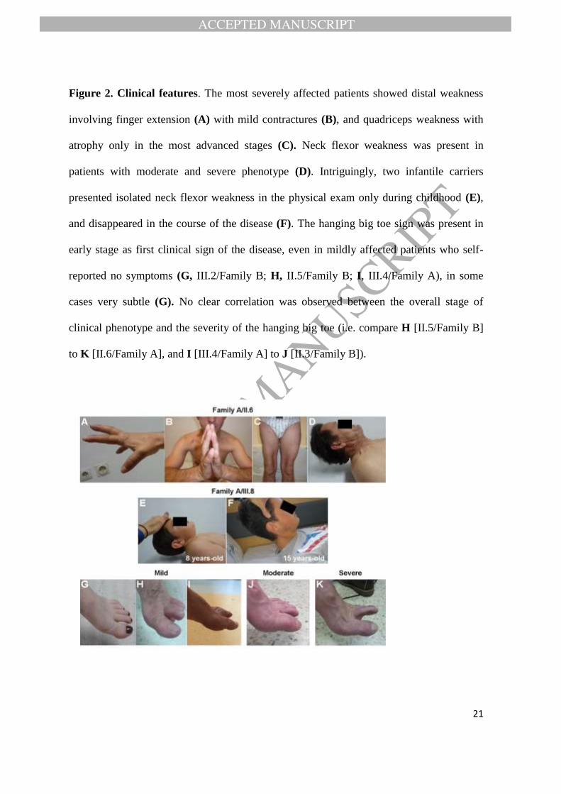
Figure 2. Clinical features. The most severely affected patients showed distal weakness
involving finger extension (A) with mild contractures (B), and quadriceps weakness with
atrophy only in the most advanced stages (C). Neck flexor weakness was present in
patients with moderate and severe phenotype (D). Intriguingly, two infantile carriers
presented isolated neck flexor weakness in the physical exam only during childhood (E),
and disappeared in the course of the disease (F). The hanging big toe sign was present in
early stage as first clinical sign of the disease, even in mildly affected patients who self-
reported no symptoms (G, III.2/Family B; H, II.5/Family B; I, III.4/Family A), in some
cases very subtle (G). No clear correlation was observed between the overall stage of
clinical phenotype and the severity of the hanging big toe (i.e. compare H [II.5/Family B]
to K [II.6/Family A], and I [III.4/Family A] to J [II.3/Family B]).

ACCEPTED MANUSCRIPT
ACCEPTED MANUSCRIP
T
22
Figure 3. Muscle histology. Muscle biopsies from patient II.14/Family A, tibialis anterior
(A-D) and patient II.3/Family B, gastrocnemius (E-H) were stained with haematoxylin and
eosin (A, B, E and F), succinate dehydrogenase (SDH) (C and G) and ATPase pH 4.5 (D
and H). Atrophy, variability in fibre size, increased endomysial connective tissue, and
necrotic fibers (arrow) (A and E), and groups of small angulated atrophic fibers with
nuclear clumps (arrowheads) (B and F) were present in both cases. SDH staining shows
prominent sarcoplasmic disorganization including core (black arrowhead), core with poor
defined borders (open arrow), and multiminicore (black arrow) lesions (C and G). Type I
predominance was observed (D). Scale bar is 50 m in all panels (A, B, C, E, F and G x
200 magnification; D and H x 100 magnification).

ACCEPTED MANUSCRIPT
ACCEPTED MANUSCRIP
T
23
Figure 4. Muscle MRI. Muscle MRI findings of patients displaying severe (A, II.6/
Family A), moderate (B, II.14/ Family A) and mild (C, II.5/ Family B) clinical phenotype.
Asymptomatic carriers can show mild sartorius muscle infiltration (D, III.1/ Family B) and
mild affected patients normal MRI (E, III.2/ Family B). The vastus intermedius and
lateralis muscles are involved only in severe stages (A). The tibialis anterior was the most
severely degenerated muscle in symptomatic patients (A-C), and usually the only affected
muscle in the lower legs. The most striking finding in our study was that the sartorius
muscle (arrowhead) was the earliest involved, even in asymptomatic carriers (D), although
it does not show evidence of significant progression (A-C).

ACCEPTED MANUSCRIPT
ACCEPTED MANUSCRIP
T
24
Figure 5. Haplotype analysis in the two families carrying the MYH7 p.R1560P
mutation. Genomic position for genotyped microsatellite and SNP markers relative to the
MYH7 gene, according to UCSC Genome Browser, Human GRCh37/hg19 (A). Haplotypes
for families A and B (B, C). Haplotype analysis using microsatellite markers D14S50,
D14S283, D14S990, D14S972, D14S64 and D14S264 in all available family members
revealed a different haplotype cosegregating with the mutation in each family. To narrow
down the studied genomic interval, we genotyped six SNP markers, rs365990, rs434273,
rs2331979, rs2277475, rs7157716 and rs11621360 close to or within the MYH7 gene. The
analysis of SNP genotypes revealed a shared haplotype for markers rs365990, rs434273,
rs2331979 and rs2277475 in all carriers of the p.R1560P mutation that defined a common
region of 306 Kb. The shared common haplotype is boxed. Genotype for mutation
p.R1560P is denoted by a + (wild-type) and M (mutant) sign. A question mark indicates
unknown genotype. Solid black symbols denote carriers of the MYH7 p.R1560P mutation.

ACCEPTED MANUSCRIPT
ACCEPTED MANUSCRIP
T
25