Embed Size (px)
Citation preview

AA
PMRAa
b
c
Egd
Oe
sI
AraafciscmnsKdpKtb
KK
Edgsttktn1
*EAarro
Neuroscience 157 (2008) 577–587
0d
NOVEL KCNA1 MUTATION IDENTIFIED IN AN ITALIAN FAMILY
FFECTED BY EPISODIC ATAXIA TYPE 1btsija2ewpnp
ddcaa2altavi12
sscittnte
ddo2iFwmmm
. IMBRICI,e1 F. GUALANDI,a1 M. C. D’ADAMO,e
. TADDEI MASIERI,a P. CUDIA,b D. DE GRANDIS,b
. MANNUCCI,c I. NICOLETTI,c S. J. TUCKER,d
. FERLINIa AND M. PESSIAe*
Section of Medical Genetics, University of Ferrara, Italy
Department of Neuroscience, Hospital of Rovigo, Italy
Section of Internal Medicine and Oncology, Department of Clinical andxperimental Medicine, University of Perugia, School of Medicine, Peru-ia, Italy
Department of Physics, Clarendon Laboratory, University of Oxford,xford, UK
Section of Human Physiology, Department of Internal Medicine, Univer-ity of Perugia School of Medicine, Via del Giochetto, I-06126 Perugia,taly
bstract—Episodic ataxia type 1 (EA1) is a rare human neu-ological syndrome characterized by continuous myokymiand attacks of generalized ataxia that can be triggered bybrupt movements, emotional stress and fatigue. An Italianamily has been identified where related members displayedontinuous myokymia, episodes of ataxia, attacks character-
zed by myokymia only, and neuromyotonia. A novel mis-ense mutation (F414C), in the C-terminal region of the K�
hannel Kv1.1, was identified in the affected individuals. Theutant homotetrameric channels were non-functional in Xe-
opus laevis oocytes. In addition, heteromeric channels re-ulting from the co-expression of wild-type Kv1.1 andv1.1(F414C), or wild-type Kv1.2 and Kv1.1(F414C) subunitsisplayed reduced current amplitudes and altered gatingroperties. This indicates that the pathogenic effect of thisCNA1 mutation is likely to be related to the defective func-
ional properties we have identified. © 2008 IBRO. Publishedy Elsevier Ltd. All rights reserved.
ey words: episodic ataxia, ion channel gene defects, Kv1.1,v1.2 cerebellum, Xenopus oocytes, confocal microscopy.
pisodic ataxia type 1 (EA1) is an autosomal dominantisorder characterized by myokymia and attacks of ataxicait that may be brought on by fever, startle, emotionaltress, and fatigue (Van Dyke et al., 1975). The first symp-oms typically manifest during childhood and can persisthroughout adult life. Since the description of the first EA1indred by Van Dyke et al. (1975), the phenotypic spec-rum of the disorder has widened considerably and it isow apparent that phenotypic differences exist not only
These authors contributed equally to this work.Corresponding author. Tel: �39-075-5857375; fax: �39-075-5857371.-mail address: [email protected] (M. Pessia).bbreviations: AP, action potentials; d.o.b., date of birth; EA1, episodictaxia type 1; GFP, green fluorescent protein; PCR, polymerase chaineaction; SNP, single nucleotide polymorphism; STR, short tandem
cepeats; TEVC, two-electrode voltage-clamp; VNTRs, variable numberf tandem repeats; WT, wild-type.
306-4522/08 © 2008 IBRO. Published by Elsevier Ltd. All rights reserved.oi:10.1016/j.neuroscience.2008.09.022
577
etween different families, but also between individuals withinhe same family. Patients have been reported with unusualymptoms such as partial epilepsy, EA1 without myokymia,
solated neuromyotonia, neuromuscular findings includingoint contractures, postural abnormalities, skeletal deformitiesnd paroxysmal dyspnea (Scheffer et al., 1998; Klein et al.,004; Kinali et al., 2004; Shook et al., 2008). Some patientsxperience severe ataxia more than 15 times per day,hereas other individuals experience attacks less than onceer month (Van Dyke et al., 1975). There is also heteroge-eity in their response to treatment, with some kindreds beingarticularly resistant to drugs (Eunson et al., 2000).
Linkage studies in several EA1 families led to theiscovery of a number of point mutations in the voltage-ependent potassium channel gene KCNA1 (Kv1.1), onhromosome 12p13 (Browne et al., 1994, 1995; Comu etl., 1996; Litt et al., 1994; Eunson et al., 2000; Scheffer etl., 1998; Klein et al., 2004; Lee et al., 2004; Shook et al.,008). The amino acid residues mutated in EA1 patientsre at positions which are highly conserved in the de-
ayed-rectifier potassium channel gene family, and func-ional studies have shown that these mutations gener-lly impair Kv1.1 channel function, although with quiteariable effects on channel assembly, trafficking and kinet-
cs (Eunson et al., 2000; Adelman et al., 1995; Zerr et al.,998; Zuberi et al., 1999; D’Adamo et al., 1998; Rea et al.,002; Imbrici et al., 2003; Cusimano et al., 2004).
The voltage-gated potassium channel Kv1.1 showsignificant cell-type and regional diversity in the nervousystem where it forms both homomeric and heteromerichannels (e.g. with Kv1.2) and is extensively modulated by
ntracellular and extracellular factors. Recently, studies at-empting to reproduce the in vivo condition have shownhat EA1 mutations may also affect heteromeric Kv1 chan-el activity, thus widening the molecular consequences of
he disease (D’Adamo et al., 1999; Rea et al., 2002; Mayliet al., 2002; Imbrici et al., 2007).
Despite several lines of evidence which indicate thatefective delayed-rectifier K� channels cause cerebellarysfunction, the molecular and neurological mechanismsf EA1 are still not completely understood (Kullmann et al.,001; Rajakulendran et al., 2007). Here we report a Sicil-
an family displaying typical and atypical EA1 symptoms.urthermore, by haplotype sharing and mutation analysise have identified a new KCNA1 mutation in the affectedembers that causes a complete loss-of-function of ho-otetrameric channels, and markedly reduced hetero-eric current amplitudes as well as producing important
hanges in their intrinsic biophysical properties.

N
Nipbslatfit
aatRc
GR
Tbtgrntpsso
lt(wPhdacrn(a21
KmanGc(
saftXsKIaa
FKDapD
P. Imbrici et al. / Neuroscience 157 (2008) 577–587578
EXPERIMENTAL PROCEDURES
europhysiology
erve conduction studies were performed using surface stimulat-ng and recording electrodes. Median and ulnar motor actionotentials (AP) were recorded bilaterally from the abductor pollicisrevis and the abductor digiti minimi muscles respectively, withtimulation at the wrist and elbow. Median and ulnar F wave
atencies were recorded. Sural sensory nerve AP were recordedntidromically at the ankles following stimulation in the calf. Elec-
romyography was performed with a concentric needle electroderom the biceps brachii and opponens pollicis muscles. Localschemia was applied by inflation of a blood pressure cuff aroundhe upper arm to 220 mm Hg for 3 min.
The experiments were undertaken with the understandingnd written consent of each subject, with the approval of theppropriate local ethics committee, and in compliance with na-
ional legislation and the Code of Ethical Principles for Medicalesearch Involving Human Subjects of the World Medical Asso-iation (Declaration of Helsinki).
enetics, molecular biology, oocyte preparation andNA injection
otal genomic DNA was isolated from peripheral blood leukocytesy BIOROBOT (Qiagen, Hilden, Germany) and analyzed as de-
ailed in Fig. 1. The coding sequence of the intronless KCNA1ene was amplified from genomic DNA by polymerase chaineaction (PCR) in five amplicons overlapping by at least 130ucleotides (Table 1S). PCR reactions were performed according
o the manufacturer’s protocols (Takara, Shiga, Japan). PCRroducts were purified and then sequenced by ABI Prism 3130equencer (Applied Biosystems, Toroed, Norway). Haplotypeharing analysis was performed by using VNTRs (variable numberf tandem repeats) surrounding the KCNA1 locus. Fluorescently-
I:1
II:1 II:2 II:3
243255253203279199
233261253215275203
233261253215275203
241251249217291201
243255253203279199
243255253203279203
241251249217291201
ig. 1. Haplotype sharing of 12p13 region. The 12p13 haplotype shaCNA1 locus lies between D12S1050 and D12S99 markers. The ut12S1050 and D12S99) are ordered according to their position in 1ffected males shared an identical haplotype in the region D12S1608-D
aternal chromosomes occurred in affected subjects, both without phenotypic e12S374 and D12S1728 also occurred in a nonaffected II:1 female, preservinabeled PCR primers were used to amplify a set of six shortandem repeats (STR) loci belonging to chromosome 12p13D12S1608, D12S1694, D12S1050, D12S99, D12S374, D12S1728)hich span a 15 Mb region in 12p, including the KCNA1 locus.rimer sequences and STR mapping information are available atttp://www.ncbi.nlm.nih.gov/genome/sts/. PCR conditions were asescribed above. All PCR products were analyzed on an ABI3130utomated sequencer. The allele size was determined using GeneS-an and Genotyper software. Sequence analysis also revealed seg-egation of four known KCNA1 synonymous changes such as singleucleotide polymorphysms (SNPs) rs1063289 (Arg-Arg), rs1048500Cys-Cys), rs22279120 (Thr-Thr) and rs4766309 (Thr-Thr) those arevailable at http://www.ncbi.nlm.nih.gov/projects/SNP/. Analysis of00 control chromosomes failed to identify TTC�TGC mutation at nt241.
The EA1 mutation (F414C) was introduced into the humanv1.1 cDNA in the pBF oocyte expression vector by site-directedutagenesis. Wild-type (WT) and mutant Kv1.1 constructs werelso tagged with green fluorescent protein (GFP) at the C-termi-us by removing the stop codon and fusing them in frame withFP excised from the pEGFP-C1 vector (Clontech). Capped
RNAs were synthesized in vitro, as previously describedD’Adamo et al., 1998).
Xenopus laevis were deeply anesthetized with an aeratedolution containing 3-aminobenzoic acid ethyl ester methansulfon-te salt (5 mM) and sodium bicarbonate (60 mM), pH�7.3. To
urther reduce their suffering, Xenopus laevis underwent no morehan two surgeries, separated by at least 3 weeks. Stages V–VIenopus oocytes were isolated, injected with 50 nl cRNAs andtored at 16 °C in fresh ND96 medium containing (mM): NaCl 96,Cl 2, MgCl2 1, CaCl2 1.8, Hepes 5, gentamicin 50 �g/ml (Sigma,
taly). Procedures involving Xenopus laevis and their care were inccordance with the regulations of the Italian Animal Welfare Actnd were approved by the local Authority Veterinary Service, and
Utilised markers :
D12 S1608
D12 S1694
D12 S1050
D12 S99
D12 S374
D12 S1728
I:2
II:4 II:5
241251249217291201
233261253215279199
243255265207267201
233261253215275199
243255265207267201
KCNA1
e four affected subjects (I:1, II:2, II:4, II:5) is depicted by a blue bar.s spanning 15 MB region around the KCNA1 locus (lying between
telomere to centromere. Six VNTRs were informative. Only the fourwhich includes the KCNA1 locus. Two recombination events between
243255265207267201
243255265207267201
red by thilized STR2p from12S99,
ffect being distal to the KCNA1 locus. A recombination event betweeng the WT KCNA1 locus in the inherited paternal chromosome.

wL
E
TfaCieMM2wLs
C
CGsolpsJpswS
C
wpasodtalfibsdCHdtehs
cittrt
ea
gmrqotpmbdandtrvtCmesw
po
(iawqw
vhabpcH
Gc
HatlgcTamt
P. Imbrici et al. / Neuroscience 157 (2008) 577–587 579
ere in accordance with the NIH Guide for the Care and Use ofaboratory Animals. The minimal number of animals was used.
lectrophysiology
wo-electrode voltage-clamp (TEVC) recordings were performedrom oocytes at �22 °C and 1–8 days after injection (D’Adamo etl., 1998). A GeneClamp 500 amplifier (Axon Instruments, Fosterity, CA, USA) interfaced to a PC computer with an ITC-16
nterface (Instrutech Corporation, Longmont, CO, USA). Micro-lectrodes were filled with KCl 3 M and had resistances of 0.1–0.5�. The extracellular solution contained (mM): NaCl 96, KCl 2,gCl2 1, CaCl2 1.8, Hepes 5, pH�7.4. Recordings were filtered atkHz and acquired at 5 kHz with Pulse software and analyzedith either PulseFit (HEKA, Germany) or IGOR (WaveMetrics,ake Oswego, OR, USA). Leak and capacitative currents wereubtracted using a P/4 protocol.
onfocal microscopy
onfocal microscopy analysis of Xenopus oocytes, injected withFP-tagged constructs, was performed on a Zeiss LSM 510 laser
canning confocal microscope (Jena, Germany) using a 10�bjective. GFP fluorescence was excited with a 488-nm argon
aser beam. Emissions were collected using a 515-565-nm band-ass filter. All images were acquired with the same experimentalettings (laser power, pinhole diameter and PMT gain) and Imagewas used for the quantification of fluorescence emission of theixels corresponding to the oocyte’s membrane. Three-dimen-ional re-construction of representative images was performedith the Surpass module of Imaris software (Bitplane, Zurich,witzerland).
RESULTS
ase report
Case II:5. The proband (date of birth, d.o.b., 1974)as born to unrelated parents after a normal full-termregnancy. He had normal motor development, but sincen accidental shoulder injury at the age of 26, developedymptoms of intermittent cerebellar ataxia with suddennset gait unsteadiness, slurred speech and leg rigidityuring attacks; consciousness remained unaltered and
here was no vertigo, nausea, diplopia, tinnitus or head-che; cognition was also normal. Generally, the attacks
asted from seconds to a few minutes and occurred four tove times a month. They could occur without precipitation,ut happened most frequently during fever, exertion,tress or startle. Treatment with acetazolamide 500 mg/ay and oxcarbamazepine 900 mg/day was ineffective.lonazepam 0.8 mg/day produced partial improvement.owever, during a period of severe stress (both parentsied in 2004) the frequency of attacks increased. Since
hen, the patient reported daily attacks of ataxia as well aspisodes (lasting 5–10 min) of intense myokymia of theand muscles without other neurological deficits and epi-odes of intense interictal neuromyotonia.
Since no reduction in attack severity occurred duringlonazepam treatment, the drug was discontinued. Neurolog-
cal examination, performed between attacks, revealed con-inuous myokymia in the small hand muscles and a tendencyo keep his right fist clenched. Interictal deambulation did noteveal pathological features. Eye movements and coordina-
ion were normal. Nystagmus was absent. Full blood count, srythrocyte sedimentation rate, blood urea and electrolytesnd thyroid function tests were normal.
Neurophysiological tests revealed resting electromyo-raphic activity in the small hand and proximal upper limbsuscles. Spontaneous myokymic activity consisted of
hythmically repeated motor unit discharges with a fre-uency of 4·s�1 (Fig. 1S). The amplitude of the spontane-usly occurring potentials was of the same amplitude as
he motor unit potentials. The duration of the bursts wasroportional to the number of spikes and ranged from 10s to 25 ms. The shape of the separate spikes was usuallyiphasic or polyphasic. During ischemia the myokymicischarges showed a gradual rise in frequency. The i.v.dministration of 1 g of ionized calcium produced no sig-ificant variation of resting activity. F wave latency of me-ian and ulnar nerves could not be detected because of
he presence of continuous myokymic activity after the Mesponse which disrupts the silent period necessary toisualize these waves. Motor and sensory nerve conduc-ion velocities and distal motor latencies were normal.ortical magnetic stimulation revealed that the central timeotor conduction was normal. Moreover, somatosensoryvoked potentials were within normal limits. Muscle andural nerve biopsies performed prior to hospitalizationere normal.
Case I:1. The proband’s deceased father was re-orted to have finger myokymia and an occasional episodef gait unsteadiness precipitated by a stressful event.
Cases II:2 and II:4. The proband’s two older brothersd.o.b., 1966 and 1971, respectively) both had symptoms ofntermittent ataxia and dysarthria since the age of 25. Thettacks, precipitated only by fever or abrupt postural changes,ere rare and very brief. For this reason none of them re-uired medical treatment. Neurological interictal examinationas unremarkable, except for mild finger myokymia.
Case III:7. The proband’s nephew (d.o.b., 1994) de-eloped attacks of ataxia at the age of 18–24 months whene first started walking. Currently the episodes, also char-cterized by dysarthria, are very rare and precipitated onlyy fever or profound physical efforts. The patient, at age 8,resented an isolated photosensitive generalized tonic–lonic seizure, which was not treated with anti epileptic drugs.e is still not under medication related to this condition.
enetic analysis and expression pattern of F414Channels
aplotype sharing analysis revealed that only the fourffected males had an identical haplotype in common in
he region D12S1608-D12S99, which includes the KCNA1ocus (Fig. 1). By mutation analysis a novel heterozygousenetic defect was identified in all affected patients thathanged nucleotide 1241 in the coding region from T�G.his mutation results in a highly conserved phenylalaninet position 414 at the end of the S6 transmembrane do-ain being changed into a cysteine (Fig. 2). Interestingly,
his region is important for the control of gating in potas-
ium channels. We therefore investigated the functional
cFociteFtD
tgmntasfTsw
3ed
cKafcmbttaKwGKtcf
FSamINKiK
P. Imbrici et al. / Neuroscience 157 (2008) 577–587580
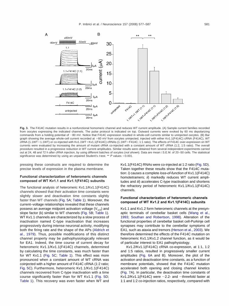
onsequences of this F414C mutation on Kv1.1 function;414C cRNA was in vitro transcribed and injected intoocytes and whole-cell currents recorded under TEVConfiguration. However, no detectable currents were elic-
ted by membrane depolarizations up to 170 mV, lasting upo 300 ms, from �100 oocytes injected with F414C cRNA,ven at higher cRNA concentrations (�twofold increase;ig. 3). By contrast, WT Kv1.1 cRNA injection gave rise to
ypical delayed-rectifier K� currents (Fig. 3; c.f. Fig. 1 in’Adamo et al., 1998).
However, EA1 is an autosomal dominant disorder andhe patients, harboring the F414C mutation, are heterozy-ous. Therefore, heteromeric channels composed of aixture of WT and mutant subunits might be formed if bothormal and mutated alleles are coexpressed. To address
his issue, coexpression studies were carried out; equalmounts of WT and mutant RNAs were coinjected in theame oocyte and the results compared with those obtainedrom cells injected with an equivalent amount of WT RNA.he co-injection of WT and F414C cRNAs (1:1 ratio) re-ulted in an average current amplitude, at �60 mV, that
I:1
II:1 II:6 II:2 II:3
III:2 III:3 III:4III:1
A
B
nt 1241 TTC->TGC
F414C
ig. 2. Pedigree of the EA1 family and sequence analysis of the KCNicily affected by EA1. All affected patients harbor the novel mutation Frepresentative patient shows a double peak (arrow) at nucleotide 124utation co-segregates with the pathological phenotype. Sequencing o
I:4, II:5 and III:7 of the EA1 family for mutation detection. This analysM_000217 (nucleotide 1241 from KCNA1 translation start codon), lev1.1 subunit. (C) Conventional membrane topology of a human Kv1.1
ncluding the F414C, is indicated. Four of such subunits are assemblev1 subfamily, may form heterotetrameric channels.
as �1.7-fold smaller than the control WT current (Fig. a
B). Moreover, �twofold and �fivefold F414C cRNA over-xpression resulted in �5.7-fold, �12-fold WT current re-uction, respectively (Fig. 3B; 1:2, 1:5 ratios).
These results indicate that F414C homotetramerichannel is nonfunctional, suggesting that the mutatedv1.1 subunit may be recognized as an anomalous proteinnd degraded by the trafficking machinery of the cell be-
ore reaching the plasma membrane. Alternatively, thehannel protein may be delivered normally to the plasmaembrane, but could have a ‘gating’ defect and thereforee unable to open correctly. In order to discriminate be-
ween these possibilities we examined the expression pat-ern of F414C channels in Xenopus laevis oocytes, bynalyzing the fluorescence intensity of cells injected withv1.1-GFP or Kv1.1(F414C)-GFP RNAs. Imaging studiesith confocal microscopy suggest that the Kv1.1(F414C)-FP construct displays an expression pattern similar tov1.1-GFP (Fig. 4). Furthermore, the biophysical proper-
ies of these GFP-tagged constructs were identical to theorresponding untagged channels (not shown). However,uture studies using a combination of biochemical assays
I:2
II:4 II:8 II:5 II:9
III:6 III:7 III:8 III:9 III:10
g region. (A) Schematic drawing of the three-generation family fromthe KCNA1 gene. (B) Sequence chromatogram of genomic DNA fromanges T�G, resulting in F414C mutation. This novel missense F414CKCNA1 coding region was performed in subjects I:1, I:2, II:1, II:2, II:3,
us to identify an heterozygous T�G transition at nucleotide 2346 inPhe to Cys substitution at codon 414 in the C-terminal region of the
The position of the mutations that have been identified in EA1 patients,a functional homotetrameric channel. Different subunits, belonging to
II:7
III:5
C
A1 codin414C in1 that chf the full
is allowedading to asubunit.d to form
nd confocal microscopy analysis of mammalian cells ex-
pp
Fc
TcsfcpsWipbacfhbfpcFccT
KTthttc
Fc
Ka1fsEtho
aaama(K
Ffcgccpo of oocytes P values
P. Imbrici et al. / Neuroscience 157 (2008) 577–587 581
ressing these constructs are required to determine therecise levels of expression in the plasma membrane.
unctional characterization of heteromeric channelsomposed of WT Kv1.1 and Kv1.1(F414C) subunits
he functional analysis of heteromeric Kv1.1/Kv1.1(F414C)hannels showed that their activation time constants werelightly slower and deactivation time constants slightlyaster than WT channels (Fig. 5A; Table 1). Moreover, theurrent–voltage relationships revealed that these channelsossess an average midpoint activation voltage (V1/2) andlope factor (k) similar to WT channels (Fig. 5B; Table 1).T Kv1.1 channels are characterized by a slow process of
nactivation named C-type inactivation that increasesrogressively during intense neuronal activity, modifyingoth the firing rate and the shape of the APs (Aldrich etl., 1979). Thus, possible modifications of this distincthannel property may be pathophysiologically relevantor EA1. Indeed, the time course of current decay foreteromeric Kv1.1/Kv1.1(F414C) channels, determinedy calculating the time constants, was much faster than
or WT Kv1.1 (Fig. 5C; Table 1). This effect was moreronounced when a constant amount of WT cRNA wasoinjected with a higher amount of F414C cRNA (1:2 ratio;ig. 5C). Furthermore, heteromeric Kv1.1/Kv1.1(F414C)hannels recovered from C-type inactivation with a timeourse significantly faster than for WT Kv1.1 (Fig. 5D;
ig. 3. The F414C mutation results in a nonfunctional homomeric charom oocytes expressing the indicated channels. The pulse protocoommands from a holding potential of �80 mV. Notice that F414C exraph showing the average whole-cell current recorded at �60 mV frRNA (1.1WT�1.1WT) or co-injected with Kv1.1WT�Kv1.1(F414C) cRurrents were evaluated by increasing the amount of mutant cRNArocedure resulted in a progressive reduction in WT current amplitudeut at 24, 48 and 72 h after cRNA injection, by using different batchesignificance was determined by using an unpaired Student’s t-test. **
able 1). This recovery was even faster when WT and 1
v1.1(F414C) RNAs were co-injected at 1:2 ratio (Fig. 5D).aken together these results show that the F414C muta-
ion: i) causes a complete loss-of-function of Kv1.1(F414C)omotetramers; ii) markedly reduces WT current ampli-
udes and iii) accelerates C-type inactivation and shortenshe refractory period of heteromeric Kv1.1/Kv1.1(F414C)hannels.
unctional characterization of heteromeric channelsomposed of WT Kv1.2 and Kv1.1(F414C) subunits
v1.1 and Kv1.2 form heteromeric channels at the presyn-ptic terminals of cerebellar basket cells (Wang et al.,993; Southan and Robertson, 1998). Alteration of the
unctional properties of cerebellar basket cell-Purkinje cellynapses may contribute to the cerebellar symptoms ofA1, such as ataxia and tremors (Herson et al., 2003). We
herefore determined the effects of the F414C mutation oneteromeric Kv1.1/Kv1.2 channel function, as it would bef particular interest to EA1 pathophysiology.
Kv1.2/Kv1.1(F414C) cRNA co-expression, at 1:1, 1:2nd 1:5 ratios, resulted in progressively smaller currentmplitudes (Fig. 6A and B). Moreover, the plot of thectivation and deactivation time constants, as a function ofembrane potentials, showed that the F414C mutationccelerated both opening and closing channel kineticsFig. 7A). In particular, the deactivation time constants ofv1.2/Kv1.1(F414C) were �2.2- and �threefold faster at
reduces WT current amplitude. (A) Sample current families recordedted on top. Outward currents were evoked by 60 ms depolarizingresulted in whole-cell currents similar to uninjected oocytes. (B) Bar
es uninjected, injected with either Kv1.1(F414C) cRNA (F414C), WTWT�F414C; 1:1 ratio). The effects of F414C over-expression on WTd with a constant amount of WT cRNA (1:2, 1:5 ratio). The overallresults were obtained from several independent experiments carrieds (not shown). Data are mean�S.E.M. of 20–50 cells. The statistical0.001.
nnel andl is indicapression
om oocytNAs (1.1
co-injectes. Similar
:1 and 1:2 co-injection ratios, respectively, compared with

KTtaptstifmmirtctrp
Ht
t1tsiannwmIlhN2afsdecs
Fsato
P. Imbrici et al. / Neuroscience 157 (2008) 577–587582
v1.2/Kv1.1WT (Fig. 7A; see inset for direct comparison;able 2). Another pronounced effect of the F414C muta-
ion was an approximately 18 mV right-shift of the midpointctivation voltage (V1/2) of heteromeric channels com-ared with WT (Fig. 7B; Table 2). By contrast, the V1/2 ofhe heteromeric Kv1.1/Kv1.1(F414C) channels, was right-hifted by only 2–3 mV compared with WT (Table 1). Both,he time course of C-type inactivation and recovery fromnactivation, for the mutated heteromeric channels, wereaster than WT Kv1.1 and increased with the amount ofutant cRNA injected (Fig. 7C and D; Table 2). In sum-ary, these results show that the F414C mutation also
mpairs heteromeric Kv1.1/Kv1.2 channel function by: i)educing WT current amplitudes; ii) markedly speeding uphe deactivation kinetics of the channel; iii) shifting theurrent–voltage relationships to more depolarized poten-ials; and iv) accelerating C-type inactivation, which furthereduces channel availability and shortens the refractoryeriod of these channels.
DISCUSSION
ere, we have described a Sicilian family affected by EA1
A1
B1
C1
A2
B2
C2
0
10
20
30
40
50
60
0
Flu
ores
cenc
e In
tens
ity
(arb
itrar
y un
its)
0
10
20
30
40
50
60
0 1 2 3
Flu
ores
cenc
e In
tens
ity
(arb
itrar
y un
its)
mm
ig. 4. Confocal microscopy analysis of Kv1.1(F414C) channels exprurface profile of a representative uninjected oocyte (A1) and of those incquired at the equator, of the corresponding oocytes shown on top. N
o Kv1.1WT, whereas, the fluorescence intensity of uninjected oocytesf corresponding images in B1–3.
hat is characterized by typical and atypical clinical symp- i
oms (Van Dyke et al., 1975; Brunt and van Weerden,990; Eunson et al., 2000). A large degree of variability in
he onset of the disease, frequency of the attacks, clinicalymptoms and high drug resistance were reported by fam-
ly members, as has been described for many other EA1-ffected patients (Rajakulendran et al., 2007). Interictaleuromyotonia, leg rigidity during attacks (neuromyoto-ia), episodes of intense myokymic activity during attacksithout ataxia and other neurological deficits are less com-only observed among the EA1 patients described so far.
ndeed, clinical symptoms such as distal weakness, pro-onged neuromyotonia and stiffness with inability to walkave been described in only a few cases (Gancher andutt, 1986; Brunt and van Weerden, 1990; Eunson et al.,000; Klein et al., 2004). Furthermore, the proband showsvery specific phenotype, characterized by increasing
requency of attacks with ageing, as well as a relation-hip between both the onset and the worsening of theisease with a specific traumatic physical or emotionalvent. The isolated photosensitive generalized tonic–lonic seizure reported by the proband’s nephew is notufficient to make a diagnosis of epilepsy, although the
A3
B3
C3
2 30
10
20
30
40
50
60
0 1 2 3
Flu
ores
cenc
e In
tens
ity
(arb
itrar
y un
its)
mm
Xenopus laevis oocytes. (A) 3D Reconstruction of the fluorescenceth Kv1.1WT (A2) and F414C (A3) RNAs. (B1–3) Fluorescence intensity,e Kv1.1(F414C)-GFP construct showed an expression pattern similarost absent. (C1–3) Fluorescence intensity plots of line profile analysis
1mm
ession injected wi
ote that thwas alm
nvolvement of the new genetic defect cannot be excluded.

btcs
wGt
mcct(ma
F(mrdtwtKtVrCdtci(i(dToic
P. Imbrici et al. / Neuroscience 157 (2008) 577–587 583
However, genetic analysis of this family that includedoth a new linkage study and mutation screening, resulted in
he identification of a novel mutation in the KCNA1 potassiumhannel gene that results in the substitution of a highly con-erved phenylalanine with a cysteine at position 414.
Although the Kv1.1(F414C) homotetrameric channelsere nonfunctional, confocal imaging analysis of Kv1.1-FP and Kv1.1(F414C)-GFP injected oocytes suggests
ig. 5. The F414C mutation speeds up the C-type inactivation anA) The activating and deactivating current traces were fitted withutation on the activation and deactivation kinetics of the channel.
atio) and Kv1.1�F414C (�; 1:2 ratio) channels were calculated aata points with the equation: ��� V1/2 exp{(V�V1/2)/k}, where � V1/2
he voltage dependence of the time constants. This plot shows thathich can be directly observed also from the representative current
he co-injection of WT and F414C cRNAs (1:1 and 1:2 ratio; insv1.1�F414C (Œ; 1:1 ratio) and Kv1.1�F414C (�; 1:2 ratio) was d
o several voltages. The normalized current–voltage data points wer
1/2 and slope factor k were calculated. The inset, on the right hanecorded at �50 mV. Notice that the mutation changes very little-type inactivation kinetics, a test pulse to �60 mV for 3.5 min wecaying phase of the current was fitted with a double-exponential f
he normalized current traces resulting from either the expressionRNA at 1:1 (solid line) and 1:2 (dotted line) ratios. The bar graph sh
nactivation for the indicated channels. These results clearly show thD) The recovery from C-type inactivation was determined by usinncreasing duration (range: 0.010 –21.87 s). The current amplituconditioning), normalized and plotted as a function of the interpouble-exponential function from which the time constants were calche inset shows sample current traces evoked by the two-pulse prof the peak current, evoked by the test pulse, with a double-ex
nactivation. A careful evaluation of the biophysical properties of chaurrent reduction. Data are means�S.E.M. of 10 –15 cells. Studen
hat the mutated channels are delivered to the plasma m
embrane, at least in part, but unable to open correctly. Byontrast, the R417X truncation mutant, which is locatedlose to the F414C, results in a protein that is nonfunc-ional, but which is preferentially retained in the cytoplasmRea et al., 2002). However, the R417X truncation re-oves the C-terminus (and associated trafficking signals)nd is therefore likely to result in a highly aberrant protein.
Even though co-expression with the Kv1.1(F4141C)
ry from inactivation of heteromeric Kv1.1/Kv1.1(F414C) channels.exponential function, in order to assess the effects of the F414Cant time constants for the human Kv1.1 (�), Kv1.1�F414C (Œ; 1:1d as a function of the test pulse. The solid line shows the fit of the
e constant at the V1/2 of the channels and k is the slope factor fortion exerts little effect on the activation– deactivation of the channelorded at �60 mV for WT channels, overlain with that resulting fromThe voltage-dependence of channel activation for Kv1.1WT (�),d by recording tail currents at �50 mV, after prepulse commands
ith the Boltzmann function I�1/1�exp{�(V�V1/2)/k} from which thehows representative tail current families for the indicated channelsge-dependence of heterozygous channels. (C) To determine thered to oocytes expressing WT and heteromeric channels and theom which the time constants were calculated. The left panel showsalone (dashed line) or from the co-expression of WT and F414Cfast (white bar) and slow (dashed bar) time constants of the C-typeutation speeds up the inactivation kinetics of heteromeric channels.le-pulse protocol to �60 mV, separated by interpulse intervals ofked by the second pulse (test) were divided by the first pulserval. The solid curves indicate the fit of the data points with ar Kv1.1WT (�), Kv1.1�F414C (Œ; 1:1) and Kv1.1�F414C (�; 1:2).the indicated channels. The superimposed solid line shows the fit
l function. Note that the mutation speeds up the recovery fromsulting from injections at 1:5 ratio, was not possible due to marked* P values 0.05; ** P values 0.001.
d recovea single
The relevnd plotteis the tim
the mutatrace recet). (B)etermine
e fitted wd side, sthe volta
as deliveunction frof Kv1.1ows theat the mg a doubdes evoulse inteulated fotocol for
ponentiannels, re
t’s t-test:
utant subunit resulted in a reduction in the current am-

prnirssaocmtV
s
pots1daamfct
Pb
Fowap*
T
V
KK
K
a**
P. Imbrici et al. / Neuroscience 157 (2008) 577–587584
litudes of WT Kv1.1 and heteromeric Kv1.1/Kv1.2, thiseduction was not enough to be considered a dominantegative effect. Another peculiarity of the F414C mutation
s its ability to impair the biophysical properties of cur-ents resulting from the co-expression of Kv1.1/Kv1.2ubunits more dramatically than those of WT Kv1.1ubunits. In fact, the activation– deactivation kineticsnd the voltage-dependence of WT Kv1.1 channels werenly slightly affected by the mutant F414C subunit. Byontrast, the deactivation time constant of mutant hetero-eric Kv1.1(F414C)/Kv1.2 currents was �2.2-fold faster
han their WT equivalent and the mutation right-shifted the
1/2 by �18 mV.The overexpression study showed that the mutated
ubunit reduces current amplitudes and impairs their bio-
ig. 6. The F414C mutation reduces the current amplitude of heterof current recorded from oocytes expressing the indicated channelshole-cell current recorded from oocytes: uninjected, injected withnd with 1:1, 1:2 and 1:5 ratios of Kv1.2 to mutant cRNA. The curren
able 1. Biophysical parameters of Kv1.1WT and Kv1.1WT/Kv1.1F41
oltage Voltage dependenceof activation
Kinetic ofactivation,�V1/2 (ms)
Kinetic odeactivat�V1/2 (ms
V1/2 (mV) k (mV)
v1.1 �26.4�0.4 6.0�0.2 6.4�0.1 17.6�1.0v1.1�Kv1.1F414C(1:1)
�23.8�0.4 7.3�0.2 8.6�0.2 15.3�1.5
v1.1�Kv1.1F414C(1:2)
�23.4�0.4 6.8�0.2 9.9�0.2 15.0�1.5
The data are the means�S.E.M. of 10–15 cells. The values in pares percentages.P values 0.05, Student’s t-test.
* P values 0.001, Student’s t-test.
otential of �80 mV. Data are mean�S.E.M. of 6 to 10 cells. The statisticalP values 0.05.
hysical properties in a way that is dependent on the ratiof co-injected cRNAs. These results strongly suggest that
he degree of impairment is related to the number of F414Cubunits incorporated into the channels (D’Adamo et al.,998), which strictly depends on the relative amounts of theifferent cRNAs co-injected (Kavanaugh et al., 1992; Rea etl., 2002). Possibly, individual variability in transcriptionnd translation processing of the WT and mutated genesay generate channels with different stoichiometries and
unctional defects. Interestingly, this mechanism may ac-ount for the high degree of inter- and intrafamilial symp-omatic variation.
The functionality of specific circuits within the CNS andNS may also be impacted by these phenomena. It haseen postulated that the attacks of cerebellar ataxia may
annels composed of Kv1.2 and Kv1.1 subunits. (A) Sample familieslse protocol is indicated on top. (B) Bar graph showing the average14C) cRNA (F414C), with 1:1 ratio of Kv1.2 and Kv1.1WT cRNAs
evoked by 60 ms depolarizing command at �60 mV, from a holding
nels
type inactivation Recovery from C-typeinactivation
st (s) �slow (s) �fast (ms) �slow (s)
�0.7 (22%) 50.0�1.9 (78%) 134�30 (50%) 4.8�1.4 (50%)�0.3* (26%) 38.5�2.0** (74%) 102�23 (49%) 2.8�0.6 (51%)
�0.6* (38%) 29.0�4.4** (62%) 73�13* (56%) 1.7�0.3 (44%)
re the mean amplitudes of the fast and slow components, expressed
meric ch. The puKv1.1(F4ts were
4C chan
fion,)
C-
�fa
7.65.8
5.1
ntheses a
significance was determined by using an unpaired Student’s t-test.

rc
cf
FcctKttKbN(mTmKfc oked by( * P value
T
V
KKKK
a**
P. Imbrici et al. / Neuroscience 157 (2008) 577–587 585
esult from altered GABAergic transmission at the basketell–Purkinje cell synapse due to impaired Kv1.1/Kv1.2
ig. 7. The F414C mutation alters the kinetics of activation–deactivahannels. (A) Activating and deactivating current traces were recordedalculate the relative time constants for Kv1.2 (Œ), Kv1.2�Kv1.1 (�), Khem as a function of the test pulse. The data points were fitted as in Fv1.2�Kv1.1 channels normalized and overlain with those resulting fr
he heteromeric channels carrying the mutation deactivate much fasteail currents recorded at �50 mV as a function of the pre-pulse pv1.2�F414C (�; 1:2 ratio) channels. The solid curves indicate the fiy the mutation. The inset, on the right hand side, shows representaormalized current traces evoked by depolarizing steps at �60 mV,
dashed line) or Kv1.2�F414C (1:1 and 1:2 ratio; solid and dotted linutated channels decayed faster than the WT. Therefore, they were fihe bar graph shows the fast (white bar) and slow (dashed bar) averutation, concentration dependently. (D) Recovery from C-type inav1.2�F414C (�; 1:2 ratio) channels determined as detailed in Fig. 5
unction from which the time constants were calculated. The exproncentration dependently. The inset shows sample current traces evsolid line). Data are means�S.E.M. of 10–15 cells. Student’s t-test:
able 2. Biophysical parameters of Kv1.2�Kv1.1WT and Kv1.2WT/K
oltage Voltage dependence ofactivation
Kinetic ofactivation,�V1/2 (ms)
Kinetdeac�V1/2
V1/2 (mV) k (mV)
v1.2 �8.3�0.5 10.0�0.5 5.3�0.1 23.0�
v1.2�Kv1.1 �18.3�0.5 12.0�0.5 3.3�0.1 15.0�
v1.2�Kv1.1F414C (1:1) �0.3�1.3** 12.5�1.1 1.7�0.06 6.7�
v1.2�Kv1.1F414C (1:2) 2.3�1.4** 13.0�1.1 2.1�0.05 4.9�
The data are the means�S.E.M. of 10–15 cells. The values in pares percentages.P values 0.05, Student’s t-test.
* P values 0.001, Student’s t-test.
hannel function. Indeed, electrophysiological recordingsrom knock-in mice, harboring the EA1 mutation V408A
age-dependence and C-type inactivation in heteromeric Kv1.1/Kv1.2ent potentials and fitted with a single exponential function, in order to14C (Œ; 1:1 ratio) and Kv1.2�F414C (�; 1:2 ratio) channels and plothe inset shows representative current traces recorded at �60 mV for-injection of Kv1.2 with F414C cRNAs (1:1 and 1:2 ratio). Notice thatWT. (B) Current–voltage relationships obtained by plotting the peak
or Kv1.2 (Œ), Kv1.2�Kv1.1 (�), Kv1.2�F414C (Œ; 1:1 ratio) andta points with the Boltzmann function that is significantly right-shifted
current families recorded at �50 mV for the indicated channels. (C).5 min, and delivered to oocytes co-expressing either Kv1.2�Kv1.1ectively; left panel). The currents resulting from the activation of thedouble-exponential functions to calculate the relevant time constants.e constants of the C-type inactivation, which were decreased by thefor Kv1.2 (Œ), Kv1.2�Kv1.1 (�), Kv1.2�F414C (Œ; 1:1 ratio) andlid curves indicate the fit of the data points with a double-exponentialf the mutated subunit accelerates the recovery from inactivation,the two-pulse protocol and then fit with a double exponential functions 0.05; ** P values 0.001.
C channels
C-type inactivation Recovery from C-typeinactivation
�fast (s) �slow (s) �fast (ms) �slow (s)
5.4�0.8 (23%) 33.0�1.4 (77%) 278�5 (54%) 1.5�0.2 (38%)5.3�1.3 (24%) 29.1�2.1 (76%) 311�86 (62%) 2.8�0.6 (38%)4.7�0.6 (51%) 15.1�1.5** (49%) 255�40 (71%) 2.4�0.3 (29%)1.8�0.3* (23%) 7.6�0.6** (77%) 120�28* (71%) 1.8�0.3 (29%)
re the mean amplitudes of the fast and slow components, expressed
tion, voltat differ
v1.2�F4ig. 5A. T
om the cor than theotential ft of the dative taillasting 3es, resp
tted withaged timctivationD. The soession o
v1.1F414
ic oftivation,(ms)
1.21.70.7**0.3**
ntheses a

(qcermtcbtk2
Iitfainiomn
AnPtISBDgtP
A
A
B
B
B
C
C
D
D
E
G
H
I
I
IK
K
K
K
L
L
M
R
R
S
S
S
P. Imbrici et al. / Neuroscience 157 (2008) 577–587586
V408A/�), revealed an increased amplitude and fre-uency of GABAergic IPSCs onto cerebellar Purkinje cells,ompared with WT animals (Herson et al., 2003). Thisvidence supports the hypothesis that an increased GABAelease from presynaptic basket cells onto Purkinje cellsay alter the output of the cerebellar cortex which provides
he signals required for motor planning, execution andoordination (Ito, 1984). Likely, the gating defects causedy the F414C mutation on Kv1.1/Kv1.2 currents may alter
he GABAergic inputs from cerebellar basket cells to Pur-inje cells as reported for V408A/� mice (Herson et al.,003).
CONCLUSION
n conclusion, a novel F414C mutation has been identifiedn an Italian family displaying distinct phenotypic charac-eristics. Both homomeric and heteromeric Kv1 channelunction is severely affected by this mutation, but althoughll affected patients harbor the same genetic defect, the
ntrafamilial symptoms are highly variable. The mecha-isms accounting for such a phenotypic variability, its ep-
sodic nature, and how different stimuli trigger the attacksf ataxia and epilepsy are mostly unknown and remain aajor challenge for future research into this and othereurological diseases.
cknowledgments—The financial support of Telethon-Italy (granto. GGP030159), of MIUR-COFIN 2005, of COMPAGNIA di Sanaolo (Turin), the Fondazione Cassa di Risparmio di Perugia, and
he Royal Society (London, UK) is gratefully acknowledged. Paolambrici is the recipient of a Ph.D. fellowship from COMPAGNIA dian Paolo (Turin). We thank Ezio Mezzasoma and Domenicoambagioni for outstanding technical assistance and Andria’Orazio for initial work. Finally, we would also like to express ourratitude to the Italian Red Cross (women’s section of Perugia) for
he donation of scientific equipment to the Section of Humanhysiology for this research.
REFERENCES
delman JP, Bond CT, Pessia M, Maylie J (1995) Episodic ataxiaresults from voltage-dependent potassium channels with alteredfunctions. Neuron 15:1449–1454.
ldrich RW Jr, Getting PA, Thompson SH (1979) Inactivation of de-layed outward current in molluscan neurone somata. J Physiol291:507–530.
rowne DL, Gancher ST, Nutt JG, Brunt ER, Smith EA, Kramer P, LittM (1994) Episodic ataxia/myokymia syndrome is associated withpoint mutations in the human potassium channel gene, KCNA1.Nat Genet 8:136–140.
rowne DL, Brunt ER, Griggs RC, Nutt JG, Gancher ST, Smith EA, LittM (1995) Identification of two new KCNA1 mutations in episodicataxia/myokymia families. Hum Mol Genet 4:1671–1672.
runt ERP, van Weerden TW (1990) Familial paroxysmal kinesigenicataxia and continuous myokymia. Brain 113:1361–1382.
omu S, Giuliani M, Narayanan V (1996) Episodic ataxia and myokymiasyndrome: a new mutation of potassium channel gene Kv1.1. AnnNeurol 40:684–687.
usimano A, D’Adamo MC, Pessia M (2004) An episodic ataxia type-1mutation in the S1 segment sensitises the hKv1.1 potassium chan-nel to extracellular Zn2�. FEBS Lett 576:237–244.
’Adamo MC, Imbrici P, Sponcichetti F, Pessia M (1999) Mutations in
the KCNA1 gene associated with episodic ataxia type-1 syndromeimpair heteromeric voltage-gated K(�) channel function. FASEB J13:1335–1345.
’Adamo MC, Liu Z, Adelman JP, Maylie J, Pessia M (1998) Episodicataxia type-1 mutations in the hKv1.1 cytoplasmic pore region alterthe gating properties of the channel. EMBO J 17:1200–1207.
unson LH, Rea R, Zuberi SM, Youroukos S, Panayiotopoulos CP,Liguori R, Avoni P, McWilliam RC, Stephenson JB, Hanna MG, Kull-mann DM, Spauschus A (2000) Clinical, genetic, and expressionstudies of mutations in the potassium channel gene KCNA1 revealnew phenotypic variability. Ann Neurol 48:647–656.
ancher ST, Nutt JG (1986) Autosomal dominant episodic ataxia: aheterogeneous syndrome. Mov Disord 1:239–53.
erson PS, Virk M, Rustay NR, Bond CT, Crabbe JC, Adelman JP,Maylie J (2003) A mouse model of episodic ataxia type-1. NatNeurosci 6:378–383.
mbrici P, Cusimano A, D’Adamo MC, De Curtis A, Pessia M (2003)Functional characterization of an episodic ataxia type-1 mutationoccurring in the S1 segment of hKv1.1 channels. Pflugers Arch446:373–379.
mbrici P, D’Adamo MC, Cusimano A, Pessia M (2007) Episodic ataxiatype 1 mutation F184C alters Zn2�-induced modulation of thehuman K� channel Kv1.4-Kv1.1/Kvbeta1.1. Am J Physiol CellPhysiol 292:C778–C787.
to M (1984) The cerebellum and neural control. New York: Raven.avanaugh MP, Hurst RS, Yakel J, Varnum MD, Adelman JP, North
RA (1992) Multiple subunits of a voltage-dependent potassiumchannel contribute to the binding site for tetraethylammonium.Neuron 8:493–497.
inali M, Jungbluth H, Eunson LH, Sewry CA, Manzur AY, Mercuri E,Hanna MG, Muntoni F (2004) Expanding the phenotype of potas-sium channelopathy: severe neuromyotonia and skeletal deformi-ties without prominent episodic ataxia. Neuromuscul Disord14:689–693.
lein A, Boltshauser E, Jen J, Baloh RW (2004) Episodic ataxia type1 with distal weakness: a novel manifestation of a potassiumchannelopathy. Neuropediatrics 35:147–149.
ullmann DM, Rea R, Spauschus A, Jouvenceau A (2001) The inher-ited episodic ataxias: how well do we understand the diseasemechanisms? Neuroscientist 7:80–88.
ee H, Wang H, Jen JC, Sabatti C, Baloh RW, Nelson SF (2004) A novelmutation in KCNA1 causes episodic ataxia without myokymia. HumMutat 24:536–542.
itt M, Kramer P, Browne D, Gancher S, Brunt ER, Root D, Phrom-chotikul T, Dubay CJ, Nutt J (1994) A gene for episodic ataxia/myokymia maps to chromosome 12p13. Am J Hum Genet55:702–709.
aylie B, Bissonnette E, Virk M, Adelman JP, Maylie JG (2002)Episodic ataxia type 1 mutations in the human Kv1.1 potassiumchannel alter hKvbeta 1-induced N-type inactivation. J Neurosci22:4786–4793.
ajakulendran S, Schorge S, Kullmann DM, Hanna MG (2007) Epi-sodic ataxia type 1: a neuronal potassium channelopathy. Neuro-therapeutics 2:258–266.
ea R, Spauschus A, Eunson L, Hanna MG, Kullmann DM (2002)Variable K� channel subunit dysfunction in inherited mutations ofKCNA1. J Physiol 538:5–23.
cheffer H, Brunt ER, Mol GJ, van der Vlies P, Stulp RP, Verlind E,Mantel G, Averyanov YN, Hofstra RM, Buys CH (1998) Threenovel KCNA1 mutations in episodic ataxia type I families [pub-lished erratum appears in Hum Genet 1998, 102, 713]. Hum Genet102:464–466.
hook SJ, Mamsa H, Jen JC, Baloh RW, Zhou L (2008) Novel muta-tion in KCNA1 causes episodic ataxia with paroxysmal dyspnea.Muscle Nerve 37:399–402.
outhan AP, Robertson B (1998) Patch-clamp recordings form cere-bellar basket cell bodies and their presynaptic terminals reveal anasymmetric distribution of voltage-gated potassium channels.
J Neurosci 18:948–955.
V
W
Z
Z
S
S
P. Imbrici et al. / Neuroscience 157 (2008) 577–587 587
an Dyke DH, Griggs RC, Murphy MJ, Goldstein MN (1975) Heredi-tary myokymia and periodic ataxia. J Neurol Sci 25:109–118.
ang H, Kunkel DD, Martin TM, Schwartzkroin PA, Tempel BL (1993)Heteromultimeric K� channels in terminal and juxtaparanodal re-gions of neurons. Nature 365:75–79.
err P, Adelman JP, Maylie J (1998) Episodic ataxia mutations inKv1.1 alter potassium channel function by dominant negative ef-fects or haploinsufficiency. J Neurosci 18:2842–2848.
uberi SM, Eunson LH, Spauschus A, De Silva R, Tolmie J, Wood
NW, McWilliam RC, Stephenson JP, Kullmann DM, Hanna MG t(1999) A novel mutation in the human voltage-gated potassiumchannel gene (Kv1.1) associates with episodic ataxia type 1 andsometimes with partial epilepsy. Brain 122:817–825.
APPENDIX
upplementary data
upplementary data associated with this article can be found, in
he online version, at doi:10.1016/j.neuroscience.2008.09.022.(Accepted 11 September 2008)(Available online 24 September 2008)