Embed Size (px)
Citation preview
RESEARCH ARTICLE Open Access
A novel interaction between kinaseactivities in regulation of cilia formationNicole DeVaul2, Katerina Koloustroubis1, Rong Wang1 and Ann O. Sperry1*
Abstract
Background: The primary cilium is an extension of the cell membrane that encloses a microtubule-based axoneme.Primary cilia are essential for transmission of environmental cues that determine cell fate. Disruption of primarycilia function is the molecular basis of numerous developmental disorders. Despite their biological importance,the mechanisms governing their assembly and disassembly are just beginning to be understood. Cilia growthand disassembly are essential events when cells exit and reenter into the cell cycle. The kinases never in mitosis-kinase 2 (Nek2) and Aurora A (AurA) act to depolymerize cilia when cells reenter the cell cycle from G0.
Results: Coexpression of either kinase with its kinase dead companion [AurA with kinase dead Nek2 (Nek2 KD) orNek2 with kinase dead AurA (AurA KD)] had different effects on cilia depending on whether cilia are growing orshortening. AurA and Nek2 are individually able to shorten cilia when cilia are growing but both are required whencilia are being absorbed. The depolymerizing activity of each kinase is increased when coexpressed with the kinasedead version of the other kinase but only when cilia are assembling. Additionally, the two kinases act additively whencilia are assembling but not disassembling. Inhibition of AurA increases cilia number while inhibition of Nek2significantly stimulates cilia length. The complex functional relationship between the two kinases reflects theirphysical interaction. Further, we identify a role for a PP1 binding protein, PPP1R42, in inhibiting Nek2 andincreasing ciliation of ARPE-19 cells.
Conclusion: We have uncovered a novel functional interaction between Nek2 and AurA that is dependent onthe growth state of cilia. This differential interdependence reflects opposing regulation when cilia are growingor shortening. In addition to interaction between the kinases to regulate ciliation, the PP1 binding protein PPP1R42directly inhibits Nek2 independent of PP1 indicating another level of regulation of this kinase. In summary, we demonstratea complex interplay between Nek2 and AurA kinases in regulation of ciliation in ARPE-19 cells.
Keywords: Cilia, AurA, Nek2, PP1, PPP1R42
BackgroundPrimary cilia are microtubule-based organelles that pro-trude from the cell membrane to receive and transduceenvironmental signals. Interference with the formationand/or stability of cilia disrupts signaling pathways essen-tial for normal development and maintenance of the dif-ferentiated state ([1–3]; as reviewed in [4, 5]). A diversecollection of developmental disorders, ciliopathies, stemdirectly from disruption of ciliary function and display awide range of abnormalities from cystic kidney to obesity(as reviewed in [6, 7]). Virtually all cells form primary cilia,
which are structurally analogous to flagella. Cilia assemblewhen cells exit the cell cycle. As cells reenter the cell cycleand begin to proliferate, cilia disassemble, the basal bodydetaches from the plasma membrane, and centrosomesduplicate to form the mitotic spindle. Although the inven-tory of proteins that constitute cilia is increasing, themechanisms regulating their formation and disassemblyare just beginning to be defined.Cilia display regulated growth and retraction when enter-
ing and exiting the cell cycle. Cilia grow as cells enter G0
and are absorbed prior to reentry into the cell cycle. Twoimportant regulators of cilia absorption are the kinasesNek2 (NIMA related kinase 2) and AurA (Aurora A). Nek2is a serine/threonine kinase that is localized to the distalportion of the mother centriole and functions in both cilia
* Correspondence: [email protected] and Cell Biology, East Carolina University, Brody School ofMedicine, Greenville, NC, USAFull list of author information is available at the end of the article
© The Author(s). 2017 Open Access This article is distributed under the terms of the Creative Commons Attribution 4.0International License (http://creativecommons.org/licenses/by/4.0/), which permits unrestricted use, distribution, andreproduction in any medium, provided you give appropriate credit to the original author(s) and the source, provide a link tothe Creative Commons license, and indicate if changes were made. The Creative Commons Public Domain Dedication waiver(http://creativecommons.org/publicdomain/zero/1.0/) applies to the data made available in this article, unless otherwise stated.
DeVaul et al. BMC Cell Biology (2017) 18:33 DOI 10.1186/s12860-017-0149-5
shortening and centrosome duplication [8, 9]. Nek2controls cilia disassembly; depletion of Nek2 causesan increase in the number of ciliated cells [8]. Nek2has been linked directly to disruption of left-rightasymmetry, a biological consequence of cilia dysfunc-tion [10]. Nek2 also regulates intraflagellar transport(IFT) through phosphorylation of the kinesin KIF24to stimulate cilia depolymerization [11]. Additionally,Nek2 induces centrosome separation prior to celldivision. Overexpression of active Nek2 causes pre-mature splitting of centrosomes [12] due to phos-phorylation and destabilization of centrosomal linkerproteins [9].AurA, another serine/threonine kinase, is essential for
maintenance of cilia length and cilia retraction prior tocell cycle reentry in diverse organisms [13, 14] as well ascentrosome maturation, duplication, and spindle assem-bly (as reviewed by [15]). Pioneering work in Chlamydo-monas reinhardtii provided the first indication thatAurA regulates the length of the flagellum of this bifla-gellate alga [16, 17]. AurA is localized to and activated atthe basal body of cilia when cilia disassemble. Overex-pression of AurA in ciliated mammalian cells inducescilia disassembly through activation of a tubulin deacety-lase [13]. Like Nek2, AurA participates in preparation ofcentrosomes for cell division (reviewed in [18–20]).PP1, a serine/threonine phosphatase, is a common
regulator of both kinases in control of centrosomeseparation prior to spindle formation at mitosis; how-ever, its role in cilia biogenesis has not been investi-gated [19–22]. PP1 activity is itself regulated by bothpositive and negative regulatory subunits. The nega-tive regulator PPP1R2 (I2) inhibits PP1 activity inboth centrosome separation and cilia acetylation andstabilization [19, 23]. We have previously identified aPP1 binding protein, PPP1R42 that is involved incentrosome separation [24]; however, its role in cili-ation is not known.Our study provides evidence that Nek2 and AurA
interact differentially depending on cilia growth status.We demonstrate that Nek2 and AurA interact on sev-eral levels. They appear to share positive and negativefactors to enhance or inhibit depolymerization activitywhen cilia are disassembling or assembling, respect-ively. Nek2 and AurA act independently when cilia aregrowing but both are required to depolymerize cilia.Furthermore, we demonstrate that these two kinasesact additively to depolymerize cilia when cilia are grow-ing and are independently involved in cilia number andlength. These findings represent a novel functionalinteraction between two kinases involved in cilia disas-sembly. In addition, we identify inhibition of Nek2 byPPP1R42, a PP1 binding protein, which is independentof PP1.
ResultsRequirement for kinase activity is dependent on ciliagrowth stateWe investigated the interaction between AurA and Nek2by overexpressing the kinases and their kinase dead coun-terparts either alone or in combination in cells eithergrowing cilia after serum starvation or absorbing cilia afterreintroduction of serum (Fig. 1). The kinase dead versionsof Nek2 and AurA have been shown to localize to thecentrosome and to have a dominant negative effect on en-dogenous kinase function by sequestering substrates andupstream regulators of the kinases (Dr. Andrew Fry, per-sonal communication and [12, 25, 26]). Expressed proteinis maintained throughout the time course of treatment(Additional file 1: Figure S1) with a transfection efficiencyof 90% on average (Additional file 2: Figure S2) and cellsshow little toxicity after transfection. These experimentsexamine a time window between formation of cilia after celldivision and before the approximate onset of the next div-ision. The effect of experimental manipulation on cilia incell populations has precedent and results of such studieshave expanded our knowledge of cilia biology [13, 27–29].We first compared cilia number and length in cells ex-
pressing either kinase active or kinase dead versions ofAurA and Nek2 in cells assembling or disassemblingcilia to determine if the effect on cilia depolymerizationdiffered depending on cilia growth status (Fig. 1, com-pare Empty (control) vs AurA, and Empty (control) vsNek2). Previous studies have shown that AurA andNek2 induce cilia depolymerization [11, 13]; therefore,we expected that cells transfected with the active kinaseswould be less ciliated than control. This was true forcells assembling cilia (Fig. 1A, left panel, AurA vs con-trol, p = 0.008; Fig. 1B left panel, Nek2 vs control,p = 0.0009) but not for cells disassembling cilia. OnlyNek2 transfected cells were significantly less ciliatedthan control (Fig. 1B, right panel, p = 0.03). There wasno significant difference in cilia length between activekinase and control (Fig. 1C, AurA vs control; Fig. 1D,Nek2 vs control). The stronger effect of the active kinasecompared to control in cells assembling cilia may reflecta shift in the balance from assembling to disassemblingcatalyzed by the active kinases. The active kinases arenot as effective at further depolymerizing cilia when ciliaare already disassembling (Fig. 1A and B, right panels).We expected that cells transfected with the kinase dead
enzymes would have more and longer cilia compared tocontrol and wild-type (compare Fig. 1 control and AurAvs AurAKD, and control and Nek2 vs Nek2KD). However,the kinase dead enzymes of AurA and Nek2 did not in-crease cilia number compared to control either when ciliaare assembling or disassembling which may suggest thatciliation is at a maximum and the kinase dead version can-not further stimulate cilia formation above control levels
DeVaul et al. BMC Cell Biology (2017) 18:33 Page 2 of 18
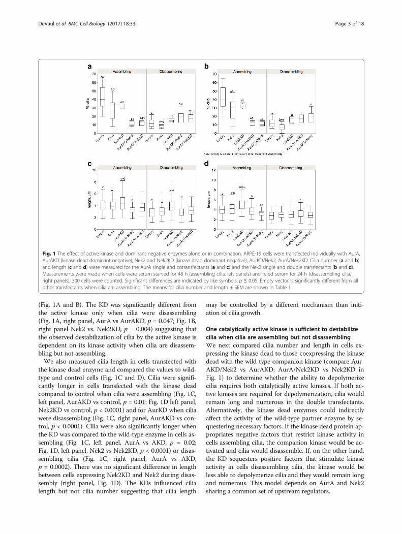
(Fig. 1A and B). The KD was significantly different fromthe active kinase only when cilia were disassembling(Fig. 1A, right panel, AurA vs AurAKD, p = 0.047; Fig. 1B,right panel Nek2 vs. Nek2KD, p = 0.004) suggesting thatthe observed destabilization of cilia by the active kinase isdependent on its kinase activity when cilia are disassem-bling but not assembling.We also measured cilia length in cells transfected with
the kinase dead enzyme and compared the values to wild-type and control cells (Fig. 1C and D). Cilia were signifi-cantly longer in cells transfected with the kinase deadcompared to control when cilia were assembling (Fig. 1C,left panel, AurAKD vs control, p = 0.01; Fig. 1D left panel,Nek2KD vs control, p < 0.0001) and for AurKD when ciliawere disassembling (Fig. 1C, right panel, AurAKD vs con-trol, p < 0.0001). Cilia were also significantly longer whenthe KD was compared to the wild-type enzyme in cells as-sembling (Fig. 1C, left panel, AurA vs AKD, p = 0.02;Fig. 1D, left panel, Nek2 vs Nek2KD, p < 0.0001) or disas-sembling cilia (Fig. 1C, right panel, AurA vs AKD,p = 0.0002). There was no significant difference in lengthbetween cells expressing Nek2KD and Nek2 during disas-sembly (right panel, Fig. 1D). The KDs influenced cilialength but not cilia number suggesting that cilia length
may be controlled by a different mechanism than initi-ation of cilia growth.
One catalytically active kinase is sufficient to destabilizecilia when cilia are assembling but not disassemblingWe next compared cilia number and length in cells ex-pressing the kinase dead to those coexpressing the kinasedead with the wild-type companion kinase (compare Aur-AKD/Nek2 vs AurAKD; AurA/Nek2KD vs Nek2KD inFig. 1) to determine whether the ability to depolymerizecilia requires both catalytically active kinases. If both ac-tive kinases are required for depolymerization, cilia wouldremain long and numerous in the double transfectants.Alternatively, the kinase dead enzymes could indirectlyaffect the activity of the wild-type partner enzyme by se-questering necessary factors. If the kinase dead protein ap-propriates negative factors that restrict kinase activity incells assembling cilia, the companion kinase would be ac-tivated and cilia would disassemble. If, on the other hand,the KD sequesters positive factors that stimulate kinaseactivity in cells disassembling cilia, the kinase would beless able to depolymerize cilia and they would remain longand numerous. This model depends on AurA and Nek2sharing a common set of upstream regulators.
Fig. 1 The effect of active kinase and dominant negative enzymes alone or in combination. ARPE-19 cells were transfected individually with AurA,AurAKD (kinase dead dominant negative), Nek2 and Nek2KD (kinase dead dominant negative), AurKD/Nek2, AurA/Nek2KD. Cilia number (a and b)and length (c and d) were measured for the AurA single and cotransfectants (a and c) and the Nek2 single and double transfectants (b and d).Measurements were made when cells were serum starved for 48 h (assembling cilia, left panels) and refed serum for 24 h (disassembling cilia,right panels). 300 cells were counted. Significant differences are indicated by like symbols; p ≤ 0.05. Empty vector is significantly different from allother transfectants when cilia are assembling. The means for cilia number and length ± SEM are shown in Table 1
DeVaul et al. BMC Cell Biology (2017) 18:33 Page 3 of 18
Cells cotransfected with AurAKD/Nek2 or Nek2KD/AurA had significantly fewer and shorter cilia than thecorresponding kinase dead when cilia were assembling(Fig. 1). Cells co-expressing AurAKD/Nek2 were signifi-cantly less ciliated than cells transfected with AurAKDalone and the control (AurAKD/Nek2 vs AurAKD,p = 0.02, Fig. 1A, left panel; AurAKD/Nek2 vs control,p < 0.0001). Cells co-expressing AurA/Nek2KD weresignificantly less ciliated than cells expressing Nek2KDalone and control (Fig. 1B, left panel, AurA/Nek2KD vsNek2KD alone, p = 0.01; AurA/Nek2KD vs control,p < 0.0001) when cells are assembling cilia. Similarly, thecilia of cells co-expressing AurAKD/Nek2 were shorterthan cells expressing AurAKD alone (Fig. 1C, left panel,p < 0.0001) and those co-expressing AurA/Nek2KDwere shorter than cells expressing Nek2KD alone andcontrol (Fig. 1D, left panel AurA/Nek2KD vs Nek2KD,p < 0.0001, AurA/Nek2KD vs control, p = 0.002).Ciliation was not reduced in the double transfectants in
cells disassembling cilia. Cilia number of the cotransfec-tants was significantly increased (compare AurAKD/Nek2vs AurA KD; AurA/Nek2KD vs Nek2KD, Fig. 1A and B,right panels). The active enzymes could not reduce cilia
number in the presence of the KD of the partner kinase(Fig. 1A and B, right panels). Cells coexpressing AurAKD/Nek2 were significantly more ciliated than those expressingAurAKD alone but not different from control (Fig. 1A,right panel, p = 0.02). AurAKD/Nek2 were significantlyshorter than AurAKD alone but not different from control(Fig. 1C, right panel). There was no significant differencein cilia number or length between cells expressing AurA/Nek2KD and Nek2KD during disassembly. Together, thesedata demonstrate that the wild-type kinases can destabilizecilia in the presence of the catalytically inactive companionkinase when cilia are assembling but not disassembling.These data do not discriminate between a differential re-quirement for both kinases depending on cilia growth, oractivation/inhibition of the companion kinase due to se-questration of common negative factors when cilia are as-sembling or positive factors when cilia are disassembling.
The KD proteins enhance the activity of the partner wild-type kinase but only when cilia are assemblingTo determine whether the KD might directly or indir-ectly affect the activity of the wild- type partner kinase,we compared cilia number and length between cells
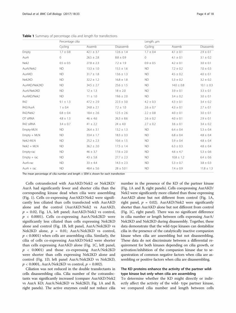
Table 1 Summary of percentage cilia and length for transfections
Percentage cilia Length, μm
Cycling Assemb Disassemb Cycling Assemb Disassemb
Empty 1.7 ± 0.8 42.1 ± 3.7 12.6 ± 1.4 1.7 ± 0.4 4.1 ± 0.1 2.9 ± 0.1
AurA 0 26.5 ± 2.8 8.8 ± 0.9 0 4.1 ± 0.1 3.1 ± 0.2
Nek2 0.5 ± 0.5 27.8 ± 2.3 7.2 ± 1.9 0.9 ± 0.5 4.2 ± 0.1 3.0 ± 0.1
AurA/Nek2 ND 13.3 ± 1.0 15.5 ± 1.6 ND 7.2 ± 0.2 7.0 ± 0.3
AurAKD ND 31.7 ± 1.8 13.6 ± 1.3 ND 4.5 ± 0.2 4.0 ± 0.1
Nek2KD ND 32.2 ± 1.2 16.8 ± 1.8 ND 5.3 ± 0.2 3.2 ± 0.2
AurAKD/Nek2KD ND 34.5 ± 2.7 25.6 ± 1.5 ND 14.0 ± 0.8 10.1 ± 0.3
AurA/Nek2KD ND 12 ± 1.3 18 ± 2.0 ND 3.9 ± 0.1 3.3 ± 0.1
AurAKD/Nek2 ND 11 ± 1.0 19.6 ± 2.0 ND 3.4 ± 0.2 3.0 ± 0.1
R42 9.1 ± 1.3 47.2 ± 2.9 22.3 ± 3.0 4.2 ± 0.3 4.3 ± 0.1 3.4 ± 0.2
R42/AurA 1 ± 0.4 24.8 ± 2.1 7.2 ± 1.0 2.6 ± 0.7 4.3 ± 0.1 2.7 ± 0.1
R42/Nek2 0.8 ± 0.4 18.4 ± 2.6 12.5 ± 2.6 2.2 ± 0.8 4.0 ± 0.1 3.0 ± 0.1
OT siRNA 4.8 ± 1.3 46 ± 4.6 26.3 ± 8.6 2.6 ± 0.2 4.0 ± 0.1 2.9 ± 0.1
R42 siRNA 3.4 ± 0.7 41 ± 2.2 24 ± 4.0 2.7 ± 0.2 3.6 ± 0.1 3.4 ± 0.2
Empty-MLN ND 26.4 ± 3.1 13.2 ± 1.3 ND 6.4 ± 0.4 5.3 ± 0.4
Empty + MLN ND 33.4 ± 1.7 18.3 ± 3.3 ND 6.8 ± 0.4 4.8 ± 0.4
Nek2-MLN ND 25.2 ± 2.3 10.6 ± 1.5 ND 5.9 ± 0.4 4.8 ± 0.4
Nek2 + MLN ND 36.2 ± 2.0 17.5 ± 1.4 ND 6.3 ± 0.3 4.8 ± 0.4
Empty-rac ND 46 ± 3.7 17.6 ± 2.0 ND 4.6 ± 4.7 5.3 ± 0.6
Empty + rac ND 43 ± 5.8 27.7 ± 2.3 ND 10.8 ± 1.2 6.4 ± 0.6
AurA-rac ND 33 ± 4.4 14.3 ± 2.3 ND 5.3 ± 0.7 3.8 ± 0.3
AurA + rac ND 48.4 ± 5.6 28 ± 5.0 l ND 7.4 ± 0.9 11.8 ± 1.3
The mean percentage of cilia number and length ± SEM is shown for each transfection
DeVaul et al. BMC Cell Biology (2017) 18:33 Page 4 of 18
transfected with either wild-type kinase alone or thedouble transfectants (compare AurA/Nek2KD vs AurA;AurAKD/Nek2 vs Nek2, Fig. 1). Cells expressing AurA/Nek2KD were significantly less ciliated than cells ex-pressing AurA alone or control when cilia are growing(Fig. 1A, left panel, AurA/Nek2KD vs AurA, p = 0.002;AurA/Nek2KD vs control, p < 0.0001). Similarly, Aur-AKD enhanced the depolymerization activity of Nek2(Fig. 1B, left panel, AurAKD/Nek2 vs Nek2, p = 0.002).Only cells transfected with AurAKD/Nek2 displayed adecrease in cilia length compared to Nek2 during ciliaassembly (Fig. 1D, left panel, p = 0.0001).In contrast, coexpression with the kinase dead com-
panion kinase did not result in increaseddepolymerization when cilia were disassembling com-pared to wild-type alone (Fig. 1A and B, right panels).Instead of stimulating depolymerization, the catalyticallyinactive kinase suppressed cilia depolymerization by thecompanion kinase. Cells dissembling cilia and coexpres-sing AurA/Nek2KD were significantly more ciliated thancells expressing AurA alone and control (Fig. 1A, rightpanel AurA/Nek2KD vs AurA, p = 0.002; AurA/Nek2KD vs control, p = .02). Cells coexpressing Aur-AKD/Nek2 were significantly more ciliated than Nek2alone and control (Fig. 1B, right panel, AurAKD/Nek2vs Nek2, p < 0.0004; AurKD/Nek2 vs control, p = 0.007).Cilia length was not significantly different in cotransfec-tants compared to wild-type or control.Our results support differential effects of the KDs on
the wild-type kinases depending on whether cilia are as-sembling or disassembling. Coexpression of the domin-ant negative enzyme with the wild-type companionkinase stimulates activity of the other kinase when ciliaare growing but inhibits its activity when cilia are disas-sembling. We conclude from these experiments that thedominant negative enzymes sequester factors that inhibitdepolymerization by the kinases when cilia are assem-bling and factors that stimulate depolymerization whencilia are disassembling. This result supports the exist-ence of regulatory proteins common to the two kinases.
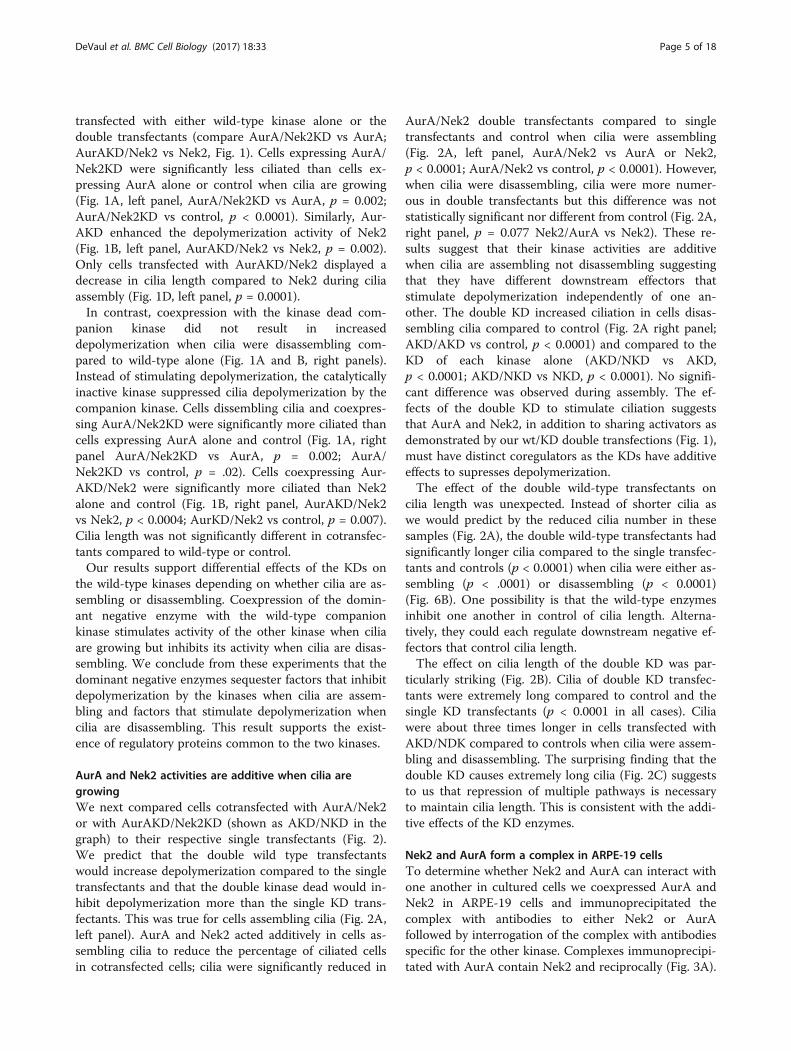
AurA and Nek2 activities are additive when cilia aregrowingWe next compared cells cotransfected with AurA/Nek2or with AurAKD/Nek2KD (shown as AKD/NKD in thegraph) to their respective single transfectants (Fig. 2).We predict that the double wild type transfectantswould increase depolymerization compared to the singletransfectants and that the double kinase dead would in-hibit depolymerization more than the single KD trans-fectants. This was true for cells assembling cilia (Fig. 2A,left panel). AurA and Nek2 acted additively in cells as-sembling cilia to reduce the percentage of ciliated cellsin cotransfected cells; cilia were significantly reduced in
AurA/Nek2 double transfectants compared to singletransfectants and control when cilia were assembling(Fig. 2A, left panel, AurA/Nek2 vs AurA or Nek2,p < 0.0001; AurA/Nek2 vs control, p < 0.0001). However,when cilia were disassembling, cilia were more numer-ous in double transfectants but this difference was notstatistically significant nor different from control (Fig. 2A,right panel, p = 0.077 Nek2/AurA vs Nek2). These re-sults suggest that their kinase activities are additivewhen cilia are assembling not disassembling suggestingthat they have different downstream effectors thatstimulate depolymerization independently of one an-other. The double KD increased ciliation in cells disas-sembling cilia compared to control (Fig. 2A right panel;AKD/AKD vs control, p < 0.0001) and compared to theKD of each kinase alone (AKD/NKD vs AKD,p < 0.0001; AKD/NKD vs NKD, p < 0.0001). No signifi-cant difference was observed during assembly. The ef-fects of the double KD to stimulate ciliation suggeststhat AurA and Nek2, in addition to sharing activators asdemonstrated by our wt/KD double transfections (Fig. 1),must have distinct coregulators as the KDs have additiveeffects to supresses depolymerization.The effect of the double wild-type transfectants on
cilia length was unexpected. Instead of shorter cilia aswe would predict by the reduced cilia number in thesesamples (Fig. 2A), the double wild-type transfectants hadsignificantly longer cilia compared to the single transfec-tants and controls (p < 0.0001) when cilia were either as-sembling (p < .0001) or disassembling (p < 0.0001)(Fig. 6B). One possibility is that the wild-type enzymesinhibit one another in control of cilia length. Alterna-tively, they could each regulate downstream negative ef-fectors that control cilia length.The effect on cilia length of the double KD was par-
ticularly striking (Fig. 2B). Cilia of double KD transfec-tants were extremely long compared to control and thesingle KD transfectants (p < 0.0001 in all cases). Ciliawere about three times longer in cells transfected withAKD/NDK compared to controls when cilia were assem-bling and disassembling. The surprising finding that thedouble KD causes extremely long cilia (Fig. 2C) suggeststo us that repression of multiple pathways is necessaryto maintain cilia length. This is consistent with the addi-tive effects of the KD enzymes.
Nek2 and AurA form a complex in ARPE-19 cellsTo determine whether Nek2 and AurA can interact withone another in cultured cells we coexpressed AurA andNek2 in ARPE-19 cells and immunoprecipitated thecomplex with antibodies to either Nek2 or AurAfollowed by interrogation of the complex with antibodiesspecific for the other kinase. Complexes immunoprecipi-tated with AurA contain Nek2 and reciprocally (Fig. 3A).
DeVaul et al. BMC Cell Biology (2017) 18:33 Page 5 of 18
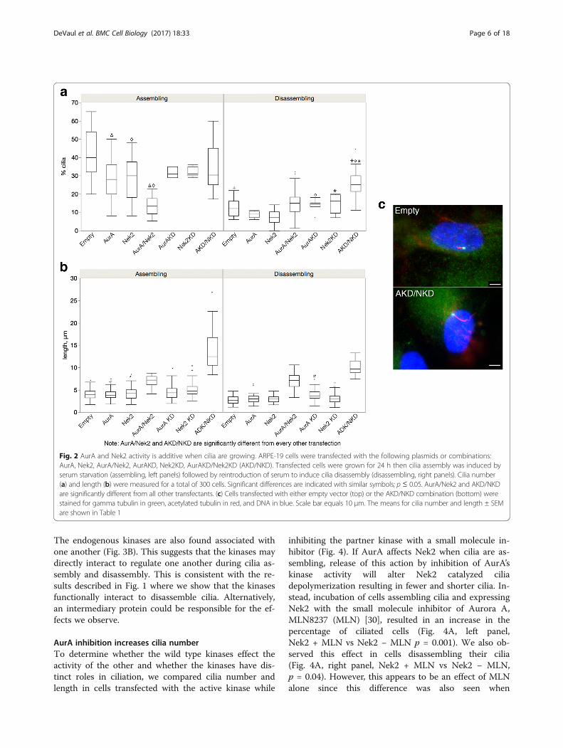
The endogenous kinases are also found associated withone another (Fig. 3B). This suggests that the kinases maydirectly interact to regulate one another during cilia as-sembly and disassembly. This is consistent with the re-sults described in Fig. 1 where we show that the kinasesfunctionally interact to disassemble cilia. Alternatively,an intermediary protein could be responsible for the ef-fects we observe.
AurA inhibition increases cilia numberTo determine whether the wild type kinases effect theactivity of the other and whether the kinases have dis-tinct roles in ciliation, we compared cilia number andlength in cells transfected with the active kinase while
inhibiting the partner kinase with a small molecule in-hibitor (Fig. 4). If AurA affects Nek2 when cilia are as-sembling, release of this action by inhibition of AurA’skinase activity will alter Nek2 catalyzed ciliadepolymerization resulting in fewer and shorter cilia. In-stead, incubation of cells assembling cilia and expressingNek2 with the small molecule inhibitor of Aurora A,MLN8237 (MLN) [30], resulted in an increase in thepercentage of ciliated cells (Fig. 4A, left panel,Nek2 + MLN vs Nek2 − MLN p = 0.001). We also ob-served this effect in cells disassembling their cilia(Fig. 4A, right panel, Nek2 + MLN vs Nek2 − MLN,p = 0.04). However, this appears to be an effect of MLNalone since this difference was also seen when
Fig. 2 AurA and Nek2 activity is additive when cilia are growing. ARPE-19 cells were transfected with the following plasmids or combinations:AurA, Nek2, AurA/Nek2, AurAKD, Nek2KD, AurAKD/Nek2KD (AKD/NKD). Transfected cells were grown for 24 h then cilia assembly was induced byserum starvation (assembling, left panels) followed by reintroduction of serum to induce cilia disassembly (disassembling, right panels). Cilia number(a) and length (b) were measured for a total of 300 cells. Significant differences are indicated with similar symbols; p ≤ 0.05. AurA/Nek2 and AKD/NKDare significantly different from all other transfectants. (c) Cells transfected with either empty vector (top) or the AKD/NKD combination (bottom) werestained for gamma tubulin in green, acetylated tubulin in red, and DNA in blue. Scale bar equals 10 μm. The means for cilia number and length ± SEMare shown in Table 1
DeVaul et al. BMC Cell Biology (2017) 18:33 Page 6 of 18
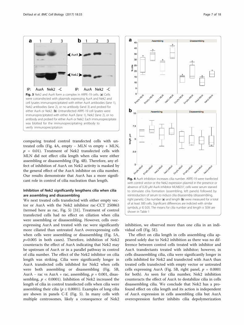
comparing treated control transfected cells with un-treated cells (Fig. 4A, empty − MLN vs empty + MLN,p = 0.01). Treatment of Nek2 transfected cells withMLN did not effect cilia length when cilia were eitherassembling or disassembling (Fig. 4B). Therefore, any ef-fect of inhibition of AurA on Nek2 activity is masked bythe general effect of the AurA inhibitor on cilia number.Our results demonstrate that AurA has a more signifi-cant role in control of cilia nucleation than length.
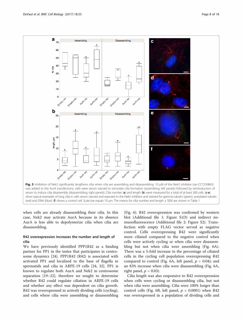
Inhibition of Nek2 significantly lengthens cilia when ciliaare assembling and disassemblingWe next treated cells transfected with either empty vec-tor or AurA with the Nek2 inhibitor rac-CCT 250863(termed here as rac, Fig. 5) [31]. Treatment of controltransfected cells had no effect on ciliation when ciliawere assembling or disassembling. However, cells over-expressing AurA and treated with rac were significantlymore ciliated than untreated AurA overexpressing cellswhen cells were assembling or disassembling (Fig. 5A,p=0.005 in both cases). Therefore, inhibition of Nek2counteracts the effect of AurA indicating that Nek2 maybe upstream of AurA or in a parallel pathway in controlof cilia number. The effect of the Nek2 inhibitor on cilialength was striking. Cilia were significantly longer inAurA transfected cells inhibited for Nek2 when cellswere both assembling or disassembling (Fig. 5B,AurA − rac vs AurA + rac, assembling, p = 0.001, disas-sembling, p < 0.0001). Inhibition of Nek2 increased thelength of cilia in control transfected cells when cilia wereassembling their cilia (p ≤ 0.0001). Examples of long ciliaare shown in panels C-E (Fig. 5). In many cells withmultiple centrosomes, likely a consequence of Nek2
inhibition, we observed more than one cilia in an indi-vidual cell (Fig. 5E).The effect on cilia length in cells assembling cilia ap-
peared solely due to Nek2 inhibition as there was no dif-ference between control cells treated with inhibitor andAurA transfectants treated with inhibitor; however, incells disassembling cilia, cilia were significantly longer incells inhibited for Nek2 and transfected with AurA thantreated cells transfected with empty vector or untreatedcells expressing AurA (Fig. 5B, right panel, p = 0.0001for both). As seen for cilia number, Nek2 inhibitioncounteracts the effect of AurA to destabilize cilia in cellsdisassembling cilia. We conclude that Nek2 has a pro-found effect on cilia length and its action is independentof AurA expression in cells assembling cilia but AurAoverexpression further inhibits cilia depolymerization
Fig. 3 Nek2 and AurA form a complex in ARPE-19 cells. (a) Cellswere cotransfected with plasmids expressing AurA and Nek2 andcell lysates immunoprecipitated with either AurA antibodies (lane 1),Nek2 antibodies (lane 2), or no antibody (land 3) and probed foreither AurA or Nek2. (b) Untransfected ARPE-19 cell lysates wereimmunoprecipitated with either AurA (lane 1), Nek2 (lane 2), or noantibody and probed for either AurA or Nek2. Each immunoprecipitatewas blotted for the immunoprecipitating antibody toverify immunoprecipitation
Fig. 4 AurA inhibition increases cilia number. ARPE-19 were tranfectedwith control vector or the Nek2 expression plasmid in the presence orabsence of 0.20 μM AurA inhibitor MLN8237, cells were serum starvedto stimulate cilia formation (assembling, left panels) followed byreintroduction of serum to induce cilia disassembly (disassembling,right panels). Cilia number (a) and length (b) were measured for a totalof at least 300 cells. Significant differences are indicted with similarsymbols, p ≤ 0.05. The means for cilia number and length ± SEM areshown in Table 1
DeVaul et al. BMC Cell Biology (2017) 18:33 Page 7 of 18
when cells are already disassembling their cilia. In thiscase, Nek2 may activate AurA because in its absenceAurA is less able to depolymerize cilia when cilia aredisassembling.
R42 overexpression increases the number and length ofciliaWe have previously identified PPP1R42 as a bindingpartner for PP1 in the testes that participates in centro-some dynamics [24]. PPP1R42 (R42) is associated withactivated PP1 and localized to the base of flagella inspermatids and cilia in ARPE-19 cells [24, 32]. PP1 isknown to regulate both AurA and Nek2 in centrosomeseparation [19–22], therefore we sought to determinewhether R42 could regulate ciliation in ARPE-19 cellsand whether any effect was dependent on cilia growth.R42 was overexpressed in actively dividing cells (cycling),and cells where cilia were assembling or disassembling
(Fig. 6). R42 overexpression was confirmed by westernblot (Additional file 1: Figure S1D) and indirect im-munofluorescence (Additional file 2: Figure S2). Trans-fection with empty FLAG vector served as negativecontrol. Cells overexpressing R42 were significantlymore ciliated compared to the negative control whencells were actively cycling or when cilia were disassem-bling but not when cilia were assembling (Fig. 6A).There was a 5-fold increase in the percentage of ciliatedcells in the cycling cell population overexpressing R42compared to control (Fig. 6A, left panel, p = 0.04); andan 83% increase when cilia were disassembling (Fig. 6A,right panel, p = 0.03).Cilia length was also responsive to R42 overexpression
when cells were cycling or disassembling cilia, but notwhen cilia were assembling. Cilia were 100% longer thancontrol cells (Fig. 6B, left panel, p < 0.0001) when R42was overexpressed in a population of dividing cells and
Fig. 5 Inhibition of Nek2 significantly lengthens cilia when cilia are assembling and disassembling. 10 μM of the Nek2 inhibitor (rac-CCT250863)was added to the AurA transfections, cells were serum starved to stimulate cilia formation (assembling, left panels) followed by reintroduction ofserum to induce cilia disassembly (disassembling, right panels). Cilia number (a) and length (b) were measured for a total of at least 300 cells. (c-e)show typical examples of long cilia in cells serum starved and exposed to the Nek2 inhibitor and stained for gamma tubulin (green), acetylated tubulin(red) and DNA (blue); (f) shows a control cell. Scale bar equals 10 μm. The means for cilia number and length ± SEM are shown in Table 1
DeVaul et al. BMC Cell Biology (2017) 18:33 Page 8 of 18
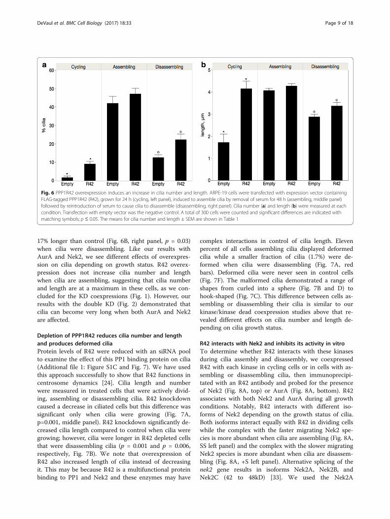
17% longer than control (Fig. 6B, right panel, p = 0.03)when cilia were disassembling. Like our results withAurA and Nek2, we see different effects of overexpres-sion on cilia depending on growth status. R42 overex-pression does not increase cilia number and lengthwhen cilia are assembling, suggesting that cilia numberand length are at a maximum in these cells, as we con-cluded for the KD coexpressions (Fig. 1). However, ourresults with the double KD (Fig. 2) demonstrated thatcilia can become very long when both AurA and Nek2are affected.
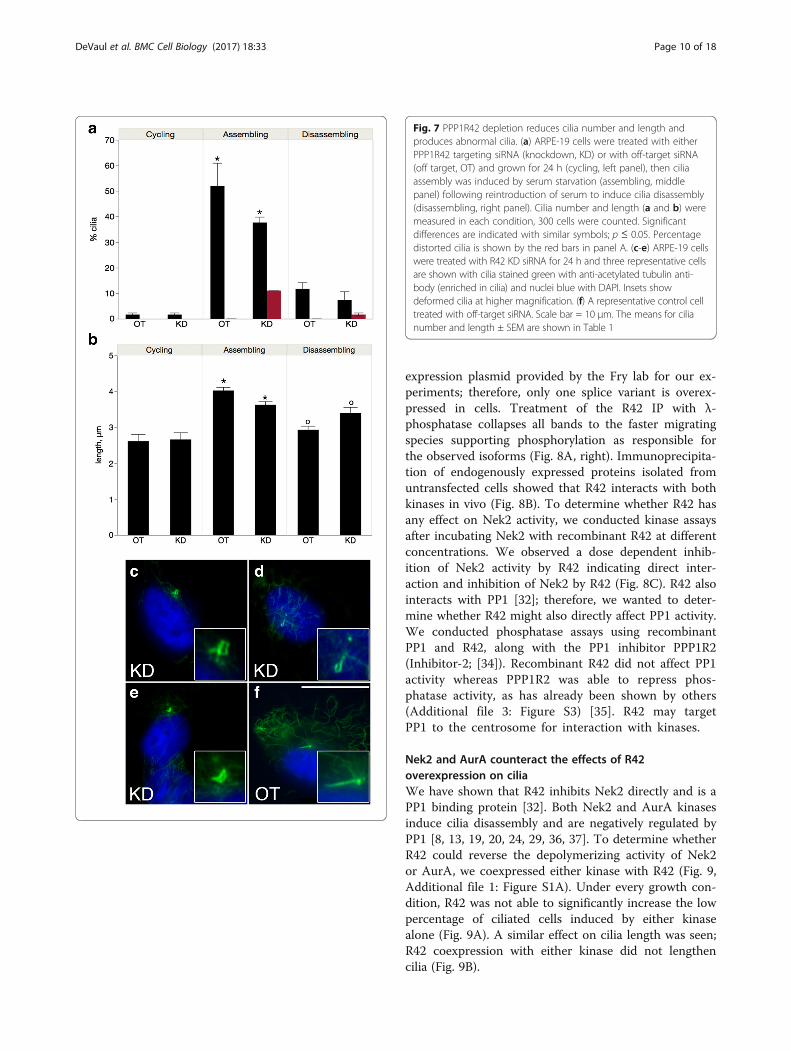
Depletion of PPP1R42 reduces cilia number and lengthand produces deformed ciliaProtein levels of R42 were reduced with an siRNA poolto examine the effect of this PP1 binding protein on cilia(Additional file 1: Figure S1C and Fig. 7). We have usedthis approach successfully to show that R42 functions incentrosome dynamics [24]. Cilia length and numberwere measured in treated cells that were actively divid-ing, assembling or disassembling cilia. R42 knockdowncaused a decrease in ciliated cells but this difference wassignificant only when cilia were growing (Fig. 7A,p=0.001, middle panel). R42 knockdown significantly de-creased cilia length compared to control when cilia weregrowing; however, cilia were longer in R42 depleted cellsthat were disassembling cilia (p = 0.001 and p = 0.006,respectively, Fig. 7B). We note that overexpression ofR42 also increased length of cilia instead of decreasingit. This may be because R42 is a multifunctional proteinbinding to PP1 and Nek2 and these enzymes may have
complex interactions in control of cilia length. Elevenpercent of all cells assembling cilia displayed deformedcilia while a smaller fraction of cilia (1.7%) were de-formed when cilia were disassembling (Fig. 7A, redbars). Deformed cilia were never seen in control cells(Fig. 7F). The malformed cilia demonstrated a range ofshapes from curled into a sphere (Fig. 7B and D) tohook-shaped (Fig. 7C). This difference between cells as-sembling or disassembling their cilia is similar to ourkinase/kinase dead coexpression studies above that re-vealed different effects on cilia number and length de-pending on cilia growth status.
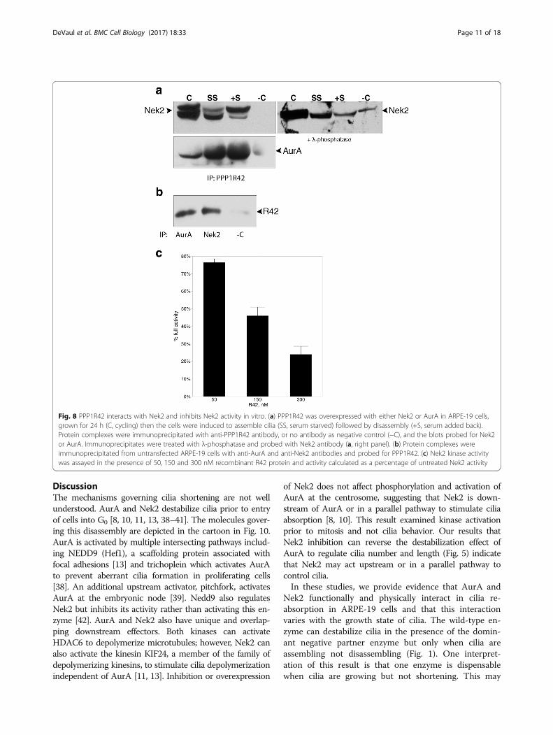
R42 interacts with Nek2 and inhibits its activity in vitroTo determine whether R42 interacts with these kinasesduring cilia assembly and disassembly, we coexpressedR42 with each kinase in cycling cells or in cells with as-sembling or disassembling cilia, then immunoprecipi-tated with an R42 antibody and probed for the presenceof Nek2 (Fig. 8A, top) or AurA (Fig. 8A, bottom). R42associates with both Nek2 and AurA during all growthconditions. Notably, R42 interacts with different iso-forms of Nek2 depending on the growth status of cilia.Both isoforms interact equally with R42 in dividing cellswhile the complex with the faster migrating Nek2 spe-cies is more abundant when cilia are assembling (Fig. 8A,SS left panel) and the complex with the slower migratingNek2 species is more abundant when cilia are disassem-bling (Fig. 8A, +S left panel). Alternative splicing of thenek2 gene results in isoforms Nek2A, Nek2B, andNek2C (42 to 48kD) [33]. We used the Nek2A
Fig. 6 PPP1R42 overexpression induces an increase in cilia number and length. ARPE-19 cells were transfected with expression vector containingFLAG-tagged PPP1R42 (R42), grown for 24 h (cycling, left panel), induced to assemble cilia by removal of serum for 48 h (assembling, middle panel)followed by reintroduction of serum to cause cilia to disassemble (disassembling, right panel). Cilia number (a) and length (b) were measured at eachcondition. Transfection with empty vector was the negative control. A total of 300 cells were counted and significant differences are indicated withmatching symbols; p ≤ 0.05. The means for cilia number and length ± SEM are shown in Table 1
DeVaul et al. BMC Cell Biology (2017) 18:33 Page 9 of 18
expression plasmid provided by the Fry lab for our ex-periments; therefore, only one splice variant is overex-pressed in cells. Treatment of the R42 IP with λ-phosphatase collapses all bands to the faster migratingspecies supporting phosphorylation as responsible forthe observed isoforms (Fig. 8A, right). Immunoprecipita-tion of endogenously expressed proteins isolated fromuntransfected cells showed that R42 interacts with bothkinases in vivo (Fig. 8B). To determine whether R42 hasany effect on Nek2 activity, we conducted kinase assaysafter incubating Nek2 with recombinant R42 at differentconcentrations. We observed a dose dependent inhib-ition of Nek2 activity by R42 indicating direct inter-action and inhibition of Nek2 by R42 (Fig. 8C). R42 alsointeracts with PP1 [32]; therefore, we wanted to deter-mine whether R42 might also directly affect PP1 activity.We conducted phosphatase assays using recombinantPP1 and R42, along with the PP1 inhibitor PPP1R2(Inhibitor-2; [34]). Recombinant R42 did not affect PP1activity whereas PPP1R2 was able to repress phos-phatase activity, as has already been shown by others(Additional file 3: Figure S3) [35]. R42 may targetPP1 to the centrosome for interaction with kinases.
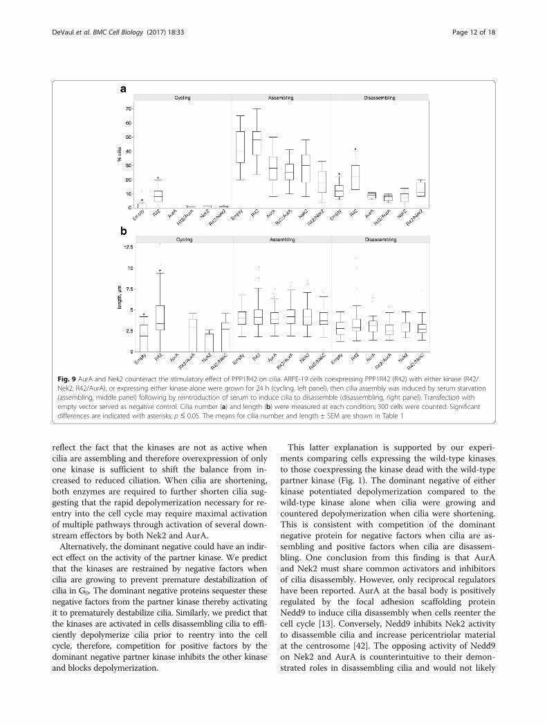
Nek2 and AurA counteract the effects of R42overexpression on ciliaWe have shown that R42 inhibits Nek2 directly and is aPP1 binding protein [32]. Both Nek2 and AurA kinasesinduce cilia disassembly and are negatively regulated byPP1 [8, 13, 19, 20, 24, 29, 36, 37]. To determine whetherR42 could reverse the depolymerizing activity of Nek2or AurA, we coexpressed either kinase with R42 (Fig. 9,Additional file 1: Figure S1A). Under every growth con-dition, R42 was not able to significantly increase the lowpercentage of ciliated cells induced by either kinasealone (Fig. 9A). A similar effect on cilia length was seen;R42 coexpression with either kinase did not lengthencilia (Fig. 9B).
Fig. 7 PPP1R42 depletion reduces cilia number and length andproduces abnormal cilia. (a) ARPE-19 cells were treated with eitherPPP1R42 targeting siRNA (knockdown, KD) or with off-target siRNA(off target, OT) and grown for 24 h (cycling, left panel), then ciliaassembly was induced by serum starvation (assembling, middlepanel) following reintroduction of serum to induce cilia disassembly(disassembling, right panel). Cilia number and length (a and b) weremeasured in each condition, 300 cells were counted. Significantdifferences are indicated with similar symbols; p ≤ 0.05. Percentagedistorted cilia is shown by the red bars in panel A. (c-e) ARPE-19 cellswere treated with R42 KD siRNA for 24 h and three representative cellsare shown with cilia stained green with anti-acetylated tubulin anti-body (enriched in cilia) and nuclei blue with DAPI. Insets showdeformed cilia at higher magnification. (f) A representative control celltreated with off-target siRNA. Scale bar = 10 μm. The means for cilianumber and length ± SEM are shown in Table 1
DeVaul et al. BMC Cell Biology (2017) 18:33 Page 10 of 18
DiscussionThe mechanisms governing cilia shortening are not wellunderstood. AurA and Nek2 destabilize cilia prior to entryof cells into G0 [8, 10, 11, 13, 38–41]. The molecules gover-ing this disassembly are depicted in the cartoon in Fig. 10.AurA is activated by multiple intersecting pathways includ-ing NEDD9 (Hef1), a scaffolding protein associated withfocal adhesions [13] and trichoplein which activates AurAto prevent aberrant cilia formation in proliferating cells[38]. An additional upstream activator, pitchfork, activatesAurA at the embryonic node [39]. Nedd9 also regulatesNek2 but inhibits its activity rather than activating this en-zyme [42]. AurA and Nek2 also have unique and overlap-ping downstream effectors. Both kinases can activateHDAC6 to depolymerize microtubules; however, Nek2 canalso activate the kinesin KIF24, a member of the family ofdepolymerizing kinesins, to stimulate cilia depolymerizationindependent of AurA [11, 13]. Inhibition or overexpression
of Nek2 does not affect phosphorylation and activation ofAurA at the centrosome, suggesting that Nek2 is down-stream of AurA or in a parallel pathway to stimulate ciliaabsorption [8, 10]. This result examined kinase activationprior to mitosis and not cilia behavior. Our results thatNek2 inhibition can reverse the destabilization effect ofAurA to regulate cilia number and length (Fig. 5) indicatethat Nek2 may act upstream or in a parallel pathway tocontrol cilia.In these studies, we provide evidence that AurA and
Nek2 functionally and physically interact in cilia re-absorption in ARPE-19 cells and that this interactionvaries with the growth state of cilia. The wild-type en-zyme can destabilize cilia in the presence of the domin-ant negative partner enzyme but only when cilia areassembling not disassembling (Fig. 1). One interpret-ation of this result is that one enzyme is dispensablewhen cilia are growing but not shortening. This may
Fig. 8 PPP1R42 interacts with Nek2 and inhibits Nek2 activity in vitro. (a) PPP1R42 was overexpressed with either Nek2 or AurA in ARPE-19 cells,grown for 24 h (C, cycling) then the cells were induced to assemble cilia (SS, serum starved) followed by disassembly (+S, serum added back).Protein complexes were immunoprecipitated with anti-PPP1R42 antibody, or no antibody as negative control (−C), and the blots probed for Nek2or AurA. Immunoprecipitates were treated with λ-phosphatase and probed with Nek2 antibody (a, right panel). (b) Protein complexes wereimmunoprecipitated from untransfected ARPE-19 cells with anti-AurA and anti-Nek2 antibodies and probed for PPP1R42. (c) Nek2 kinase activitywas assayed in the presence of 50, 150 and 300 nM recombinant R42 protein and activity calculated as a percentage of untreated Nek2 activity
DeVaul et al. BMC Cell Biology (2017) 18:33 Page 11 of 18
reflect the fact that the kinases are not as active whencilia are assembling and therefore overexpression of onlyone kinase is sufficient to shift the balance from in-creased to reduced ciliation. When cilia are shortening,both enzymes are required to further shorten cilia sug-gesting that the rapid depolymerization necessary for re-entry into the cell cycle may require maximal activationof multiple pathways through activation of several down-stream effectors by both Nek2 and AurA.Alternatively, the dominant negative could have an indir-
ect effect on the activity of the partner kinase. We predictthat the kinases are restrained by negative factors whencilia are growing to prevent premature destabilization ofcilia in G0. The dominant negative proteins sequester thesenegative factors from the partner kinase thereby activatingit to prematurely destabilize cilia. Similarly, we predict thatthe kinases are activated in cells disassembling cilia to effi-ciently depolymerize cilia prior to reentry into the cellcycle, therefore, competition for positive factors by thedominant negative partner kinase inhibits the other kinaseand blocks depolymerization.
This latter explanation is supported by our experi-ments comparing cells expressing the wild-type kinasesto those coexpressing the kinase dead with the wild-typepartner kinase (Fig. 1). The dominant negative of eitherkinase potentiated depolymerization compared to thewild-type kinase alone when cilia were growing andcountered depolymerization when cilia were shortening.This is consistent with competition of the dominantnegative protein for negative factors when cilia are as-sembling and positive factors when cilia are disassem-bling. One conclusion from this finding is that AurAand Nek2 must share common activators and inhibitorsof cilia disassembly. However, only reciprocal regulatorshave been reported. AurA at the basal body is positivelyregulated by the focal adhesion scaffolding proteinNedd9 to induce cilia disassembly when cells reenter thecell cycle [13]. Conversely, Nedd9 inhibits Nek2 activityto disassemble cilia and increase pericentriolar materialat the centrosome [42]. The opposing activity of Nedd9on Nek2 and AurA is counterintuitive to their demon-strated roles in disassembling cilia and would not likely
Fig. 9 AurA and Nek2 counteract the stimulatory effect of PPP1R42 on cilia. ARPE-19 cells coexpressing PPP1R42 (R42) with either kinase (R42/Nek2; R42/AurA), or expressing either kinase alone were grown for 24 h (cycling, left panel), then cilia assembly was induced by serum starvation(assembling, middle panel) following by reintroduction of serum to induce cilia to disassemble (disassembling, right panel). Transfection withempty vector served as negative control. Cilia number (a) and length (b) were measured at each condition; 300 cells were counted. Significantdifferences are indicated with asterisks; p ≤ 0.05. The means for cilia number and length ± SEM are shown in Table 1
DeVaul et al. BMC Cell Biology (2017) 18:33 Page 12 of 18
counteract one another [8, 10, 11, 13]. It is important tonote that the report concerning Nek2 inhibition byNedd9 was not obtained from cells disassembling ciliabut from cycling cells and may reflect differential regula-tion in cycling vs disassembling cells which is consistentwith our work [42]. No common activators or inhibitorsof AurA and Nek2 have been identified, however, wewould predict that associated positive regulatory factorswould be enriched in cells disassembling cilia and nega-tive regulators in cells assembling cilia. This functionalinteraction is consistent with our finding that the kinasesare associated with one another (Fig. 3) and the work ofothers demonstrating that the two kinases colocalize tothe basal body [8, 42].Although Nek2 and AurA appear to share regulatory
proteins in common, their inhibition has different effectson cilia number and length. We show that AurA plays animportant role in regulating cilia number (Fig. 4) whileNek2 inhibition significantly impacts cilia length (Fig. 5).This suggests that nucleation of cilia is inhibited by AurAin cells either assembling or disassembling cilia whileNek2 functions primarily to maintain the steady statelength of cilia. Our finding contrasts with a previous resultthat flagella length in Clamydomonas reinhardtii is regu-lated by an AurA-like protein [16]. However, our experi-ments examine cilia length under dynamic rather thansteady state conditions that may involve different mecha-nisms of regulation. Additionally, we show that Nek2
activates AurA in control of cilia number because Nek2inhibition inhibits depolymerization by AurA (Fig. 5).However, our results with the double wild-type transfec-tants indicate that the functions of the kinases are overlap-ping. Cells cotransfected with active AurA and Nek2 hadsignificantly fewer cilia compared to each kinase trans-fected separately when cilia were assembling; however,cilia length was increased in the double wild-type transfec-tion. When cilia are disassembling and the kinases are ac-tivated, expression of both dominant negative kinasessuppresses depolymerization and increases both cilianumber and length above the single KD alone but onlywhen cilia are disassembling. This suggests that the mech-anism of AurA and Nek2 in regulation of cilia length dif-fers from their control over cilia number.A particularly intriguing finding was our observation
that cilia were extremely long in cells transfected withAKD/NKD (Fig. 2). HDAC6 is a downstream effector ofboth Nek2 and AurA, therefore we would expect thataxonemal microtubules might be stabilized in the doubleKD and that this effect would be augmented comparedto the single KD transfectants [10, 13]. Furthermore, thedepolymerizing kinesin KIF24 is activated by Nek2 toensure cilia disassembly and the Nek2-KIF24 pathway istemporally and functionally distinct from the AurA-HDAC6 pathway [11]. We would expect interruption ofboth pathways to be additive. Ciliary growth is mediatedby intraflagellar transport (IFT) via anterograde motors
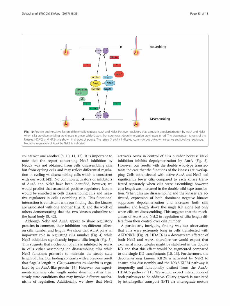
Fig. 10 Positive and negative factors differentially regulate AurA and Nek2. Positive regulators that stimulate depolymerization by AurA and Nek2when cilia are disassembling are shown in green while factors that counteract depolymerization are shown in red. The downstream targets of thekinases, HDAC6 and KIF24 are shown in shades of purple. The letters X and Y indicated common but unknown negative and positive regulators.Negative regulation of AurA by Nek2 is indicated
DeVaul et al. BMC Cell Biology (2017) 18:33 Page 13 of 18
that move protein and membrane to the ciliary tip andretrograde motors that return cargo to the cilia base[43]. Cilia length is in a state of “dynamic stability” withassembly in balance with disassembly [44–46]. One ex-planation of our result is that by increasing the length ofthe microtubule “track” upon which motors transportcargos to the ciliary tip, the cilia is extended beyond itssteady state length. This indicates that the length of themicrotubule track is one of the limiting factors for IFT.Cilia length in Chlamydomonas reinhardtii is regulatedby phosphorylation of CALK, an AurA-like protein. In-hibition of CALK activity increases flagella length totwice their normal length [47]. In our system, Nek2 hasa more profound effect on cilia length than does AurA.Nek2 is proposed to act as a switch between cilia
growth and resorption in the establishment of left rightasymmetry [10]. Increased amounts of Nek2 shift thebalance to cilia depolymerization while decreased Nek2causes centrosome defects; therefore, it is proposed thatNek2 promotes cilia biogenesis and homeostasis. Wehave identified a new inhibitor of Nek2, the PP1 bindingprotein PPP1R42. R42 inhibits the activity of Nek2 invitro and binds to Nek2 in cultured cells. This is consist-ent with our finding that R42 overexpression increasesciliation presumably by inhibiting cilia depolymerizationby Nek2. We conclude that R42 is partially responsiblefor inhibition of Nek2 in addition to dephosphorylationby PP1 [20, 22]. Our data establishes that different phos-phorylated isoforms of Nek2 interact with PPP1R42 de-pending on cilia growth state and further supports ourproposal that Nek2 has different activities when cilia areassembling or disassembling.We have previously shown that R42 interacts with PP1;
however, R42 does not directly affect PP1 activity in vitro(Additional file 3: Figure S3). R42 may target PP1 to Nek2to facilitate dephosphorylation and inhibition of Nek2; ex-periments are planned to determine whether a ternarycomplex exits containing Nek2, PP1, and R42.
ConclusionsFigure 10 presents a model for cilia assembly (top) anddisassembly (bottom) based on this work and that ofothers. In this diagram, positive factors that stimulatedepolymerization are shown in green and negative regu-lators in red and flank either AurA or Nek2. Factorsidentified by others are Nedd9, trichoplein, pitchfork(pifo) and PP1R2 [10, 13, 38, 48]. HDAC6 and KIF24 aredownstream targets of AurA and Nek2 that act todestabilize the microtubules of the axoneme [11, 13].The work described here makes several unique contribu-tions. First, we propose that AurA and Nek2 are acti-vated and inhibited by positive (green) and negative(red) regulators which control AurA and Nek2 activitydifferently depending on the growth state of cilia. We
predict that depolymerization is restrained by negativefactors that are active when cilia are assembling (top)and that polymerization is enhanced by positive factorswhen cilia are disassembling (bottom). Second, AurAand Nek2 exist in a complex and share positive andnegative regulators, indicated by unknown factors X andY. This conclusion is supported by our data that the kin-ase dead of one enzyme affects the activity of the otherpresumably by sequestering regulatory factors (Fig. 1).Third, AurA and Nek2 have distinct effects on cilianumber and length; AurA inhibition increases cilia num-ber with no effect on cilia length while Nek2 inhibitionincreases cilia length (Figs. 4 and 5).Fourth, the depolymerizing ability of AurA and Nek2
is additive when cilia are assembling. In addition, thedouble wild-type transfectants had longer but fewer cilia(Fig. 2). Finally, we show that PPP1R42, a PP1 bindingprotein, binds to and inhibits Nek2 in vivo and increasesciliation when cilia are shortening but not growing. Thisdependence on cilia growth status mirrors our resultswith the kinases. PPP1R42 may actively promote assem-bly when cilia are disassembling or prevent cilia fromshortening. We also show that PPP1R42 does not acti-vate PP1 suggesting that its inhibition of Nek2 is inde-pendent of its interaction with PP1.This work establishes a complex web of regulation of
cilia depolymerization that involves both positive andnegative effectors that are differentially regulated in cellsassembling cilia compared to disassembling cilia. We havealso identified a new negative regulator of Nek2 whichdoes not act through PP1 but binds directly to the kinaseand represents a candidate molecule for differentialmodulation in cells assembling or disassembling cilia.
MethodsCell culture and nucleic acid transfectionHuman retinal pigmented epithelial cells (ARPE-19)were obtained from American Type Tissue Collection(Manassas, VA) and grown in DMEM-F12 media supple-mented with 10% fetal bovine serum and 1% penicillin-streptomycin as directed by the supplier. To drive cellsinto quiescence, cells were washed three times in PBSand grown in culture media without fetal bovine serumfor 48 h. To release cells from the quiescence, 10%serum was added back to the culture media for 24 h. Tostudy the effect of protein overexpression on ciliagrowth and retraction, cells were transfected with ex-pression vectors encoding PPP1R42, AurA, AurA kinasedead mutant, Nek2, or Nek2 kinase dead mutant, and24 h later serum was removed from the media to haltgrowth and drive cells into G0 to stimulate cilia growth.Following incubation in serum free media for 48 h,serum was added to the growth media to induce ciliadisassembly and cells were observed for cilia number
DeVaul et al. BMC Cell Biology (2017) 18:33 Page 14 of 18
and length 24 h later. Protein expression was confirmedby western analysis (Additional file 1: Figure S1).Overexpression of PPP1R42 in ARPE-19 cells was per-
formed using a PPP1R42-FLAG expression vector con-structed by insertion of the PPP1R42 cDNA downstreamand in frame with the FLAG tag of 3XFLAG CMV-14(Sigma-Aldrich; St. Louis, MO). Overexpression of Nek2was performed using a Nek2-myc expression vectorwhile inhibition of Nek2 activity was achieved using aNek2-K37R-myc expression vector, a dominant negativeform of Nek2 (kind gifts of Dr. Andrew Fry, [20]). Over-expression of AurA was performed using an AurA-mycexpression vector while inhibition of AurA kinase activ-ity was achieved using an AurA-K162R-myc expressionvector, a dominant negative form of AurA (kind gifts ofDr. Erich Nigg, [26]). Cells were transfected using Lipo-fectamine 2000® (Life Technologies; Lincoln, NE) ac-cording to the manufacturer’s recommendations. Briefly,cells were transfected for 24 h and overexpression wasconfirmed by western blot (Additional file 1: Figure S1).90–95% transfection efficiency of the FLAG plasmidswas determined using immunofluorescence. Depletion ofPPP1R42 in ARPE-19 cells was performed using theON-TARGETplus SMARTpool siRNA LOC286187 fromDharmacon/Thermo Scientific (Pittsburgh, PA) as previ-ously described [24]. Cells were transfected using theLipofectamine-RNAiMAX reagent (Life Technologies;Lincoln, NE) according to manufacturer’s recommenda-tions. Briefly, cells were transfected for 48 h with a finalconcentration of 20 μM siRNA. Following incuba-tion, knockdown was confirmed by western blot(Additional file 1: Figure S1).The AurA inhibitor MLN8237 (0.25 μm; Selleckchem,
Houston, TX) was added to cells transfected with theNek2 plasmid, grown for 24 h, serum removed to inducecilia formation for 48 h and serum reintroduced to in-duce cilia disassembly. This procedure was repeated withthe AurA plasmid and 10 μm Nek2 inhibitor (rac-CCT250863, Tocris Biosciences, Bristol, UK).
Western blotARPE-19 total cell lysates were prepared as previouslydescribed [24]. Briefly, cells were resuspended in celllysis buffer (50 mM Tris, pH 7.5, 1 mM EDTA, 1 mMEGTA, and 1% NP-40) with protease and phosphataseinhibitors. Protein was separated using SDS-PAGE andproteins were transferred from the gel to polyvinylidenedifluoride (PVDF) membrane (BIO-RAD Laboratories;Hercules, CA). PPP1R42-FLAG was detected using anti-FLAG antibody (1:1000; F1804; Sigma-Aldrich; St. Louis,MO) and confirmed with anti-human PPP1R42 antibody(1:1000; HPA028628; Sigma-Aldrich; St. Louis, MO).Nek2-myc and AurA-myc were detected using anti-mycantibody (1:1000; TA325701; Origene Technologies;
Rockville, MD). Immune complexes bound to the mem-brane were detected with horseradish peroxidase-conjugated donkey secondary antibody (711–035-152;Jackson ImmunoResearch Inc.; West Grove, PA) and de-veloped with SuperSignal® West Pico ChemiluminescentSubstrate according to directions of the manufacturer(Thermo Fisher Scientific; Asheville, NC).
Indirect immunofluorescenceARPE-19 cells were grown on coverslips, fixed andpermeabilized with methanol at −20 °C for 10 min, andthen non-specific sites were blocked by incubation with3% BSA in Tris-buffered saline and Triton X-100(TBST) (20 mM Tris, pH 7.5, 150 mM NaCl, 2 mMEGTA, 0.1% Triton X-100) for 30 min. The cells wereincubated with anti-acetylated-tubulin antibody to detectprimary cilia (1:1000; T6793; Sigma-Aldrich; St. Louis,MO). Acetylated tubulin is a well-accepted marker forprimary cilia. Cells were then incubated with FITC-conjugated donkey anti-mouse secondary antibody(1:200; 715–095-150; Jackson ImmunoResearch Inc.;West Grove, PA). DNA was stained with 4′,6-diami-dino-2-phenylindole (DAPI) incorporated into themounting media (Vector Labs; Burlingame, CA). Theintracellular localization of proteins was observed with aNikon E600 fluorescence microscope, Pan Fluor 100×objective (N.A. 0.5–1.3) or Pan Fluor 40× objective(N.A. 0.75), fit with appropriate filters and images cap-tured with an Orca II CCD camera, model C4742–95(Hamamatsu; Middlesex, NJ) and Metamorph imageanalysis and acquisition software (Molecular Devices;Sunnyvale, CA, USA). Images were exported to Photo-shop (Adobe; San Jose, CA) and only linear adjustmentsto brightness and/or contrast were performed.
Morphometric and statistical analysisCaptured images of cells containing cilia, verified by cost-aining with anti-acetlyated tubulin and anti-gamma tubu-lin for the centrosome (Thermo Scientific, PA5–34815;Asheville, NC) were captured by Metamorph and enlargedto visualize cilia clearly. The length of cilia was obtainedusing the line tool calibrated for the 100X objective. Foreach treatment, 300 cells were measured. The data forcilia quantification and length are expressed as box andwhiskers plots for Figs. 1-2, 4-5 and 10 and as mean ± SEMfor Figs. 6 and 7. The differences between groups were an-alyzed using the unpaired Student’s t-test. A p-value of≤0.05 was considered significant.
CoimmunoprecipitationProtein complexes were collected by immunoprecipita-tion. Briefly, affinity purified antibody to PPP1R42 wasincubated with precleared cell lysate (1 mg protein)followed by anti-rabbit IgG beads. After transfer to
DeVaul et al. BMC Cell Biology (2017) 18:33 Page 15 of 18
membrane, immunoprecipated proteins were detectedwith anti-Nek2 (1:500; sc-33,167; Santa Cruz Biotechnol-ogy; Dallas, TX) or anti-AurA antibodies (1:500; PC742;EMD Millipore, Billerica, MA), and Veriblot anti-rabbitHRP (Abcam; Cambridge, MA). Use of the Veriblot sec-ondary prevents detection of IgG heavy chain. Negativecontrol for coimmunoprecipitation was precleared lysateincubated with no antibody.
Kinase assayKinase assays were conducted in kinase buffer (5 mMMOPS, pH 7.2, 2.5 mM β-glycerophosphate, 1 mMEGTA, 0.4 mM EDTA, 5 mM MgCl2, 0.05 mM DTT)with 1 μg myelin basic protein as substrate and 5 μCi/μl[32P] ATP. 50 nM Nek2 (Thermo Fisher; Waltham, MA)was incubated with varying concentrations of recombin-ant PPP1R42 (Biomatik; Wilmington, DE). After the des-ignated time, reactions were terminated by spotting ontophosphocellulose P82 paper, washed extensively with 1%phosphoric acid, and the trapped radioactivity measuredby scintillation counting.
Phosphatase assayInhibition or activation of PP1 by R42 and R2 was ac-complished using the fluorescence based RediPlate96©enzcheck serine/threonine phosphatase assay kit fromFisher Scientific (Pittsburgh, PA). Appropriate amountsof recombinant R2 or R42 (Biomatik; Wilmington, DE)were mixed in reaction buffer containing 2 mM DTTand 200 μM MnCl2 and added to wells containing thefluorescent phosphatase substrate. After incubation at30 °C for 30 min, fluorescence measured at excitation/emission 358/452 nm.
Additional files
Additional file 1: Figure S1. Protein expression in transfected cells. Inall cases cell lysates were prepared from transfected cells that weregrown for 24 h, serum starved for 48 h followed by reintroduction ofserum for 24 h. Proteins were resolved by SDS-PAGE, transferred to mem-brane and probed with the indicated antibodies. (A) Cells were trans-fected with the indicated expression plasmids or combinations andproteins probed with anti-FLAG (R42) or anti-myc (AurA and Nek2). TheAurA and Nek2 plasmids were verified by sequencing, we were unable toresolve these proteins using this gel system; however, both AurA andNek2 have been reported as doublets in PAGE [13, 49]. Untransfectedcells served as negative control (−C). (B) Cells were transfected with theindicated expression plasmids or combinations and proteins probed withanti-myc (AurA and Nek2). Untransfected cells served as negative control(−C). All panels for each section were exposed to film for the samelength of time. (C) Cells were treated with off target (OT) or PPP1R42(R42) targeting siRNA (KD) and membrane probed with anti-R42 and anti-actin. (D) Cells were transfected with R42-FLAG tagged vector or emptyvector (−C) and proteins probed with anti-FLAG. Expressed proteins aremaintained in the cell throughout the course of the experiment withreduction when cells are metabolically inactive after starvation. Blots wereprobed for actin as a loading control. (TIFF 5922 kb)
Additional file 2: Figure S2. Expression plasmids transfect ARPE-19 athigh efficiency. ARPE-19 cells were transfected with plasmids expressingeither FLAG tagged R42 or myc tagged kinase constructs and grown for24 h in complete media. Cells were stained with anti-FLAG or anti-mycantibody and detected with Alex Fluor 594 secondary antibody (red).Nuclei were stained with DAPI (blue) (A). 100 cells were counted for eachcondition and the efficiency of transfection for all constructs was about90%. Scale bars equal 10 μm. (B) Proteins lysates from cells transfectedwith either Nek2, AurA, Nek2KD, AurAKD, and R42, were separated bySDS-PAGE transferred to membrane and probed with the appropriate anti-bodies. Proteins from untransfected cells were loaded to indicate thelevel of endogenous protein (Ne, Ae, and R42e). (TIFF 16425 kb)
Additional file 3: Figure S3. PPP1R42 does not enhance PP1 activity invitro. Recombinant PP1 (USBiologicals; Salem, MA) was incubated withvarying concentrations of recombinant R2 (A) or R42 (B) (Biomatik;Wilmington, DE) and phosphatase activity measured as described inMaterials and Methods. (TIFF 2235 kb)
AbbreviationsAurA KD: Aurora A kinase dead; AurA: Aurora A; KD: kinase dead, knockdown; Nek2 KD: Never in mitosis kinase-2 kinase dead; Nek2: Never in mitosiskinase-2; OT: Off target; PPP1R2 or R2: Phosphoprotein phosphatase 1regulatory subunit 2 or Inhibitor-2; PPP1R42 or R42: Phosphoproteinphosphatase 1 regulatory subunit 42
AcknowledgementsThe authors wish to thank Dr. Andrew Fry and Dr. Erich Nigg for gifts of theexpression plasmids used in this work.
FundingThis work was supported by grant HD080151 (AOS) from the NationalInstitutes of Health; however, this funding body had no input into thedesign collection analysis, and interpretation, and in writing the manuscript.
Availability of data and materialsAll primary data and materials generated by this work are available uponrequest.
Authors’ contributionsThe authors contributed to the work in the following ways: NDconducted transfections followed by immunofluorescence and ciliamorphometrics and drafted the manuscript. RW conducted transfectionsfollowed by immunofluorescence and cilia morphometrics, western blot,immunoprecipitation and phosphatase assays, KK conducted transfections. AOSconceived and oversaw the experiments, wrote the manuscript and conductedkinase assays. All authors read and approved the final manuscript.
Ethics approval and consent to participateThese studies involve no human or animal subjects; therefore, the consentform and ethics approval are not applicable.
Consent for publicationBecause these studies do not make use of any external individual’s data,consent for publication does not apply.
Competing interestsThe Authors declare they have no competing interests.
Publisher’s NoteSpringer Nature remains neutral with regard to jurisdictional claims in publishedmaps and institutional affiliations.
Author details1Anatomy and Cell Biology, East Carolina University, Brody School ofMedicine, Greenville, NC, USA. 2Laboratory of Biochemistry and Genetics,National Institute of Diabetics and Digestive and Kidney Diseases, NationalInstitutes of Health, Bethesda, MD, USA.
DeVaul et al. BMC Cell Biology (2017) 18:33 Page 16 of 18
Received: 26 July 2017 Accepted: 1 November 2017
References1. Schneider L, Clement CA, Teilmann SC, Pazour GJ, Hoffmann EK, Satir P,
Christensen ST. PDGFRalphaalpha signaling is regulated through theprimary cilium in fibroblasts. Curr Biol. 2005;15(20):1861–6.
2. Simons M, Gloy J, Ganner A, Bullerkotte A, Bashkurov M, Kronig C, SchermerB, Benzing T, Cabello OA, Jenny A, Mlodzik M, Polok B, Driever W, Obara T,Walz G. Inversin, the gene product mutated in nephronophthisis type II,functions as a molecular switch between Wnt signaling pathways. NatGenet. 2005;37(5):537–43.
3. Huangfu D, Liu A, Rakeman AS, Murcia NS, Niswander L, Anderson KV.Hedgehog signalling in the mouse requires intraflagellar transport proteins.Nature. 2003;426(6962):83–7.
4. Veland IR, Awan A, Pedersen LB, Yoder BK, Christensen ST. Primary cilia andsignaling pathways in mammalian development, health and disease.Nephron Physiol. 2009;111(3):p39–53.
5. Basten SG, Giles RH. Functional aspects of primary cilia in signaling, cellcycle and tumorigenesis. Cilia. 2013;2(1):6.
6. Quinlan RJ, Tobin JL, Beales PL. Modeling ciliopathies: primary cilia indevelopment and disease. Curr Top Dev Biol. 2008;84:249–310.
7. Badano JL, Mitsuma N, Beales PL, Katsanis N. The ciliopathies: an emergingclass of human genetic disorders. Annu Rev Genomics Hum Genet. 2006;7:125–48.
8. Spalluto C, Wilson DI, Hearn T. Nek2 localises to the distal portion of themother centriole/basal body and is required for timely cilium disassembly atthe G2/M transition. Eur J Cell Biol. 2012;91(9):675–86.
9. Fry AM, Mayor T, Meraldi P, Stierhof YD, Tanaka K, Nigg EA. C-Nap1, a novelcentrosomal coiled-coil protein and candidate substrate of the cell cycle-regulated protein kinase Nek2. J Cell Biol. 1998;141(7):1563–74.
10. Endicott SJ, Basu B, Khokha M, Brueckner M. The NIMA-like kinase Nek2 is akey switch balancing cilia biogenesis and resorption in the development ofleft-right asymmetry. Development. 2015;142(23):4068–79.
11. Kim S, Lee K, Choi JH, Ringstad N, Dynlacht BD. Nek2 activation of Kif24ensures cilium disassembly during the cell cycle. Nat Commun. 2015;6:8087.
12. Fry AM, Meraldi P, Nigg EA. A centrosomal function for the human Nek2protein kinase, a member of the NIMA family of cell cycle regulators. EMBOJ. 1998;17(2):470–81.
13. Pugacheva EN, Jablonski SA, Hartman TR, Henske EP, Golemis EA. HEF1-dependent aurora a activation induces disassembly of the primary cilium.Cell. 2007;129(7):1351–63.
14. Pan J, Wang Q, Snell WJ. An aurora kinase is essential for flagellar disassemblyin Chlamydomonas. Dev Cell. 2004;6(3):445–51.
15. Marumoto T, Zhang D, Saya H. Aurora-a - a guardian of poles. Nat RevCancer. 2005;5(1):42–50.
16. Luo M, Cao M, Kan Y, Li G, Snell W, Pan J. The phosphorylation state of anaurora-like kinase marks the length of growing flagella in Chlamydomonas.Curr Biol. 2011;21(7):586–91.
17. Cao M, Li G, Pan J. Regulation of cilia assembly, disassembly, and length byprotein phosphorylation. Methods Cell Biol. 2009;94:333–46.
18. Wang G, Jiang Q, Zhang C. The role of mitotic kinases in coupling thecentrosome cycle with the assembly of the mitotic spindle. J Cell Sci. 2014;127(Pt 19):4111–22.
19. Eto M, Elliott E, Prickett TD, Brautigan DL. Inhibitor-2 regulates proteinphosphatase-1 complexed with NimA-related kinase to induce centrosomeseparation. J Biol Chem. 2002;277(46):44013–20.
20. Helps NR, Luo X, Barker HM, Cohen PT. NIMA-related kinase 2 (Nek2), a cell-cycle-regulated protein kinase localized to centrosomes, is complexed toprotein phosphatase 1. Biochem J. 2000;349(Pt 2):509–18.
21. Katayama H, Zhou H, Li Q, Tatsuka M, Sen S. Interaction and feedbackregulation between STK15/BTAK/aurora-a kinase and protein phosphatase 1through mitotic cell division cycle. J Biol Chem. 2001;276(49):46219–24.
22. Mi J, Guo C, Brautigan DL, Larner JM. Protein phosphatase-1alpha regulatescentrosome splitting through Nek2. Cancer Res. 2007;67(3):1082–9.
23. Wang H, Brautigan DL. A novel transmembrane Ser/Thr kinase complexeswith protein phosphatase-1 and inhibitor-2. J Biol Chem. 2002;277(51):49605–12.
24. DeVaul N, Wang R, Sperry AO. PPP1R42, a PP1 binding protein, regulatescentrosome dynamics in ARPE-19 cells. Biol Cell. 2013;105(8):359–71.
25. Faragher AJ, Fry AM. Nek2A kinase stimulates centrosome disjunction and isrequired for formation of bipolar mitotic spindles. Mol Biol Cell. 2003;14(7):2876–89.
26. Meraldi P, Nigg EA. Centrosome cohesion is regulated by a balance ofkinase and phosphatase activities. J Cell Sci. 2001;114(Pt 20):3749–57.
27. Dere R, Perkins AL, Bawa-Khalfe T, Jonasch D, Walker CL. Beta-catenin linksvon Hippel-Lindau to aurora kinase a and loss of primary cilia in renal cellcarcinoma. J Am Soc Nephrol. 2015;26(3):553–64.
28. Ou Y, Ruan Y, Cheng M, Moser JJ, Rattner JB, van der Hoorn FA. Adenylatecyclase regulates elongation of mammalian primary cilia. Exp Cell Res. 2009;315(16):2802–17.
29. Plotnikova OV, Nikonova AS, Loskutov YV, Kozyulina PY, Pugacheva EN,Golemis EA. Calmodulin activation of aurora-a kinase (AURKA) is requiredduring ciliary disassembly and in mitosis. Mol Biol Cell. 2012;23(14):2658–70.
30. Manfredi MG, Ecsedy JA, Chakravarty A, Silverman L, Zhang M, Hoar KM,Stroud SG, Chen W, Shinde V, Huck JJ, Wysong DR, Janowick DA, Hyer ML,Leroy PJ, Gershman RE, Silva MD, Germanos MS, Bolen JB, Claiborne CF,Sells TB. Characterization of alisertib (MLN8237), an investigational small-molecule inhibitor of aurora a kinase using novel in vivo pharmacodynamicassays. Clin Cancer Res. 2011;17(24):7614–24.
31. Innocenti P, Cheung KM, Solanki S, Mas-Droux C, Rowan F, Yeoh S, Boxall K,Westlake M, Pickard L, Hardy T, Baxter JE, Aherne GW, Bayliss R, Fry AM,Hoelder S. Design of potent and selective hybrid inhibitors of the mitotickinase Nek2: structure-activity relationship, structural biology, and cellularactivity. J Med Chem. 2012;55(7):3228–41.
32. Wang R, Sperry AO. PP1 forms an active complex with TLRR (lrrc67), aputative PP1 regulatory subunit, during the early stages of spermiogenesisin mice. PLoS One. 2011;6(6):e21767.
33. Hames RS, Fry AM. Alternative splice variants of the human centrosomekinase Nek2 exhibit distinct patterns of expression in mitosis. Biochem J.2002;361(Pt 1):77–85.
34. Huang FL, Glinsmann WH. Separation and characterization of twophosphorylase phosphatase inhibitors from rabbit skeletal muscle. Eur JBiochem. 1976;70(2):419–26.
35. Huang FL, Glinsmann W. A second heat-stable protein inhibitor ofphosphorylase phosphatase from rabbit muscle. FEBS Lett. 1976;62(3):326–9.
36. Ohashi S, Sakashita G, Ban R, Nagasawa M, Matsuzaki H, Murata Y, TaniguchiH, Shima H, Furukawa K, Urano T. Phospho-regulation of human proteinkinase aurora-a: analysis using anti-phospho-Thr288 monoclonal antibodies.Oncogene. 2006;25(59):7691–702.
37. Hilton LK, Gunawardane K, Kim JW, Schwarz MC, Quarmby LM. The kinasesLF4 and CNK2 control ciliary length by feedback regulation of assembly anddisassembly rates. Curr Biol. 2013;23(22):2208–14.
38. Inoko A, Matsuyama M, Goto H, Ohmuro-Matsuyama Y, Hayashi Y, EnomotoM, Ibi M, Urano T, Yonemura S, Kiyono T, Izawa I, Inagaki M. Trichoplein andaurora a block aberrant primary cilia assembly in proliferating cells. J CellBiol. 2012;197(3):391–405.
39. Kinzel D, Boldt K, Davis EE, Burtscher I, Trumbach D, Diplas B, Attie-Bitach T,Wurst W, Katsanis N, Ueffing M, Lickert H. Pitchfork regulates primary ciliadisassembly and left-right asymmetry. Dev Cell. 2010;19(1):66–77.
40. Plotnikova OV, Pugacheva EN, Golemis EA. Aurora a kinase activityinfluences calcium signaling in kidney cells. J Cell Biol. 2011;193(6):1021–32.
41. Inaba H, Goto H, Kasahara K, Kumamoto K, Yonemura S, Inoko A, Yamano S,Wanibuchi H, He D, Goshima N, Kiyono T, Hirotsune S, Inagaki M. Ndel1suppresses ciliogenesis in proliferating cells by regulating the trichoplein-aurora a pathway. J Cell Biol. 2016;212(4):409–23.
42. Pugacheva EN, Golemis EA. The focal adhesion scaffolding protein HEF1regulates activation of the aurora-a and Nek2 kinases at the centrosome.Nat Cell Biol. 2005;7(10):937–46.
43. Kozminski KG, Johnson KA, Forscher P, Rosenbaum JL. A motility in theeukaryotic flagellum unrelated to flagellar beating. Proc Natl Acad Sci U S A.1993;90(12):5519–23.
44. Keeling J, Tsiokas L, Maskey D. Cellular mechanisms of ciliary length control.Cell. 2016;5(1)
45. Stephens RE. Synthesis and turnover of embryonic sea urchin ciliaryproteins during selective inhibition of tubulin synthesis and assembly. MolBiol Cell. 1997;8(11):2187–98.
46. Marshall WF, Rosenbaum JL. Intraflagellar transport balances continuousturnover of outer doublet microtubules: implications for flagellar lengthcontrol. J Cell Biol. 2001;155(3):405–14.
47. Cao M, Meng D, Wang L, Bei S, Snell WJ, Pan J. Activation loopphosphorylation of a protein kinase is a molecular marker of organelle size
DeVaul et al. BMC Cell Biology (2017) 18:33 Page 17 of 18
that dynamically reports flagellar length. Proc Natl Acad Sci U S A. 2013;110(30):12337–42.
48. Satinover DL, Leach CA, Stukenberg PT, Brautigan DL. Activation of aurora-akinase by protein phosphatase inhibitor-2, a bifunctional signaling protein.Proc Natl Acad Sci U S A. 2004;101(23):8625–30.
49. Ha Kim Y, Yeol Choi J, Jeong Y, Wolgemuth DJ, Rhee K. Nek2 localizes tomultiple sites in mitotic cells, suggesting its involvement in multiple cellularfunctions during the cell cycle. Biochem Biophys Res Commun. 2002;290(2):730–6.
• We accept pre-submission inquiries
• Our selector tool helps you to find the most relevant journal
• We provide round the clock customer support
• Convenient online submission
• Thorough peer review
• Inclusion in PubMed and all major indexing services
• Maximum visibility for your research
Submit your manuscript atwww.biomedcentral.com/submit
Submit your next manuscript to BioMed Central and we will help you at every step:
DeVaul et al. BMC Cell Biology (2017) 18:33 Page 18 of 18