Embed Size (px)
Citation preview
博士論文(要約)
A Novel Function of a Transmembrane Protein Yip1A
in the Unfolded Protein Response
(小胞体ストレス応答における膜貫通タンパク質 Yip1Aの新規機能)
田口 由起

i
Contents
List of abbreviations
1. Introduction
1.1 Unfolded protein response
1.2 Autophagy
1.3 Brucella spp.
1.3.1 Intracellular trafficking of Brucella spp.
1.3.2 Brucella effectors and host factors
1.4 Yip1A and COPII vesicle biogenesis at ERES
1.5 The purpose of this study
2. Materials and methods
2.1 Cell Culture
2.2 Antibodies
2.3 Plasmids
2.4 siRNA
2.5 Bacterial Strains
2.6 Transfections
2.7 SDS-PAGE and Western Blotting
2.8 Immunofluorescence Microscopy
2.9 Immunoprecipitation
2.10 Native PAGE
1
1
3
6
6
7
8
11
12
12
12
14
14
14
14
15
15
16
16

ii
2.11 Infections
2.12 Determination of colony forming unit
2.13 Electron Microscopy
2.14 RNA isolation and RT-PCR
2.15未発表の共同研究内容が含まれるので未掲載
2.16未発表の共同研究内容が含まれるので未掲載
2.17未発表の共同研究内容が含まれるので未掲載
2.18 Statistical analysis
3. Results
3.1 Infection of HeLa cells with Brucella abortus
3.1.1 Brucella infection activates the IRE1 pathway of the
UPR
3.1.2 Brucella infection leads to the upregulation of the COPII
vesicle components Sar1, Sec23, and Sec24D
3.2 Activation of the IRE1 pathway of the UPR under tunicamycin
treatment
3.2.1 Yip1A interacts with pIRE1 at ERES
3.2.2 Yip1A is responsible for the phosphorylation of IRE1
and the upregulation of the COPII components Sar1,
Sec23, and Sec24D
3.2.3 Yip1A mediates a high-order assembly of IRE1
molecules
17
17
17
18
18
18
18
19
20
20
20
21
22
22
24
26
iii
3.2.4 Yip1A-knockdown has little effect on the ER localization
of IRE1
3.3 Formation of autophagosome-like vacuoles under tunicamycin
treatment
3.3.1 Yip1A mediates the formation of large vacuoles
through the IRE1 pathway
3.3.2 Yip1A mediates the formation of autophagosome-like
vacuoles through the IRE1 pathway
3.4 Activation of the IRE1 pathway of the UPR during infection
with B. abortus
3.4.1 Depletion of Yip1A with siRNA
3.4.2 Yip1A is responsible for the activation of the IRE1
pathway of the UPR
3.4.3 Yip1A is responsible for the upregulation of Sar1, Sec23,
and Sec24D
3.5 Intracellular replication of B. abortus
3.5.1 Depletion of Yip1A or IRE1 with siRNA
3.5.2 Yip1A-knockdown suppresses the intracellular growth of
B. abortus
3.6 Maturation of B. abortus into ER-derived BCVs
3.6.1 Ultrastructural analysis of BCVs by electron
microscopy
27
28
28
28
29
29
30
31
32
32
32
34
34

iv
3.6.2 Characterization of vacuoles by Immunofluorescence
microscopy
3.6.3 Lamp2-positive BCVs
3.6.4 Formation of autophagosomes
3.7未発表の共同研究内容が含まれるので未掲載
3.7.1未発表の共同研究内容が含まれるので未掲載
3.7.2未発表の共同研究内容が含まれるので未掲載
3.7.3未発表の共同研究内容が含まれるので未掲載
3.7.4未発表の共同研究内容が含まれるので未掲載
3.7.5未発表の共同研究内容が含まれるので未掲載
3.8 Proposed model of how B. abortus matures into ER-derived
replicative BCVs
4. Discussion
4.1 Yip1A functions in the activation of the IRE1 pathway of the
UPR
4.2 Yip1A may coordinate COPII vesicle transport between the
secretory pathway and the autophagy pathway
4.3 Yip1A may transduce signals through the IRE1-JNK or IRE1-
NF-κB pathway
4.4 Intracellular replication of B. abortus
4.5未発表の共同研究内容が含まれるので未掲載
4.6 UPR and autophagy in therapeutic aspects
35
36
36
37
37
37
37
38
38
38
39
39
40
42
43
45
45

v
5. Figures
Figure 1. Unfolded protein response in mammalian cells
Figure 2.Autophagy pathway
Figure 3. Brucella intracellular trafficking in host cells
Figure 4. Human Yip1A protein
Figure 5. Rab small GTPase
Figure 6. COPII vesicle biogenesis at ERES
Figure 7. Replication of B. abortus within HeLa cells
Figure 8. The IRE1 pathway of the UPR was activated by infection
with B. abortus
Figure 9. The PERK and ATF6 pathways of the UPR were not
activated by infection with B. abortus
Figure 10. The COPII components Sar1, Sec23, and Sec24D were
upregulated by infection with B. abortus
Figure 11. Yip1A specifically interacts with pIRE1
Figure 12. Yip1A co-localizes with pIRE1 at ERES
Figure 13. Depletion of Yip1A with siRNA
Figure 14. Yip1A is responsible for the activation of the IRE1
pathway of the UPR
Figure 15. Yip1A knockdown had no effect on the activation of
PERK or ATF6
Figure 16. The upregulation of Sar1, Sec23, and Sec24D was
47
47
48
49
50
51
52
53
54
55
56
57
58
59
60
61

vi
triggered during Tm treatment through the IRE1 pathway
Figure 17. Yip1A mediates the oligomerization of IRE1 molecules
under Tm treatment
Figure 18. Yip1A mediates the formation of high-order species of
IRE1 molecules under Tm treatment
Figure 19. The ER localization of IRE1 was not affected by
Yip1A-knockdown
Figure 20. Yip1A mediates the formation of large vacuoles through
the IRE1 pathway under Tm treatment
Figure 21. Yip1A mediates the formation of autophagosome-like
vacuoles through the IRE1 pathway under Tm treatment
Figure 22. Depletion of Yip1A with siRNA during infection with B.
abortus
Figure 23. Yip1A mediates the activation of the IRE1 pathway of
the UPR during infection with B. abortus
Figure 24. The PERK and ATF6 pathways were not affected by
Yip1A-knockdown during infection with B. abortus
Figure 25. The upregulation of COPII components Sar1, Sec23,
and Sec24D was suppressed by Yip1A-knockdown during
infection with B. abortus
Figure 26. The upregulation of COPII components Sar1, Sec23,
and Sec24D was suppressed by IRE1-knockdown during
62
63
64
65
66
67
68
69
70
71

vii
infection with B. abortus
Figure 27. Depletion of Yip1A or IRE1 with siRNA in HeLa cells
infected with B. abortus
Figure 28. Yip1A-knockdown suppresses the intracellular growth
of B. abortus
Figure 29. Yip1A-knockdown suppresses the intracellular
replication of B. abortus
Figure 30. Yip1A is required for maturation of B. abortus into
ER-derived replicative BCVs
Figure 31. Vacuoles adjacent to replicative BCVs originate from
the ER and the endosomes/lysosomes
Figure 32. Large vacuoles have an autophagosome-like nature
Figure 33. Yip1A-knockdown confined BCVs within
Lamp2-positive compartments
Figure 34. Yip1A-knockdown prevents the formation of
autophagosomes
Figure 35. 未発表の共同研究内容が含まれるので未掲載
Figure 36. 未発表の共同研究内容が含まれるので未掲載
Figure 37. 未発表の共同研究内容が含まれるので未掲載
Figure 38. 未発表の共同研究内容が含まれるので未掲載
Figure 39. 未発表の共同研究内容が含まれるので未掲載
Figure 40. 未発表の共同研究内容が含まれるので未掲載
72
73
74
75
76
77
78
79
80
81
81
81
81
81
81
viii
Figure 41. 未発表の共同研究内容が含まれるので未掲載
Figure 42. 未発表の共同研究内容が含まれるので未掲載
Figure 43. 未発表の共同研究内容が含まれるので未掲載
Figure 44. 未発表の共同研究内容が含まれるので未掲載
Figure 45. 未発表の共同研究内容が含まれるので未掲載
Figure 46. Proposed model of how B. abortus matures into
ER-derived replicative BCVs
6. References
Acknowledgments
81
81
81
81
81
82
83
100

ix
List of abbreviations
ATF4: activating transcription factor-4
ATF6: activating transcription factor-6
ATG: autophagy-related
B. abortus: Brucella abortus
BCV: Brucella-containing vacuole
Bip: immunoglobulin binding protein
B. melitensis: Brucella melitensis
BSA: bovine serum albumin
Brucella spp.: Brucella species
B. suis: Brucella suis
CFU: colony forming unit
COPII: coat protein complex II
CstA: conserved Sec24A-targeted protein A
DMEM: Dulbecco’s Modified Eagle’s Medium
Dot: defect in organelle trafficking
eIF2α: eukaryotic translation-initiation factor 2α
EM: electron microscopy
ER: endoplasmic reticulum
ERES: ER exit sites
ERGIC: ER-Golgi intermediate compartment

x
ERK: extracellular signal-regulated kinase
ERSE: ER stress response element
FCS: fetal calf serum
GAP: GTPase activating protein
GAPDH: glyceraldehyde-3-phosphate dehydrogenase
GDF: GDI displacement factor
GDI: guanine nucleotide dissociation inhibitor
GEF: guanine nucleotide exchange factor
GFP: green fluorescent protein
HRP: horseradish peroxidase
Icm: intracellular multiplication
IP: immunoprecipitation
IRE1: inositol-requiring enzyme 1
JNK: c-Jun NH2-terminal kinase
kDa: kilodalton
Lamp2: lysosome-associated membrane protein 2
LC3: microtubule-associated protein 1 light chain 3
LCV: Legionella-containing vacuole
LidA: lowered viability in the presence of dotA
L. pneumophila: Legionella pneumophila
MOI: multiplicity of infection
NF-κB: nuclear factor kappa light chain enhancer of activated B cells
xi
PAGE: polyacrylamide gel electrophoresis
PBS: phosphate-buffered saline
PERK: protein kinase RNA (PKR)-like ER kinase
p.i.: post infection
pIRE1: phosphorylated IRE
pPERK: phosphorylated PERK
RicA: Rab2 interacting conserved protein A
S1P: site-1 protease
S2P: site-2 protease
siRNA: small interfering RNA
SLO: streptolysin O
T4SS: type IV secretion system
TBS: Tris-buffered saline
TcpB: TIR domain containing-protein B
Tm: tunicamycin
TRAF2: tumor-necrosis factor receptor associated factor 2
UPR: unfolded protein response
XBP1: X box-binding protein 1
Yip1A: Ypt-interacting protein 1A
YIPF5: Ypt1p-interacting protein 1 domain family, member 5

1
1. Introduction
1.1 Unfolded protein response
The endoplasmic reticulum (ER) is an essential organelle that functions in protein
synthesis, folding and secretion, lipid and sterol synthesis, and calcium homeostasis
(Baumann and Walz, 2001). Properly folded proteins are transported from the ER to
their target organelles, whereas misfolded or unfolded proteins are retained and
degraded. When misfolded or unfolded proteins accumulate in the ER, ER stress is
induced. To restore homeostasis in the ER, cells trigger intracellular signaling pathway
called unfolded protein response (UPR) (Liu and Kaufman, 2003). The UPR alleviates
the ER stress by reducing global protein synthesis, by increasing the folding capacity of
the ER, and by removing misfolded or unfolded proteins from the ER.
In mammalian cells, the UPR is composed of three pathways that are initiated by
distinct ER sensors: inositol-requiring enzyme 1 (IRE1), protein kinase RNA
(PKR)-like ER kinase (PERK), and activating transcription factor-6 (ATF6) (Liu and
Kaufman, 2003, Schröder and Kaufman, 2005, Ron and Walter, 2007) (Figure 1). These
sensors are usually held in an inactive state by the ER chaperone immunoglobulin
binding protein (Bip). Under conditions of ER stress, Bip is released from the ER
sensors, which allows activation of the UPR. Activation of each sensor produces an
active transcription factor, which in turn induces the transcription of downstream target
genes to restore ER homeostasis. IRE1 oligomerizes upon release of Bip, which drives
trans-autophosphorylation (Korennykh et al., 2009). Activated IRE1 removes a short

2
intron from X box-binding protein 1 (XBP1) mRNA, which results in the production of
an active transcription factor, spliced-XBP1 protein (Cox and Walter, 1996, Yoshida et
al., 2001). Spliced-XBP1 activates the transcription of genes encoding proteins that are
involved in ER biogenesis (Sriburi et al., 2007) and ER quality control (Yoshida et al.,
2001, Hetz, 2012). PERK homodimerizes and undergoes trans-autophosphorylation in
response to ER stress. The activation of the PERK pathway transiently inhibits protein
synthesis. Activated PERK phosphorylates eukaryotic translation-initiation factor 2α
(eIF2α), which suppresses global mRNA translation, but activates translation of
activating transcription factor-4 (ATF4) (Harding et al., 1999). ATF4 is a transcription
factor that translocates to the nucleus and activates UPR target genes (Harding et al.,
2000). Activated ATF6 transits from the ER to the Golgi, where it is cleaved
proteolytically by site-1 protease (S1P) and site-2 protease (S2P) (Haze et al., 1999, Ye
et al., 2000). The cleaved-ATF6 translocates to the nucleus and induces genes that
contain the ER stress response element (ERSE) (Yoshida et al., 1998). Recently, the
UPR has been reported to induce autophagy (Ogata et al., 2006, Yorimitsu et al., 2006,
Hoyer-Hansen, 2007, Suh et al., 2012, Shinohara et al, 2013). During the UPR,
transcription of autophagy-related genes is upregulated and autophagosome formation is
facilitated to remove protein aggregates and damaged organelles (Kouroku et al., 2007,
Hoyer-Hansen, 2007). Although the UPR mediated a pro-survival response, when the
UPR fails to restore ER homeostasis, apoptotic cell death is induced to eliminate the
stressed cells (Walter and Ron, 2011).
The UPR has been implicated in the pathogenesis of several viral and bacterial

3
infections, such as those of influenza A virus (Hassan et al., 2012), hepatitis C virus
(Tardif et al., 2004), Japanese encephalitis virus (Su et al., 2002), Dengue virus (Pena
and Harris, 2011), West Nile virus (Medigeshi et al., 2007), Mycobacterium
tuberculosis (Seimon et al, 2010), and group A Streptococcus (Baruch et al., 2014).
These pathogens modulate individual pathways of the UPR in distinct ways to establish
more favorable environment for their replication in host cells. For example, West Nile
virus activates chaperone production and membrane biogenesis by the UPR for their
benefit. Hepatitis C virus has been demonstrated to activate all three UPR signaling
pathways followed by the induction of autophagy (Shinohara et al., 2013). However, the
precise role of the UPR in the intracellular life of pathogens, and the mechanism by
which pathogens modulates the UPR remain to be elucidated.
1.2 Autophagy
Autophagy is a cellular catabolic process that is highly conserved among organisms.
Cytoplasmic contents are sequestered into double-membrane vesicles known as
autophagosomes, which then fuse with the endosomal-lysosomal system to generate
autolysosomes. The sequestered contents are degraded by lysosomal proteases and
released into the cytoplasm. Autophagy is induced to maintain cellular homeostasis in
response to stresses such as nutrient starvation, pathogenic protein aggregation, and
invading pathogens. To date, a number of autophagy-related (ATG) genes have been
identified (Mizushima et al., 2011). Autophagy consists of four steps: nucleation,
elongation, maturation, and degradation (Dreux and Chisar, 2010, Lamb et al., 2013)

4
(Figure 2). First, the isolation membrane, which is a cup-shaped structure of a double
membrane cisterna, is formed to initiate autophagy. The P150-VPS34-Beclin1 complex
involves in this nucleation step. During elongation step, the Atg5-Atg12-Atg16L
complex and the microtubule-associated protein 1 light chain 3 (LC3)-Atg4 complex
are recruited to the isolation membrane. Cytosolic LC3 is conjugated to phosphatidyl
ethanolamine and inserted into isolation membranes. The ATG proteins dissociate from
the membrane before the closure of the autophagosome, but LC3 remains on the inner
surface of the autophagosome and therefore serves as a marker for autophagosome
formation. Eventually, autophagosomes mature into autolysosomes by fusing with
endocytic compartments including late endosomes and lysosomes. The inner membrane
and contents of autophagosome are degraded by lysosomal proteases.
Various organelles including the ER (Simonsen and Stenmark, 2008, Axe et al.,
2008, Hayashi-Nishino et al., 2009, Yla-Anttila et al., 2009), the ER-Golgi intermediate
compartment (ERGIC) (Ge et al., 2013), the Golgi (van der Vaart et al., 2010, Ohashi
and Munro, 2010), mitochondria (Hailey et al., 2010), the plasma membrane
(Ravikumar et al., 2010) and recycling endosomes (Longatti et al., 2012) have been
implicated to supply membrane source for autophagosomes. In mammalian cells, the
most plausible origin of the isolation membrane under nutrient starvation is a
subdomain of the ER, and other organelles contribute to supply membrane for
elongation of the isolation membrane (Lamb et al., 2013). Several studies have
demonstrated that ER exit sites (ERES) and the ERGIC play important roles in
autophagy (Zoppino et al., 2010, Ge et al., 2013, Graef et al., 2013). Functional ERES

5
and coat protein complex II (COPII) vesicle trafficking from the ER to the ERGIC are
required for autophagosome formation, and the membranes enriched in ERGIC markers
enhance LC3-II lipidation. ERES are subdomains of the ER where dynamic membrane
fission events occur, and closely associated with the ERGIC. Thus autophagy is
interconnected with the secretory pathway.
Autophagy can function as a host defense system against viral and bacterial
infections to eliminate those pathogens, while certain viruses and bacteria such as
hepatitis C virus (Dreux and Chisari, 2011, Shinohara et al., 2013), Japanese
encephalitis virus (Jin et al., 2013), Dengue virus (Khakpoor et al., 2009, Panyasrivanit
et al., 2009), poliovirus (Jackson et al., 2005), Staphylococcus aureus (Schnaith et al.,
2007), Coxiella burnetii (Gutierrez et al., 2005, Romano et al., 2007), Legionella
pneumophila (L. pneumophila) (Amer and Swanson, 2005), Brucella abortus (B.
abortus) (Pizarro-Cerdá et al., 1998a, 1998b), Brucella melitensis (B. melitensis) (Guo
et al., 2012) have evolved strategies to subvert the autophagy machinery for their
intracellular survival. For instance, hepatitis C virus exploits UPR-autophagy pathways
to generate the membrane structures that are required for its replication and progeny
production (Dreux et al., 2011, Shinohara et al., 2013). Some bacterial pathogens such
as Brucella species (Brucella spp.) and L. pneumophila exploit autophagy to establish
their safe replication niche. These bacteria are sequestered into autophagosomes, but
prevent autophagosome fusion with lysosomes, which results in replicative
bacterium-containing vacuoles.

6
1.3 Brucella spp.
1.3.1 Intracellular trafficking of Brucella spp.
Brucella spp. is a gram-negative facultative pathogen that infect many mammalian
species, including cows, goats, sheep, dogs and pigs, as well as humans (Pappa et al.,
2005). The pathogens cause a zoonotic disease known as brucellosis, which is
characterized by abortion and sterility in animals, and debilitating disorders in humans.
B. abortus, B. melitensis, and Brucella suis (B. suis) are most pathogenic species for
human, and the pathogen can be transmitted through contacts with infected animals or
ingestion of contaminated food products. Infection with Brucella spp. results in a
significant economic and health burden due to its high infectivity, chronic nature, and
difficulties in vaccine production. Better understanding of the host-pathogen interplay
that supports Brucella replication is essential for the development of effective
treatments for brucellosis.
Brucella spp. can replicate in both phagocytic and non-phagocytic cells. After being
internalized within host cells, it resides in a membrane-bound compartment, the
Brucella-containing vacuole (BCV). BCVs undergo a series of interactions with
vesicular trafficking pathways in host cells (Figure 3). They transiently interact with
early and late endosomes (Comerci et al., 2001, Celli et al., 2003), and then lysosomes
in a limited way (Starr et al., 2008). Following the interaction with the endocytic
compartments, BCVs are targeted to the ER, where they interact with ERES (Celli et al.,
2003). The interaction leads to fusogenic events between the BCVs and ER membranes,
generating ER-derived replicative BCVs (Pizarro-Cerda et al., 1998a, 1998b, Celli et al.,

7
2005, Star et al., 2008). Celli et al. (2005) suggested that functional ERES and specific
interaction with COPII compartments at ERES are required for the biogenesis of
replicative BCVs. However, the mechanisms by which the bacteria are sequestered into
such vacuoles and obtain a continuous membrane supply for their replication remain
unknown.
1.3.2 Brucella effectors and host factors
Intracellular Brucella spp. secrete effector molecules into the host cytoplasm or onto
the BCV membrane through a unique secretion system, and modulate intracellular
trafficking to establish a safe replication niche. The VirB type IV secretion system
(T4SS) is known to be required for fusion of BCVs with ER membranes (Comerci et al.,
2001, Celli et al., 2003). A VirB mutant strain of Brucella spp. cannot interact with the
ER and fails to survive and replicate.
To date, several Brucella effectors have been reported. For example, VceA and
VceC are translocated into the host cytoplasm (de Jong et al., 2008), and VceC triggers
a host inflammatory response by inducing UPR-dependent NF-κB signaling (de Jong
et al., 2013). RicA (Rab2 interacting conserved protein A) interacts with host Rab2, and
affects the trafficking of BCVs (de Barsy et al., 2011). CstA (conserved
Sec24A-targeted protein A) interacts with Sec24A (de Barsy et al., 2012), whereas
BspA, BspB, and BspF are targeted to the compartments of the secretory pathway
(Myeni et al., 2013). TcpB (TIR domain containing-protein B) induces the upregulation
of UPR target genes and structural reorganization of the ER (Smith et al., 2013). The

8
molecular functions of these effectors in Brucella replication need to be further
characterized.
Host factors that are involved in the ER-Golgi vesicular transport pathways, such as
Sar1 (Celli et al., 2005), Rab2, and glyceraldehyde-3-phosphate dehydrogenase
(GAPDH) (Fugier et al., 2009) have been shown to be required for intracellular
replication of B. abortus. The bacteria exploit Sar1 at ERES for BCVs to fuse with the
ER (Celli et al., 2005). GAPDH and Rab2 are recruited onto BCV membranes, which
indicates that BCVs intercept retrograde trafficking and interact with the ERGIC
(Fugier et al., 2009). Recently, several studies have suggested that Brucella infection
might also induce the UPR (Qin et al., 2008, de Jong et al., 2013, Smith et al., 2013).
Qin et al. (2008) demonstrated that Brucella replication is suppressed following the
knockdown of IRE1 in insect cells and murine embryonic fibroblasts. De Jong et al.
(2013) suggested that B. abortus infection activated the IRE1 pathway, whereas Smith
et al. (2013) showed that all three UPR pathways were induced in infection of murine
macrophages with B. melitensis. Therefore, the mechanistic link between the UPR and
Brucella infection remains controversial. The precise role of the UPR in the intracellular
life of Brucella spp., the host factors involved in replication processes, and the
mechanism by which Brucella modulates the UPR remain to be elucidated.
1.4 Yip1A and COPII vesicle biogenesis at ERES
Human Yip1A (Ypt-interacting protein 1A, also known as Ypt1p-interacting protein
1 domain family, member 5 (YIPF5)) is a 257 amino acids multi-pass transmembrane

9
protein belonging to the Yip1 family (Figure 4A and 4B). The Yip1 family proteins are
highly conserved throughout the evolution and share several features (Yang et al., 1998,
Tang et al, 2001, Calero et al., 2002, Shakoori et al., 2003). First, they have similar
membrane topology with the hydrophilic N-terminus facing the cytoplasm and the
hydrophobic C-terminus embedded in membranes. Second, Yip1 family proteins have
an ability to interact with prenylated Rab proteins (Calero et al., 2003). Third, members
of Yip1 family associate with each other (Yang et al., 1998, Ito et al., 2001, Calero et al.,
2002), suggesting that they have potential to form a higher-order complex.
Rab proteins are small GTPases that function in vesicle formation, budding,
transport, tethering, docking, and membrane fusion, and control vesicle trafficking
(Stenmark, 2009). Rab proteins cycle between two forms (Figure 5). The GDP-bound
form of Rab binds a guanine nucleotide dissociation inhibitor (GDI) in the cytoplasm. A
GDI displacement factor (GDF) dissociates GDI from Rab, which leads to insertion of
the Rab into the membrane via a prenyl group. The prenylated Rab is activated by a
guanine nucleotide exchange factor (GEF) to the GTP-bound form. The GTP-bound
Rab interacts with distinct effectors that mediate vesicle trafficking. Finally, a GTPase
activating protein (GAP) catalyzes the hydrolysis of GTP to GDP, and the
GDP-bound Rab is released from the membrane into the cytoplasm.
The yeast homolog Yip1p is first identified as an interacting protein with Rab
GTPases Ypt1p and Ypt31p, the yeast homologs of Rab1 and Rab11 using a yeast
two-hybrid system (Yang et al., 1998). Yip1p is essential for yeast cell viability and
functions in ER-to-Golgi membrane transport. Although Yip1p has been shown to

10
interact with several Rab proteins (Calero et al., 2003, Chen et al., 2004), Yip1A has not
been reported to interact with Rab proteins, suggesting that it may function independent
of Rab proteins or indirectly interact with them. Yip1A is localized to ERES and the
Golgi (Tang et al., 2001), and to the ERGIC (Yoshida et al., 2008, Kano et al., 2009).
The distinct localization of Yip1A indicates that they might have specific function with
binding partners where they are compartmentalized. Yip1A has been implicated in
several trafficking steps between the ER and the Golgi, including COPII vesicle
budding at ERES (Tang et al., 2001), vesicle tethering to the Golgi membrane (Jin et al.,
2005), and COPI-independent retrograde vesicle transport (Kano et al., 2009). Yip1A
has also been implicated in the maintenance of ER morphology (Dykstra et al., 2010).
The COPII vesicles are assembled at ERES (Lee et al., 2004, Lee and Miller, 2007)
(Figure 6). First, the small GTPase Sar1 is recruited on the ER membrane through the
membrane bound GEF Sec12 that converts the GDP-bound form of Sar1 into
GTP-bound form. Activated Sar1 induces membrane bending, and leads to recruitment
of the inner coat components Sec23-Sec24 by directly binding Sec23 (Bi et al., 2002).
Sec24 binds cargo proteins and concentrates them (Miller et al., 2002), thus forming a
‘pre-budding complex’. Then the outer coat components Sec13-Sec31 polymerize to
collect pre-budding complexes and shape the vesicles. Finally, a transport vesicle buds
from the ER and traffic to the ERGIC. Yip1A was shown to bind to the Sec23/Sec24
complex of COPII and antibodies against Yip1A inhibited the COPII vesicle budding
from ERES (Tang et al., 2001). Dykstra et al. (2010) reported that the knockdown of
Yip1A slowed the COPII-mediated protein export from the ER. However, others

11
demonstrated that Yip1A-knockdown did not affect anterograde transport (Yoshida et
al., 2008, Kano et al., 2009). These studies raise the possibility that Yip1A might be
involved in the biogenesis of the COPII vesicles at ERES that are not destined for the
secretory pathway.
1.5 The purpose of this study
In recent years, multiple links between cellular signaling pathways of host cells and
intracellular pathogens have been revealed. Better understanding of the host-pathogen
interplay that supports the survival of pathogens is essential for the development of
effective treatments. While the UPR and autophagy allows cells to adapt detrimental
physiological or pathological conditions and to restore cellular homeostasis, certain
pathogens exploit these host machineries for their intracellular growth.
Brucella species replicate within host cells in the form of ER-derived vacuoles.
The mechanisms by which the bacteria are sequestered into such vacuoles and obtain a
continuous membrane supply for their replication remain to be elucidated. In the present
study, I investigated a potential role of the UPR and autophagy in intracellular life of B.
abortus, and unveiled a novel function of Yip1A in the activation of the IRE1 pathway
of the UPR and the subsequent formation of autophagosome-like vacuoles.(未発表の
共同研究内容が含まれるので未掲載)On the basis of these findings, I proposed a
model for intracellular Brucella replication that exploits the host UPR and autophagy
machineries, both of which are critical for B. abortus to establish a safe replication
niche. Yip1A is indispensable for these processes.

12
2. Materials and Methods
2.1 Cell Culture
HeLa cells were cultured at 37°C in a 5% CO2 atmosphere in Dulbecco’s Modified
Eagle’s Medium (DMEM; Nissui) supplemented with 10% fetal calf serum (FCS) and
penicillin/streptomycin (Gibco). For transfection, HeLa cells were seeded in 35-mm
culture dishes. For confocal microscopy, cells were plated onto coverslips in 35-mm
culture dishes. For infection, cells were inoculated into DMEM supplemented with 10%
FCS (DMEM-10%FCS) in 6-well tissue culture plates 24 hr before infection. To induce
the UPR, HeLa cells were treated with 5 μg/ml tunicamycin (Sigma) in DMEM and
incubated at 37°C in a 5% CO2 atmosphere.
2.2 Antibodies
The primary antibodies used were: mouse monoclonal anti-ERGIC53 (Alexis),
rabbit monoclonal anti-GM130 (Abcam), rabbit polyclonal anti-Rab1 (Santa Cruz),
mouse monoclonal anti-Rab2 (Abcam), rabbit polyclonal anti-Sec23 (Abcam), rabbit
polyclonal anti-Sec24A (Proteintech), rabbit polyclonal anti-Sec24B (Sigma), rabbit
polyclonal anti-Sec24C (Sigma), rabbit polyclonal anti-Sec24D (Sigma), mouse
monoclonal anti-Sec31A (BD Biosciences), goat polyclonal anti-Sec61α (Abcam),
mouse monoclonal anti-HSP47 (Enzo Life Sciences), rat monoclonal anti-HA (Roche),
mouse monoclonal anti-GAPDH (Millipore), rabbit polyclonal anti-IRE1 (phospho
S724; Abcam), rabbit polyclonal anti-IRE1 (Abcam), rabbit monoclonal

13
anti-phospho-PERK (Thr980; Cell Signaling Technology), rabbit polyclonal anti-ATF6
(Abcam), rabbit polyclonal anti-XBP1 (Abcam), rabbit polyclonal anti-Sar1 (Abcam),
mouse monoclonal anti-lysosome-associated membrane protein 2 (Lamp2) (developed
by J. T. August and J. E. K. Hildreth, obtained from the Developmental Studies
Hybridoma Bank, created by the NICHD of the NIH, and maintained at The University
of Iowa), rabbit polyclonal LC3 (Cell Signaling Technology), rabbit polyclonal
anti-green fluorescent protein (GFP) (MBL), mouse monoclonal anti-β-tubulin (Sigma),
rabbit polyclonal anti-Myc-tag (Cell Signaling Technology) and mouse monoclonal
anti-calnexin (BD Biosciences) antibodies. The rabbit polyclonal anti-Yip1A antibody
was raised as described in Kano et al. (2009). The guinea pig polyclonal anti-Yip1A
antibody was generated by MBL (Medical and Biological Laboratories) against the
Yip1A peptide MMQPQQPYTGQIYQPTQC. The polyclonal anti-Brucella abortus
antibody was purified from rabbit serum immunized with formalin-inactivated whole
cells of B. abortus 544. The secondary antibodies used for immunofluorescence were:
Alexa Fluor® 488 Goat Anti-Rabbit IgG (H+L) (Life Technologies), Alexa Fluor® 488
Goat Anti-Mouse IgG (H+L) (Life Technologies), Alexa Fluor® 488 Goat Anti-rat IgG
(H+L) (Life Technologies), Cy3-conjugated Goat Anti-Rabbit IgG (Chemicon),
Cy3-conjugated Goat Anti-Mouse IgG (Chemicon), Alexa Fluor® 647 Goat
Anti-mouse IgG (H+L) (Life Technologies), and Alexa Fluor® 647 Goat Anti-Guinea
Pig IgG (H+L) (Life Technologies) antibodies. The secondary antibodies used for
western blotting were: Horse Radish Peroxidase (HRP)-conjugated Goat Anti-Mouse
IgG (Promega), HRP-conjugated Anti-Goat IgG (Santa Cruz), and HRP-conjugated
14
Goat Anti-Rabbit IgG (Cell Signaling) antibodies. Normal rabbit IgG was purchased
from Santa Cruz.
2.3 Plasmids
Plasmids pEGFP-C1 and pCMV-Myc were purchased from Clontech. Plasmid
pEU-E01-His-TEV-MCS-N1 was from CellFree Sciences Co., Ltd.
2.4 siRNA
Small interfering RNA (siRNA) against human Yip1A (ID 127564), siRNA against
human IRE1 (s200430) and negative control siRNA (Silencer® Negative Control 1
siRNA) were obtained from Ambion.
2.5 Bacterial Strains
Brucella abortus strain 544 was obtained from the National Institute of Animal
Health, Ibaraki, Japan and cultured on trypticase soy agar with 5% sheep blood (Nippon
Becton Dickinson) at 37°C in a 10% CO2 atmosphere.
2.6 Transfections
FuGENE® HD Transfection Reagent (Roche) was used for plasmid transfection,
and Lipofectamine™ 2000 Transfection Reagent (Invitrogen) was used for siRNA
transfection.

15
2.7 SDS-PAGE and Western Blotting
HeLa cells were scraped into RIPA buffer (50 mM Tris, pH 7.4, 150 mM NaCl, 1%
NP 40, 0.25% sodium deoxycholate, 1 mM EDTA) that contained protease inhibitor
cocktail (Roche) and passed 30 times through a 27-gauge needle. The cell lysates were
mixed with 2× SDS sample buffer and boiled for 5 min. Proteins were separated on a 5–
20% SDS polyacrylamide gel, and transferred onto PVDF membrane (Millipore). The
membrane was blocked for 1 hr at room temperature with Tris-buffered saline (TBS)
that contained 0.1% Tween 20 (TBST) and 5% BSA, and then incubated with the
respective primary antibody in blocking buffer overnight at 4°C. After washing three
times with TBST, the membrane was incubated with the respective secondary antibody
in blocking buffer for 1 hr at room temperature. After washing three times with TBST,
protein bands were detected using the ECL Western Blotting Detection Kit (Amersham)
and a LAS-4000 mini imaging system (FUJIFILM). The intensity of the bands was
quantified using the MultiGauge software (FUJIFILM).
2.8 Immunofluorescence Microscopy
HeLa cells were washed twice with phosphate-buffered saline (PBS), fixed and
permeabilized with methanol-acetone (1:1, v/v) for 6.5 min at 4°C, and then washed
three times with PBS. The cells were blocked for 30 min in PBS that contained 3%
bovine serum albumin (BSA) and incubated with the respective primary antibody in
blocking buffer for 2 hr at room temperature. After washing three times with PBS, the
cells were incubated with the respective secondary antibody in blocking buffer for 1 hr

16
at room temperature. After washing three times with PBS, the coverslips were mounted
in SlowFade Gold antifade reagent (Invitrogen) and examined under oil immersion on a
Zeiss LSM 510 laser scanning confocal microscope.
2.9 Immunoprecipitation
HeLa cells were scraped into ice-cold lysis buffer (50 mM Tris, pH 8.0, 150 mM
NaCl, 1% NP 40, 0.1% SDS, 0.5% sodium deoxycholate) that contained protease
inhibitor cocktail and passed 15 times through a 27-gauge needle. The cells were
incubated for 30 min at 4°C with rotation and centrifuged for 20 min at 15,000 rpm. The
supernatant was immunoprecipitated with rabbit anti-IRE1 (phospho S724) antibody or
normal rabbit IgG for 3 hr at 4°C followed by Protein G Sepharose 4 Fast Flow (GE
Healthcare) overnight at 4°C. After centrifugation at 13,000 rpm for 5 s, the precipitates
were washed three times with lysis buffer and then boiled in 2× SDS sample buffer for
5 min. The immunoprecipitates were separated by SDS-PAGE and immunoblotted with
rabbit anti-Yip1A antibody.
2.10 Native PAGE
HeLa cells were scraped into 50mM TBS that contained 1% Triton X-100 and
protease inhibitor cocktail, and passed 30 times through a 27-gauge needle. The cell
lysates were mixed with 2× Native PAGE loading buffer (Cosmo Bio). The same
amounts of protein were loaded in each lane of a 5%~20% native gel. The
electrophoresis ran at 10mA for 2.5hr at 4℃, and then the gel was subjected to western

17
blot analysis with a pIRE1 antibody.
2.11 Infections
HeLa cells were infected with log-phase cultures of B. abortus at a multiplicity of
infection (MOI) of 400. The culture plates were centrifuged at 1,000 × g for 10 min at
20°C and then incubated for 1 hr at 37°C in a 5% CO2 atmosphere. After washing twice
with DMEM-10%FCS, the cells were incubated for 1 hr in DMEM-10%FCS
supplemented with 50 μg/ml gentamicin to kill extracellular bacteria. Thereafter, the
culture medium was replaced by DMEM-10%FCS supplemented with 10 μg/ml
gentamicin.
2.12 Determination of colony forming unit
To evaluate intracellular Brucella growth, infected cells were washed three times
with PBS and lysed with 0.5 ml of 0.1% Triton X-100 in PBS. Serial dilutions of the
lysates were plated onto Thayer-Martin Agar (Nippon Becton Dickinson) and incubated
for 3 days at 37°C in a 5% CO2 atmosphere before colony forming units (CFUs) were
counted.
2.13 Electron Microscopy
The ultrastructure of HeLa cells infected with B. abortus was examined by
transmission electron microscopy. Infected HeLa cells were prefixed with 2.5%
18
glutaraldehyde and 2% paraformaldehyde in 0.1 M phosphate buffer, pH 7.4 for 2 hr at
room temperature, post-fixed in 1% osmium tetroxide, and embedded in Epon.
Ultrathin sections were stained with uranyl acetate and lead citrate, and then observed
under a transmission electron microscope (H-7650, Hitachi Ltd.) at 80 kV.
2.14 RNA isolation and RT-PCR
Total RNA was purified from Brucella-infected HeLa cells using an RNeasy Mini
Kit (Qiagen) and reverse-transcribed with the use of a ReverTra Ace® qPCR RT Kit
(TOYOBO Co. Ltd.). One-step PCR was carried out using Fast SYBR® Green Master
Mix (Applied Biosystems) and a StepOnePlus™ Real-Time PCR System (Applied
Biosystems). The primer pairs used were:(未発表の共同研究内容が含まれるので未
掲 載 ) forward, 5’-GCGAATTCTCATCCAGTTTGGCTATGTA-3’ and reverse
5’-GCGTCGACTCACTGTCCTTCCATGGCTAA-3’ for Yip1A, and forward,
5’-GCCATCAATGACCCCTTCATTGACC-3’ and reverse,
5’-CGCCTGCTTCACCACCTTCTTGATG-3’ for GAPDH. GAPDH was used as an
internal standard.
2.15 未発表の共同研究内容が含まれるので未掲載
2.16 未発表の共同研究内容が含まれるので未掲載
2.17 未発表の共同研究内容が含まれるので未掲載

19
2.18 Statistical analysis
Differences between individual sets of data were assessed using a Welch’s t-test.
Differences were considered significant at p < 0.05.

20
3. Results
3.1 Infection of HeLa cells with Brucella abortus
3.1.1 Brucella infection activates the IRE1 pathway of the UPR
The unfolded protein response (UPR) has been implicated in the pathogenesis of
several viral and bacterial infections (Seimon et al, 2010, Baruch et al., 2014, Hassan et
al., 2012, Tardif et al., 2004, Su et al., 2002, Pena and Harris, 2011, Medigeshi et al.,
2007). Recent studies have suggested that Brucella spp. may also induce the UPR
during infection (Qin et al., 2008, de Jong et al., 2013, Smith et al., 2013). However,
none of these studies have directly shown the activation of the UPR sensors, and the
precise role of the UPR in the intracellular life of Brucella spp. remains unknown. Here,
I have characterized the activation of three UPR sensors IRE1, PERK, and ATF6 during
infection of HeLa cells with B. abortus. HeLa cells have been widely used for in vitro
studies of Brucella infection.
First, I monitored the intracellular replication of B. abortus in HeLa cells. HeLa
cells were infected with B. abortus (strain 544), and extracellular bacteria were
eliminated by gentamicin treatment. The number of colony forming units (CFUs) was
determined at indicated time points after infection (Figure 7A). Consistent with
previous reports (Pizarro-Cerda et al., 1998a, Celli et al., 2003), a significant increase in
the number of CFUs was observed at 24hr post infection (p.i.). To further confirm the
intracellular replication, I detected B. abortus within infected cells by using
immunofluorescence microscopy with an anti-B. abortus antibody (Figure 7B).

21
Extensive intracellular replication of B. abortus was identified at 24hr p.i., indicating
that the bacteria had already established a safe niche for their replication.
To investigate the induction of the UPR during Brucella infection, HeLa cells were
infected or not with B. abortus, and the activation of three UPR sensors (IRE1, PERK,
and ATF6) was analyzed by western blotting (Figure 8A and 9A). IRE1 undergoes
trans-autophosphorylation when activated (Korennykh et al., 2009, Li et al., 2010). As
shown by the increase in phosphorylated IRE1 (pIRE1), Brucella infection triggered the
activation of IRE1 (Figure 8B). At early time points (4 hr and 8 hr p.i.), and then later
(16 hr p.i. onwards), a drastic increase in pIRE1 was observed in B. abortus-infected
cells. Phosphorylated IRE1 removes a short intron from XBP1 mRNA, which results in
the production of spliced-XBP1 protein (Cox and Walter, 1996, Yoshida et al., 2001).
Spliced-XBP1 increased over time during Brucella infection (Figure 8C).
Upon activation, PERK is also trans-autophosphorylated (Harding et al., 1999),
while ATF6 is transported from the ER to the Golgi and cleaved proteolytically (Haze
et al., 1999, Ye et al., 2000), thus generating cleaved-ATF6. The amount of
phosphorylated PERK (pPERK) and cleaved-ATF6 has not changed significantly over
time both in control cells and in infected cells (Figure 9B and 9C), which indicates that
the PERK and ATF6 pathways were not activated by Brucella infection. These results
demonstrate that the infection with B. abortus preferentially activates the IRE1 pathway
of the UPR, but not the PERK and ATF6 pathways, in HeLa cells.
3.1.2 Brucella infection leads to the upregulation of the COPII vesicle
22
components Sar1, Sec23, and Sec24D
Spliced-XBP1 translocates into the nucleus and serves as a transcription factor (Cox
and Walter, 1996, Yoshida et al., 2001). It upregulates a wide range of downstream
target genes (Sriburi et al., 2007, Yoshida et al., 2001, Hetz, 2012). To confirm the
activation of the IRE1-XBP1 pathway during infection with B. abortus, I examined the
expression of several genes that are involved in the early secretory pathway, because
Brucella trafficking appears to interplay with the early secretory pathway to establish its
replicative niche (Celli et al. 2005, Fugier et al., 2009).
HeLa cells were treated as described in the previous section (3.1.1). At 24hr p.i., cell
lysates were prepared and analyzed by western blotting (Figure 10A). I found that the
expression of Sar1, Sec23, and Sec24D was enhanced significantly in Brucella-infected
cells compared to uninfected control cells (Figure 10B). These molecules are all
involved in the formation of COPII vesicles at ERES (D'Arcangelo et al., 2013). Sar1
controls the organization of ERES, and Sec23/Sec24D complex constitutes the inner
coat of COPII vesicles. Thus, infection with B. abortus leads to the upregulation of the
COPII vesicle components Sar1, Sec23, and Sec24D through the activation of the IRE1
pathway of the UPR.
3.2 Activation of the IRE1 pathway of the UPR under tunicamycin treatment
3.2.1 Yip1A interacts with pIRE1 at ERES
Functional ERES and COPII vesicles have been implicated in the intracellular
replication of B. abortus (Celli et al., 2005). The upregulation of the COPII vesicle

23
components Sar1, Sec23, and Sec24D that follows the activation of the IRE1 pathway
of the UPR (Figure 10B) suggested that a host factor that links the UPR and COPII
vesicle biogenesis may play an important role in the intracellular replication of B.
abortus. To search for such a host factor, I performed an immunoprecipitation (IP)
assay using an anti-pIRE1 antibody against HeLa cells that were treated with
tunicamycin (Tm), a compound that causes the UPR by inhibiting N-linked
glycosylation. The immunoprecipitates were analyzed by western blotting with a panel
of antibodies against molecules involved in the ER-Golgi vesicular transport pathways
(Figure 11A). Intriguingly, the inner components of the COPII coat (Sec23, Sec24A,
Sec24B, Sec24C and Sec24D), Rab1, and Yip1A were found to interact with pIRE1. In
contrast, a component of the outer coat (Sec31A), Rab2, Sar1, as well as some ER-
(Sec61α, HSP47 and calnexin), ERGIC- (ERGIC53) and cis-Golgi- (GM130) resident
proteins showed no specific interaction with pIRE1 (Figure 11A).
Among the test panel, Yip1A was included as a candidate interacting partner for
pIRE1, because it localizes to ERES, binds to the Sec23/Sec24 complex, and is
involved in COPII vesicle biogenesis (Tang et al., 2001). Since Yip1A had not
previously been implicated in the UPR or in the intracellular replication of B. abortus, I
decided to focus on this protein. To further confirm the specificity of the interaction
between Yip1A and pIRE1, the IP assay was repeated but with an anti-Yip1A antibody,
and pIRE1 was identified to bind to Yip1A (Figure 11B). The interaction of Yip1A with
pIRE1 was enhanced upon Tm treatment (Figure 11C), and thus dependent on the
induction of the UPR.

24
To confirm the IP results, I examined the localization of Yip1A and pIRE1 in HeLa
cells that were treated with Tm to induce the UPR. Under the UPR condition, IRE1
molecules cluster into oligomers, and undergo trans-autophosphorylation. Accordingly,
pIRE1 can be detected as large foci with an anti-pIRE1 antibody by
immunofluorescence microscopy (Kimata et al., 2007, Korennykh et al., 2009, Li et al.,
2010). HeLa cells were double-stained for pIRE1 and Yip1A after Tm treatment
(Figure 12A). Large pIRE1 foci were detected throughout the cytoplasm and
co-localized with Yip1A. Yip1A is localized to ERES and the Golgi (Tang et al., 2001),
and to the ERGIC (Yoshida et al., 2008, Kano et al., 2009). Given that the large pIRE1
foci were co-stained with Sec31, a marker for ERES (Figure 12B), Yip1A and pIRE1
were located at ERES upon the induction of the UPR.
3.2.2 Yip1A is responsible for the phosphorylation of IRE1 and the upregulation
of the COPII components Sar1, Sec23, and Sec24D
I assumed that Yip1A at ERES might be involved in the activation of IRE1. To
address this, I knocked down the expression of Yip1A by using small interfering RNA
(siRNA) and investigated the effect on the activation of the UPR. First, the depletion of
Yip1A was evaluated by western blot analysis (Figure 13A). HeLa cells were
transfected with control scramble siRNA or Yip1A siRNA for 24 hr. The expression of
Yip1A was reduced by 72.5% (Figure 13A). The knockdown of Yip1A was further
confirmed by immunofluorescence microscopy (Figure 13B).
The cells transfected with siRNA were then treated with Tm to induce the UPR.
25
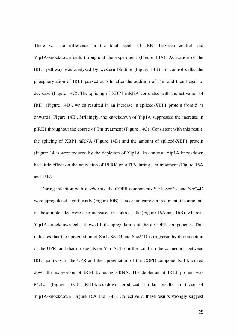
There was no difference in the total levels of IRE1 between control and
Yip1A-knockdown cells throughout the experiment (Figure 14A). Activation of the
IRE1 pathway was analyzed by western blotting (Figure 14B). In control cells, the
phosphorylation of IRE1 peaked at 5 hr after the addition of Tm, and then began to
decrease (Figure 14C). The splicing of XBP1 mRNA correlated with the activation of
IRE1 (Figure 14D), which resulted in an increase in spliced-XBP1 protein from 5 hr
onwards (Figure 14E). Strikingly, the knockdown of Yip1A suppressed the increase in
pIRE1 throughout the course of Tm treatment (Figure 14C). Consistent with this result,
the splicing of XBP1 mRNA (Figure 14D) and the amount of spliced-XBP1 protein
(Figure 14E) were reduced by the depletion of Yip1A. In contrast, Yip1A knockdown
had little effect on the activation of PERK or ATF6 during Tm treatment (Figure 15A
and 15B).
During infection with B. abortus, the COPII components Sar1, Sec23, and Sec24D
were upregulated significantly (Figure 10B). Under tunicamycin treatment, the amounts
of these molecules were also increased in control cells (Figure 16A and 16B), whereas
Yip1A-knockdown cells showed little upregulation of these COPII components. This
indicates that the upregulation of Sar1, Sec23 and Sec24D is triggered by the induction
of the UPR, and that it depends on Yip1A. To further confirm the connection between
IRE1 pathway of the UPR and the upregulation of the COPII components, I knocked
down the expression of IRE1 by using siRNA. The depletion of IRE1 protein was
84.3% (Figure 16C). IRE1-knockdown produced similar results to those of
Yip1A-knockdown (Figure 16A and 16B). Collectively, these results strongly suggest

26
that the upregulation of the COPII components Sar1, Sec23, and Sec24D depends on
the activation of IRE1 that is mediated by Yip1A.
3.2.3 Yip1A mediates a high-order assembly of IRE1 molecules
Upon the induction of the UPR, IRE1 molecules oligomerize into higher-order
species. This causes the autophosphorylation of IRE1, resulting in the appearance of
large pIRE1 foci throughout the cytoplasm (Kimata et al., 2007, Korennykh et al., 2009,
Li et al., 2010). I hypothesized that the deficiency in IRE1 phosphorylation observed in
Yip1A-knockdown cells may be attributed to the inability of IRE1 molecules to form
oligomers. To examine this possibility, the formation of large pIRE1 foci was assessed
by immunofluorescence microscopy under Tm treatment (Figure 17A and 17B). HeLa
cells were transfected with scramble siRNA or Yip1A siRNA for 24 hr, and then treated
with Tm to induce the UPR. The number of pIRE1 foci per cell was counted in these
cells. In control cells, time-dependent appearance of pIRE1 foci was observed: the
number of foci increased during the first 6 hr of Tm treatment, and then started to
decrease (Figure 17A, upper panels, 17B), consistent with the result obtained in the
western blot analysis of pIRE1 (Figure 14C). By contrast, in Yip1A knockdown cells,
pIRE1 foci were hardly observed throughout the Tm treatment (Figure 17A, lower
panels, 17B). These results support the idea that IRE1 molecules fail to assemble into
cluster in the absence of Yip1A under Tm treatment.
The above effect of Yip1A-knockdown on the oligomeric state of IRE1 was further
demonstrated by native polyacrylamide gel electrophoresis (PAGE), which permits the

27
separation of multi-protein complexes under native conditions. Phosphorylated IRE1
molecules were resolved as two high-order complexes with apparent molecular weights
of approximately 500kDa and 1000kDa (termed pIRE1-I and pIRE1-II, respectively)
(Figure 18A). In control cells, the amount of pIRE1-I was increased after 4 hr of Tm
treatment and then decreased (Figure 18A, lanes labeled ‘S’ and 18B), which coincides
with the results of the western blot analysis of pIRE1 (Figure 14C) or the formation of
pIRE1 foci (Figure 17A and 17B). The amount of pIRE1-II remained constant
throughout the Tm treatment. In Yip1A knockdown cells, the amount of both high-order
complexes was reduced significantly (Figure 18A, lanes labeled ‘Y’, and 18B). These
data support the idea that Yip1A is responsible for the phosphorylation of IRE1 via the
high-order assembly of IRE1 molecules under the UPR condition.
3.2.4 Yip1A-knockdown has little effect on the ER localization of IRE1
Yip1A has been implicated in the maintenance of ER structure (Dykstra et al., 2010).
The deficiency in oligomerization or high-order assembly of IRE1 molecules caused by
Yip1A-knockdown may be attributed to morphological deformation of the ER
membrane where IRE1 localizes. To evaluate this possibility, the localization of total
IRE1 was examined by immunofluorescence microscopy. HeLa cells were transfected
with scramble siRNA or Yip1A siRNA for 24 hr, and then treated with Tm for 5 hr to
induce the UPR. Several large vacuoles were observed in control cells, but not in
Yip1A-knockdown cells (Figure 19). Otherwise, IRE1 was stained throughout the
cytoplasm in a reticular pattern both in control and in Yip1A-knockdown cells,

28
indicating its intrinsic localization in the ER. Concentric whorl structures of ER
membrane reported by Dykstra et al. (2010) were not observed by the depletion of
Yip1A. I therefore concluded that the ER localization of IRE1 was not affected by
Yip1A-knockdown.
3.3 Formation of autophagosome-like vacuoles under tunicamycin treatment
3.3.1 Yip1A mediates the formation of large vacuoles through the IRE1 pathway
During Tm treatment, large vacuoles were observed in control cells but not in
Yip1A-knockdown cells (Figure 19). To determine whether vacuolization induced by
Tm treatment is also dependent on the Yip1A-mediated IRE1 activation, I investigated
the effect of Yip1A- or IRE1-knockdown on vacuolization under Tm treatment. HeLa
cells were transfected with each siRNA and then treated with Tm to induce the UPR.
The ER structure was visualized by immunofluorescence microscopy with an
anti-calnexin antibody (Figure 20A). Whereas large vacuoles were formed after Tm
treatment in control cell (Figure 20A, left-hand panels, arrows), such vacuolization was
not seen in Yip1A-knockdown (Figure 20A, middle panels) or IRE1-knockdown
(Figure 20A, right-hand panels) cells. The percentage of cells with vacuoles was
significantly lower in Yip1A- or IRE1-knockdown cells than in control cells after Tm
treatment (Figure 20B). Thus, there is likely to be a link between Yip1A-mediated
activation of IRE1 and the formation of large vacuoles under Tm treatment.
3.3.2 Yip1A mediates the formation of autophagosome-like vacuoles through the

29
IRE1 pathway.
The activation of the UPR has been implicated in the induction of autophagy
(Bernales et al., 2006, Hoyer-Hansen et al., 2007). In addition, the COPII vesicles
budding from ERES have been shown to supply membrane for autophagosome
formation (Graef et al., 2013, Tan et al., 2013, Wang et al., 2014). Given these recent
findings, I assumed that the formation of large vacuoles might be related to ER-derived
autophagy that is triggered by the UPR. I characterized the large vacuoles by using LC3
as a marker for autophagosome formation. When autophagy is induced, cytosolic LC3
(LC3-I) becomes lipidated (LC3-II) and translocated onto isolation membranes. These
structures can be visualized as dots by fluorescence microscopy. An expression
construct for GFP-LC3 was co-transfected with siRNA. In control cells, a number of
large GFP-LC3 dots appeared after Tm treatment (Figure 21, left-hand panels), and
some were detected along the periphery of large vacuoles (Figure 21, left-hand panel,
inset, arrowheads), indicating that these Tm-induced vacuoles have an autophagic
nature. Notably, the knockdown of Yip1A or IRE1 significantly reduced the number of
GFP-LC3 dots (Figure 21, middle and right-hand panels). Taking together, these
findings suggest that Yip1A mediates the formation of large autophagosome-like
vacuoles via the activation of IRE1 under Tm treatment.
3.4 Activation of the IRE1 pathway of the UPR during infection with B. abortus
3.4.1 Depletion of Yip1A with siRNA.

30
In the present study, the IRE1 pathway of the UPR was preferentially induced by
infection with B. abortus (Figure 8) and Yip1A was responsible for the activation of
IRE1 under Tm treatment (Figure 14). To determine whether Yip1A has the same
function during infection with B. abortus, I assessed the effect of Yip1A-knockdown on
the activation of the IRE1 pathway during Brucella infection. HeLa cells were infected
with B. abortus, and then transfected with scramble siRNA or Yip1A siRNA at 1 hr p.i.
Infection with B. abortus preceded the siRNA transfection to eliminate any effects of
Yip1A knockdown on the internalization of B. abortus. To evaluate the depletion of
Yip1A during infection, RT-PCR and western blotting were performed for Yip1A.
Yip1A mRNA was reduced by approximately 80% from 12 hr p.i. onwards (Figure
22A). Yip1A protein was reduced by 72.0% at 12 hr p.i., and by more than 80% at16 hr
p.i. onwards (Figure 22B and 22C), and this knockdown of Yip1A protein was
considered to be sufficient to demonstrate the role of Yip1A on the intracellular
replication of B. abortus at these later time points. At 4 hr or 8 hr p.i., the knockdown of
Yip1A protein was 16.8% or 46.1%, and thus the effects of Yip1A knockdown at these
time points were likely to be limited.
3.4.2 Yip1A is responsible for the activation of the IRE1 pathway of the UPR
Then the activation of the UPR sensors IRE1, PERK, and ATF6 was analyzed by
western blotting (Figure 23B and 24A). There was little difference in the total levels of
IRE1 between control and Yip1A-knockdown cells throughout the experiment (Figure
23A). Control cells (Figure 23C, 24B and 24C) showed activation kinetics for these
31
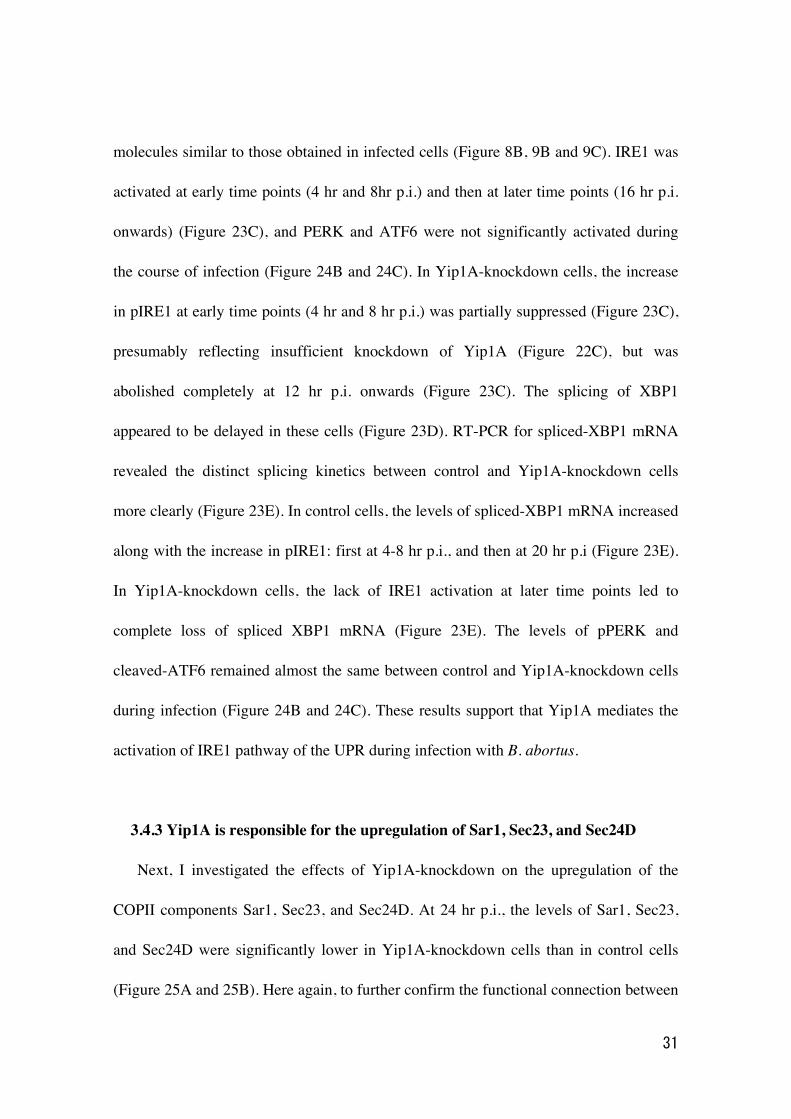
molecules similar to those obtained in infected cells (Figure 8B, 9B and 9C). IRE1 was
activated at early time points (4 hr and 8hr p.i.) and then at later time points (16 hr p.i.
onwards) (Figure 23C), and PERK and ATF6 were not significantly activated during
the course of infection (Figure 24B and 24C). In Yip1A-knockdown cells, the increase
in pIRE1 at early time points (4 hr and 8 hr p.i.) was partially suppressed (Figure 23C),
presumably reflecting insufficient knockdown of Yip1A (Figure 22C), but was
abolished completely at 12 hr p.i. onwards (Figure 23C). The splicing of XBP1
appeared to be delayed in these cells (Figure 23D). RT-PCR for spliced-XBP1 mRNA
revealed the distinct splicing kinetics between control and Yip1A-knockdown cells
more clearly (Figure 23E). In control cells, the levels of spliced-XBP1 mRNA increased
along with the increase in pIRE1: first at 4-8 hr p.i., and then at 20 hr p.i (Figure 23E).
In Yip1A-knockdown cells, the lack of IRE1 activation at later time points led to
complete loss of spliced XBP1 mRNA (Figure 23E). The levels of pPERK and
cleaved-ATF6 remained almost the same between control and Yip1A-knockdown cells
during infection (Figure 24B and 24C). These results support that Yip1A mediates the
activation of IRE1 pathway of the UPR during infection with B. abortus.
3.4.3 Yip1A is responsible for the upregulation of Sar1, Sec23, and Sec24D
Next, I investigated the effects of Yip1A-knockdown on the upregulation of the
COPII components Sar1, Sec23, and Sec24D. At 24 hr p.i., the levels of Sar1, Sec23,
and Sec24D were significantly lower in Yip1A-knockdown cells than in control cells
(Figure 25A and 25B). Here again, to further confirm the functional connection between
32
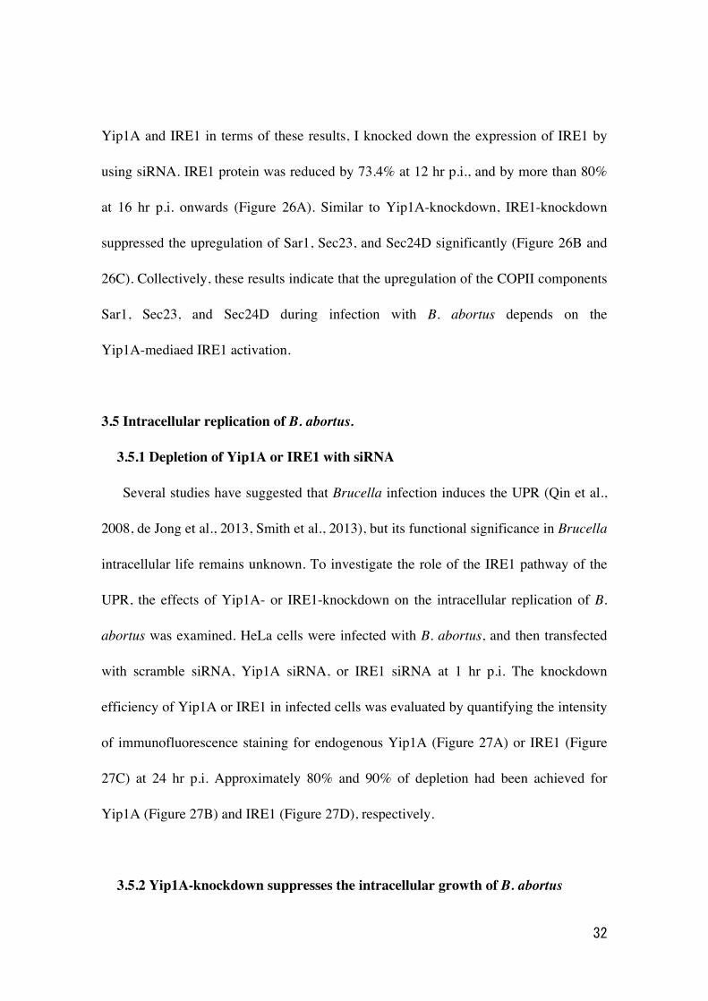
Yip1A and IRE1 in terms of these results, I knocked down the expression of IRE1 by
using siRNA. IRE1 protein was reduced by 73.4% at 12 hr p.i., and by more than 80%
at 16 hr p.i. onwards (Figure 26A). Similar to Yip1A-knockdown, IRE1-knockdown
suppressed the upregulation of Sar1, Sec23, and Sec24D significantly (Figure 26B and
26C). Collectively, these results indicate that the upregulation of the COPII components
Sar1, Sec23, and Sec24D during infection with B. abortus depends on the
Yip1A-mediaed IRE1 activation.
3.5 Intracellular replication of B. abortus.
3.5.1 Depletion of Yip1A or IRE1 with siRNA
Several studies have suggested that Brucella infection induces the UPR (Qin et al.,
2008, de Jong et al., 2013, Smith et al., 2013), but its functional significance in Brucella
intracellular life remains unknown. To investigate the role of the IRE1 pathway of the
UPR, the effects of Yip1A- or IRE1-knockdown on the intracellular replication of B.
abortus was examined. HeLa cells were infected with B. abortus, and then transfected
with scramble siRNA, Yip1A siRNA, or IRE1 siRNA at 1 hr p.i. The knockdown
efficiency of Yip1A or IRE1 in infected cells was evaluated by quantifying the intensity
of immunofluorescence staining for endogenous Yip1A (Figure 27A) or IRE1 (Figure
27C) at 24 hr p.i. Approximately 80% and 90% of depletion had been achieved for
Yip1A (Figure 27B) and IRE1 (Figure 27D), respectively.
3.5.2 Yip1A-knockdown suppresses the intracellular growth of B. abortus

33
First, intracellular bacterial growth was evaluated by counting CFUs over 24 hr
following infection. The kinetics of Brucella replication in control cells agreed with
those obtained in previous studies (Pizarro-Cerda, 1998a, Celli et al., 2003) (Figure 28,
solid bars). Robust increase in CFU occurred at 16 hr p.i. onwards, which indicates that
B. abortus undergoes extensive replication. At 24hr p.i., 51.8-fold increase in CFU was
observed. Intriguingly, Yip1A-knockdown inhibited bacterial growth, which resulted in
about a 40% reduction in CFUs at 24 hr p.i. (Figure 28, open bars). IRE1-knockdown
suppressed the increase in CFU in a similar manner to Yip1A-knockdown, and caused
an about 50% reduction in CFU at 24 hr p.i. (Figure 28, solid gray bars).
To confirm further the effect on intracellular replication, the number of B. abortus
within infected cells was examined by using immunofluorescence microscopy. Fixed
cells were stained with an anti-B. abortus antibody. Consistent with the results in the
CFU counting (Figure 28), only a few bacteria were observed in each siRNA-treated
cells at 8hr p.i. (Figure 29A, upper panels). In control cells, the onset of bacterial
replication could be seen at 16 hr p.i., and the cytoplasm of an infected cell was filled
with robustly replicating bacteria at 24 hr p.i. (Figure 29, left-hand panels; also Figure
27A and 27C, left-hand panels). In contrast, Yip1A-knockdown cells (Figure 29A,
middle panels; also and Figure 27A, right-hand panel) or IRE1-knockdown cells (Figure
29A, right-hand panels; also Figure 27C, right-hand panel) contained a considerably
small number of B. abortus at 24 hr p.i. To assess the replication efficiency, the
percentage of infected cells with fewer than ten B. abortus was determined. As can be
seen in Figure 29B, significantly less bacteria were observed in Yip1A- or

34
IRE1-knockdown cells. Altogether, these results indicate that the activation of the IRE1
pathway is critical for B. abortus to establish a safe replication niche, and that Yip1A is
indispensable for this process during infection.
3.6 Maturation of B. abortus into ER-derived BCVs.
3.6.1 Ultrastructural analysis of BCVs by electron microscopy
To characterize the deficiency in intracellular replication observed in Yip1A- or
IRE1-knockdown cells, an ultrastructural analysis of BCVs by electron microscopy
(EM) was performed at 24 hr p.i. (Figure 30A, 30B, and 30C). In control cells, infection
with B. abortus generated a significant number of replicative BCVs with vacant
vacuoles in their vicinity (Figure 30A). These membrane-bound compartments were
derived from the ER, because ribosomes lined their surface (Figure 30A, inset,
arrowheads). The lumens of the vacuoles were dilated, which resulted in massive ER
expansion. As compared with control cells, Yip1A-knockdown cells (Figure 30B)
displayed distinct morphological features. Only a few bacteria were observed within the
cells, and enlarged vacuoles were rarely seen. Notably, most BCVs were not enclosed in
ER-derived membranes (Figure 30B, inset). Similar results were obtained in
IRE1-knockdown cells (Figure 30C).
Thus, the EM analysis of infected cells revealed two forms of BCVs, one with an
outermost ER-derived membrane (Figure 30A, inset; defined as I), and the other devoid
of the ER-derived membrane (Figure 30B, inset; defined as II). The percentage of these
two types of BCVs was determined. At 24 hr p.i., about 85% of BCVs in control cells
35
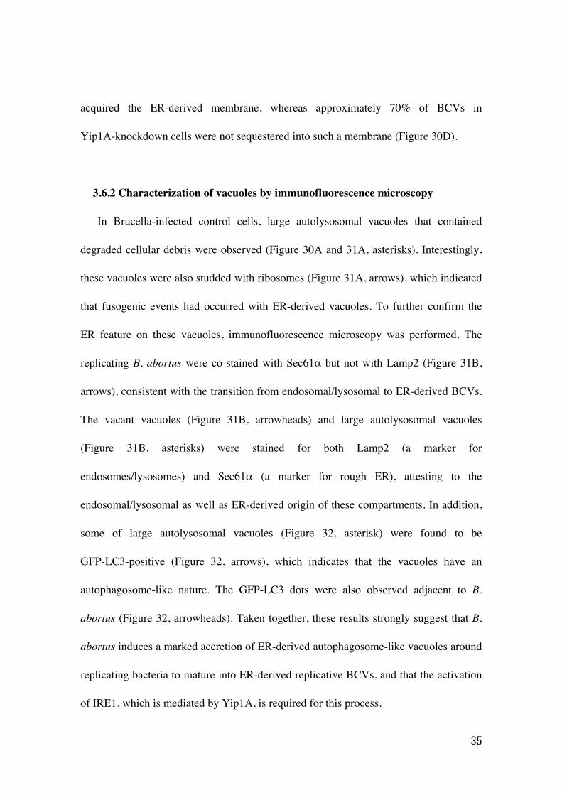
acquired the ER-derived membrane, whereas approximately 70% of BCVs in
Yip1A-knockdown cells were not sequestered into such a membrane (Figure 30D).
3.6.2 Characterization of vacuoles by immunofluorescence microscopy
In Brucella-infected control cells, large autolysosomal vacuoles that contained
degraded cellular debris were observed (Figure 30A and 31A, asterisks). Interestingly,
these vacuoles were also studded with ribosomes (Figure 31A, arrows), which indicated
that fusogenic events had occurred with ER-derived vacuoles. To further confirm the
ER feature on these vacuoles, immunofluorescence microscopy was performed. The
replicating B. abortus were co-stained with Sec61α but not with Lamp2 (Figure 31B,
arrows), consistent with the transition from endosomal/lysosomal to ER-derived BCVs.
The vacant vacuoles (Figure 31B, arrowheads) and large autolysosomal vacuoles
(Figure 31B, asterisks) were stained for both Lamp2 (a marker for
endosomes/lysosomes) and Sec61α (a marker for rough ER), attesting to the
endosomal/lysosomal as well as ER-derived origin of these compartments. In addition,
some of large autolysosomal vacuoles (Figure 32, asterisk) were found to be
GFP-LC3-positive (Figure 32, arrows), which indicates that the vacuoles have an
autophagosome-like nature. The GFP-LC3 dots were also observed adjacent to B.
abortus (Figure 32, arrowheads). Taken together, these results strongly suggest that B.
abortus induces a marked accretion of ER-derived autophagosome-like vacuoles around
replicating bacteria to mature into ER-derived replicative BCVs, and that the activation
of IRE1, which is mediated by Yip1A, is required for this process.
36
3.6.3 Lamp2-positive BCVs
In Yip1A-knockdown cells, ER-derived autophagosome-like vacuoles were absent
and BCVs were not sequestered into an ER-derived membrane (Figure 30B). This can
be attributed to the defect in trafficking of BCVs from endosomal/lysosomal
compartments to the ER or to the deficiency in the formation of ER-derived
autophagosome-like vacuoles. First, the intracellular trafficking of BCVs in
Yip1A-knockdown cells was examined by immunofluorescence microscopy for Lamp2,
a marker for late endosomes/lysosomes. Co-localization of BCVs with Lamp2-positive
vacuoles was assessed over time (Figure 33A and 33B). In control cells, BCVs left
Lamp2-positive compartments in a time-dependent manner, and 92% of BCVs were
Lamp2-negative at 24 hr p.i. (Figure 33B). By contrast, about 50% of BCVs were
co-localized with Lamp2 in Yip1A-knockdown cells (Figure 33B), suggesting that these
BCVs were confined within endosomal/lysosomal compartments. This implies that
Yip1A may play an additional role in trafficking from the endosomal/lysosomal
compartments to the ER to generate ER-derived BCVs. However, IRE1-knockdown
cells showed kinetics similar to those of Yip1A-knockdown cells, and about half of
BCVs were still retained in Lamp2-positive compartments at 24 hr i.p. (Figure 33B).
Therefore, it is not likely that the absence of the ER-derived membrane around BCVs is
due to the trafficking defects caused by Yip1A-knockdown.
3.6.4 Formation of autophagosomes

37
Next, the formation of autophagosomes was examined by fluorescence microscopy
at 24hr p.i. An expression construct for GFP-LC3 was co-transfected with siRNA. In
control cells infected with B. abortus, a number of large GFP-LC3 dots were observed
in the vicinity of replicating bacteria (Figure 34A, left-hand panel); by contrast, the
fluorescence staining of GFP-LC3 was faint and diffuse throughout the cytoplasm in
Brucella-infected Yip1A-knockdown cells (Figure 34A, middle panel). A similar result
was obtained by the knockdown of IRE1 (Figure 34A, right-hand panel). The number of
GFP-LC3 dots was significantly low in Yip1A- or IRE1-knockdown cells (Figure 34B).
These results demonstrate that HeLa cells induce autophagosomes during infection with
B. abortus, and that there is a possible link between autophagosome formation and the
Yip1A-mediated activation of IRE1. The transition from endosomal/lysosomal to
ER-derived BCVs are likely to occur not via trafficking to the ER but via a fusogenic
event with ER-derived autophagosome-like vacuoles, and Yip1A-mediated activation of
IRE1 is required for the induction of autophagosome formation.
3.7未発表の共同研究内容が含まれるので未掲載
3.7.1 未発表の共同研究内容が含まれるので未掲載
3.7.2 未発表の共同研究内容が含まれるので未掲載
3.7.3 未発表の共同研究内容が含まれるので未掲載

38
3.7.4 未発表の共同研究内容が含まれるので未掲載
3.7.5 未発表の共同研究内容が含まれるので未掲載
3.8 Proposed model of how B. abortus matures into ER-derived replicative BCVs
On the basis of the findings in the present study, I propose a model for the
maturation of B. abortus into ER-derived replicative BCVs (Figure 46A). During
infection, B. abortus triggers the activation of IRE1, presumably by secreting effector
molecules(未発表の共同研究内容が含まれるので未掲載)into the cytoplasm of
host cells. IRE1 molecules form high-order complexes at ERES with the aid of Yip1A,
and are activated by trans-autophosphorylation. The activated IRE1 in turn triggers the
biogenesis of ER-derived autophagosome-like vacuoles. These vacuoles then fuse with
endolysosomal vesicles. B. abortus might intercept this UPR-induced ER-derived
autophagy process to acquire ER-derived membranes. Since the bacteria that have
reached the ER are located in late endosomal/lysosomal compartments (Starr et al.,
2008), they would be able to fuse with these vacuoles. Once they have acquired the
ER-derived membrane, BCVs retain functional features of the ER, and replication of B.
abortus in individual vacuoles might be supported through continual accretion of ER
membranes derived from the IRE1-specific UPR. In contrast, the knockdown of Yip1A
(Figure 46B) or IRE1 (Figure 46C) prevents the activation of IRE1, and therefore
ER-derived membranes are not available for Brucella replication. Consequently, BCVs
remain in endosomal/lysosomal compartments.
39
4. Discussion
4.1 Yip1A functions in the activation of the IRE1 pathway of the UPR
In the present study, I identified a novel function of Yip1A in the activation of the
IRE1 pathway of the UPR using tunicamycin treatment and infection with B. abortus.
The activation of the IRE1 pathway leads to the biogenesis of ER-derived
autophagosome-like vacuoles, which is required for B. abortus to establish its
ER-derived replicative niche. The finding of Yip1A as a regulatory protein for the UPR
and autophagy was unexpected. Yeast homolog of Yip1A, Yip1p, was first identified as
a binding partner for Rab proteins (Yang et al., 1998), and Yip1A has been proposed to
be involved in vesicle trafficking. To date, several functions in membrane trafficking
have been suggested for Yip1A, including involvement in COPII vesicle budding at
ERES (Tang et al., 2001), vesicle tethering to the Golgi membrane (Jin et al., 2005),
and COPI-independent retrograde vesicle transport (Kano et al., 2009). However, in
mammalian cells, interaction with Rab proteins has not been reported for Yip1A, and its
function is still controversial. Recently, Dykstra et al. (2010) reported that ER
morphology was affected by the depletion of Yip1A, but I did not observe such whorled
ER formation, presumably because the event occurs after long-term treatment with
Yip1A siRNA (48-72 hr) in contrast to the shorter-term treatment in this study (24 hr).
The precise mechanism for the involvement of Yip1A in the activation of IRE1
remains to be determined. However, several results in the present study suggest that the
role of Yip1A in IRE1 activation is direct. First, the interaction between Yip1A and

40
pIRE1 is specific (Figure 11A and 11B, Figure 12) and depends on the induction of the
UPR (Figure 11C). Second, the formation of large pIRE1 foci under Tm treatment is
severely impaired by the depletion of Yip1A (Figure 17), which indicates that IRE1
fails to assemble into cluster under the UPR condition in the absence of Yip1A. Finally,
Yip1A-knockdown prevents the formation of high-order complexes of pIRE1 (Figure
18). IRE1 is usually held in its inactive state by binding the ER chaperon protein Bip.
Under conditions of ER stress, Bip is released from IRE1. However, the dissociation of
Bip is not sufficient to fully activate IRE1 (Pincus et al., 2010), and unfolded proteins
are required to promote oligomerization (Kimata et al., 2007). Yip1A may function as
an additional regulatory molecule to stabilize high-order oligomeric state of IRE1 by
limiting membrane diffusion or some other mechanisms.
4.2 Yip1A may coordinate COPII vesicle transport between the secretory pathway
and the autophagy pathway
A model that I proposed for Brucella intracellular replication (Figure 46A) is in line
with previous studies that demonstrate an intriguing link between the UPR and
autophagic vacuole formation (Bernales et al., 2006, Hoyer-Hansen et al., 2008, Ogata
et al., 2006, Li et al., 2008). Ogata et al. (2006) demonstrated that the IRE1 signaling
pathway is required for the activation of autophagy under the UPR. They showed that
the PERK and ATF6 pathways are not needed for the activation of autophagy. Our data
also strongly suggest that the IRE1 pathway can regulate autophagic events
independently from the other ER sensors.

41
In the present study, both tunicamycin treatment and infection with B. abortus
upregulated the COPII vesicle components Sar1, Sec23, and Sec24D. These proteins
assemble at ERES and lead to the curvature of the ER membrane. This might enhance
the capacity of COPII vesicles to export from ERES by promoting to pinch off vesicles
from the ERES. Several recent studies suggest that ER-derived COPII vesicles are
destined not only for the early secretory pathway to the Golgi, but also for autophagy
(Graef et al., 2013, Tan et al., 2013, Wang et al., 2014). Autophagosomes are formed at
ERES, and newly budded COPII vesicles might function as a structural core and/or
membrane source for autophagosome formation (Graef et al., 2013, Wang et al., 2014).
Tan et al. (2013) indicate that COPII vesicles are rerouted from the secretory pathway to
the autophagy pathway under starvation, and might supply membrane for
autophagosome formation. They showed that distinct effectors of Ypt1 (the yeast
homolog of Rab1) direct COPII vesicles to different pathways.
Brucella spp. might modulate these intracellular trafficking via multiple effectors.
Myeni et al. (2013) demonstrated that the Brucella effectors BspB and BspF inhibit the
host early secretory pathway prior to the biogenesis of replicative BCVs.(未発表の共
同研究内容が含まれるので未掲載)These effectors might act coordinately to
interrupt the host secretory pathway and to redirect trafficking to the ER-derived
autophagy pathway. The upregulation of the COPII components Sar1, Sec23, and
Sec24D during Brucella infection could facilitate the formation of autophagosomes.
Given the dual function of Yip1A in COPII vesicle formation or budding at ERES
(Tang et al., 2001) and in regulating the activation of IRE1 pathway of the UPR, which
42
has been documented in this study, Yip1A might coordinate COPII vesicle transport
between the secretory pathway and the autophagy pathway.
4.3 Yip1A may transduce signals through the IRE1-JNK or IRE1-NF-κB
pathway
In the present study, the levels of spliced-XBP1 were not reduced as drastically as
IRE1 phosphorylation upon knockdown of Yip1A. This might indicate that other
downstream pathways of IRE1 are affected by Yip1A-knockdown. Smith et al. (2013)
suggest that the IRE1- c-Jun NH2-terminal kinase (JNK) signaling pathway, rather than
the IRE1-XBP1 pathway, supports Brucella replication in macrophages. In addition to
the splicing of XBP1 mRNA, activated IRE1 also transmits signals through the
tumor-necrosis factor receptor associated factor 2 (TRAF2) and JNK pathway. It also
modulates the nuclear factor kappa light chain enhancer of activated B cells (NF-κB)
and extracellular signal-regulated kinase (ERK) signaling pathways. Bernales et al.
(2006) showed that expression of Hac1, the yeast homolog of XBP1, was insufficient to
induce autophagosome formation, which suggests that other signaling pathways besides
XBP1 are required. Ogata et al. (2006) suggest that activation of autophagy during the
UPR is mediated by the IRE1-TRAF2-JNK pathway. I have found that the
phosphorylation of JNK and NF-κB was reduced significantly in Yip1A-knockdown
cells (unpublished data). Therefore, Yip1A may transduce signals through the
IRE1-JNK or IRE1-NF-κB pathways.

43
4.4 Intracellular replication of B. abortus
Brucella spp. replicates within an ER-derived membrane-bound compartment in
host cells. However, the molecular mechanisms by which the pathogen establishes the
replicative niche remain unclear. In the present study, I demonstrated several lines of
evidence that clarify the mechanism by which B. abortus acquires the ER-derived
membrane. First, during Brucella infection, the IRE1 pathway, but not the PERK and
ATF6 pathways, of the UPR was activated, and the COPII vesicle components Sar1,
Sec23, and Sec24D were upregulated. Second, biogenesis of ER-derived
autophagosome-like vacuoles was observed in Brucella-infected cells. Third, Yip1A
was identified as a novel host factor that is required for the activation of IRE1 and the
subsequent formation of ER-derived autophagosome-like vacuoles. In
Yip1A-knockdown cells, B. abortus failed to be sequestered within an ER-derived
membrane, and remained in an endosomal/lysosomal compartment. (未発表の共同研
究内容が含まれるので未掲載)On the basis of these findings, I proposed a model for
Brucella maturation into ER-derived replicative BCVs. (Figure 46).
The proposed model explains many previous findings. For example, Brucella
exploits the host autophagy machinery to reach its replication compartment
(Pizarro-Cerdá et al., 1998b, Celli et al., 2003). Functional ERES, but not the
subsequent secretory pathway, are required for the biogenesis of replicative BCVs and
Sar1 mediates the fusion event between BCVs and the ER at ERES. COPII complexes
are formed in close proximity to BCVs (Celli et al., 2005). These earlier reports did not
address the mechanism by which the interaction of BCVs with Sar1/ERES and COPII

44
complexes enables the bacteria to mature into ER-derived replicative BCVs. The
extensive Brucella replication is linked to an accretion of the ER (Celli et al., 2005).
Mutants of the Brucella virB operon, encoding the T4SS, are unable to sustain
interaction with the ER, which suggests that the translocation of the Brucella VirB
effector is involved in this step (Celli et al., 2003). In the present study, I characterized
the interplay between the host and pathogen at the molecular level, thereby showing
how B. abortus subverts the host UPR and autophagy machineries to mature into
ER-derived replicative BCVs.
The depletion of Yip1A confined BCVs within Lamp2-positive compartments. This
implies that Yip1A may play an additional role in trafficking from the
endosomal/lysosomal compartments to the ER to generate ER-derived BCVs. Kano et
al. (2009) proposed that Yip1A regulates retrograde trafficking to the ER, which is
associated with membrane recruitment of Rab6. Chen and Machner (2013)
demonstrated that L. pneumophila secretes an effector protein LidA (Lowered viability
in the presence of dotA) through its Dot (Defective for organelle trafficking)/Icm
(Intracellular multiplication) type IV secretion system to recruit Rab6 on
Legionella-containing vacuoles (LCVs), which is required for efficient intracellular
replication of the pathogen. In the present study, however, the knockdown of IRE1
yielded similar effects to those of Yip1A-knockdown: BCVs were locked in a
Lamp2-positive stage devoid of ER-derived membrane, the upregulation of the COPII
components was suppressed, and ER-derived autophagosome-like vacuoles were
diminished. These results indicate that the transition from endosomal/lysosomal to

45
ER-derived BCVs occurs not via trafficking to the ER but via a fusogenic event with
ER-derived autophagosome-like vacuoles, and that Yip1A-mediated activation of IRE1
at ERES is required for the induction of the ER-derived autophagy.
4.5 未発表の共同研究内容が含まれるので未掲載
4.6 UPR and autophagy in therapeutic aspects.
In the present study, I provided the first evidence showing that Yip1A plays a
pivotal role in the activation of the IRE1 pathway of the UPR and the subsequent
formation of ER-derived autophagosome-like vacuoles. Functional interaction between
two cellular machineries, the UPR and autophagy, was demonstrated in the context of
infection of HeLa cells with B. abortus as well as under tunicamycin treatment. The
challenge for the future is to characterize the interaction between Yip1A and IRE1, and
to elucidate the molecular mechanisms of how Yip1A regulates the activation of the
IRE1 pathway of the UPR and transits signals to the autophagy pathway.
Both the UPR and autophagy function to alleviate adverse physiological or
pathological conditions and to restore cellular homeostasis, which determines whether
cells survive or die. Currently, the UPR and autophagy represent promising pathways
for treatment of a number of human diseases including cancers, diabetes, atherosclerosis,
and neurodegenerative diseases (Suh et al., 2012, Cao and Kaufman, 2013). On the
other hand, the UPR and autophagy may play a causative role in a variety of disorders.
For example, cancer cells are constitutively under high oxidative stress and the

46
activation of the UPR and autophagy has been reported in several cancers. Molecules
that modulate the UPR and autophagy can be therapeutic candidates for diverse diseases.
The characterization of the function of Yip1A will provide new insights into the
molecular mechanisms of these diseases, and contributes to design therapeutic strategies
against these diseases.
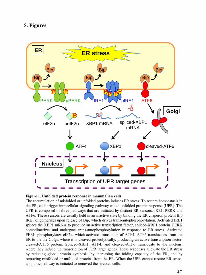
Figure 1. Unfolded protein response in mammalian cells
The accumulation of misfolded or unfolded proteins induces ER stress. To restore homeostasis in
the ER, cells trigger intracellular signaling pathway called unfolded protein response (UPR). The
UPR is composed of three pathways that are initiated by distinct ER sensors: IRE1, PERK and
ATF6. These sensors are usually held in an inactive state by binding the ER chaperon protein Bip.
IRE1 oligomerizes upon release of Bip, which drives trans-autophosphorylation. Activated IRE1
splices the XBP1 mRNA to produce an active transcription factor, spliced-XBP1 protein. PERK
homodimerizes and undergoes trans-autophosphorylation in response to ER stress. Activated
PERK phosphorylates eIF2α, which activates translation of ATF4. ATF6 translocates from the
ER to the the Golgi, where it is cleaved proteolytically, producing an active transcription factor,
cleaved-ATF6 protein. Spliced-XBP1, ATF4, and cleaved-ATF6 translocate to the nucleus,
where they induce the transcription of UPR target genes. These responses alleviate the ER stress
by reducing global protein synthesis, by increasing the folding capacity of the ER, and by
removing misfolded or unfolded proteins from the ER. When the UPR cannot restore ER stress,
apoptotic pathway is initiated to removed the stressed cells.
PPP PPP
XBP1
PERK pPERK IRE1 pIRE1 ATF6
Golgi
ER
Nucleus
XBP1 mRNA spliced-XBP1
mRNA
P
eIF2α peIF2α
ATF4 cleaved-ATF6
Bip
Bip
Bip
Bip
Bip
Bip
ER stress
Transcription of UPR target genes
47
5. Figures
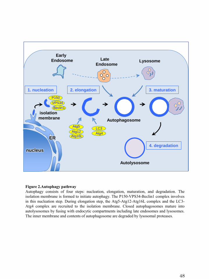
Figure 2.Autophagy pathway
Autophagy consists of four steps: nucleation, elongation, maturation, and degradation. The
isolation membrane is formed to initiate autophagy. The P150-VPS34-Beclin1 complex involves
in this nucleation step. During elongation step, the Atg5-Atg12-Atg16L complex and the LC3-
Atg4 complex are recruited to the isolation membrane. Closed autophagosomes mature into
autolysosomes by fusing with endocytic compartments including late endosomes and lysosomes.
The inner membrane and contents of autophagosome are degraded by lysosomal proteases.
48
Early
Endosome Late
EndosomeLysosome
ER
nucleus
isolation
membrane
4. degradation
2. elongation1. nucleation 3. maturation
Autolysosome
Autophagosome
P150
VPS34
Beclin1
LC3 Atg5
Atg4Atg12
Atg16L

Figure 3. Brucella intracellular trafficking in host cells
Once inside host cells, Brucella resides in a membrane-bound compartment, the Brucella-containing
vacuole (BCV). BCVs undergo a series of interactions with vesicular trafficking pathways in host
cells. They transiently interact early and late endosomes, and then lysosomes in a limited way.
Following the interaction with the endocytic compartments, BCVs are targeted to the ER, where
they interact with ERES. The interaction leads to fusogenic events between the BCVs and ER
membranes, generating ER-derived replicative BCVs. However, the mechanisms by which the
bacteria are sequestered into such vacuoles and obtain a continuous membrane supply for their
replication remain unknown. Host factors that are necessary for Brucella replication are shown.
49
GolgiTGN
ERGIC
Early
Endosome
Late
Endosome
Lysosome
Lipid raft
GAPDH
Rab2
ER-derived
replicative BCVs
ER
nucleus
Sar1
Sec23/24
?
ERESIRE1

A.
N C
1 257
1 MSGFENLNTD FYQTSYSIDD QSQQSYDYGG SGGPYSKQYA
41 GYDYSQQGRF VPPDMMQPQQ PYTGQIYQPT QAYTPASPQP
81 FYGNNFEDEP PLLEELGINF DHIWQKTLTV LHPLKVADGS
121 IMNETDLAGP MVFCLAFGAT LLLAGKIQFG YVYGISAIGC
161 LGMFCLLNLM SMTGVSFGCV ASVLGYCLLP MILLSSFAVI
201 FSLQGMVGII LTAGIIGWCS FSASKIFISA LAMEGQQLLV
241 AYPCALLYGV FALISVF
B.
Figure 4. Human Yip1A protein
(A) Schematic representation of human Yip1A protein. Yip1A is a 257 amino acids multi-pass
transmembrane protein with the hydrophilic N-terminus and the hydrophobic C-terminus.
represents hydrophilic regions, and shows hydrophobic regions.
(B) The amino acid sequences of human Yip1A. Hydrophobic regions are shaded in red. The
region that interacts with Sec23 is underlined (amino acid residues 75-106, Tang et al, 2001).
50

Figure 5. Rab small GTPase
The inactive GDP-bound form of Rab binds a guanine nucleotide dissociation inhibitor (GDI) in
the cytoplasm. A GDI displacement factor (GDF) dissociates GDI from the Rab, which leads to
insertion of the Rab into the membrane via a prenyl group. The prenylated Rab is activated by a
guanine nucleotide exchange factor (GEF) to the GTP-bound form. The GTP-bound Rab interacts
with effectors that mediate vesicle trafficking. Finally, a GTPase activating protein (GAP)
catalyzes the hydrolysis of GTP to GDP, and the GDP-bound Rab is released from the membrane
into the cytoplasm.
GDP-Rab
GDI
GDF GDP-Rab GTP-Rab
GEF
Effector GAP
GDP-Rab
GDI
organelle membrane
lumen
cytoplasm
51

Figure 6. COPII vesicle biogenesis at ERES
The small GTPase Sar1 is recruited on the ER membrane through the membrane bound GEF
Sec12 that converts the GDP-bound form of Sar1 into GTP-bound form. Activated Sar1 induces
membrane bending, and leads to recruitment of the inner coat components Sec23-Sec24 by
directly binding Sec23. Sec24 binds cargo proteins and concentrates them, thus forming a ‘pre-
budding complex’. Then the outer coat components Sec13-Sec31 polymerize to collect pre-
budding complexes and shape the vesicles. Finally, a transport vesicle buds from the ER and
delivers cargo proteins to the ERGIC. Yip1A cycles between the ER and the cis-Golgi. At ERES,
Yip1A interacts with the Sec23/Sec24 complex. Yip1A might be involved in biogenesis of COPII
vesicles that are not destined for the secretory pathway.
52
secretory pathway
other pathway
?
Sec12
Sar1GDP
GTP
Sar1
Sec23Sec24
Sec13/Sec31
ERESlumen
cytoplasm
cargo
protein
Yip1A
Traffic to the ERGIC

A.
0
10
20
30
40
1 12 24
fold
in
cre
as
e in
CF
U
time post-infection (hr)
B.
Figure 7. Replication of B. abortus within HeLa cells
(A) Intracellular growth of B. abortus within HeLa cells. HeLa cells were infected with B.
abortus and extracellular bacteria were eliminated by treatment with gentamicin for 1 hr. CFUs
were determined at 1, 12, and 24 hr p.i. A significant increase in the number of CFUs was
observed at 24hr p.i. Data are means± SD from three independent experiments.
(B) Representative confocal micrograph of HeLa cells infected with B. abortus at 1, 12, and 24
hr p.i. Fixed cells were stained for B. abortus (green). Extensive intracellular replication of B.
abortus was observed at 24hr p.i. The infected cell is outlined with white dashed lines. Scale bar
is 10 μm.
24 hr p.i.
B. abortusB. abortus B. abortus
1 hr p.i. 12 hr p.i.
53

0
1
2
3
4
5
0 4 8 12 16 20 24
rela
tive
pro
tein
le
ve
l
time post-infection (hr)
pIRE1
control
B. abortus
0
1
2
3
4
5
0 4 8 12 16 20 24
rela
tive
pro
tein
le
ve
l
time post-infection (hr)
spliced-XBP1
control
B. abortus
B.
C.
- spliced-XBP1
control .
0 4 8 12 16 20 24
B. abortus .
0 4 8 12 16 20 24 (hr p.i.) - pIRE1
- GAPDH
A.
Figure 8. The IRE1 pathway of the UPR was activated by infection with B. abortus
HeLa cells were uninfected (‘control’) or infected with B. abortus (‘B. abortus’). Cell lysates
were collected at the indicated time points after infection and analyzed by western blotting.
(A) Representative immunoblots for phosphorylated IRE1 (pIRE1), spliced-XBP1, and GAPDH.
GAPDH was used for normalization. The intensity of the bands was quantified using the
MultiGauge software.
(B) Relative protein levels of pIRE1 in uninfected control (open circles) and Brucella-infected
(solid circles) cells. The protein levels at time 0 hr were assigned the value 1. A drastic increase
in pIRE1 was observed in B. abortus-infected cells at early time points (4 hr and 8 hr p.i.), and
then later (16 hr p.i. onwards). Data are means± SD from three independent experiments.
(C) Relative protein levels of spliced-XBP1 in uninfected control (open circles) and Brucella-
infected (solid circles) cells. The protein levels at time 0 hr were assigned the value 1. Spliced-
XBP1 increased over time in B. abortus-infected cells. Data are means ± SD from three
independent experiments.
54
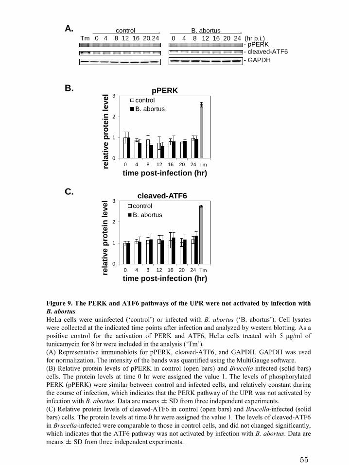
- pPERK- cleaved-ATF6
0
1
2
3
0 4 8 12 16 20 24rela
tive
pro
tein
le
ve
l
time post-infection (hr)
pPERK
control
B. abortus
Tm
0
1
2
3
0 4 8 12 16 20 24
rela
tive
pro
tein
le
ve
l
time post-infection (hr)
cleaved-ATF6
control
B. abortus
Tm
B.
C.
control .
Tm 0 4 8 12 16 20 24
B. abortus .
0 4 8 12 16 20 24 (hr p.i.)
- GAPDH
A.
Figure 9. The PERK and ATF6 pathways of the UPR were not activated by infection with
B. abortus
HeLa cells were uninfected (‘control’) or infected with B. abortus (‘B. abortus’). Cell lysates
were collected at the indicated time points after infection and analyzed by western blotting. As a
positive control for the activation of PERK and ATF6, HeLa cells treated with 5 μg/ml of
tunicamycin for 8 hr were included in the analysis (‘Tm’).
(A) Representative immunoblots for pPERK, cleaved-ATF6, and GAPDH. GAPDH was used
for normalization. The intensity of the bands was quantified using the MultiGauge software.
(B) Relative protein levels of pPERK in control (open bars) and Brucella-infected (solid bars)
cells. The protein levels at time 0 hr were assigned the value 1. The levels of phosphorylated
PERK (pPERK) were similar between control and infected cells, and relatively constant during
the course of infection, which indicates that the PERK pathway of the UPR was not activated by
infection with B. abortus. Data are means± SD from three independent experiments.
(C) Relative protein levels of cleaved-ATF6 in control (open bars) and Brucella-infected (solid
bars) cells. The protein levels at time 0 hr were assigned the value 1. The levels of cleaved-ATF6
in Brucella-infected were comparable to those in control cells, and did not changed significantly,
which indicates that the ATF6 pathway was not activated by infection with B. abortus. Data are
means± SD from three independent experiments.
55

0
1
2
3
4
5
rela
tive
pro
tein
le
ve
l Sar1
**
A.
0
1
2
3
rela
tive
pro
tein
le
ve
l Sec24D
*
control B. abortus control B. abortus control B. abortus0
1
2
3
4
5
rela
tive
pro
tein
le
ve
l Sec23
**
- Sar1
- Sec23
- Sec24D
- GAPDH
control B. abortus
B.
Figure 10. The COPII components Sar1, Sec23, and Sec24D were upregulated by infection
with B. abortus
HeLa cells were uninfected (‘control’) or infected with B. abortus (‘B. abortus’). Cell lysates
were collected at 24hr p.i. and analyzed by western blotting.
(A) Representative immunoblots for Sar1, Sec23, Sec24D, and GAPDH. GAPDH was used for
normalization. The intensity of the bands was quantified using the MultiGauge software.
(B) Relative protein levels of Sar1, Sec23, and Sec24D in uninfected control (open bars) and
Brucella-infected (solid bars) cells. The protein levels in control cells were assigned the value 1.
The expression of Sar1, Sec23, and Sec24D was increased significantly in Brucella-infected cells.
Data are means± SD from three independent experiments. *: p<0.05; **: p<0.01.
56

Figure 11. Yip1A specifically interacts with pIRE1
(A) Representative immunoblots showing co-immunoprecipitation with pIRE1. After 5 hr of Tm
treatment, immunoprecipitation was performed on HeLa cell lysates with an anti-pIRE1 antibody
(lanes labeled ‘pIRE1’) or control anti-rabbit IgG (lanes labeled ‘IgG’), and the
immunoprecipitates were analyzed by western blotting for a panel of molecules known to be
involved in the ER-Golgi vesicular transport pathways or for ER- (Sec61α, HSP47, and calnexin),
ERGIC- (ERGIC53) and cis-Golgi- (GM130) resident proteins. Phosphorylated IRE1 interacts
with the inner components of the COPII coat (Sec23, Sec24A, Sec24B, Sec24C, and Sec24D),
Rab1, and Yip1A, but not with a component of the outer coat (Sec31A), Rab2, Sar1, and some
ER- (Sec61α, HSP47 and calnexin), ERGIC- (ERGIC53) and cis-Golgi- (GM130) resident
proteins.
(B) Representative immunoblot showing the co-immunoprecipitation of pIRE1 with Yip1A.
After 5 hr of Tm treatment, immunoprecipitation was performed on HeLa cell lysates with an
anti-Yip1A antibody (lane labeled ‘Yip1A’) or control anti-rabbit IgG (lane labeled ‘IgG’), and
the immunoprecipitates were analyzed by western blotting with an anti-pIRE1 antibody.
Phosphorylated IRE1 was identified to bind to Yip1A.
(C) Representative immunoblot showing the co-immunoprecipitation of Yip1A with pIRE1.
After 0 hr or 5 hr of Tm treatment, immunoprecipitation was performed on HeLa cell lysates
with an anti-pIRE1 antibody (lane labeled ‘pIRE1’) or control anti-rabbit IgG (lane labeled
‘IgG’), and the immunoprecipitates were analyzed by western blotting with an anti-Yip1A
antibody. The intensity of the bands was quantified using the MultiGauge software, and the
results are shown in the bar graph. The protein levels at 0 hr of Tm treatment were assigned the
value 1. The interaction of Yip1A with pIRE1 was enhanced upon Tm treatment. Data are means
± SD from three independent experiments. *: p<0.05.
57
A.
- a-Sec23
- a-Sec24A
- a-Sec24B
- a-Sec24C
- a-Sec24D
- a-Sar1
- a-Sec31
- a-Rab2
- a-calnexin
- a-Sec61α
- a-Hsp47
- a-ERGIC53
- a-GM130
B.
Tm: 0 5 (hr)
Input IgG pIRE1 input IgG pIRE1
WB: a-Yip1A0
1
2
3
4
0 5rela
tive
pro
tein
le
ve
l
Tm (hr)
Yip1A
*
C.
- a-Rab1
- a-Yip1A
WB: a-pIRE1
IP .
input IgG Yip1A
IP .
input IgG pIRE1 WB
IP .
input IgG pIRE1 WB
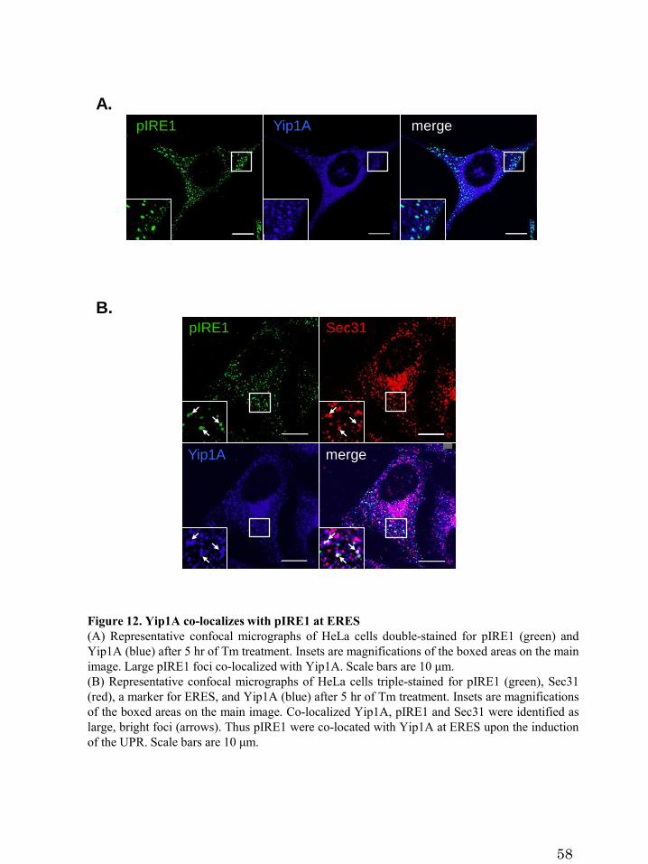
B.
A.
pIRE1 Yip1A merge
pIRE1 Sec31
mergeYip1A
Figure 12. Yip1A co-localizes with pIRE1 at ERES
(A) Representative confocal micrographs of HeLa cells double-stained for pIRE1 (green) and
Yip1A (blue) after 5 hr of Tm treatment. Insets are magnifications of the boxed areas on the main
image. Large pIRE1 foci co-localized with Yip1A. Scale bars are 10 μm.
(B) Representative confocal micrographs of HeLa cells triple-stained for pIRE1 (green), Sec31
(red), a marker for ERES, and Yip1A (blue) after 5 hr of Tm treatment. Insets are magnifications
of the boxed areas on the main image. Co-localized Yip1A, pIRE1 and Sec31 were identified as
large, bright foci (arrows). Thus pIRE1 were co-located with Yip1A at ERES upon the induction
of the UPR. Scale bars are 10 μm.
58

Figure 13. Depletion of Yip1A with siRNA
(A) Representative immunoblots showing the knockdown efficiency of Yip1A in HeLa cells at
24 hr after siRNA transfection. GAPDH was used for normalization. The intensity of the bands
was quantified using the MultiGauge software, and the results are shown in the bar graph. The
protein levels in control cells were assigned the value 1. The expression of Yip1A was reduced
by 72.5%. Data are means± SD from three independent experiments. **: p<0.01.
(B) Representative confocal micrographs of control (left-hand panel) and Yip1A-knockdown
(right-hand panel) cells stained for Yip1A, showing the depletion of Yip1A at 24 hr after siRNA
transfection. Cells are outlined with white dashed lines. Scale bars are 10 μm.
A.
B.
scramble Yip1AsiRNA siRNA
- GAPDH
- Yip1A
scramble Yip1AsiRNA siRNA
0
0.2
0.4
0.6
0.8
1
rela
tive
pro
tein
le
ve
l Yip1A
**
scramble siRNA Yip1A siRNA
Yip1A Yip1A
59

C.
B.
0
1
2
3
4
0 1 2 3 4 5 6 7 8rela
tive
pro
tein
le
ve
l
Tm (hr)
pIRE1
scramble siRNA
Yip1A siRNA
0
1
2
3
4
0 1 2 3 4 5 6 7 8rela
tive
pro
tein
le
ve
lTm (hr)
spliced-XBP1
scramble siRNA
Yip1A siRNA
0
20
40
60
80
100
0 2 4 6 8rela
tive
mR
NA
le
ve
l
Tm (hr)
spliced-XBP1 mRNAscramble siRNA
Yip1A siRNA
E.
scramble siRNA Yip1A siRNA .
Tm: 0 1 2 3 4 5 6 7 8 0 1 2 3 4 5 6 7 8 (hr)
- β-tubulin
- p-IRE1 - spliced-XBP1
D.
Figure 14. Yip1A is responsible for the activation of the IRE1 pathway of the UPR
HeLa cells were transfected with control scramble siRNA or Yip1A siRNA for 24 hr, and then
treated with Tm to induce the UPR. Cell lysates were prepared at the indicated time points and
analyzed by western blotting.
(A) Representative immunoblots for IRE1 and GAPDH. GAPDH was used for normalization.
The intensity of the bands was quantified using the MultiGauge software. Relative protein levels
of IRE1 in control (solid circles) and Yip1A-knockdown (open circles) cells during Tm treatment
are shown in the line graph. The protein levels in control cells at the beginning of the Tm
treatment were assigned the value 1. There was no difference in the total levels of IRE1 between
control and Yip1A-knockdown cells throughout the experiment. Data are means ± SD from
three independent experiments.
(B) Representative immunoblots for pIRE1, spliced-XBP1, and β-tubulin. β-tubulin was used for
normalization. The intensity of the bands was quantified using the MultiGauge software.
(C-E) Relative protein levels of pIRE1 (C), relative mRNA levels of spliced-XBP1 (D), and
relative protein levels of spliced-XBP1 (E) in control (solid circles) and Yip1A-knockdown
(open circles) cells. The protein or mRNA levels in control cells at the beginning of the Tm
treatment were assigned the value 1. The phosphorylation of IRE1 peaked at 5 hr after the
addition of Tm, and then began to decrease (C). The splicing of XBP1 mRNA correlated with the
activation of IRE1 (D), which resulted in an increase in spliced-XBP1 protein from 5 hr onwards
(E). Knockdown of Yip1A diminished the increase in pIRE1 during Tm treatment (C). The
splicing of XBP1 mRNA (D) and the amount of spliced-XBP1 protein (E) were reduced by the
depletion of Yip1A. Data are means± SD from three independent experiments.
A.
0
1
2
3
4
0 2 4 6 8rela
tive
pro
tein
le
ve
l
Tm (hr)
IRE1scramble siRNA
Yip1A siRNA- IRE1
scramble siRNA Yip1A siRNA .
Tm: 0 2 4 6 8 0 2 4 6 8 (hr)
- GAPDH
60

0
1
2
3
4
5
0 2 4 6 8rela
tive
pro
tein
le
ve
l
Tm (hr)
pPERK
scramble siRNA
Yip1A siRNA
- GAPDH
- pPERK
- cleaved-ATF6
0
1
2
3
4
5
0 2 4 6 8rela
tive
pro
tein
le
ve
l
Tm (hr)
cleaved-ATF6scramble siRNA
Yip1A siRNA
scramble siRNA Yip1A siRNA .
Tm: 0 2 4 6 8 0 2 4 6 8 (hr)
Figure 15. Yip1A knockdown had no effect on the activation of PERK or ATF6
HeLa cells were transfected with control scramble siRNA or Yip1A siRNA for 24 hr, and then
treated with Tm to induce the UPR. Cell lysates were prepared at the indicated time points and
analyzed by western blotting.
(A) Representative immunoblots for pPERK, cleaved-ATF6 and GAPDH. GAPDH was used for
normalization. The intensity of the bands was quantified using the MultiGauge software.
(B) Relative protein levels of pPERK and cleaved-ATF6 in control (solid circles) and Yip1A-
knockdown (open circles) cells during Tm treatment. The protein levels in control cells at the
beginning of the Tm treatment were assigned the value 1. There is no significant difference
between control and Yip1A-knockdown cells in the activation of PERK or ATF6. Data are
means± SD from three independent experiments.
A.
B.
61

0
1
2
3
0 2 4 6 8
rela
tive
pro
tein
le
ve
l
Tm (hr)
Sec24D
scramble siRNA
Yip1A siRNA
IRE1 siRNA
0
1
2
3
0 2 4 6 8
rela
tive
pro
tein
le
ve
l
Tm (hr)
Sec23scramble siRNAYip1A siRNAIRE1 siRNA
0
1
2
3
4
5
6
0 2 4 6 8rela
tive
pro
tein
le
ve
l
Tm (hr)
Sar1
scramble siRNAYip1A siRNAIRE1 siRNA
- Sar1
- GAPDH
- Sec23- Sec24D
scramble siRNA Yip1A siRNA IRE1 siRNA .
Tm: 0 2 4 6 8 0 2 4 6 8 0 2 4 6 8
(hr)
A.
scramble IRE1siRNA siRNA
- GAPDH
- IRE1
0
0.2
0.4
0.6
0.8
1
1.2
rela
tive
pro
tein
le
ve
l IRE1
scramble IRE1siRNA siRNA
**C.
B.
Figure 16. The upregulation of Sar1, Sec23, and Sec24D was triggered during Tm
treatment through the IRE1 pathway
HeLa cells were transfected with control scramble siRNA or Yip1A siRNA for 24 hr, and then
treated with Tm to induce the UPR. Cell lysates were prepared at the indicated time points and
analyzed by western blotting.
(A) Representative immunoblots for Sar1, Sec23, Sec24D, and GAPDH. GAPDH was used for
normalization. The intensity of the bands was quantified using the MultiGauge software.
(B) Relative protein levels of Sar1, Sec23, and Sec24D in control (solid circles), Yip1A-
knockdown (open circles), and IRE1-knockdown (solid gray circles) cells. The protein levels in
control cells at the beginning of the Tm treatment were assigned the value 1. Tm treatment
induced the upregulation of Sar1, Sec23, and Sec24D. Yip1A- or IRE1-knockdown suppressed
the upregulation of these molecules, suggesting that the upregulation of these COPII components
under Tm treatment depends on the activation of IRE1 that is mediated by Yip1A. Data are
means± SD from three independent experiments.
(C) Representative immunoblot showing the depletion of IRE1 in HeLa cells at 24 hr after
siRNA transfection. GAPDH was used for normalization. The intensity of the bands was
quantified using the MultiGauge software, and the results are shown in the bar graph. The protein
levels in control cells were assigned the value 1. The depletion of IRE1 protein was 84.3%. Data
are means± SD from three independent experiments. **: p<0.01.
62

A.
2hr
pIRE1 pIRE1
pIRE1 pIRE1
scramble siRNA
Yip1A siRNA
pIRE1 pIRE1 pIRE1
0
10
20
30
40
50
0 2 4 6 8
# o
f p
IRE
1 f
oci
pe
r c
ell
Tm (hr)
scramble siRNA
Yip1A siRNA
pIRE1
0 hr 2 hr 4 hr 6 hr 8 hr
B.
pIRE1 pIRE1 pIRE1pIRE1
pIRE1
pIRE1
Figure 17. Yip1A mediates the oligomerization of IRE1 molecules under Tm treatment
(A) Representative confocal micrographs of control (upper panels) or Yip1A-knockdown (lower
panels) cells during the Tm treatment. Fixed cells at the indicated time points were stained for
pIRE1. Cells are outlined with white dashed lines. In control cells, the number of foci increased
during the first 6 hr of Tm treatment, and then started to decrease. In Yip1A knockdown cells,
pIRE1 foci were hardly observed throughout the Tm treatment. Scale bars are 10 μm.
(B) The numbers of pIRE1 foci per cell were counted, and are shown in the line graph. Data are
means± SD (N=30).
63

Tm: 0 4 8 (hr)
S Y S Y S Y
A.
0
1
2
3
4
0 4 8
rela
tive
pro
tein
le
ve
l
Tm (hr)
pIRE1-I
scramble siRNA
Yip1A siRNA
**
****
0
1
2
3
0 4 8
rela
tive
pro
tein
le
ve
l
Tm (hr)
pIRE1-II
scramble siRNA
Yip1A siRNA
******
242kDa -
720kDa -
1048kDa -
480kDa -
- pIRE1-I
- pIRE1-II
WB: a-pIRE1
Figure 18. Yip1A mediates the formation of high-order species of IRE1 molecules under
Tm treatment
(A) Representative immunoblot for pIRE1 after native PAGE, showing two high-order
complexes of pIRE1 (termed pIRE1-I and pIRE1-II). Lane ‘S’ represents lysate from HeLa cells
transfected with control scramble siRNA, and lane ‘Y’ represents lysate from HeLa cells
transfected with Yip1A siRNA. Numbers on the left-hand side correspond to the standard
molecular weight. The intensity of the bands was quantified by using the MultiGauge software.
(B) Relative protein levels of pIRE1-1 and pIRE1-II in control (solid bars) and Yip1A-
knockdown (open bars) cells during Tm treatment. Protein levels in control cells at the beginning
of the Tm treatment were assigned the value 1. In control cells, the amount of pIRE1-I was
increased after 4 hr of Tm treatment and then decreased. The amount of pIRE1-II remained
constant throughout the Tm treatment. In Yip1A knockdown cells, the amount of both high-order
complexes was reduced significantly. Data are means± SD from three independent experiments.
**: p<0.01.
B.
64

IRE1IRE1
scramble siRNA Yip1A siRNA
Figure 19. The ER localization of IRE1 was not affected by Yip1A-knockdown
Representative confocal micrographs showing the localization of total IRE1 in control (left-hand
panel) or Yip1A-knockdown (right-hand panel) cells. HeLa cells were transfected with each
siRNA for 24 hr, and then treated with Tm for 5 hr to induce the UPR. Fixed cells were stained
for IRE1 (green). Several large vacuoles were observed in control cells (arrows), but not in
Yip1A-knockdown cells. Otherwise, IRE1 was stained throughout the cytoplasm in a reticular
pattern both in control and in Yip1A-knockdown cells, indicating its intrinsic localization in the
ER. Scale bars are 10 μm.
65

A.
calnexin calnexin
4hr 4hr
calnexin
calnexin calnexin calnexin
scramble siRNA Yip1A siRNA IRE1 siRNA Tm
0 hr
5 hr
**
0
20
40
60
0 5
% o
f c
ell
s
wit
h v
ac
uo
les
Tm (hr)
scramble siRNA
Yip1A siRNA
IRE1 siRNA
**B.
Figure 20. Yip1A mediates the formation of large vacuoles through the IRE1 pathway
under Tm treatment
HeLa cells were transfected with scramble siRNA, Yip1A siRNA or IRE1 siRNA for 24 hr, and
then treated with Tm for 5 hr to induce the UPR.
(A) Representative confocal micrographs of control (left-hand panels), Yip1A-knockdown
(middle panels), and IRE1-knockdown (right-had panels) cells after 0 hr or 5 hr of Tm treatment.
The ER structure was visualized with an anti-calnexin antibody. Large vacuoles were observed in
control cells (arrows), but not in Yip1A- or IRE1-knockdown cells. Scale bars are 10 μm.
(B) The percentage of cells with vacuoles was counted, and is shown in the bar graph. The
percentage of cells with vacuoles was significantly lower in Yip1A- or IRE1-knockdown cells
than in control cells after Tm treatment. Data are means ± SD from three independent
experiments (N=100). **: p<0.01.
66

.
GFP-LC3 GFP-LC3 GFP-LC3
GFP-LC3 GFP-LC3 GFP-LC3
Figure 21. Yip1A mediates the formation of autophagosome-like vacuoles through the
IRE1 pathway under Tm treatment
Representative confocal micrographs of control (left-hand panels; magnification of the boxed
area is shown in inset), Yip1A-knockdown (middle panels), and IRE1-knockdown (right-hand
panels) cells after 0 hr or 5 hr of Tm treatment. An expression construct for GFP-LC3 was co-
transfected with siRNA. Cells are outlined with white dashed lines. In control cells, a number of
large GFP-LC3 dots appeared after Tm treatment, and some were detected along the periphery of
large vacuoles (arrowheads), indicating that these Tm-induced vacuoles have an autophagic
nature. Scale bars are 10 μm.
scramble siRNA Yip1A siRNA IRE1 siRNA Tm
0 hr
5 hr
67

0
0.5
1
0 4 8 12 16 20 24rela
tive
mR
NA
le
ve
ltime post-infection (hr)
Yip1A mRNA
scramble siRNA
Yip1A siRNA
A.
scramble siRNA .
0 4 8 12 16 20 24
Yip1A siRNA .
0 4 8 12 16 20 24 (hr p.i.)
- GAPDH
- Yip1A
0
0.2
0.4
0.6
0.8
1
0 4 8 12 16 20 24rela
tive
pro
tein
le
ve
l
time post-infection (hr)
Yip1A
scramble siRNA
Yip1A siRNA
B.
Figure 22. Depletion of Yip1A with siRNA during infection with B. abortus
HeLa cells were infected with B. abortus, and then transfected with scramble siRNA or Yip1A
siRNA at 1 hr p.i.
(A) Relative mRNA levels of Yip1A in control (solid circles) and Yip1A-knockdown (open
circles) cells during infection with B. abortus. Total RNA was extracted at the indicated time
points and RT-PCR was carried out as described in Materials and Methods. The mRNA levels at
time 0 hr were assigned the value 1. Yip1A mRNA was reduced by approximately 80% from 12
hr p.i. onwards. Data are means± SD from three independent experiments.
(B) Representative immunoblots for Yip1A and GAPDH. Cell lysates were collected at the
indicated time points, and analyzed by western blotting. GAPDH was used for normalization.
The intensity of the bands was quantified using the MultiGauge software.
(C) Relative protein levels of Yip1A in control (solid circles) and Yip1A-knockdown (open
circles) cells during infection with B. abortus. The protein levels at time 0 hr were assigned the
value 1. Yip1A protein was reduced by 72.0% at 12 hr p.i., and by more than 80% at16 hr p.i.
onwards. Data are means± SD from three independent experiments.
C.
68

Figure 23. Yip1A mediates the activation of the IRE1 pathway of the UPR during infection
with B. abortus
HeLa cells were infected with B. abortus, and then transfected with scramble siRNA or Yip1A
siRNA at 1 hr p.i. Cell lysates were collected at the indicated time points, and analyzed by
western blotting.
(A) Representative immunoblots for IRE1 and GAPDH. GAPDH was used for normalization.
The intensity of the bands was quantified using the MultiGauge software. Relative protein levels
of IRE1 in control (solid circles) and Yip1A-knockdown (open circles) cells during during
Brucella infection are shown in the line graph. The protein levels at time 0 hr were assigned the
value 1. There was little difference in the total levels of IRE1 between control and Yip1A-
knockdown cells throughout the experiment. Data are means ± SD from three independent
experiments.
(B) Representative immunoblots for pIRE1, spliced-XBP1, and GAPDH. GAPDH was used for
normalization. The intensity of the bands was quantified using the MultiGauge software.
(C-E) Relative protein levels of pIRE1 (C) and spliced-XBP1 (D), and relative mRNA levels of
spliced-XBP1 (E) in control (solid circles) and Yip1A-knockdown (open circles) cells. The
protein or mRNA levels in control cells at time 0hr were assigned the value 1. In Yip1A-
knockdown cells, the increase in pIRE1 was abolished completely at 12 hr p.i. onwards (C). The
splicing of XBP1 appeared to be delayed in these cells (D). The distinct splicing kinetics was
observed between control and Yip1A-knockdown cells (E). The lack of IRE1 activation at later
time points led to complete loss of spliced XBP1 mRNA. Data are means ± SD from three
independent experiments.
C.
0
1
2
3
4
5
0 4 8 12 16 20 24rela
tive
pro
tein
le
ve
l
time post-infection (hr)
pIRE1
scramble siRNA
Yip1A siRNA
0
1
2
3
4
5
0 4 8 12 16 20 24rela
tive
pro
tein
le
ve
l
time post-infection (hr)
spliced-XBP1scramble siRNA
Yip1A siRNA
D.
0
1
2
3
4
5
0 4 8 12 16 20 24rela
tive
mR
NA
le
ve
l
time post-infection (hr)
spliced-XBP1 mRNA
scramble siRNA
Yip1A siRNA
B.
- GAPDH
- spliced-XBP1
- pIRE1
E.
scramble siRNA Yip1A siRNA .
0 4 8 12 16 20 24 0 4 8 12 16 20 24 (hr p.i.)
A.
0
1
2
3
4
5
0 4 8 12 16 20 24rela
tive
pro
tein
le
ve
l
time post-infection (hr)
IRE1
scramble siRNA
Yip1A siRNA
- GAPDH
- IRE1
scramble siRNA .
0 4 8 12 16 20 24
Yip1A siRNA .
0 4 8 12 16 20 24 (hr p.i.)
69

- cleaved-ATF6 - pPERK
- GAPDH
0
1
2
3
0 4 8 12 16 20 24rela
tive
pro
tein
le
ve
l
time post-infection (hr)
pPERKscramble siRNA
Yip1A siRNA
Tm
0
1
2
3
0 4 8 12 16 20 24
rela
tive
pro
tein
le
ve
l
time post-infection (hr)
cleaved-ATF6scramble siRNA
Yip1A siRNA
Tm
scramble siRNA Yip1A siRNA .
0 4 8 12 16 20 24 0 4 8 12 16 20 24 Tm (hr p.i.)
B.
C.
A.
Figure 24. The PERK and ATF6 pathways were not affected byYip1A-knockdown during
infection with B. abortus
HeLa cells were infected with B. abortus, and then transfected with scramble siRNA or Yip1A
siRNA at 1 hr p.i. Cell lysates were collected at the indicated time points, and analyzed by
western blotting. As a positive control for the activation of PERK and ATF6, HeLa cells treated
with 5 μg/ml of tunicamycin for 8 hr were included in the analysis (‘Tm’).
(A) Representative immunoblots for pPERK, cleaved-ATF6, and GAPDH. GAPDH was used
for normalization. The intensity of the bands was quantified using the MultiGauge software.
(B) Relative protein levels of pPERK in control (solid bars) and Yip1A-knockdown (open bars)
cells during infection with B. abortus. The protein levels at time 0 hr were assigned the value 1.
The levels of pPERK were similar between control and Yip1A-knockdown cells during the
course of infection, which indicates that the PERK pathway was not affected by Yip1A-
knockdown. Data are means± SD from three independent experiments.
(C) Relative protein levels of cleaved-ATF6 in control (solid bars) and Yip1A-knockdown (open
bars) cells during infection with B. abortus. The protein levels at time 0 hr were assigned the
value 1. The levels of cleaved-ATF6 were similar between control and Yip1A-knockdown cells
during the course of infection, which indicates that the ATF6 pathway was not affected by
Yip1A-knockdown.
70
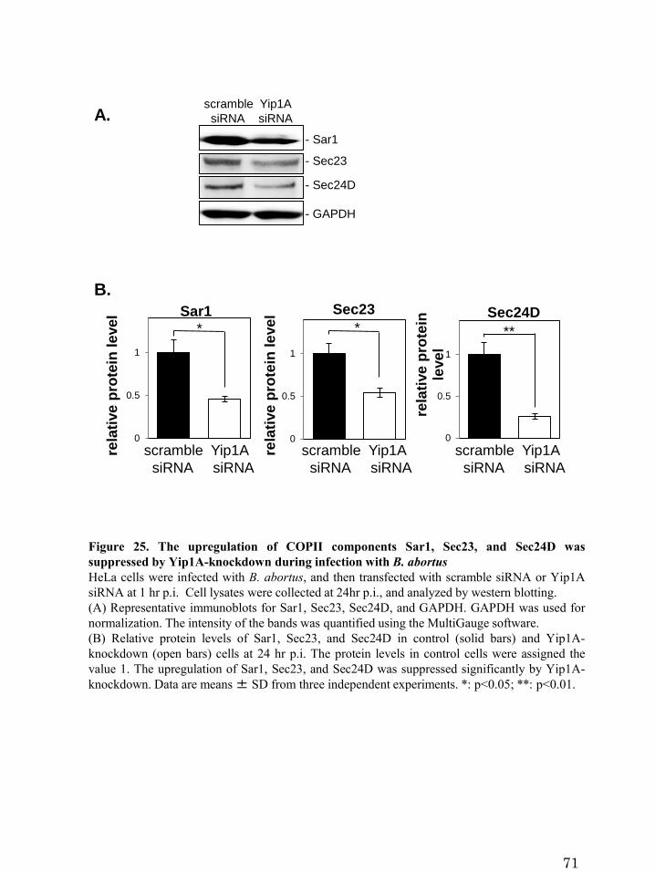
*
0
0.5
1
rela
tive
pro
tein
le
ve
l Sec23
scramble Yip1A
siRNA siRNA
**
0
0.5
1
rela
tive
pro
tein
le
ve
l
Sec24D
- Sar1
- Sec23
- Sec24D
- GAPDH
*
0
0.5
1
rela
tive
pro
tein
le
ve
l Sar1
scramble Yip1A
siRNA siRNA
scramble Yip1A
siRNA siRNA
scramble Yip1A
siRNA siRNA
Figure 25. The upregulation of COPII components Sar1, Sec23, and Sec24D was
suppressed by Yip1A-knockdown during infection with B. abortus
HeLa cells were infected with B. abortus, and then transfected with scramble siRNA or Yip1A
siRNA at 1 hr p.i. Cell lysates were collected at 24hr p.i., and analyzed by western blotting.
(A) Representative immunoblots for Sar1, Sec23, Sec24D, and GAPDH. GAPDH was used for
normalization. The intensity of the bands was quantified using the MultiGauge software.
(B) Relative protein levels of Sar1, Sec23, and Sec24D in control (solid bars) and Yip1A-
knockdown (open bars) cells at 24 hr p.i. The protein levels in control cells were assigned the
value 1. The upregulation of Sar1, Sec23, and Sec24D was suppressed significantly by Yip1A-
knockdown. Data are means± SD from three independent experiments. *: p<0.05; **: p<0.01.
A.
B.
71

0
0.5
1
rela
tive
pro
tein
le
ve
l Sar1
*
0
0.5
1
rela
tive
pro
tein
le
ve
l Sec24D
**
0
0.5
1
rela
tive
pro
tein
le
ve
l Sec23
*
scramble IRE1
siRNA siRNA
scramble IRE1
siRNA siRNA
scramble IRE1
siRNA siRNA
- GAPDH- IRE1
0
0.2
0.4
0.6
0.8
1
0 4 8 12 16 20 24rela
tive
pro
tein
le
ve
l
time post-infection (hr)
IRE1
scramble siRNA
IRE1 siRNA
scramble siRNA .
0 4 8 12 16 20 24
IRE1 siRNA .
0 4 8 12 16 20 24 (hr p.i.)
Figure 26. The upregulation of COPII components Sar1, Sec23, and Sec24D was
suppressed by IRE1-knockdown during infection with B. abortus
HeLa cells were infected with B. abortus, and then transfected with scramble siRNA or IRE1
siRNA at 1 hr p.i. Cell lysates were collected at 24hr p.i., and analyzed by western blotting.
(A) Representative immunoblots for IRE1 and GAPDH, showing the depletion of IRE1 in HeLa
cells during infection with B. abortus. GAPDH was used for normalization. The intensity of the
bands was quantified using the MultiGauge software. Relative protein levels of IRE1 in control
(solid circles) and IRE1-knockdown (open circles) cells are shown in the line graph. The protein
levels at time 0 hr were assigned the value 1. IRE1 protein was reduced by 73.4% at 12 hr p.i.,
and by more than 80% at16 hr p.i. onwards. Data are means ± SD from three independent
experiments.
(B) Representative immunoblot for Sar1, Sec23, Sec24D, and GAPDH. GAPDH was used for
normalization. The intensity of the bands was quantified using the MultiGauge software.
(C) Relative protein levels of Sar1, Sec23, and Sec24D in control (solid bars) and IRE1-
knockdown (open bars) cells at 24 hr p.i. The protein levels in control cells were assigned the
value 1. The upregulation of Sar1, Sec23, and Sec24D was suppressed significantly by IRE1-
knockdown. Data are means± SD from three independent experiments. *: p<0.05; **: p<0.01.
C.
- Sar1
- Sec23
- Sec24D
- GAPDH
scramble IRE1
siRNA siRNAB.
A.
72

A. B.
C. D.
IRE1 siRNA
Yip1A siRNA
scramble siRNA
scramble siRNA
Yip1A
B. abortus
Yip1A
B. abortus
IRE1
B. abortusIRE1
B. abortus
0
0.5
1
scramblesiRNA
IRE1siRNA
rela
tive
pro
tein
le
ve
l IRE1
**
0
0.5
1
scramblesiRNA
Yip1AsiRNA
rela
tive
pro
tein
le
ve
l Yip1A
**
Figure 27. Depletion of Yip1A or IRE1 with siRNA in HeLa cells infected with B. abortus
(A) Representative confocal micrographs of control (left-hand panel) and Yip1A-
knockdown(right-hand panel) cells at 24 hr p.i. Fixed cells were double-stained for Yip1A
(green) and B. abortus (red). The infected cells are outlined with white dashed lines. Scale bars
are 10 μm.
(B) The knockdown efficiency of Yip1A in infected cells was evaluated by quantifying the
intensity of immunofluorescence staining for Yip1A, and the result is shown in the bar graph.
Approximately 80% of depletion had been achieved for Yip1A. Data are means ± SD (N=30).
**: p<0.01.
(C) Representative confocal micrographs of control (left-hand panel) and IRE1-
knockdown(right-hand panel) cells at 24 hr p.i. Fixed cells were double-stained for IRE1 (green)
and B. abortus (red). The infected cells are outlined with white dashed lines. Scale bars are 10
μm.
(D) The knockdown efficiency of IRE1 in infected cells was evaluated by quantifying the
intensity of immunofluorescence staining for IRE1, and the result is shown in the bar graph.
Approximately 90% of depletion had been achieved for IRE1. Data are means± SD (N=30). **:
p<0.01.
73

Figure 28. Yip1A-knockdown suppresses the intracellular growth of B. abortus
HeLa cells were infected with B. abortus, and then transfected with scramble siRNA (solid bars),
Yip1A siRNA (open bars) or IRE1 siRNA (solid gray bars) at 1 hr p.i. CFUs were enumerated at
the indicated time points. In control cells, robust increase in CFU occurred at 16 hr p.i. onwards,
which indicates that B. abortus undergoes extensive replication (solid bars). At 24hr p.i., 51.8-
fold increase in CFU was observed. Yip1A-knockdown inhibited bacterial growth, which
resulted in about a 40% reduction in CFUs at 24 hr p.i. (open bars). IRE1-knockdown suppressed
the increase in CFU similar to Yip1A-knockdown, and caused about a 50% reduction in CFU at
24 hr p.i. (solid gray bars). Data are means ± SD from three independent experiments. *:
p<0.05; **: p<0.01.
0
20
40
60
1 4 8 12 16 20 24
fold
in
cre
as
e in
CF
U
time post-infection (hr)
scramble siRNA
Yip1A siRNA
IRE1 siRNA**
*
*
**
74

B.
0
20
40
60
80
100
% o
f in
fec
ted
ce
lls
W
ith
<1
0 B
.ab
ort
us
**
**
Scramble Yip1A IRE1
siRNA siRNA siRNA
Figure 29. Yip1A-knockdown suppresses the intracellular replication of B. abortus
(A) Representative confocal micrographs of control (left-hand panels), Yip1A-knockdown
(middle panels), and IRE1-knockdown (right-hand panels) cells at 8 hr, 16 hr, and 24 hr p.i.
Fixed cells were stained for B. abortus. The infected cells are outlined with white dashed lines. In
control cells, the onset of bacterial replication could be seen at 16 hr p.i., and the cytoplasm of an
infected cell was filled with robustly replicating bacteria at 24 hr p.i. In contrast, Yip1A- or
IRE1-knockdown cells contained a considerably small number of B. abortus at 24 hr p.i. Scale
bars are 10 μm.
(B) The percentage of infected cells with fewer than ten B. abortus was determined, and the
result is shown in the bar graph. Yip1A- or IRE1-knockdown significantly suppressed the
intracellular replication of B. abortus.
8 hr p.i. 8 hr p.i. 8 hr p.i.
Yip1A siRNA
B. abortus B. abortus B. abortus
B. abortus B. abortus B. abortus
B. abortus B. abortus B. abortus
A. scramble siRNA IRE1 siRNA
16 hr p.i. 16 hr p.i. 16 hr p.i.
24 hr p.i. 24 hr p.i. 24 hr p.i.
75

A.
Yip1A siRNAscramble siRNA
I II
IRE1 siRNA
Figure 30. Yip1A is required for maturation of B. abortus into ER-derived replicative
BCVs
(A-C) Representative electron micrographs of Brucella-infected control (A), Yip1A-knockdown
(B), and IRE1-knockdown (C) cells at 24 hr p.i. Insets are magnifications of the boxed areas on
the main image, showing the typical forms of BCVs. In control cells, a significant number of
replicative BCVs can be observed with vacant vacuoles in their vicinity. These BCVs are in the
form of ER-derived membrane-bound compartments (inset in (A), defined as ‘I’). Note the
presence of ribosomes on the membrane (arrowheads). Large autolysosome-like vacuoles that
contained degraded cellular debris were also observed (asterisks). In Yip1A-knockdown cells,
the bacteria were not sequestered into such compartments (inset in (B), defined as ‘II’). Similar
results were obtained in IRE1-knockdown cells (C). Scale bars are 2 μm.
(D) The percentages of the two forms of BCVs (I and II) present in control or Yip1A-knockdown
cells at 24 hr p.i. In control cells, 85% of BCVs had acquired the ER-derived membrane, whereas
70% of BCVs were not sequestered into such a membrane in Yip1A-knockdown cells. The total
numbers of BCVs analyzed were 67 for control cells and 37 for Yip1A-knockdown cells.
C. D.
0
20
40
60
80
100
% o
f B
CV
sI II
scramble Yip1A
siRNA siRNA
B.
*
**
*
76

Figure 31. Vacuoles adjacent to replicative BCVs originate from the ER and the
endosomes/lysosomes
(A) Representative electron micrograph of Brucella-infected control cells at 24 hr p.i., showing
large autolysosomal vacuoles that contain degraded cellular debris (asterisks). The vacuoles are
studded locally with ribosomes (arrows).
(B) Representative confocal micrographs of Brucella-infected control cells at 24 hr p.i. triple-
stained for HA-Sec61α (a marker for rough ER; green), B. abortus (red), and Lamp2 (a marker
for endosomes/lysosomes; blue). An expression construct for HA-Sec61α was co-transfected
with scramble siRNA. The infected cells are outlined with white dashed lines. Magnifications of
the boxed areas are shown below the main images. B. abortus (arrows) was co-stained with
Sec61α but not with Lamp2, consistent with the transition from endosomal/lysosomal to ER-
derived BCVs. Large autolysosomal vacuoles (asterisks) and the vacuoles adjacent to replicating
bacteria (arrowheads) were stained for both Sec61α and Lamp2, indicating both the
endosomal/lysosomal and ER-derived origin of these compartments. Scale bars are 10 μm.
A.
* *
mergeB. abortusHA-Sec61α
* * *
* * ** * *
** *
B.
mergeB. abortusHA-Sec61α
77

B. abortusGFP-LC3
merge
B. abortusGFP-LC3
**
*
Figure 32. Large vacuoles have an autophagosome-like nature
Representative confocal micrographs of Brucella-infected control cells at 24 hr p.i., triple-stained
for GFP-LC3 (a marker for autophagosomes; green), B. abortus (red), and Lamp2 (blue). An
expression construct for GFP-LC3 was co-transfected with scramble siRNA. Magnifications of
the boxed areas are shown below the main images. Some of the large autolysosomal vacuoles
(asterisk) and replicating B. abortus (arrowheads) were co-stained with GFP-LC3 dots (arrows).
Scale bar is 10 μm.
78

A.
Lamp2 B. abortus merge
Lamp2 B. abortus merge
Lamp2 B. abortus merge
scramble siRNA
Yip1A siRNA
IRE1 siRNA
0
20
40
60
80
100
8 16 24% L
am
p2
-po
sit
ive
BC
V
time post-infection (hr)
scramble siRNA
Yip1A siRNA
B.
Figure 33. Yip1A-knockdown confined BCVs within Lamp2-positive compartments
(A) Representative confocal micrographs of control (upper panels), Yip1A-knockdown (middle
panels), and IRE1-knockdown (lower panels) cells double-stained for Lamp2 (a marker for late
endosomes/lysosomes; green) and B. abortus (red) at 24 hr p.i. BCVs co-localized with Lamp2
are indicated by arrowheads. The infected cells are outlined with white dashed lines. Scale bars
are 10 μm.
(B) The percentage of Lamp2-positive BCVs was determined, and is shown in the line graph. In
control cells, BCVs left Lamp2-positive compartments in a time-dependent manner, and 92% of
BCVs were Lamp2-negative at 24 hr p.i. By contrast, about 50% of BCVs were co-localized with
Lamp2 in Yip1A-knockdown cells. IRE1-knockdown cells showed kinetics similar to those of
Yip1A-knockdown cells.
79
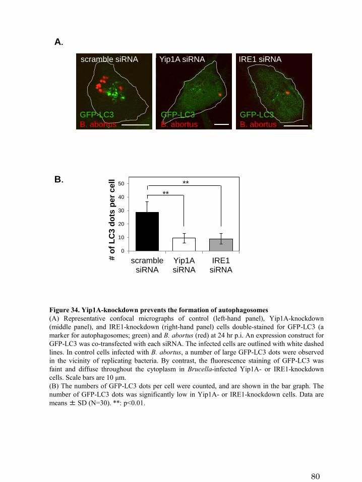
A.
B.
0
10
20
30
40
50
scramblesiRNA
Yip1AsiRNA
IRE1siRNA
# o
f L
C3
do
ts p
er
ce
ll
**
**
Figure 34. Yip1A-knockdown prevents the formation of autophagosomes
(A) Representative confocal micrographs of control (left-hand panel), Yip1A-knockdown
(middle panel), and IRE1-knockdown (right-hand panel) cells double-stained for GFP-LC3 (a
marker for autophagosomes; green) and B. abortus (red) at 24 hr p.i. An expression construct for
GFP-LC3 was co-transfected with each siRNA. The infected cells are outlined with white dashed
lines. In control cells infected with B. abortus, a number of large GFP-LC3 dots were observed
in the vicinity of replicating bacteria. By contrast, the fluorescence staining of GFP-LC3 was
faint and diffuse throughout the cytoplasm in Brucella-infected Yip1A- or IRE1-knockdown
cells. Scale bars are 10 μm.
(B) The numbers of GFP-LC3 dots per cell were counted, and are shown in the bar graph. The
number of GFP-LC3 dots was significantly low in Yip1A- or IRE1-knockdown cells. Data are
means± SD (N=30). **: p<0.01.
GFP-LC3
B. abortus
GFP-LC3
B. abortus
scramble siRNA Yip1A siRNA IRE1 siRNA
GFP-LC3
B. abortus
80

81
Figure 35. (未発表の共同研究内容が含まれるので未掲載)
Figure 36. (未発表の共同研究内容が含まれるので未掲載)
Figure 37. (未発表の共同研究内容が含まれるので未掲載)
Figure 38. (未発表の共同研究内容が含まれるので未掲載)
Figure 39. (未発表の共同研究内容が含まれるので未掲載)
Figure 40. (未発表の共同研究内容が含まれるので未掲載)
Figure 41. (未発表の共同研究内容が含まれるので未掲載)
Figure 42. (未発表の共同研究内容が含まれるので未掲載)
Figure 43. (未発表の共同研究内容が含まれるので未掲載)
Figure 44. (未発表の共同研究内容が含まれるので未掲載)
Figure 45. (未発表の共同研究内容が含まれるので未掲載)

pIRE1 Yip1A COPII T4SSBrucella
effectorIRE1
Knockdown of Yip1A
ERES
BCVsecretory
pathway
P
Sar1↑
Sec23↑
Sec24D↑
ER-derived
autophagy
(d)
(b)
(c)
(a)
ERES
BCVsecretory
pathway
ERES
Activation of IRE1 BCV
PPP P
ER-derived
autophagy
pathway
fusion
BCV
ERES
(e)
ER-derived replicative BCVs
B.A.
C.
Endosomal/lysosomal BCVs
ERES
BCVsecretory
pathway
Activation of IRE1
Endosomal/lysosomal BCVs
Knockdown of IRE1
Activation of IRE1
PPP P
Figure 46. Proposed model of how B. abortusmatures into ER-derived replicative BCVs
(A) (a) During infection, B. abortus triggers the activation of IRE1, presumably by secreting
effector molecules into the cytoplasm of host cells. (b) IRE1 molecules form high-order
complexes at ERES with the aid of Yip1A, and are activated by trans-autophosphorylation. (c)
The activated IRE1 in turn triggers the biogenesis of ER-derived autophagosome-like vacuoles.
The COPII vesicle components Sar1, Sec23, and Sec24D are upregulated, which might enhance
the capacity of COPII vesicles to export from ERES. (d) The ER-derived autophagosome-like
vacuoles then fuse with endolysosomal vesicles. Since B. abortus that have reached the ER are
located in late endosomal/lysosomal compartments, they should be able to fuse with these
vacuoles. (e) Once they have acquired the ER-derived membrane, BCVs retain functional
features of the ER, and replication of B. abortus in individual vacuoles might be supported
through continual accretion of ER membranes derived from the IRE1-specific UPR.
(B,C) Knockdown of Yip1A (B) or IRE1 (C) prevents the activation of IRE1. Consequently, ER-
derived membranes are not generated for bacterial replication and B. abortus remains in
endosomal/lysosomal compartments.
82

83
6. References
Amer AO, Swanson MS (2005) Autophagy is an immediate macrophage response to
Legionella pneumophila. Cell Microbiol 7: 765–778
Arenas GN, Staskevich AS, Aballay A, Mayorga LS (2000) Intracellular trafficking of
Brucella abortus in J774 macrophages. Infect Immun 68: 4255–4263
Axe EL, Walker SA, Manifava M, Chandra P, Roderick HL, Habermann A, Griffiths G,
Ktistakis NT (2008) Autophagosome formation from membrane compartments enriched
in phosphatidylinositol 3-phosphate and dynamically connected to the endoplasmic
reticulum. J Cell Biol 182: 685- 701.
Baruch M, Belotserkovsky I, Hertzog BB, Ravins M, Dov E, McIver KS, Le Breton YS,
Zhou Y, Cheng CY, Hanski E (2014) An extracellular bacterial pathogen modulates
host metabolism to regulate its own sensing and proliferation. Cell 156: 97-108
Baumann O, Walz B (2001) Endoplasmic reticulum of animal cells and its organization
into structural and functional domains. Int Rev Cytol 205: 149-214
Bernales S, McDonald KL, Walter P (2006) Autophagy counterbalances endoplasmic
reticulum expansion during the unfolded protein response. PLoS Biol 4: e423

84
Bi X, Corpina RA, Goldberg J (2002) Structure of the Sec23/24-Sar1 pre-budding
complex of the COPII vesicle coat. Nature 419: 271-277
Calero M, Winand NJ, Collins RN (2002) FEBS Lett 515: 89–98
Calero M, Chen CZ, Zhu W, Winand N, Havas KA, Gilbert PM, Burd CG, Collins RN
(2003) Dual prenylation is required for Rab protein localization and function. Mol Biol
Cell 14: 1852–1867
Cao SS, Kaufman RJ (2013) Targeting endoplasmic reticulum stress in metabolic
disease. Expert Opin Ther Targets 17: 437-448
Celli J, de Chastellier C, Franchini DM, Pizarro-Cerda J, Moreno E, Gorvel JP (2003)
Brucella evades macrophage killing via VirB-dependent sustained interactions with the
endoplasmic reticulum. J Exp Med 198: 545–556
Celli J, Salcedo SP, Gorvel JP (2005) Brucella coopts the small GTPase Sar1 for
intracellular replication. Proc Natl Acad Sci USA 102: 1673–1678
Chen CZ, Calero M, DeRegis CJ, Heidtman M, Barlowe C, Collins RN (2004) Genetic
analysis of yeast Yip1p function reveals a requirement for Golgi-localized rab proteins
and rab-Guanine nucleotide dissociation inhibitor. Genetics 168: 1827–1841

85
Chen Y, Machner MP (2013) Targeting of the small GTPase Rab6A’ by the Legionella
pneumophila effector LidA. Infect Immun 81: 2226-2235
Comerci DJ, Martnez-Lorenzo MJ, Sieira R, Gorvel JP, Ugalde RA (2001) Essential
role of the VirB machinery in the maturation of the Brucella abortus-containing vacuole.
Cell Microbiol 3: 159-168
Cox JS, Walter P (1996) A novel mechanism for regulating activity of a transcription
factor that controls the unfolded protein response. Cell 87: 391-404
D'Arcangelo JG, Stahmer KR, Miller EA (2013) Vesicle-mediated export from the ER:
COPII coat function and regulation. Biochim Biophys Acta 1833: 2464–2472
de Barsy M, Jamet A, Filopon D, Nicolas C, Laloux G, Rual JF, Muller A, Twizere JC,
Nkengfac B, Vandenhaute J, Hill DE, Salcedo SP, Gorvel JP, Letesson JJ, De Bolle X
(2011) Identification of a Brucella spp. secreted effector specifically interacting with
human small GTPase Rab2. Cell Microbiol 13: 1044–1058
de Barsy M, Mirabella A, Letesson JJ, De Bolle X (2012) A Brucella abortus cstA
mutant is defective for association with endoplasmic reticulum exit sites and displays
altered trafficking in HeLa cells. Microbiology 158: 2610-2618

86
de Jong MF, Sun YH, den Hartigh AB, van Dijl JM, Tsolis RM (2008) Identification of
VceA and VceC, two members of the VjbR regulon that are translocated into
macrophages by the Brucella type IV secretion system. Mol Microbiol 70: 1378–1396
de Jong MF, Starr T, Winter MG, den Hartigh AB, Child R, Knodler LA, van Dijl JM,
Celli J, Tsolis RM (2013) Sensing of Bacterial Type IV Secretion via the Unfolded
Protein Response. mBio 4: e00418
Dreux M, Chisari FV (2010) Viruses and the autophagy machinery. Cell Cycle 9: 1295–
1307
Dreux M, Chisari FV (2011) Impact of the autophagy machinery on hepatitis C virus
infection. Viruses 3: 1342-1357
Dykstra KM, Pokusa JE, Suhan J, Lee TH (2010) Yip1A structures the mammalian
endoplasmic reticulum. Mol Biol Cell 21: 1556–1568
Franco IS, Shuman HA, Charpentier X (2009) The perplexing functions and surprising
origins of Legionella pneumophila type IV secretion effectors. Cell Microbiol 11:
1435–1443
Fugier E, Salcedo SP, de Chastellier C, Pophillat M, Muller A, Arce-Gorvel V,
87
Fourquet P, Gorvel JP (2009) The glyceraldehyde-3-phosphate dehydrogenase and the
small GTPase Rab 2 are crucial for Brucella replication. PLoS Pathog 5: e1000487
Ge L, Melville D, Zhang M, Schekman R (2013) The ER–Golgi intermediate
compartment is a key membrane source for the LC3 lipidation step of autophagosome
biogenesis. Elife 2: e00947
Graef M, Friedman JR, Graham C, Babu M, Nunnari J (2013) ER exit sites are physical
and functional core autophagosome biogenesis components. Mol Biol Cell 24: 2918–
2931
Guo F, Zhang H, Chen C, Hu S, Wang Y, Qiao J, Ren Y, Zhang K, Wang Y, Du G
(2012) Autophagy favors Brucella melitensis survival in infected macrophages. Cell
Mol Biol Lett 17: 249-257
Gutierrez MG, Vázquez CL, Munafó DB, Zoppino FC, Berón W, Rabinovitch M,
Colombo MI (2005) Autophagy induction favours the generation and maturation of the
Coxiella-replicative vacuoles. Cell Microbiol 7: 981–993
Hailey DW, Rambold AS, Satpute-Krishnan P, Mitra K, Sougrat R, Kim PK,
Lippincott-Schwartz J (2010) Mitochondria supply membranes for autophagosome
biogenesis during starvation. Cell 141: 656–667

88
Harding HP, Zhang Y, Ron D (1999) Protein translation and folding are coupled by an
endoplasmic-reticulum-resident kinase. Nature 397: 271-274
Harding HP, Zhang Y, Bertolotti A, Zeng H, Ron D (2000) Perk is essential for
translational regulation and cell survival during the unfolded protein response. Mol Cell
5: 897-904
Hassan IH, Zhang MS, Powers LS, Shao JQ, Baltrusaitis J, Rutkowski DT, Legge K,
Monick MM (2012) Influenza A viral replication is blocked by inhibition of the
inositol-requiring enzyme 1 (IRE1) stress pathway. J Biol Chem 287: 4679–4689
Hayashi-Nishino M, Fujita N, Noda T, Yamaguchi A, Yoshimori T, Yamamoto A
(2009) A subdomain of the endoplasmic reticulum forms a cradle for autophagosome
formation. Nat Cell Biol 11: 1433-1437
Haze K, Yoshida H, Yanagi H, Yura T, Mori K (1999) Mammalian transcription factor
ATF6 is synthesized as a transmembrane protein and activated by proteolysis in
response to endoplasmic reticulum stress. Mol Biol Cell 10: 3787-3799
Hetz C (2012) The unfolded protein response: controlling cell fate decisions under ER
stress and beyond. Nature Rev Mol Cell Biol 13: 89–102

89
Hoyer-Hansen M and Jaattela M (2007) Connecting endoplasmic reticulum stress to
autophagy by unfolded protein response and calcium. Cell Death Differ 14: 1576– 1582
Ito T, Chiba T, Ozawa R, Yoshida M, Hattori M, Sakaki Y (2001) A comprehensive
two-hybrid analysis to explore the yeast protein interactome. Proc Natl Acad Sci USA
98: 4569–4574
Jackson WT, Giddings TH Jr, Taylor MP, Mulinyawe S, Rabinovitch M, Kopito RR,
Kirkegaard K (2005) Subversion of cellular autophagosomal machinery by RNA
viruses. PLoS Biol 3: e156
Jin C, Zhang Y, Zhu H, Ahmed K, Fu C, Yao X (2005) Human Yip1A specifies the
localization of Yif1 to the Golgi apparatus. Biochem Biophys Res Commun 334: 16-22
Jin R, Zhu W, Cao S, Chen R, Jin H, Liu Y, Wang S, Wang W, Xiao G (2013) Japanese
encephalitis virus activates autophagy as a viral immune evasion strategy. PLoS One 8:
e52909
Kano F, Yamauchi S, Yoshida Y, Watanabe-Takahashi M, Nishikawa K, Nakamura N,
Murata M (2009) Yip1A regulates the COPI-independent retrograde transport from the
Golgi complex to the ER. J Cell Sci 122: 2218–2227

90
Kano F, Nakatsu D, Noguchi Y, Yamamoto A, Murata M (2012) A resealed-cell system
for analyzing pathogenic intracellular events: Perturbation of endocytic pathways under
diabetic conditions. PLoS One 7: e44127
Khakpoor A, Panyasrivanit M, Wikan N, Smith DR (2009) A role for
autophagolysosomes in dengue virus 3 production in HepG2 cells. J Gen Virol 90:
1093-1103.
Kimata Y, Ishiwata-Kimata Y, Ito T, Hirata A, Suzuki T, Oikawa D, Takeuchi M,
Kohno K (2007) Two regulatory steps of ER-stress sensor Ire1 involving its cluster
formation and interaction with unfolded proteins. J Cell Biol 179: 75-86
Korennykh AV, Egea PF, Korostelev AA, Finer-Moore J, Zhang C, Shokat KM, Stroud
RM, Walter P (2009) The unfolded protein response signals through high-order
assembly of Ire1. Nature 457: 687–693
Kouroku Y, Fujita E, Tanida I, Ueno T, Isoai A, Kumagai H, Ogawa S, Kaufman RJ,
Kominami E, Momoi T (2007) ER stress (PERK/eIF2alpha phosphorylation) mediates
the polyglutamine-induced LC3 conversion, an essential step for autophagy formation.
Cell Death Differ 14: 230–239
Lamb CA, Yoshimori T, Tooze SA (2013) The autophagosome: origins unknown,

91
biogenesis complex. Nat Rev Mol Cell Biol 14: 759-774
Lee MC, Miller EA, Goldberg J, Orci L, Schekman R (2004) Bi-directional protein
transport between the ER and golgi. Annu Rev Cell Dev Biol 20: 87-123
Lee MC, Miller EA (2007) Molecular mechanisms of COPII vesicle formation. Semin
Cell Dev Biol 18: 424-434
Li H, Korennykh AV, Behrman SL, Walter P (2010) Mammalian endoplasmic
reticulum stress sensor IRE1 signals by dynamic clustering. Proc Natl Acad Sci USA
107: 16113-16118
Li J, Ni M, Lee B, Barron E, Hinton DR, Lee AS (2008) The unfolded protein response
regulator GRP78/BiP is required for endoplasmic reticulum integrity and stress-induced
autophagy in mammalian cells. Cell Death Differ 15: 1460–1471
Liu CY, Kaufman RJ (2003) The unfolded protein response. J Cell Sci 116: 1861-1862
Longatti A, Lamb CA, Razi M, Yoshimura S, Barr FA, Tooze SA (2012) TBC1D14
regulates autophagosome formation via Rab11 and recycling endosomes. J Cell Biol
197: 659–675

92
Marchesini MI, Herrmann CK, Salcedo SP, Gorvel JP, Comerci DJ (2011) In search of
Brucella abortus type IV secretion substrates: screening and identification of four
proteins translocated into host cells through VirB system. Cell Microbiol 13: 1261-1274
Medigeshi GR, Lancaster AM, Hirsch AJ, Briese T, Lipkin WL, Defilippis V, Früh K,
Mason PW, Nikolich-Zugich J, Nelson JA (2007) West Nile virus infection activates
the unfolded protein response, leading to CHOP induction and apoptosis. J Virol 81:
10849-10860
Miller E, Antonny B, Mamamoto S, Schekman R (2002) Cargo selection into COPII
vesicles is driven by the Sec24p subunit. EMBO J 21: 6105-6113
Mizushima N, Yoshimori T, Ohsumi Y (2011) The role of Atg proteins in
autophagosome formation. Annu Rev Cell Dev Biol 27: 107-132
Myeni S, Child R, Ng TW, Kupko JJ 3rd, Wehrly TD, Porcella SF, Knodler LA, Celli J
(2013) Brucella modulates secretory trafficking via multiple type IV secretion effector
proteins. PLoS Pathog 9: e1003556
Ogata M, Hino S, Saito A, Morikawa K, Kondo S, Kanemoto S, Murakami T,
Taniguchi M, Tanii I, Yoshinaga K, Shiosaka S, Hammarback JA, Urano F, Imaizumi
K (2006) Autophagy is activated for cell survival after endoplasmic reticulum stress.

93
Mol Cell Biol 26: 9220-9231
Ohashi, Y, Munro S (2010) Membrane delivery to the yeast autophagosome from the
Golgi–endosomal system. Mol Biol Cell 21: 3998–4008
Panyasrivanit M, Khakpoor A, Wikan N, Smith DR (2009) Co-localization of
constituents of the dengue virus translation and replication machinery with amphisomes.
J Gen Virol 90: 448-56
Pappas G, Akritidis N, Bosilkovski M, Tsianos E (2005) Brucellosis. N Engl J Med
352: 2325–2336
Pena J, Harris E (2011) Dengue virus modulates the unfolded protein response in a
time-dependent manner. J Biol Chem 286: 14226-14236
Pincus D, Chevalier MW, Aragón T, van Anken E, Vidal SE, El-Samad H, Walter P
(2010) Bip binding to the ER-stress sensor IRE1 tune the homeostatic behavior of the
unfolded protein response. PLoS Biol 8: e1000415
Pizarro-Cerda J, Meresse S, Parton RG, van der Goot G, Sola-Landa A, Lopez-Goñi I,
Moreno E, Gorvel JP (1998a) Brucella abortus transits through the autophagic pathway

94
and replicates in the endoplasmic reticulum of nonprofessional phagocytes. Infect
Immun 66: 5711–5724
Pizarro-Cerdá J, Moreno E, Sanguedolce V, Mége J-L, Gorvel JP (1998b) Virulent
Brucella abortus avoids lysosome fusion and distributes within autophagosome-like
compartments. Infect Immun 66: 2387–2392
Qin QM, Pei J, Ancona V, Shaw BD, Ficht TA, de Figueiredo P (2008) RNAi screen of
endoplasmic reticulum-associated host factors reveals a role for IRE1alpha in
supporting Brucella replication. PLoS Pathog 4: e1000110
Ravikumar B, Moreau K, Jahreiss L, Puri C, Rubinsztein DC (2010) Plasma membrane
contributes to the formation of pre-autophagosomal structures. Nature Cell Biol 12:
747–757
Romano PS, Gutierrez MG, Beron W, Rabinovitch M, Colombo MI (2007) The
autophagic pathway is actively modulated by phase II Coxiella burnetii to efficiently
replicate in the host cell. Cell Microbiol 9: 891–909
Ron D, Walter P (2007) Signal integration in the endoplasmic reticulum unfolded
protein response. Nat Rev Mol Cell Biol 8: 519–529.

95
Schnaith A, Kashkar H, Leggio SA, Addicks K, Krönke M, Krut O (2007)
Staphylococcus aureus subvert autophagy for induction of caspase-independent host
cell death. J Biol Chem 282: 2695–2706
Schröder M, Kaufman RJ (2005) The mammalian unfolded protein response. Annu Rev
Biochem 74: 739–789
Seimon TA, Kim MJ, Blumenthal A, Koo J, Ehrt S, Wainwright H (2010) Induction of
ER stress in macrophages of tuberculosis granulomas. PLoS One 5: e12772.
Shakoori A, Fujii G, Yoshimura S, Kitamura M, Nakayama K, Ito T, Ohno H,
Nakamura N (2003) Identification of a five-pass transmembrane protein family
localizing in the Golgi apparatus and the ER. Biochem Biophys Res Commun 312:
850–857
Shinohara Y, Imajo K, Yoneda M, Tomeno W, Ogawa Y, Kirikoshi H, Funakoshi K,
Ikeda M, Kato N, Nakajima A, Saito S (2013) Unfolded protein response pathways
regulate Hepatitis C Virus replication via modulation of autophagy. Biochem Biophys
Res Commun 432: 326-332
Simonsen A, Stenmark H (2008) Self-eating from an ER-associated cup. J Cell Biol
182: 621-622.

96
Smith JA, Khan M, Magnani DD, Harms JS, Durward M, Radhakrishnan GK, Liu YP,
Splitter GA (2013) Brucella induces an unfolded protein response via TcpB that
supports intracellular replication in macrophages. PLoS Pathog 9: e1003785
Sriburi R, Bommiasamy H, Buldak GL, Robbins GR, Frank M, Jackowski S, Brewer
JW (2007) Coordinate regulation of phospholipid biosynthesis and secretory pathway
gene expression in XBP-1(S)-induced endoplasmic reticulum biogenesis. J Biol Chem
282: 7024–704
Starr T, Ng TW, Wehrly TD, Knodler LA, Celli J (2008) Brucella intracellular
replication requires trafficking through the late endosomal/lysosomal compartment.
Traffic 9: 678–694
Stenmark H (2009) Rab GTPases as coordinators of vesicle traffic. Nat Rev Mol Cell
Biol 10: 513–525
Su HL, Liao CL, Lin YL (2002) Japanese encephalitis virus infection initiates
endoplasmic reticulum stress and an unfolded protein response. J Virol 76: 4162–4171
Suh DH, Kim Mi-K, Kim HS, Chung HH, Song YS (2012) Unfolded protein response
to autophagy as a promising druggable target for anticancer therapy. Ann NY Acad Sci
1271: 20-32

97
Tan D, Cai Y, Wang J, Zhang J, Menon S, Chou H-T, Ferro-Novick S, Reinisch KM,
Walz T (2013) The EM structure of the TRAPPIII complex leads to the identification of
a requirement for COPII vesicles on the macroautophagy pathway. Proc Natl Acad Sci
USA 110: 19432–19437
Tang BL, Ong YS, Huang B, Wei S, Wong ET, Qi R, Horstmann H, Hong W (2001) A
membrane protein enriched in endoplasmic reticulum exit sites interacts with COPII. J
Biol Chem 276: 40008-40017
Tardif KD, Mori K, Kaufman RJ, Siddiqui A (2004) Hepatitis C virus suppresses the
IRE1-XBP1 pathway of the unfolded protein response. J Biol Chem 279: 17158–17164
van der Vaart A, Griffith J, Reggiori F (2010) Exit from the Golgi is required for the
expansion of the autophagosomal phagophore in yeast Saccharomyces cerevisiae. Mol
Biol Cell 21: 2270–2284
Walter P, Ron D (2011) The unfolded protein response: From stress pathway to
homeostatic regulation. Science 334: 1081-1086
Wang J, Tan D, Cai Y, Reinisch KM, Walz T, Ferro-Novick S (2014) A requirement for
ER-derived COPII vesicles in phagophore initiation. Autophagy 10: 708-709
98
Yang X, Matern HT, Gallwitz D (1998) Specific binding to a novel and essential Golgi
membrane protein (Yip1p) functionally links the transport GTPases Ypt1p and Ypt31p.
EMBO J 17:4954-4963
Ye J, Rawson RB, Komuro R, Chen X, Davé UP, Prywes R, Brown MS, Goldstein JL
(2000) ER stress induces cleavage of membrane-bound ATF6 by the same proteases
that process SREBPs. Mol Cell 6: 1355–1364
Yla-Anttila P, Vihinen H, Jokitalo E, Eskelinen EL (2009) 3D tomography reveals
connections between the phagophore and endoplasmic reticulum. Autophagy 5:
1180-1185
Yorimitsu T, Nair U, Yang Z, Klionsky DJ (2006) Endoplasmic reticulum stress
triggers autophagy. J Biol Chem 281: 30299-30304
Yoshida H, Haze K, Yanagi H, Yura T, Mori K (1998) Identification of the cis-acting
endoplasmic reticulum stress response element responsible for transcriptional induction
of mammalian glucose-regulated proteins – Involvement of basic leucine zipper
transcription factors. J Biol Chem 273: 33741-33749
Yoshida H, Matsui T, Yamamoto A, Okada T, Mori K (2001) XBP1 mRNA is induced

99
by ATF6 and spliced by IRE1 in response to ER stress to produce a highly active
transcription factor. Cell 107: 881–891
Yoshida Y, Suzuki K, Yamamoto A, Sakai N, Bando M, Tanimoto K, Yamaguchi Y,
Sakaguchi T, Akhter H, Fujii G, Yoshimura S, Ogata S, Sohda M, Misumi Y,
Nakamura N (2008) YIPF5 and YIF1A recycle between the ER and the Golgi apparatus
and are involved in the maintenance of the Golgi structure. Exp Cell Res 314: 3427–
3443
Zoppino FC, Militello RD, Slavin I, Alvarez C, Colombo MI (2010) Autophagosome
formation depends on the small GTPase Rab1 and functional ER exit sites. Traffic 11:
1246–1261

100
Acknowledgments
The author wishes to express her thanks to Prof. Masayuki Murata at The University
of Tokyo for his supervision for this work. The author also wishes to express her thanks
to Dr. Fumi Kano at The University of Tokyo for her valuable suggestions throughout
this work. The author would like to thank Dr. Koichi Imaoka at National Institute of
Infectious Diseases for his technical support on infection with Brucella abortus and
experienced suggestions. The author would like to thank Dr. Akihiko Uda at National
Institute of Infectious Diseases for useful discussions on this work. The author also
thanks Ms. Michiyo Kataoka for her technical support on electron micrographs. The
assistance of the colleagues in the laboratory of the university with technical aspects of
this work is acknowledged. The author sincerely thanks her husband Dr. Satoshi
Taguchi and our three children Marin, Karin, and Rintaro for their continuous support
and encouragement.


![Eros is a novel transmembrane protein that controls the ...eprints.whiterose.ac.uk/114492/1/jem.20161382.full[1].pdf · Shaun Flint,1 James C. Lee,1 Kim Hoenderdos,2 Leanne Kane,](https://img.pdfslide.us/doc/110x75/5b3d80717f8b9a560a8dfc9a/eros-is-a-novel-transmembrane-protein-that-controls-the-1pdf-shaun-flint1.jpg)