Embed Size (px)
Citation preview
A Novel Facile Method of Labeling Octreotidewith 18F-Fluorine
Peter Laverman1, William J. McBride2, Robert M. Sharkey3, Annemarie Eek1, Lieke Joosten1, Wim J.G. Oyen1,David M. Goldenberg3, and Otto C. Boerman1
1Department of Nuclear Medicine, Radboud University Nijmegen Medical Centre, Nijmegen, The Netherlands; 2Immunomedics,Inc., Morris Plains, New Jersey; and 3Garden State Cancer Center, Center for Molecular Medicine and Immunology, Belleville,New Jersey
Several methods have been developed to label peptides with 18F.However, in general these are laborious and require a multistepsynthesis. We present a facile method based on the chelationof 18F-aluminum fluoride (Al18F) by 1,4,7-triazacyclononane-1,4,7-triacetic acid (NOTA). The method is characterized by thelabeling of NOTA-octreotide (NOTA-D-Phe-cyclo[Cys-Phe-D-Trp-Lys-Thr-Cys]-Throl (MH1 1305) [IMP466]) with 18F.Methods: Octreotide was conjugated with the NOTA chelateand labeled with 18F in a 2-step, 1-pot method. The labeling pro-cedure was optimized with regard to the labeling buffer, peptide,and aluminum concentration. Radiochemical yield, specific ac-tivity, in vitro stability, and receptor affinity were determined. Bio-distribution of 18F-IMP466 was studied in AR42J tumor–bearingmice and compared with that of 68Ga-labeled IMP466. In addi-tion, small-animal PET/CT images were acquired. Results:IMP466 was labeled with Al18F in a single step with 50% yield.The labeled product was purified by high-performance liquidchromatography to remove unbound Al18F and unlabeled pep-tide. The radiolabeling, including purification, was performed in45 min. The specific activity was 45,000 GBq/mmol, and the pep-tide was stable in serum for 4 h at 37�C. Labeling was performedat pH 4.1 in sodium citrate, sodium acetate, 4-(2-hydroxyethyl)-1-piperazineethanesulfonic acid, and 2-(N-morpholino)ethanesul-fonic acid buffer and was optimal in sodium acetate buffer. Theapparent 50% inhibitory concentration of the 19F-labeledIMP466 determined on AR42J cells was 3.6 nM. Biodistributionstudies at 2 h after injection showed a high tumor uptake of 18F-IMP466 (28.3 6 5.2 percentage injected dose per gram [%ID/g]; tumor-to-blood ratio, 300 6 90), which could be blocked byan excess of unlabeled peptide (8.6 6 0.7 %ID/g), indicatingthat the accumulation in the tumor was receptor-mediated. Bio-distribution of 68Ga-IMP466 was similar to that of 18F-IMP466.18F-IMP466 was stable in vivo, because bone uptake was only0.4 6 0.2 %ID/g, whereas free Al18F accumulated rapidly in thebone (36.9 6 5.0 %ID/g at 2 h after injection). Small-animalPET/CT scans showed excellent tumor delineation and high pref-erential accumulation in the tumor. Conclusion: NOTA-octreotidecould be labeled rapidly and efficiently with 18F using a 2-step,1-pot method. The compound was stable in vivo and showedrapid accretion in somatostatin receptor subtype 2–expressing
AR42J tumors in nude mice. This method can be used to labelother NOTA-conjugated compounds with 18F.
Key Words: octreotide; radiofluorination; NOTA; peptide; PET;aluminum fluoride
J Nucl Med 2010; 51:454–461DOI: 10.2967/jnumed.109.066902
During the past decade, radiolabeled receptor-bindingpeptides have emerged as an important class of radiophar-maceuticals that have changed radionuclide imaging.Peptides have been labeled with 111In and 99mTc forSPECT and, more recently, with positron emitters such as68Ga, 64Cu, 86Y, and 18F for PET. 18F is the most widelyused radionuclide in PET and has excellent characteristicsfor peptide-based imaging: the half-life (110 min) matchesthe pharmacokinetics of most peptides, and the lowpositron energy of 635 keV results in short ranges in tissueand excellent preclinical imaging resolution (,2 mm) (1).A wide range of methods to label peptides with 18F havebeen investigated. In general, an 18F-labeled synthon isproduced by nucleophilic substitution, which is subse-quently reacted with the functionalized peptide. One of thefirst generally applicable methods is based on conjugationof the synthon N-succinimidyl-4-18F-fluorobenzoate toa primary amino group of the peptide (2). Although widelyused, the method requires a multistep synthesis and is,therefore, time-consuming and laborious. Poethko et al.have developed an improved 18F-labeling method by react-ing 18F-fluorobenzaldehyde with an aminooxy-derivatizedpeptide, forming an oxime bond (3). Alternatively, 18F-fluorobenzaldehyde was reacted with hydrazine-nicotin-amide–conjugated peptides. However, this method resultedin increased lipophilicity of the peptide that may lead tounfavorable characteristics in vivo (4). Carbohydration ofthe peptide may counteract the enhanced lipophilicity (5).More recently, 18F-FDG was explored for the labeling ofaminooxy-derivatized peptides (6,7). This approach re-quires the use of carrier-free 18F-FDG, necessitating high-performance liquid chromatography (HPLC) purification of
Received Jun. 3, 2009; revision accepted Nov. 16, 2009.For correspondence or reprints contact: Peter Laverman, Department
of Nuclear Medicine, Radboud University Nijmegen Medical Centre, P.O.Box 9101, 6500 HB Nijmegen, The Netherlands.
E-mail: [email protected] ª 2010 by the Society of Nuclear Medicine, Inc.
454 THE JOURNAL OF NUCLEAR MEDICINE • Vol. 51 • No. 3 • March 2010
18F-FDG before conjugation with the functionalizedpeptide. Furthermore, methods based on the Huisgencycloaddition of alkynes and azides were explored for thefluorination of peptides (8–11). Recently, silicon-basedbuilding blocks were used to fluorinate bombesin peptidesfunctionalized with 2 tertiary butyl groups. However, thismethod also resulted in a lipophilic 18F peptide and loss oftumor targeting (12,13).
We reported recently that a 1,4,7-triazacyclononane-1,4,7-triacetic acid (NOTA)–conjugated pretargeting pep-tide could be labeled directly with 18F (14). To demonstratethat this 2-step, 1-pot method can be applied to otherpeptides, we report a new approach to label the somato-statin analog NOTA-octreotide with 18F. Radiolabeledsomatostatin analogs, such as diethylenetriaminepentaace-tic acid (DTPA)–octreotide, DOTA-Tyr3-octreotide, NOTA-octreotide, and DOTA-Tyr3-octreotate, can be used toimage somatostatin receptor subtype 2–expressing tumors.With this 2-step, 1-pot fluorination method, the peptidecould be stably labeled with a 50% radiochemical yield ata high specific activity within 45 min.
MATERIALS AND METHODS
GeneralThe octreotide peptide analog NOTA-D-Phe-cyclo[Cys-Phe-D-
Trp-Lys-Thr-Cys]-Throl (MH1 1305), designated IMP466, wassynthesized using standard Fmoc-based solid-phase peptide syn-thesis. After cleavage from the resin, the peptide was cyclicized byincubation with dimethyl sulfoxide overnight. The Throl resin andprotected amino acids were purchased from CreoSalus Inc. Thebis-t-butyl NOTA ligand was provided by Immunomedics, Inc. Allother chemicals were purchased from Sigma-Aldrich or FisherScientific. All buffers used for the radiolabeling procedures weremetal-free.
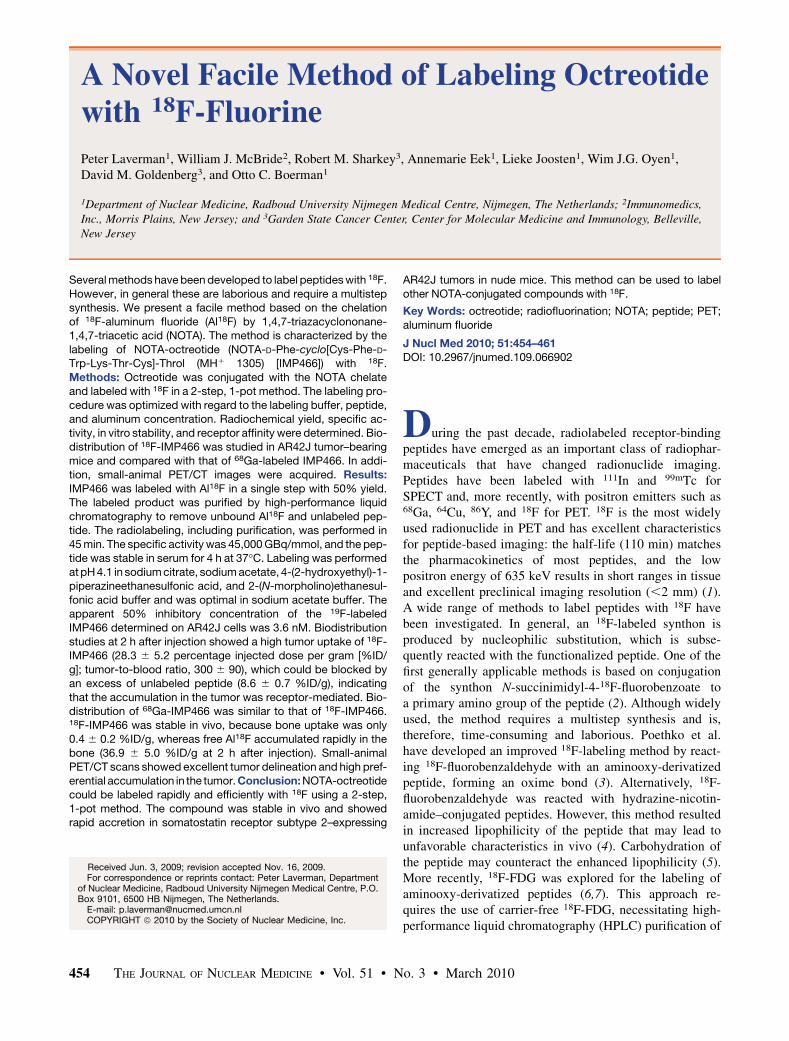
Radiolabeling18F Labeling. The 18F-labeling reaction is summarized in
Figure 1. A QMA SepPak Light cartridge (Waters) with 2–6GBq of 18F (BV Cyclotron VU) was washed with 3 mL of metal-free water. 18F was eluted from the cartridge with 0.4 M KHCO3,and fractions of 200 mL were collected. The pH of the fractionswas adjusted to 4 with 10 mL of metal-free glacial acetic acid.Three microliters of 2 mM AlCl3 in 0.1 M sodium acetate buffer,pH 4, were added. Then, 10–50 mL of IMP466 (10 mg/mL) wereadded in 0.5 M sodium acetate, pH 4.1. The reaction mixture wasincubated at 100�C for 15 min unless stated otherwise. The
radiolabeled peptide was purified on reversed-phase (RP) HPLC.The 18F-IMP466–containing fractions were collected and diluted2-fold with H2O and purified on a 1-mL Oasis HLB cartridge(Waters) to remove acetonitrile and trifluoroacetic acid. In brief,the fraction was applied on the cartridge, and the cartridge waswashed with 3 mL of H2O. The radiolabeled peptide was theneluted with 2 · 200 mL of 50% ethanol. On injection in mice, thepeptide was diluted with 0.9% NaCl.
Effect of BufferThe effect of the buffer on the labeling efficiency of IMP466
with 18F2 was investigated (n 5 3 for each buffer). IMP466 wasdissolved in sodium citrate buffer, sodium acetate buffer, 2-(N-morpholino)ethanesulfonic acid (MES) or 4-(2-hydroxyethyl)-1-piperazineethanesulfonic acid (HEPES) buffer at 10 mg/mL (7.7mM). The molarity of all buffers was 1 M, and the pH was 4.1. To200 mg (153 nmol) of IMP466, 100 mL of 18F-aluminum fluoride(Al18F) (pH 4) were added and incubated at 100�C for 15 min.Radiolabeling yield and specific activity were determined with RPHPLC as described below.
68Ga Labeling. IMP466 was labeled with 68GaCl3 eluted froma TiO2-based 1,110-MBq 68Ge/68Ga generator (Cyclotron Co.Ltd.) using 0.1 M HCl (Ultrapure; J.T. Baker). Five 1-mL fractionswere collected, and an aliquot of the second fraction was used forlabeling the peptide. IMP466 (4 mg) was dissolved in 2.5 MHEPES buffer, pH 7.0. 68Ga eluate (120–240 MBq, 4 times thevolume of the peptide) was added, and the mixture was heated at95�C for 20 min. Then 50 mM ethylenediaminetetraacetic acidwas added to a final concentration of 5 mM to complex thenonincorporated 68Ga31. The 68Ga-labeled IMP466 was purifiedon a 1-cm3 Oasis HLB cartridge and eluted with 200 mL of 50%ethanol. The specific activity of 68Ga-IMP466 was 20,000 GBq/mmol at the time of injection.
HPLC AnalysisThe radiolabeled preparations were analyzed by RP HPLC on
an Agilent 1200 system (Agilent Technologies). A C18 column(Onyx monolithic, 4.6 · 100 mm; Phenomenex) was used at a flowrate of 2 mL/min with the following buffer system: buffer A, 0.1%v/v trifluoroacetic acid in water; buffer B, 0.1% v/v trifluoroaceticacid in acetonitrile; and a gradient of 97% buffer A at 0–5 min and80% buffer A to 75% buffer A at 5–35 min. The radioactivity ofthe eluate was monitored using an in-line NaI radiodetector(Raytest GmbH). Elution profiles were analyzed using Gina-starsoftware (version 2.18; Raytest GmbH). The specific activity wasdetermined by HPLC using calibration curves based on theultraviolet signal.
FIGURE 1. Preparation of Al18F andchelation with NOTA-octreotide.
Al18F-OCTREOTIDE • Laverman et al. 455
Octanol–Water Partition Coefficient (log Poctanol/water)To determine the lipophilicity of the radiolabeled peptides,
approximately 50,000 counts per minute of the radiolabeledpeptide were diluted in 0.5 mL of phosphate-buffered saline. Anequal volume of 1-octanol was added to obtain a binary phasesystem. After stirring in a vortex mixer for 2 min, we separated the2 layers by centrifugation (100g, 5 min). Three 100-mL sampleswere taken from each layer, and radioactivity was measured ina well-type g-counter (Wallac Wizard 3"; Perkin-Elmer).
StabilityTen microliters of the 18F-labeled IMP466 were incubated in
500 mL of freshly collected human serum and incubated for 4 h at37�C. An equal volume of acetonitrile was added and stirred in avortex mixer, then followed by centrifugation at 1,000g for 5 minto pellet the precipitated serum proteins. The supernatant wasanalyzed on RP HPLC as described above.
The in vivo stability of 18F-IMP466 was examined by injecting18.5 MBq of 18F-IMP466 in a BALB/c nude mouse. After 30 min,the mouse was euthanized, and blood and urine were collected andanalyzed by HPLC.
Cell CultureThe AR42J rat pancreatic tumor cell line was cultured in
Dulbecco’s modified Eagle’s medium (Gibco Life Technologies)supplemented with 4,500 mg of D-glucose per milliliter, 10% (v/v)fetal calf serum, 2 mmol of glutamine per liter, 100 U of penicillinper milliliter, and 100 mg of streptomycin per milliliter. Cells werecultured at 37�C in a humidified atmosphere with 5% CO2.
Apparent 50% Inhibitory Concentration (IC50)Determination
The apparent IC50 for binding the somatostatin receptors onAR42J cells was determined in a competitive binding assayusing 19F-IMP466, 69Ga-IMP466, or 115In-DTPA-octreotide tocompete for the binding of 111In-DTPA-octreotide (OctreoScan;Covidien).
19F-IMP466 was formed by mixing an AlF solution (0.02 MAlCl3 in 0.5 M NaAc, pH 4, with 0.1 M NaF in 0.5 M NaAc, pH 4)with IMP466 and heating at 100�C for 15 min. The reaction mixturewas purified by RP HPLC on a C-18 column (30 · 150 mm, Sunfire;Waters), as described above.
69Ga-IMP466 was prepared by dissolving gallium nitrate (2.3 ·1028 mol) in 30 mL mixed with 20 mL of IMP466 (1 mg/mL) in10 mM NaAc, pH 5.5, and heated at 90�C for 15 min. Samples ofthe mixture were used without further purification.
115In-DTPA-octreotide was made by mixing indium chloride (1 ·1025 mol) with 10 mL of DTPA-octreotide (1 mg/mL) in 50 mMNaAc, pH 5.5, and incubated at room temperature for 15 min. Thissample was used without further purification. 111In-DTPA-octreo-tide was radiolabeled according to the manufacturer’s protocol.
AR42J cells were grown to confluency in 12-well plates andwashed twice with binding buffer (Dulbecco’s modified Eagle’smedium with 0.5% bovine serum albumin). After 10 min ofincubation at room temperature in binding buffer, 19F-IMP466,69Ga-IMP466, or 115In-DTPA-octreotide was added at a final con-centration ranging from 0.1 to 1,000 nM, together with a traceamount (10,000 counts per minute) of 111In-DTPA-octreotide(radiochemical purity . 95%). After incubation at room temperaturefor 3 h, the cells were washed twice with ice-cold phosphate-bufferedsaline. Cells were scraped, and cell-associated radioactivity wasdetermined. Under these conditions, a limited extent of internaliza-
tion may occur. Therefore, we describe the results of this competitivebinding assay as apparent IC50 values rather than IC50. The apparentIC50 was defined as the peptide concentration at which 50% ofbinding without competitor was reached. Apparent IC50 values werecalculated using GraphPad Prism software (version 4.00 for Win-dows; GraphPad Software).
Biodistribution StudiesMale nude BALB/c mice (6–8 wk old) were injected subcutane-
ously in the right flank with 0.2 mL of AR42J cell suspension of 1 ·107 cells/mL. Approximately 2 wk after inoculation, when tumorswere 5–8 mm in diameter, 370 kBq of 18F-labeled or 68Ga-labeledIMP466 (both 0.2 nmol) were administered intravenously (n 5 5).Separate groups of mice (n 5 5) were coinjected with a 1,000-foldmolar excess of unlabeled IMP466. One group of 3 mice wasinjected with unchelated Al18F. All mice were killed by CO2/O2
asphyxiation 2 h after injection. Tissues of interest were dissected,weighed, and counted in a g-counter. The percentage injected doseper gram of tissue (%ID/g) was calculated for each tissue on thebasis of a dilution of the product for injection. The animalexperiments were approved by the local animal welfare committeeand performed according to national regulations.
PET/CTMice with subcutaneous AR42J tumors were injected intrave-
nously with 10 MBq of 18F-IMP466 or 68Ga-IMP466 (both 0.7nmol) per mouse. One and 2 h after the injection of peptide, micewere scanned on an animal PET/CT scanner (Inveon; SiemensPreclinical Solutions) with an intrinsic spatial resolution of 1.5mm (1). The animals were placed supine in the scanner. PETemission scans were acquired over 15 min, followed by a CT scanfor anatomic reference (spatial resolution, 113 mm; 80 kV,500 mA). Scans were reconstructed using Inveon AcquisitionWorkplace software (version 1.2; Siemens Preclinical Solutions),using an ordered-set expectation maximization 3-dimensionalmaximum a posteriori algorithm with the following parameters:matrix, 256 · 256 · 159; pixel size, 0.43 · 0.43 · 0.8 mm3; andb-value, 0.1.
Statistical AnalysisAll mean values are given as 6SD. Statistical analysis was
performed using a Welch’s corrected unpaired Student t test or 1-way ANOVA using GraphPad InStat software (version 3.06; Graph-Pad Software). The level of significance was set at P less than 0.05.
RESULTS
18F-Labeling Procedure
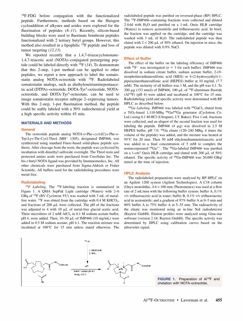
HPLC analysis of the IMP466 labeling mixture (Fig. 2)showed the presence of unbound Al18F (retention time [Rt] 5
0.8 min) and 2 radioactive peptide peaks (Rt 5 17.4 and 19.8min), indicating the formation of two 18F-IMP466 stereoiso-mers. Moreover, an ultraviolet peak of unlabeled IMP466 ispresent (Rt 5 21.4 min). After HPLC and HLB purification,both the unbound Al18F and the unlabeled IMP466 ultravi-olet peaks disappeared (Fig. 2).
Effect of Buffer. For sodium acetate, MES, or HEPES,the radiolabeling yield was 49% 6 2%, 46% 6 2%, and48% 6 3%, respectively (n 5 3 for each buffer). In sodiumcitrate, no labeling was observed. When the labelingreaction was performed in sodium acetate buffer, the
456 THE JOURNAL OF NUCLEAR MEDICINE • Vol. 51 • No. 3 • March 2010
specific activity was 32,000 6 17,000 GBq/mmol, whereasin MES and HEPES buffer, specific activities of 29,000 6
14,000 and 31,000 6 23,000 GBq/mmol, respectively, wereobtained.
Effect of Peptide Concentration. The effect of peptideconcentration on the labeling efficiency also was investi-gated. IMP466 was dissolved in sodium acetate buffer, pH4.1, at a concentration of 7.7 mM (10 mg/mL). Either 38,153, or 363 nmol of IMP466 were added to 200 mL ofAl18F (581–603 MBq) to yield a final IMP466 concentra-tions of 190, 765, and 1,815 mM, respectively. The radio-labeling yield increased with increasing amounts ofpeptide. At a concentration of 190 mM, the radiolabelingyield was 8% 6 1%; at 765 mM, the yield increasedto 42% 6 3%; and at the highest concentration, theradiolabeling yield was 50% 6 2%. The specific activityof the products obtained at each concentration was 48,000GBq/mmol.
Effect of AlCl3 Concentration. Because AlCl3 is used toform Al18F, the added amount of AlCl3 is critical in the
labeling procedure. Five stock solutions with variousAlCl3 concentrations were prepared: 0.2, 0.5, 1.0, 2.0,and 20 mM. From these solutions in sodium acetate, 3mL were added to 200 mL of 18F-fluoride, pH 4, to formAl18F, resulting in final amounts of AlCl3 added of 0.6, 1.5,3.0, 6.0, and 60 nmol, respectively. To these samples, 153nmol of IMP466 (final concentration, 765 mM) were addedand incubated for 15 min at 100�C. Radiolabeling yield wasoptimal (50% 6 2%, n 5 5) after incubation with 6 nmol ofAlCl3. Lowering the AlCl3 concentration resulted in re-duced yields, ranging from 42% at 3 nmol to 10% at 0.6nmol of AlCl3. Increasing the amount led to a similareffect. Incubation with 60 nmol of AlCl3 resulted ina radiolabeling yield of only 6%.
Octanol–Water Partition Coefficient
To determine the lipophilicity of the 18F- and 68Ga-labeled IMP466, the octanol–water partition coefficientswere determined. The log Poctanol/water value for the 18F-IMP466 was 22.44 6 0.12, and that of 68Ga-IMP466 was23.79 6 0.07, indicating that the 18F-IMP466 was slightlyless hydrophilic than 68Ga-IMP466.
IC50 Determination
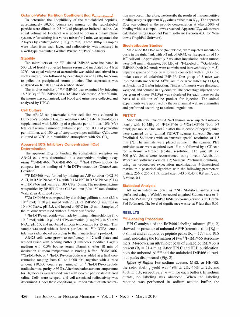
The affinity profiles are shown in Figure 3. The apparentIC50 of Al19F-labeled IMP466 was 3.6 6 0.6 nM, whereasthat for 69Ga-labeled IMP466 was 13 6 3 nM. The apparentIC50 of the reference peptide, 115In-DTPA-octreotide(OctreoScan), was 6.3 6 0.9 nM.
Stability18F-labeled IMP466 did not release Al18F after incubation
in human serum at 37�C for 4 h, indicating excellentstability of the Al18F-NOTA-octreotide. These findings wereconfirmed in vivo. After 30 min, only intact radiolabeled
FIGURE 2. RP HPLC chromatograms of IMP466 18F-labeling mix (A) and purified 18F-IMP466 (B). Red tracesrepresent radioactivity (left y-axis), and blue traces representultraviolet signal (right y-axis). In HPLC chromatogram ofcrude mixture, unbound Al18F eluted with void volume (Rt 5
0.8 min). Two radioactive peaks correspond to times ofstereoisomers of radiolabeled peptide (Rt 5 17.4 and 19.8min). Finally, unlabeled IMP466 was present in ultravioletchannel (Rt 5 21.4 min). After purification, only 2 radioactivepeptide peaks are observed, indicating formation of 2stereoisomers. cps 5 counts per second; UV 5 ultraviolet.
FIGURE 3. Competitive binding assay (apparent IC50) of19F-IMP466, 69Ga-IMP466, and 115In-DTPA-octreotide de-termined on AR42J tumor cells. Values on y-axis representbinding expressed as percentage of binding withoutcompetitor.
Al18F-OCTREOTIDE • Laverman et al. 457
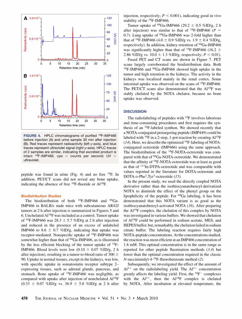
peptide was found in urine (Fig. 4) and no free 18F. Inaddition, PET/CT scans did not reveal any bone uptake,indicating the absence of free 18F-fluoride or Al18F.
Biodistribution Studies
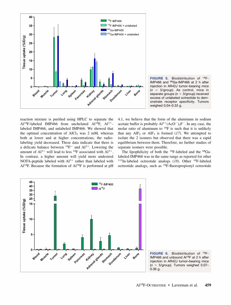
The biodistribution of both 18F-IMP466 and 68Ga-IMP466 in BALB/c nude mice with subcutaneous AR42Jtumors at 2 h after injection is summarized in Figures 5 and6. Unchelated Al18F was included as a control. Tumor uptakeof 18F-IMP466 was 28.3 6 5.7 %ID/g at 2 h after injectionand reduced in the presence of an excess of unlabeledIMP466 to 8.6 6 0.7 %ID/g, indicating that uptake wasreceptor-mediated. Nonspecific uptake of 18F-IMP466 wassomewhat higher than that of 68Ga-IMP466, as is illustratedby the less efficient blocking of the tumor uptake of 18F-IMP466. Blood levels were low (0.10 6 0.07 %ID/g, 2 hafter injection), resulting in a tumor-to-blood ratio of 300 6
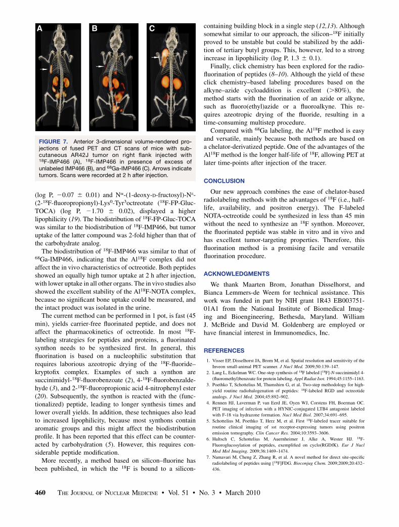
90. Uptake in normal tissues, except in the kidneys, was low,with specific uptake in somatostatin receptor subtype 2–expressing tissues, such as adrenal glands, pancreas, andstomach. Bone uptake of 18F-IMP466 was negligible, ascompared with uptake after injection of nonchelated Al18F(0.33 6 0.07 %ID/g vs. 36.9 6 5.0 %ID/g at 2 h after
injection, respectively; P , 0.001), indicating good in vivostability of the 18F-IMP466.
Tumor uptake of 68Ga-IMP466 (29.2 6 0.5 %ID/g, 2 hafter injection) was similar to that of 18F-IMP466 (P 5
0.7). Lung uptake of 68Ga-IMP466 was 2-fold higher thanthat of 18F-IMP466 (4.0 6 0.9 %ID/g vs. 1.9 6 0.4 %ID/g,respectively). In addition, kidney retention of 68Ga-IMP466was significantly higher than that of 18F-IMP466 (16.2 6
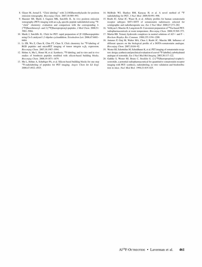
2.86 %ID/g vs. 10.0 6 1.3 %ID/g, respectively; P , 0.01).Fused PET and CT scans are shown in Figure 7. PET
scans largely corroborated the biodistribution data. Both18F-IMP466 and 68Ga-IMP466 showed high uptake in thetumor and high retention in the kidneys. The activity in thekidneys was localized mainly in the renal cortex. Someintestinal uptake was observed on the scans of 18F-IMP466.The PET/CT scans also demonstrated that the Al18F wasstably chelated by the NOTA chelator, because no boneuptake was observed.
DISCUSSION
The radiolabeling of peptides with 18F involves laboriousand time-consuming procedures and first requires the syn-thesis of an 18F-labeled synthon. We showed recently thata NOTA-conjugated pretargeting peptide (IMP449) could belabeled with 18F in a 2-step, 1-pot reaction by creating Al18F(14). Here, we describe the optimized 18F-labeling of NOTA-conjugated octreotide (IMP466) using the same approach.The biodistribution of the 18F-NOTA-octreotide was com-pared with that of 68Ga-NOTA-octreotide. We demonstratedthat the affinity of 18F-NOTA-octreotide was at least as goodas that of 111In-DTPA-octreotide and was comparable withvalues reported in the literature for DOTA-octreotate andDOTA-D-Phe1,Tyr3-octreotide (15).
In the present study, we used the directly coupled NOTAderivative rather than the isothiocyanatobenzyl-derivatizedNOTA to diminish the effect of the phenyl group on thelipophilicity of the peptide. For 68Ga labeling, it has beendemonstrated that this NOTA variant is as good as theisothiocyanatobenzyl-activated NOTA (16). After preparingthe Al18F complex, the chelation of this complex by NOTAwas investigated in various buffers. We showed that chelationof Al18F could be performed in sodium acetate, MES, andHEPES buffer, but, remarkably, the chelation failed in sodiumcitrate buffer. The labeling reaction requires fairly highNOTA-peptide concentrations. At the concentrations studied,the reaction was most efficient at an IMP466 concentration of1.8 mM. This optimal concentration is in the same range asreported for other peptide fluorination methods (3,4) butlower than the optimal concentration required in the classicN-succinimidyl-4-18F-fluorobenzoate method (2).
Subsequently, we investigated the effect of the amount ofAl31 on the radiolabeling yield. The Al31 concentrationgreatly affects the labeling yield. First, the 18F2 complexeswith the Al31, then the Al18F complex is chelatedby NOTA. After incubation at elevated temperature, the
FIGURE 4. HPLC chromatograms of purified 18F-IMP466before injection (A) and urine sample 30 min after injection(B). Red traces represent radioactivity (left y-axis), and bluetraces represent ultraviolet signal (right y-axis). HPLC tracesof 2 samples are similar, indicating that excreted product isintact 18F-IMP466. cps 5 counts per second; UV 5
ultraviolet.
458 THE JOURNAL OF NUCLEAR MEDICINE • Vol. 51 • No. 3 • March 2010
reaction mixture is purified using HPLC to separate theAl18F-labeled IMP466 from unchelated Al18F, Al31-labeled IMP466, and unlabeled IMP466. We showed thatthe optimal concentration of AlCl3 was 2 mM, whereasboth at lower and at higher concentrations, the radio-labeling yield decreased. These data indicate that there isa delicate balance between 18F2 and Al31. Lowering theamount of Al31 will lead to less 18F associated with Al31.In contrast, a higher amount will yield more undesiredNOTA-peptide labeled with Al31 rather than labeled withAl18F. Because the formation of Al18F is performed at pH
4.1, we believe that the form of the aluminum in sodiumacetate buffer is probably Al31(AcO2)3F2. In any case, themolar ratio of aluminum to 18F is such that it is unlikelythat any AlF2 or AlF3 is formed (17). We attempted toisolate the 2 isomers but observed that there was a rapidequilibrium between them. Therefore, no further studies ofseparate isomers were possible.
The lipophilicity of both the 18F-labeled and the 68Ga-labeled IMP466 was in the same range as reported for other111In-labeled octreotide analogs (18). Other 18F-labeledoctreotide analogs, such as 18F-fluoropropionyl octreotide
FIGURE 5. Biodistribution of 18F-IMP466 and 68Ga-IMP466 at 2 h afterinjection in AR42J tumor–bearing mice(n 5 5/group). As control, mice inseparate groups (n 5 3/group) receivedexcess of unlabeled octreotide to dem-onstrate receptor specificity. Tumorsweighed 0.04–0.33 g.
FIGURE 6. Biodistribution of 18F-IMP466 and unbound Al18F at 2 h afterinjection in AR42J tumor–bearing mice(n 5 5/group). Tumors weighed 0.07–0.36 g.
Al18F-OCTREOTIDE • Laverman et al. 459
(log P, 20.07 6 0.01) and Na-(1-deoxy-D-fructosyl)-Ne-(2-18F-fluoropropionyl)-Lys0-Tyr3octreotate (18F-FP-Gluc-TOCA) (log P, 21.70 6 0.02), displayed a higherlipophilicity (19). The biodistribution of 18F-FP-Gluc-TOCAwas similar to the biodistribution of 18F-IMP466, but tumoruptake of the latter compound was 2-fold higher than that ofthe carbohydrate analog.
The biodistribution of 18F-IMP466 was similar to that of68Ga-IMP466, indicating that the Al18F complex did notaffect the in vivo characteristics of octreotide. Both peptidesshowed an equally high tumor uptake at 2 h after injection,with lower uptake in all other organs. The in vivo studies alsoshowed the excellent stability of the Al18F-NOTA complex,because no significant bone uptake could be measured, andthe intact product was isolated in the urine.
The current method can be performed in 1 pot, is fast (45min), yields carrier-free fluorinated peptide, and does notaffect the pharmacokinetics of octreotide. In most 18F-labeling strategies for peptides and proteins, a fluorinatedsynthon needs to be synthesized first. In general, thisfluorination is based on a nucleophilic substitution thatrequires laborious azeotropic drying of the 18F-fluoride–kryptofix complex. Examples of such a synthon aresuccinimidyl-18F-fluorobenzoate (2), 4-18F-fluorobenzalde-hyde (3), and 2-18F-fluoropropionic acid 4-nitrophenyl ester(20). Subsequently, the synthon is reacted with the (func-tionalized) peptide, leading to longer synthesis times andlower overall yields. In addition, these techniques also leadto increased lipophilicity, because most synthons containaromatic groups and this might affect the biodistributionprofile. It has been reported that this effect can be counter-acted by carbohydration (5). However, this requires con-siderable peptide modification.
More recently, a method based on silicon–fluorine hasbeen published, in which the 18F is bound to a silicon-
containing building block in a single step (12,13). Althoughsomewhat similar to our approach, the silicon–18F initiallyproved to be unstable but could be stabilized by the addi-tion of tertiary butyl groups. This, however, led to a strongincrease in lipophilicity (log P, 1.3 6 0.1).
Finally, click chemistry has been explored for the radio-fluorination of peptides (8–10). Although the yield of theseclick chemistry–based labeling procedures based on thealkyne–azide cycloaddition is excellent (.80%), themethod starts with the fluorination of an azide or alkyne,such as fluoro(ethyl)azide or a fluoroalkyne. This re-quires azeotropic drying of the fluoride, resulting in atime-consuming multistep procedure.
Compared with 68Ga labeling, the Al18F method is easyand versatile, mainly because both methods are based ona chelator-derivatized peptide. One of the advantages of theAl18F method is the longer half-life of 18F, allowing PET atlater time-points after injection of the tracer.
CONCLUSION
Our new approach combines the ease of chelator-basedradiolabeling methods with the advantages of 18F (i.e., half-life, availability, and positron energy). The F-labeledNOTA-octreotide could be synthesized in less than 45 minwithout the need to synthesize an 18F synthon. Moreover,the fluorinated peptide was stable in vitro and in vivo andhas excellent tumor-targeting properties. Therefore, thisfluorination method is a promising facile and versatilefluorination procedure.
ACKNOWLEDGMENTS
We thank Maarten Brom, Jonathan Disselhorst, andBianca Lemmers-de Weem for technical assistance. Thiswork was funded in part by NIH grant 1R43 EB003751-01A1 from the National Institute of Biomedical Imag-ing and Bioengineering, Bethesda, Maryland. WilliamJ. McBride and David M. Goldenberg are employed orhave financial interest in Immunomedics, Inc.
REFERENCES
1. Visser EP, Disselhorst JA, Brom M, et al. Spatial resolution and sensitivity of the
Inveon small-animal PET scanner. J Nucl Med. 2009;50:139–147.
2. Lang L, Eckelman WC. One-step synthesis of 18F labeled [18F]-N-succinimidyl 4-
(fluoromethyl)benzoate for protein labeling. Appl Radiat Isot. 1994;45:1155–1163.
3. Poethko T, Schottelius M, Thumshirn G, et al. Two-step methodology for high-
yield routine radiohalogenation of peptides: 18F-labeled RGD and octreotide
analogs. J Nucl Med. 2004;45:892–902.
4. Rennen HJ, Laverman P, van Eerd JE, Oyen WJ, Corstens FH, Boerman OC.
PET imaging of infection with a HYNIC-conjugated LTB4 antagonist labeled
with F-18 via hydrazone formation. Nucl Med Biol. 2007;34:691–695.
5. Schottelius M, Poethko T, Herz M, et al. First 18F-labeled tracer suitable for
routine clinical imaging of sst receptor-expressing tumors using positron
emission tomography. Clin Cancer Res. 2004;10:3593–3606.
6. Hultsch C, Schottelius M, Auernheimer J, Alke A, Wester HJ. 18F-
Fluoroglucosylation of peptides, exemplified on cyclo(RGDfK). Eur J Nucl
Med Mol Imaging. 2009;36:1469–1474.
7. Namavari M, Cheng Z, Zhang R, et al. A novel method for direct site-specific
radiolabeling of peptides using [18F]FDG. Bioconjug Chem. 2009;2009;20:432–
436.
FIGURE 7. Anterior 3-dimensional volume-rendered pro-jections of fused PET and CT scans of mice with sub-cutaneous AR42J tumor on right flank injected with18F-IMP466 (A), 18F-IMP466 in presence of excess ofunlabeled IMP466 (B), and 68Ga-IMP466 (C). Arrows indicatetumors. Scans were recorded at 2 h after injection.
460 THE JOURNAL OF NUCLEAR MEDICINE • Vol. 51 • No. 3 • March 2010
8. Glaser M, Arstad E. ‘‘Click labeling’’ with 2-[18f]fluoroethylazide for positron
emission tomography. Bioconjug Chem. 2007;18:989–993.
9. Hausner SH, Marik J, Gagnon MK, Sutcliffe JL. In vivo positron emission
tomography (PET) imaging with an avb6 specific peptide radiolabeled using 18F-
‘‘click’’ chemistry: evaluation and comparison with the corresponding 4-
[18F]fluorobenzoyl- and 2-[18F]fluoropropionyl-peptides. J Med Chem. 2008;51:
5901–5904.
10. Marik J, Sutcliffe JL. Click for PET: rapid preparation of [F-18]fluoropeptides
using Cu-I catalyzed 1,3-dipolar cycloaddition. Tetrahedron Lett. 2006;47:6681–
6684.
11. Li ZB, Wu Z, Chen K, Chin FT, Chen X. Click chemistry for 18F-labeling of
RGD peptides and microPET imaging of tumor integrin avb3 expression.
Bioconjug Chem. 2007;18:1987–1994.
12. Hohne A, Mu L, Honer M, et al. Synthesis, 18F-labeling, and in vitro and in vivo
studies of bombesin peptides modified with silicon-based building blocks.
Bioconjug Chem. 2008;19:1871–1879.
13. Mu L, Hohne A, Schubiger PA, et al. Silicon-based building blocks for one-step18F-radiolabeling of peptides for PET imaging. Angew Chem Int Ed Engl.
2008;47:4922–4925.
14. McBride WJ, Sharkey RM, Karacay H, et al. A novel method of 18F
radiolabeling for PET. J Nucl Med. 2009;50:991–998.
15. Reubi JC, Schar JC, Waser B, et al. Affinity profiles for human somatostatin
receptor subtypes SST1-SST5 of somatostatin radiotracers selected for
scintigraphic and radiotherapeutic use. Eur J Nucl Med. 2000;27:273–282.
16. Velikyan I, Maecke H, Langstrom B. Convenient preparation of 68Ga-based PET-
radiopharmaceuticals at room temperature. Bioconjug Chem. 2008;19:569–573.
17. Martin RB. Ternary hydroxide complexes in neutral solutions of Al31 and F2.
Biochem Biophys Res Commun. 1988;155:1194–1200.
18. Antunes P, Ginj M, Walter MA, Chen J, Reubi JC, Maecke HR. Influence of
different spacers on the biological profile of a DOTA-somatostatin analogue.
Bioconjug Chem. 2007;18:84–92.
19. Wester HJ, Schottelius M, Scheidhauer K, et al. PET imaging of somatostatin recep-
tors: design, synthesisand preclinical evaluation of a novel 18F-labelled, carbohydrated
analogue of octreotide. Eur J Nucl Med Mol Imaging. 2003;30:117–122.
20. Guhlke S, Wester HJ, Bruns C, Stocklin G. (2-[18F]fluoropropionyl-(D)phe1)-
octreotide, a potential radiopharmaceutical for quantitative somatostatin receptor
imaging with PET: synthesis, radiolabeling, in vitro validation and biodistribu-
tion in mice. Nucl Med Biol. 1994;21:819–825.
Al18F-OCTREOTIDE • Laverman et al. 461