Embed Size (px)
Citation preview
NANO EXPRESS Open Access
A Novel Design of an Intelligent DrugDelivery System Based on NanoantennaParticlesM. Abbas1,3*, A. Kessentini2,4, H. Loukil1,5, P. Muneer1, V. P. Thafasal Ijyas1, S. E. Bushara1 and M. Abdul Wase1
Abstract
Compound nanoparticle drug delivery system plays an important role in the interaction with lymph nodes. Thereare three primary types of lymphocytes: B cells, T cells, and natural killer cells. When the cells of the immune systemturn carcinogenic, they assault body cells. The lymph fluid plays an important role in attacking healthy cells of thebody; hence, this paper aimed to design a drug delivery system, which can efficiently direct nanoparticles to targetthe infected cells, helping in high-speed elimination of such cells. The proposed design depends on the interactionbetween these molecules, and the intelligent nano-controller has the ability to guide the nanoparticles byanaerobic contact. The proposed design proved that the smaller the nanoparticle size and density, the less dynamicviscosity of the liquid would be, which would reflect its resistance to flow. In addition, it was concluded thathydrogen molecules play a significant role in reducing lymphatic fluid resistance due to their low density.
Keywords: Drug delivery, Nanoparticles, Fluid viscosity, Lymphatic fluid, Cancer cells
IntroductionThe current cancer treatment options include surgery,radiation, and chemotherapy. These treatment strat-egies also harm ordinary tissues and result in partialannihilation of malignant growth. Therefore, nano-technology can overcome these shortcomings by spe-cifically targeting harmful cells and neoplasm, directlyresecting tumors, and increasing the effectiveness ofradiation-based and other treatment modalities. Thiscan significantly decrease the adverse effects of thetreatment and increase the rate of survival. Nanotech-nology is a promising tool for the treatment of malig-nant growth as it offers newer and better treatmentmodalities by using nanomaterial. Nanoparticles canspecifically target many molecules differentiallyexpressed on cancer cells. The generally vast airfoil
region of nanoparticles can be functionalized with li-gands such as small particles and deoxyribonucleiccorrosive or ribonucleic corrosive chain peptide anti-bodies. The ligands are used as a drug and in thera-nostic applications. The physical properties ofnanoparticles, such as vitality distraction and reradia-tion, can likewise be used to affect ailing tissue, suchas in laser removal and hyperthermia applications [1].The innovative nanoparticle software program and ac-
tive pharmaceutical element will also enable explorationof a wider repertoire of active ingredients. Therefore, theimmunogenic cargo and surface coat are being investigatedas both adjuvants to nanoparticle-mediated and traditionalchemotherapy. This innovative strategy includes the designof nanoparticles as artificial antigen presenting on cells andin vivo depots of stimulatory factors that exert anti-tumoreffects. The nanotechnology represents an active area of re-search with many applications. Nanoparticles have gainedinterest in medical-technology due to their tunable physi-cochemical characteristics such as thawing index point,hydrophilicity, electrical and thermal conduction, catalytic
© The Author(s). 2019 Open Access This article is distributed under the terms of the Creative Commons Attribution 4.0International License (http://creativecommons.org/licenses/by/4.0/), which permits unrestricted use, distribution, andreproduction in any medium, provided you give appropriate credit to the original author(s) and the source, provide a link tothe Creative Commons license, and indicate if changes were made.
* Correspondence: [email protected] Engineering Department, College of Engineering, King KhalidUniversity, P.O.Box 960, Abha, Asir 61421, Saudi Arabia3Department of Computers and Communications, College of Engineering,Delta University for Science and Technology, Talkha, EgyptFull list of author information is available at the end of the article
Abbas et al. Nanoscale Research Letters (2019) 14:289 https://doi.org/10.1186/s11671-019-3102-z
activity, light absorption, and scattering [2]. In principle,nanomaterials are described as materials with particles inthe range of 1 to 100 nm. There are several pieces of legis-lation in the European Union and the USA with specificreference to medical research using nanomaterials. How-ever, there is no internationally accepted definition ofnanomaterials. Different organizations consider differentconcepts of nanomaterials [3]. One of the aims of thenanoparticle drug delivery system is to treat lymphatic fluidwith cancer cells. A compound nanoparticle drug deliverysystem in interaction with lymph nodes is shown in Fig. 1.The US Food and Drug Administration alludes to
nanomaterials as materials with particles in the range of1 to 100 with properties different from the bulk material[4, 5]. Nanofibers, nanoplates, nanowires, quantum dots,and other related materials have been characterized [6].Solid lipid nanoparticles (SLNs) are a type of lipid nano-particles (LN), which can be constructed by utilizingsolid lipids [7]. Subsequent versions of SLNs have beendeveloped such as nanostructured lipid carriers (NLCs),which represent the second era of LN [8]. Both SLN andNLC are built from solid lipids. The interior structure ofSLN contains solid lipids, while NLC is developed utiliz-ing a mixture of solid and liquid lipids, which produceprecious stone cross section [9, 10]. These flaws haveadditionally been reported for SLNs in light of the factthat SLNs that contain many solid lipid segments can beused in medical applications [11, 12]. Polymeric nano-particles (PN) can be built from natural polymers or in-organic materials, for example, silica [13]. The polymersor lipids shape the core of NPs, which improve stabilityand drug delivery and offer uniform shape and size [14].
PN can be described as nanocapsules or nanospheres.The nanocapsules contain oil in a vesicular structurealong with a drug [15, 16], while nanospheres containpolymeric chains without oil [17, 18]. A drug is packedin PNs through blending with the polymer. The incorp-oration of the drug is ensured in the nanoparticles at thetime of polymerization. PNs are loaded with a drug bydissolving, scattering, or artificially adsorbing it in theconstituents of the polymer network [19, 20]. Thereare three types of lymphocytes: B cells, T cells, andnatural killer cells. The B cells make antibodies thatattack invading microorganisms, while they also attackthe immune system when they become carcinogenic.Therefore, considering the important role of lymphfluid in autoimmunity, the objective of this paper wasto design an intelligent drug delivery system based onnanoantenna particles. Thus, the system containsmany nanoparticles in different quantities. The nextsection presents the design of an intelligent drug de-livery system.
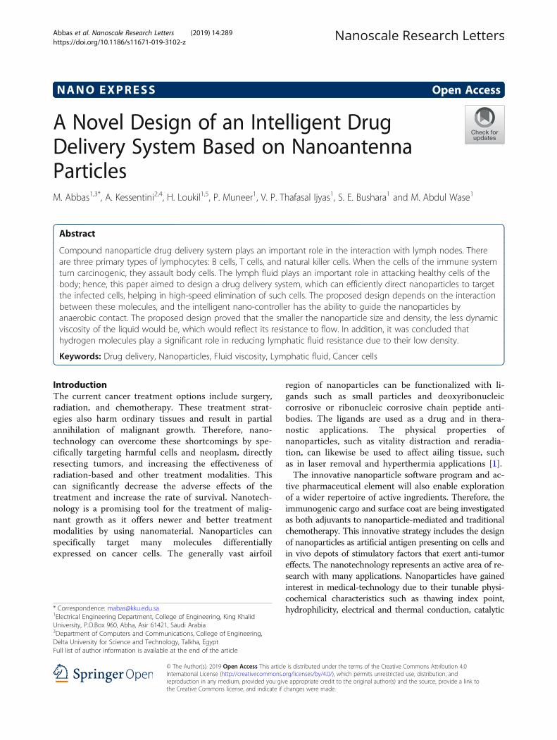
Design of a Nano-Intelligent Drug Delivery SystemThe proposed nano-intelligent drug delivery systemcontains a nano-controller operated by an electricalsource of nanoparticles made of a nano-piezoelectricmaterial. The complex repository of nanoparticles hasa number of micro-repositories. Each small repositorycontains one type of nanoparticles. A nanoparticle mol-ecule contains a nanoantenna designed to communicatewith the nano-controller. The proposed nano-intelligentdrug delivery system also contains carbon nanotubes for
Fig. 1 Compound nanoparticle drug delivery system and its interaction with lymph nodes
Abbas et al. Nanoscale Research Letters (2019) 14:289 Page 2 of 13
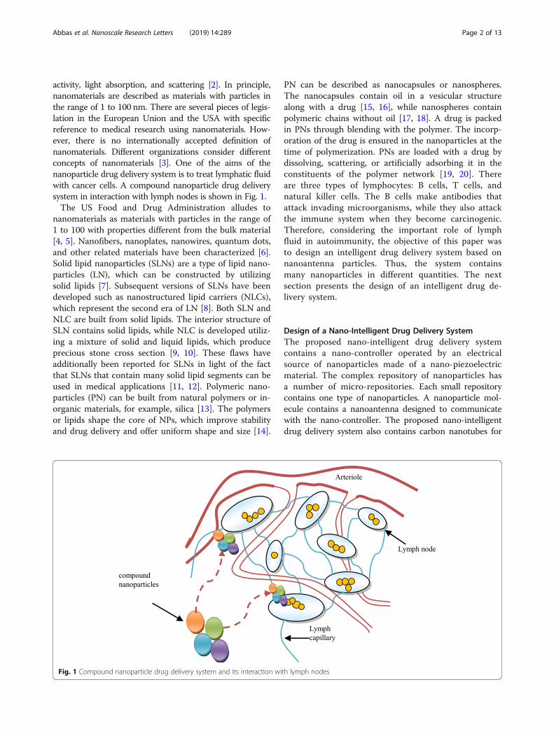
the rapid delivery of drugs to cancer cells. It can be associ-ated with the infected cells as shown in Fig. 2. The systembegins by sending nanoparticles to the cancer cells called“exploratory nanoparticles.” These molecules, by means ofanaerobic communication, send the complete picture oftheir position inside the cells to the nano-controller. Basedon the situation encountered by the exploratory nanopar-ticles, the nano-controller sends nanoparticles of differentnumber, type, and density to the cancer cells, based on theinformation collected from the exploratory nanoparticles.These nanoparticles are called “fighting nanoparticles.”This is not a random process but controlled by the
nano-controller considering several aspects and loga-rithms, which will ensure efficient and quick delivery of
the nanoparticles. In order to accurately and quicklydeliver the nanoparticles to the cancer cells, the com-pressive binary search algorithm will be employed [21].Further, nanoparticles will be delivered in different dens-ities so that the drug becomes more effective. Thesemethodologies and their modus operandi by employingthe nano-controller are illustrated in Fig. 3. The physicalstructure of the nano-controller is similar to that of thenanoparticles, but it is in the form of metal so that itcan gain electrical energy for a short period while work-ing. This metal contains a wireless antenna along with asmall memory that contains the operating codes with ananoparticle link between the nano-controller and thenanoparticle store. The nanoparticle repository contains
Fig. 2 General structure showing the association of the proposed drug system with the infected cells
Fig. 3 Sending process of the fighting nanoparticles to cancer cells
Abbas et al. Nanoscale Research Letters (2019) 14:289 Page 3 of 13

several different types of nanoparticles. The opening andclosing, as well as the duration of opening of the nano-gate, will be controlled to tailor the number of particlesto be delivered.
A Description of the Nature of the Nanoparticles Used inthe Proposed Drug SystemIn the next section, the nature of the nanoparticles uti-lized in the proposed drug delivery system is discussed.In this work, low-density anaerobic nanoparticles wereused as described in an earlier report [22].
Low-Density NanoparticlesConsider the drug delivery process of compoundnanoparticles for cancer as a penetrating process inlymph fluid, where a tumor is surrounded by thelymphatic fluid. The composition of the melanoma re-sembles the lymphatic fluid. The proposed analyticalmodel is based on a nanotube system consisting ofthree different types of nanoparticles. The nanoparti-cles are placed in a high-density lymphatic fluid. Wecan define specific nanoparticles of solid A in thespherical polar coordinates as A = (ra, ϑa, φa), wherera is the radial coordinate for the nanoparticle ofsolid A, ϑa is the zenithal coordinate for the nanopar-ticle of solid A, and φa is the azimuthal coordinatefor the nanoparticle of solid A. The correspondingcoordinates of solid B are B = (rb, ϑb, φb), respect-ively, and the corresponding coordinates of solid Nare N = (rn, ϑn, φn), respectively. Consider that thereare two properties of the lymph node, viz., tender andswollen, which are affected by the cancer cells ofHodgkin’s lymphoma. The lymph node with tenderproperty Tp can be described as Tp (N,t); this meansthat the value of Tp in association with the fluid ofnanoparticles of solid N varies with the time. Now,let us consider that the total effect of the compoundnanoparticles in the tender property is defined as:
Tpt ¼ Tp A; tð Þ þ Tp B; tð Þ þ :…………þ Tp N ; tð Þ ð1Þ
Consider the same case for the swollen property,which could be defined as:
Tst ¼ Ts A; tð Þ þ Ts B; tð Þ þ :…………þ Ts N ; tð Þ ð2Þ
From Eqs. 1 and 2, the rate of change of both proper-ties with time can be determined as:
∂ Tp A; tð Þð Þ∂t
þ ∂ Tp B; tð Þð Þ∂t
þ…∂ Tp N ; tð Þð Þ
∂t¼ ∂Tp tð Þ
∂tð3Þ
∂ Ts A; tð Þð Þ∂t
þ ∂ Ts B; tð Þð Þ∂t
þ…∂ Ts N ; tð Þð Þ
∂t¼ ∂Ts tð Þ
∂tð4Þ
The point in the lymph fluid that can be occupied byone nanoparticle of solid N is defined as:
Pon ¼ Po po; tð Þn ð5ÞLet us consider a nanoparticle of the solid N derivative
of the tender lymph fluid is defined as∂ðTpðN ;tÞÞpo
∂t , thenthe compound material derivative of the tender lymphfluid would be equal to:
∂ Tp A; tð Þð Þpo∂t
þ ∂ Tp B; tð Þð Þpo∂t
þ…∂ Tp N ; tð Þð Þpo
∂t¼ ∂Tp tð Þpo
∂tð6Þ
∂ Ts A; tð Þð Þpo∂t
þ ∂ Ts B; tð Þð Þpo∂t
þ…∂ Ts N ; tð Þð Þpo
∂t¼ ∂Ts tð Þpo
∂tð7Þ
The corresponding velocity components of the solid Nare taken as (vrn, vϑn, vφn). Then, the flow velocity of theparticles of solid N is represented using Navier-Stokesequations at dynamic viscosity dν of the lymph fluid,and p is the pressure and ρ is the density of lymph fluidas follows:
∂vrn∂t
þ vrn∂vrn∂rn
þ vϑnrn
∂vrn∂ϑn
þ vφnrn sinϑn
∂vrn∂φn
−v2ϑnrn
−v2φnrn
þ 1ρ∂p∂rn
−dν½ 1rn2
∂∂rn
rn2∂vrn∂rn
� �þ 1rn2sinϑn
∂∂ϑn
sinϑn∂vrn∂ϑn
� �
þ 1
rn2 sin2ϑn∂2vrn∂φn2
þ −2vrnrn2
−2
rn2 sinϑn∂ vϑnsinϑnð Þ
∂ϑn
−2
rn2sinϑn∂vφn∂φn
� ¼ 0
ð8Þ
The dynamic viscosity dν of the lymph fluid is calcu-lated as follows:
dν ¼
∂vrn∂t
þ vrn∂vrn∂rn
þ vϑn∂vrnrn ∂ϑn
þ vφnrn sinϑn
∂vrn∂φn
−v2ϑnrn
−v2φnrn
þ 1 ∂pρ∂rn
" #
½ 1rn2
∂∂rn
rn2∂vrn∂rn
� �þ 1rn2sinϑn
∂∂ϑn
sinϑn∂vrn∂ϑn
� �þ 1
rn2 sin2ϑn∂2vrn∂φn2
þ−2vrnrn2
−2
rn2 sinϑn∂ vϑnsinϑnð Þ
∂ϑn−
2rn2sinϑn
∂vφn∂φn
�
ð9Þ
The Navier-Stokes equations of solids A and B couldbe represented as Eqs. 8 and 9. Thus, Eq. 9 could be rep-resented as follows:
Abbas et al. Nanoscale Research Letters (2019) 14:289 Page 4 of 13
The particles are in nano-dimensions; thus, their radiiwould be very small, and for simplicity, Eq. 10 is representedas follows:
Equation 11 could be represented as follows:
There is a direct relationship between the radii of thenanoparticles and the lymph viscosity due to cancer. Iflymph becomes too static and viscous, it is unable tocarry out its function properly, which is circulating andcleaning up toxins and helping to fight cancer. If
nanoparticle size is smaller, the lymphatic cancer cellsare easy to kill. In order to describe the transport of thetotal quantity of the compound nanoparticles, we usethe continuity equation and assume the three nanoparti-cles of solids A, B, and N as follows:
dν ¼
∂vrn∂t
þ vrn∂vrn∂rn
þ vϑnrn
∂vrn∂ϑn
þ vφnrn sinϑn
∂vrn∂φn
−v2ϑnrn
−v2φnrn
þ 1ρ∂p∂rn
" #
1rn2
∂∂rn
rn2∂vrn∂rn
� �þ 1rn2sinϑn
∂∂ϑn
sinϑn∂vrn∂ϑn
� �þ 1
rn2 sin2ϑn∂2vrn∂φn2
−2vrnrn2
−2
rn2 sinϑn∂ vϑnsinϑnð Þ
∂ϑn−
2rn2sinϑn
∂vφn∂φn
� �
¼
∂vra∂t
þ vra∂vra∂ra
þ vϑara
∂vra∂ϑa
þ vφara sinϑa
∂vra∂φa
−v2ϑara
−v2φara
þ 1ρ∂p∂ra
" #
1ra2
∂∂ra
ra2∂vra∂ra
� �þ 1ra2sinϑa
∂∂ϑa
sinϑa∂vra∂ϑa
� �þ 1
ra2 sin2ϑa∂2vra∂φa2
−2vrara2
−2
ra2 sinϑa∂ vϑasinϑað Þ
∂ϑa−
2ra2sinϑa
∂vφa∂φa
� �
¼
∂vrb∂t
þ vrb∂vrb∂rb
þ vϑbrb
∂vrb∂ϑb
þ vφbrb sinϑb
∂vrb∂φb
−v2ϑbrb
−v2φbrb
þ 1ρ∂p∂rb
" #
1
rb2∂∂rb
rb2∂vrb∂rb
� �þ 1
rb2sinϑb
∂∂ϑb
sinϑb∂vrb∂ϑb
� �þ 1
rb2 sin2ϑb
∂2vrb∂φb2
−2vrbrb2
−2
rb2 sinϑb
∂ vϑbsinϑbð Þ∂ϑb
−2
rb2sinϑb
∂vφb∂φb
� �
ð10Þ
dν ¼ vϑnrn
∂vrn∂ϑn
þ vφnrn sinϑn
∂vrn∂φn
−v2ϑnrn
−v2φnrn
þ 1ρ∂p∂rn
" #=½ 1rn2
∂∂rn
rn2∂vrn∂rn
� �þ 1rn2sinϑn
∂∂ϑn
sinϑn∂vrn∂ϑn
� �þ 1
rn2 sin2ϑn∂2vrn∂φn2
−2vrnrn2
−2
rn2 sinϑn∂ vϑnsinϑnð Þ
∂ϑn−
2rn2sinϑn
∂vφn∂φn
�
¼ vϑara
∂vra∂ϑa
þ vφara sinϑa
∂vra∂φa
−v2ϑara
−v2φara
þ 1ρ∂p∂ra
" #=½ 1ra2
∂∂ra
ra2∂vra∂ra
� �þ 1ra2sinϑa
∂∂ϑa
sinϑa∂vra∂ϑa
� �þ 1
ra2 sin2ϑa∂2vra∂φa2
−2vrara2
−2
ra2 sinϑa∂ vϑasinϑað Þ
∂ϑa−
2ra2sinϑa
∂vφa∂φa
�
¼ vϑbrb
∂vrb∂ϑb
þ vφbrb sinϑb
∂vrb∂φb
−v2ϑbrb
−v2φbrb
þ 1ρ∂p∂rb
" #=
1
rb2∂∂rb
rb2∂vrb∂rb
� �þ 1
rb2sinϑb
∂∂ϑb
ðsinϑb ∂vrb∂ϑb
� �þ 1
rb2 sin2ϑb
∂2vrb∂φb2
−2vrbrb2
−2
rb2 sinϑb
∂ vϑbsinϑbð Þ∂ϑb
−2
rb2sinϑb
∂vφb∂φb
�
ð11Þ
dν ¼ rn vϑn∂vrn∂ϑn
þ vφnsinϑn
∂vrn∂φn
−v2ϑn−v2φn þ
rnρ
∂p∂rn
� �=
�∂∂rn
rn2∂vrn∂rn
� �þ 1sinϑn
∂∂ϑn
sinϑn∂vrn∂ϑn
� �þ 1
sin2ϑn∂2vrn∂φn2
−2vrn−2
sinϑn∂ vϑnsinϑnð Þ
∂ϑn−
2sinϑn
∂vφn∂φn
�
¼ ra vϑa∂vra∂ϑa
þ vφasinϑa
∂vra∂φa
−v2ϑa−v2φa þ
raρ∂p∂ra
� �=
∂∂ra
ra2∂vra∂ra
� �þ 1sinϑa
∂∂ϑa
sinϑa∂vra∂ϑa
� �þ 1
sin2ϑa∂2vra∂φa2
−2vra−2
sinϑa∂ vϑasinϑað Þ
∂ϑa−
2sinϑa
∂vφa∂φa
� �
¼ rb vϑb∂vrb∂ϑb
þ vφbsinϑb
∂vrb∂φb
−v2ϑb−v2φb þ
rbρ
∂p∂rb
� �=
∂∂rb
rb2∂vrb∂rb
� �þ 1sinϑb
∂∂ϑb
sinϑb∂vrb∂ϑb
� �þ 1
sin2ϑb∂2vrb∂φb2
−2vrb−2
sinϑb∂ vϑbsinϑbð Þ
∂ϑb−
2sinϑb
∂vφb∂φb
� �
ð12Þ
Abbas et al. Nanoscale Research Letters (2019) 14:289 Page 5 of 13

1ra2
∂∂ra
ra2vra� �þ 1
ra sinϑa∂∂ϑa
sinϑvϑað Þ þ 1ra sinϑa
∂vφa∂φa
þ 1
rb2∂∂rb
rb2vrb� �þ 1
rb sinϑb∂
∂ϑbsinϑvϑbð Þ
þ 1rb sinϑb
∂vφb∂φb
þ 1rn2
∂∂rn
rn2vrn� �þ 1
rn sinϑn∂
∂ϑnsinϑvϑnð Þ
þ 1rn sinϑn
∂vφn∂φn
¼ 0
ð13ÞThe dynamic fluid viscosity could be determined from
the following equation [23]:
Vs ¼ 29r2g ρp−ρfð Þ
dvð14Þ
where Vs is the particles’ settling velocity (m/s), r is theStokes radius of the particle (m), g is the gravitational accel-eration (m/s2), ρp is the density of the particles (kg/m3), ρfis the density of the fluid (kg/m3), and dv is the (dynamic)fluid viscosity (Pa·s). The lymph fluid is slightly heavier thanwater (lymph density = 1019 kg/m3, water density = 998.28kg/m3 at 20 °C). As a reference value, we consider the dy-namic viscosity of the water to be 1.002 × 10⁻³ kg m–1 s–1).Dynamic viscosity is the measurement of the fluid’s in-
ternal resistance to flow, while kinematic viscosity refers tothe ratio of dynamic viscosity to density. The effect of all thenanoparticles on the fluid viscosity is represented as follows:
dv ¼ 2g9
ra2 ρa−ρfð Þvsa
þ rb2 ρb−ρfð Þvsb
þ rn2 ρn−ρfð Þvn
� �ð15Þ
By comparing Eq. 12 and Eq. 15, the following equa-tion could be emerged:
2g9
ra2 ρa−ρfð Þvsa
þ rb2 ρb−ρfð Þvsb
þ rn2 ρn−ρfð Þvn
� ���������
¼ rn vϑn∂vrn∂ϑn
þ vφnsinϑn
∂vrn∂φn
−v2ϑn−v2φn þ
rnρf
∂p∂rn
� �
=
�∂∂rn
rn2∂vrn∂rn
� �þ 1sinϑn
∂∂ϑn
sinϑn∂vrn∂ϑn
� �
þ 1
sin2ϑn∂2vrn∂φn2
−2vrn−2
sinϑn∂ vϑnsinϑnð Þ
∂ϑn
−2
sinϑn∂vφn∂φn
�¼ ra
�vϑa
∂vra∂ϑa
þ vφasinϑa
∂vra∂φa
−v2ϑa−v2φa
þ raρf
∂p∂ra
�=
�∂∂ra
ra2∂vra∂ra
� �þ 1sinϑa
∂∂ϑa
sinϑa∂vra∂ϑa
� �
þ 1
sin2ϑa∂2vra∂φa2
−2vra−2
sinϑa∂ vϑasinϑað Þ
∂ϑa−
2sinϑa
∂vφa∂φa
�
¼ rb vϑb∂vrb∂ϑb
þ vφbsinϑb
∂vrb∂φb
−v2ϑb−v2φb þ
rbρf
∂p∂rb
� �
=
�∂∂rb
rb2∂vrb∂rb
� �þ 1sinϑb
∂∂ϑb
sinϑb∂vrb∂ϑb
� �
þ 1
sin2ϑb∂2vrb∂φb2
−2vrb−2
sinϑb∂ vϑbsinϑbð Þ
∂ϑb−
2sinϑb
∂vφb∂φb
�
ð16Þ
Equation 16 depicts the relationship between the dens-ity of the lymph fluid, the density of the nanoparticles ofthe compound drug system, and the radius of the nano-particles. There is a positive correlation between thedensity of the lymph fluid and the density of the nano-particles. The smaller the density and radius of nanopar-ticles, the lesser the density of the fluid will be. Asestablished earlier, the decrease in the density of thelymph fluid leads to its inability to reproduce and reducethe ferocity of the disease. It can, therefore, be con-cluded that the tumor can be cured by minimizing thesize of nanoparticles. These particles can reach in therange of up to 0.1 nm (i.e., Angstrom or picometerrange). The particles of this size can act as the nucleusof drug delivery in this drug system. Equation 16 showsthat radii of the nanoparticles in the proposed drug sys-tem are related to the effectiveness of the delivery sys-tem. Much lower sized nanoparticles can reduce thedensity of lymph fluid and the spread of the disease.
Nanoparticles with NanoantennasThis study used the nanoparticle described in an earlier re-port [22] as an emissary with a nano-microcontroller. Inthe system, the proposed transmission distance is verysmall and compatible with the composition of nanoparti-cles. Thus, the middle gap can be neglected in mid-dis-tance and is symbolized by Cd. Further, Ra and Xa are thereal part and the imaginary part of the anaerobic imped-ance. After neglecting the load of the intercellular spacebetween the nanoparticles and the nano-microcontroller,Ra and Xa can be calculated as follows [22]:
Ra ¼ ra01þ Cdwa 2xa0 þ Cd ra02 þ xa02ð Þwað Þ ð17Þ
Xa ¼ x0−Cd ra02 þ xa02ð Þwa
1þ Cdwa 2xa0 þ Cd ra02 þ xa02ð Þwað Þ ð18Þ
Thus, the load resistance value of nanotubes can bepredicted as in the following equation:
rl ¼ g2R
g2−2gSXεLωþ S2 R2 þ X2� �
εL2ω2ð19Þ
where εL is the permittivity of the loading material, g isthe size of the gap, and S is the effective cross-sectionarea of the gap. In order to simplify the equation, thevalue of g2 can be neglected as it is too low and the finalequation can be rewritten as follows:
rl ¼ g2R
−2gSXεLωþ S2 R2 þ X2� �
εL2ω2ð20Þ
Then,
Abbas et al. Nanoscale Research Letters (2019) 14:289 Page 6 of 13
rl ¼ g2R
SεLω −2gX þ S R2 þ X2� �
εLω� � ð21Þ
The optical nano-photo concept can be used as an ef-fective tool for interpreting and predicting these effectsto design and improve nanoscale parameters and in-crease the nano-sensitivity to serve better as a single mo-lecular sensor. Nanoantenna may provide optimalperformance in terms of sensitivity, efficiency, and band-width in the process. The next section presents the con-cept of searching the cancerous lymph nodes usingcompressive binary search algorithm.
Searching for the Target Lymphatic Nodes UsingCompressive Binary SearchIn order for nanoparticles to reach the cancerous cells in afast and efficient manner, we applied compressive binarysearch by the nano-microcontroller. The guided nanopar-ticles follow a specific path to quickly reach the target.This movement is based on the information obtainedfrom the “exploratory nanoparticles.” Assume that the tar-get lymph node, Tf, has exactly one nonzero entry, wherethe location of the lymph node is unknown. The algorithmdivides mt measurements into a total of St stages, whereSt refers to the stages of the lymph nodes. The measure-ments are more than one for all the stages of the lymphnodes, which is necessary for the algorithm to be executeduntil completion. Based on this measurement, the algo-rithm decides between going left or right, until the nano-particles reach the target, the cancerous lymph node.
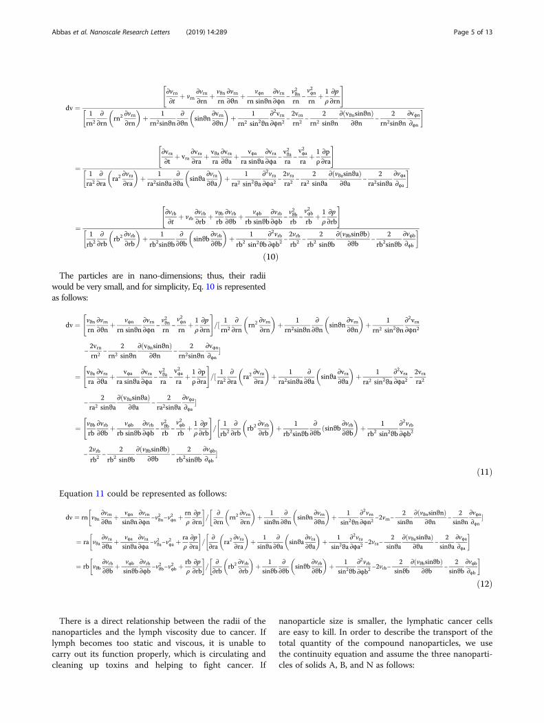
Results and DiscussionIn order to analyze the proposed design, the nanoparti-cles were applied to the following five types of mate-rials: silicone, lithium, lung, helium, and hydrogen. Thematerials were chosen because of their low density. Thelung nanoparticles were samples from nano-sized lungnodules. They appear encircling with white shadows ina chest X-ray or computerized tomography scan takenfrom the lung of the person and required to be undam-aged. The proposed idea is based on the analyticalmodel, which indicates that the smaller the density ofnanoparticles, the smaller the dynamic viscosity will be.This will result in a decrease in fluid viscosity. It isshown that the types of materials and the density ofeach particle will affect settling velocity of nanoparticlesat entry into the lymphatic fluid and the density of thelymphatic fluid. We considered the following parame-ters: acceleration of gravity (g) = 9.80665, particle
Fig. 4 Density of nanoparticles for the five selected materials
Abbas et al. Nanoscale Research Letters (2019) 14:289 Page 7 of 13
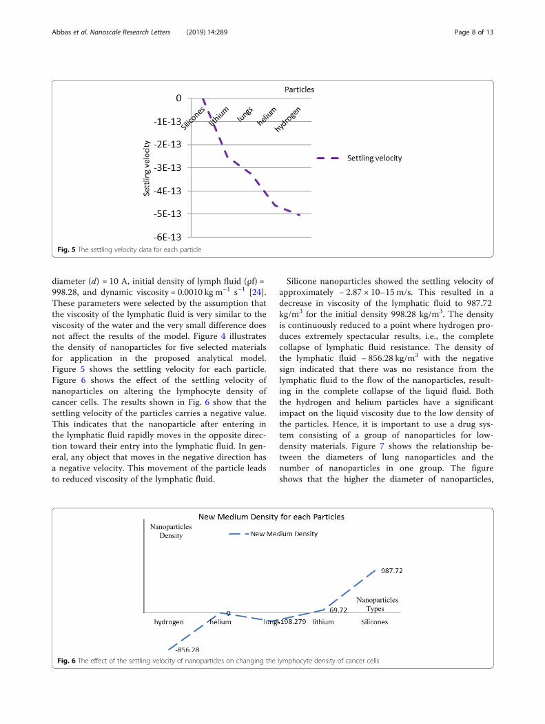
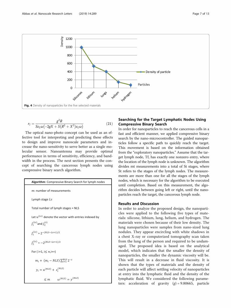
diameter (d) = 10 A, initial density of lymph fluid (ρf) =998.28, and dynamic viscosity = 0.0010 kg m–1 s–1 [24].These parameters were selected by the assumption thatthe viscosity of the lymphatic fluid is very similar to theviscosity of the water and the very small difference doesnot affect the results of the model. Figure 4 illustratesthe density of nanoparticles for five selected materialsfor application in the proposed analytical model.Figure 5 shows the settling velocity for each particle.Figure 6 shows the effect of the settling velocity ofnanoparticles on altering the lymphocyte density ofcancer cells. The results shown in Fig. 6 show that thesettling velocity of the particles carries a negative value.This indicates that the nanoparticle after entering inthe lymphatic fluid rapidly moves in the opposite direc-tion toward their entry into the lymphatic fluid. In gen-eral, any object that moves in the negative direction hasa negative velocity. This movement of the particle leadsto reduced viscosity of the lymphatic fluid.
Silicone nanoparticles showed the settling velocity ofapproximately − 2.87 × 10⁻¹⁵ m/s. This resulted in adecrease in viscosity of the lymphatic fluid to 987.72kg/m3 for the initial density 998.28 kg/m3. The densityis continuously reduced to a point where hydrogen pro-duces extremely spectacular results, i.e., the completecollapse of lymphatic fluid resistance. The density ofthe lymphatic fluid − 856.28 kg/m3 with the negativesign indicated that there was no resistance from thelymphatic fluid to the flow of the nanoparticles, result-ing in the complete collapse of the liquid fluid. Boththe hydrogen and helium particles have a significantimpact on the liquid viscosity due to the low density ofthe particles. Hence, it is important to use a drug sys-tem consisting of a group of nanoparticles for low-density materials. Figure 7 shows the relationship be-tween the diameters of lung nanoparticles and thenumber of nanoparticles in one group. The figureshows that the higher the diameter of nanoparticles,
Fig. 5 The settling velocity data for each particle
Fig. 6 The effect of the settling velocity of nanoparticles on changing the lymphocyte density of cancer cells
Abbas et al. Nanoscale Research Letters (2019) 14:289 Page 8 of 13
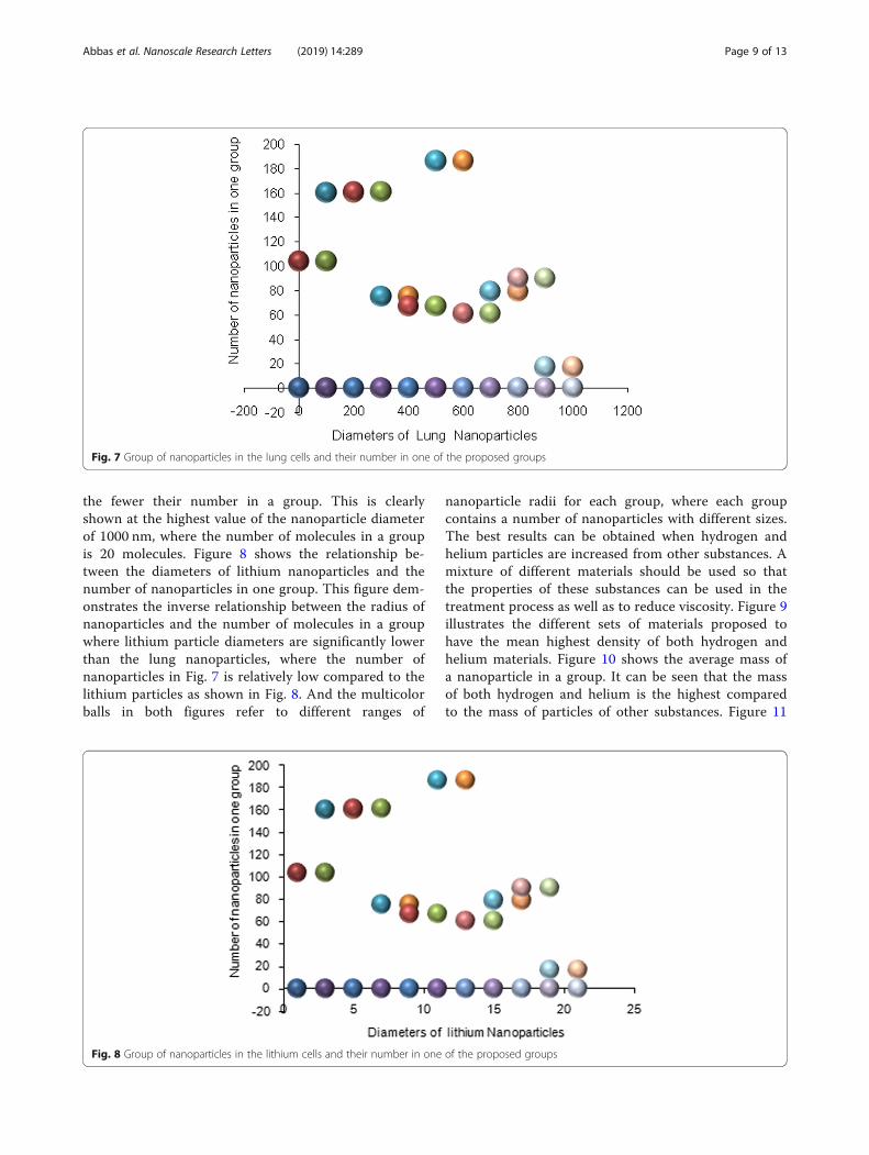
the fewer their number in a group. This is clearlyshown at the highest value of the nanoparticle diameterof 1000 nm, where the number of molecules in a groupis 20 molecules. Figure 8 shows the relationship be-tween the diameters of lithium nanoparticles and thenumber of nanoparticles in one group. This figure dem-onstrates the inverse relationship between the radius ofnanoparticles and the number of molecules in a groupwhere lithium particle diameters are significantly lowerthan the lung nanoparticles, where the number ofnanoparticles in Fig. 7 is relatively low compared to thelithium particles as shown in Fig. 8. And the multicolorballs in both figures refer to different ranges of
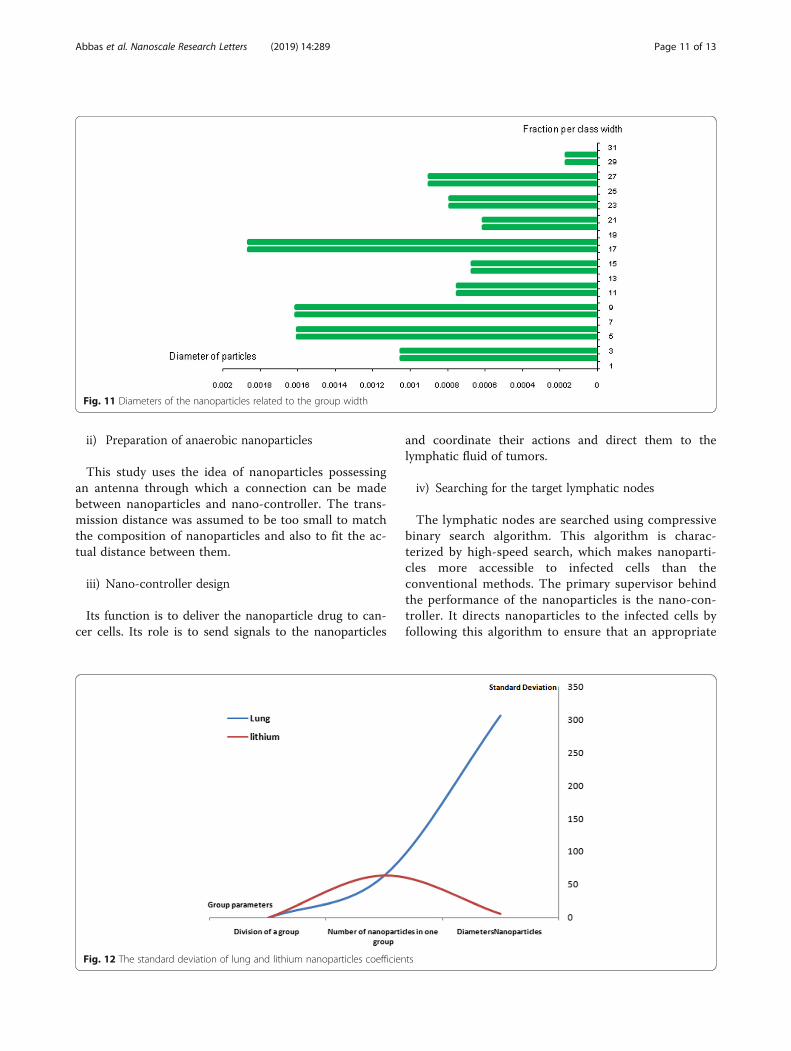
nanoparticle radii for each group, where each groupcontains a number of nanoparticles with different sizes.The best results can be obtained when hydrogen andhelium particles are increased from other substances. Amixture of different materials should be used so thatthe properties of these substances can be used in thetreatment process as well as to reduce viscosity. Figure 9illustrates the different sets of materials proposed tohave the mean highest density of both hydrogen andhelium materials. Figure 10 shows the average mass ofa nanoparticle in a group. It can be seen that the massof both hydrogen and helium is the highest comparedto the mass of particles of other substances. Figure 11
Fig. 7 Group of nanoparticles in the lung cells and their number in one of the proposed groups
Fig. 8 Group of nanoparticles in the lithium cells and their number in one of the proposed groups
Abbas et al. Nanoscale Research Letters (2019) 14:289 Page 9 of 13
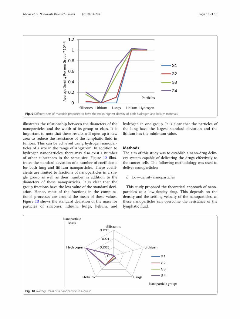
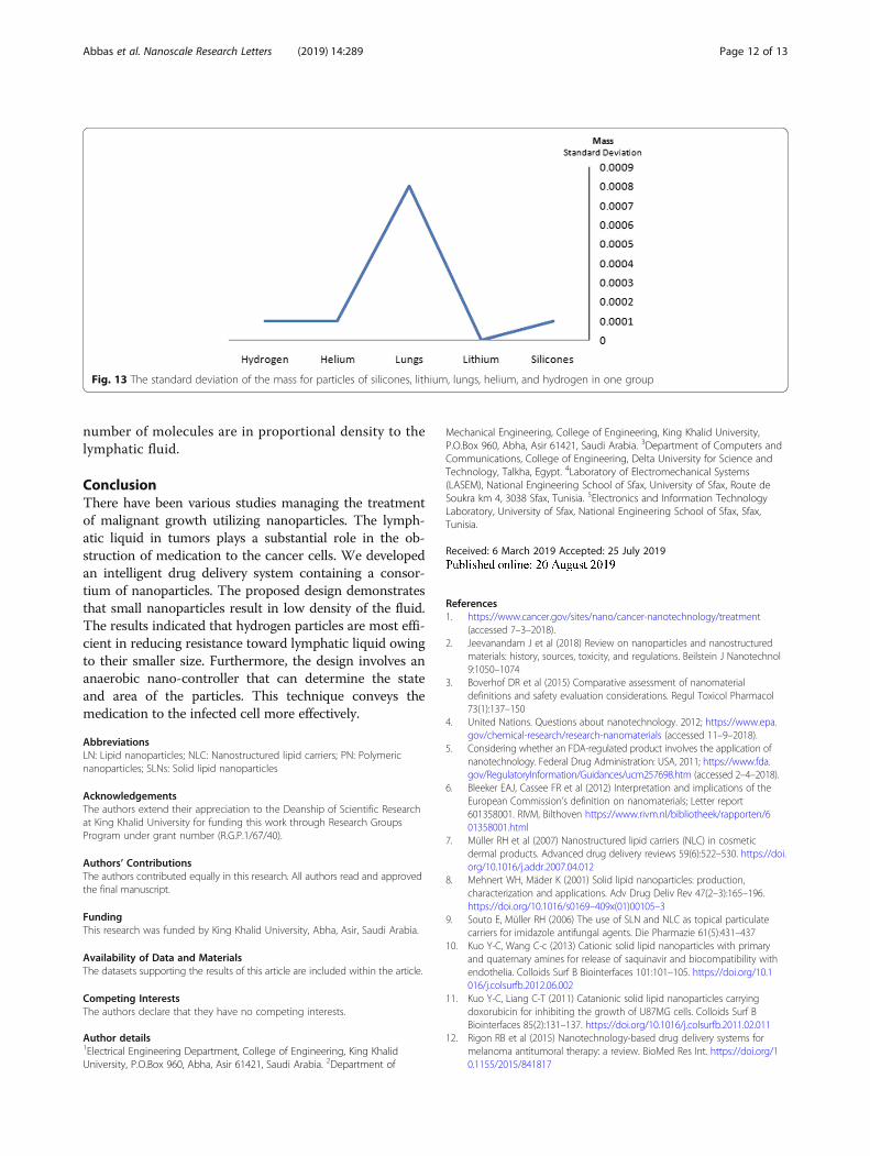
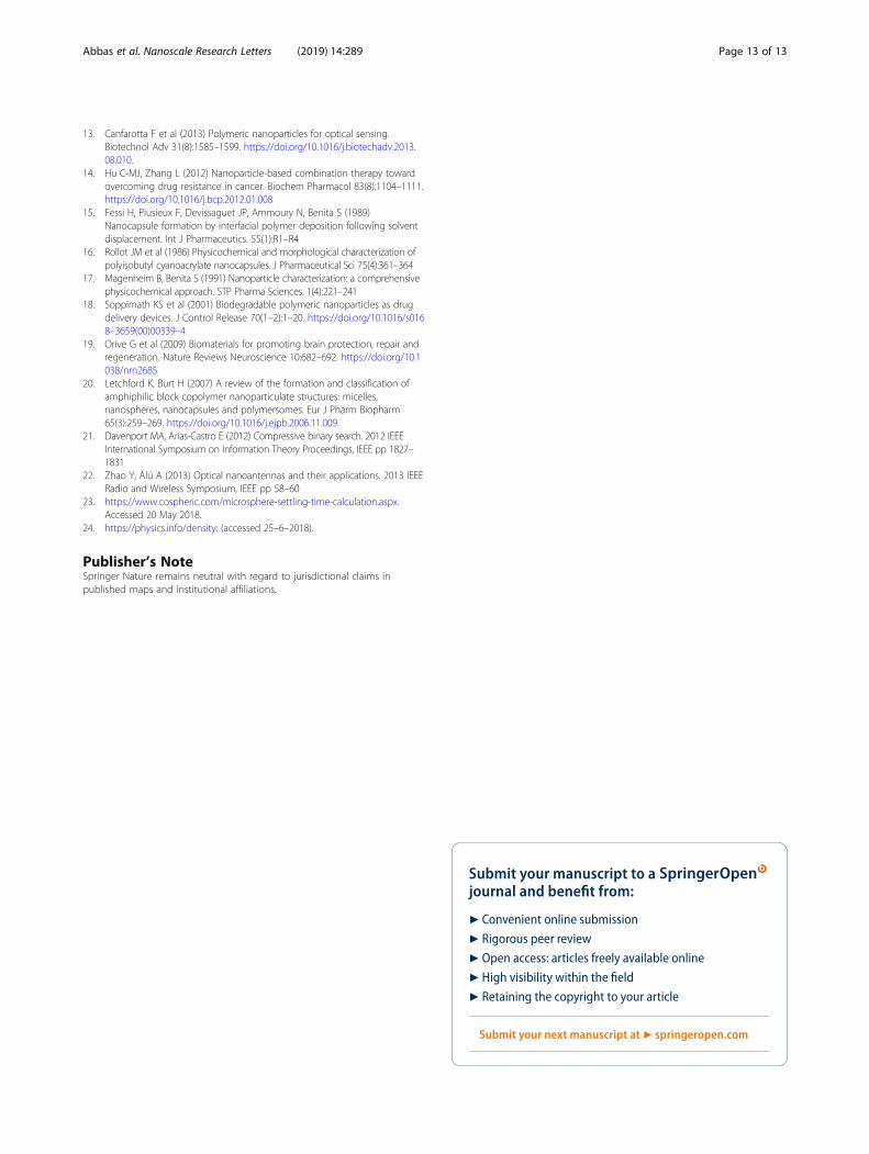
illustrates the relationship between the diameters of thenanoparticles and the width of its group or class. It isimportant to note that these results will open up a newarea to reduce the resistance of the lymphatic fluid intumors. This can be achieved using hydrogen nanopar-ticles of a size in the range of Angstrom. In addition tohydrogen nanoparticles, there may also exist a numberof other substances in the same size. Figure 12 illus-trates the standard deviation of a number of coefficientsfor both lung and lithium nanoparticles. These coeffi-cients are limited to fractions of nanoparticles in a sin-gle group as well as their number in addition to thediameters of these nanoparticles. It is clear that thegroup fractions have the less value of the standard devi-ation. Hence, most of the fractions in the computa-tional processes are around the mean of these values.Figure 13 shows the standard deviation of the mass forparticles of silicones, lithium, lungs, helium, and
hydrogen in one group. It is clear that the particles ofthe lung have the largest standard deviation and thelithium has the minimum value.
MethodsThe aim of this study was to establish a nano-drug deliv-ery system capable of delivering the drugs effectively tothe cancer cells. The following methodology was used todeliver nanoparticles:
i) Low-density nanoparticles
This study proposed the theoretical approach of nano-particles as a low-density drug. This depends on thedensity and the settling velocity of the nanoparticles, asthese nanoparticles can overcome the resistance of thelymphatic fluid.
Fig. 9 Different sets of materials proposed to have the mean highest density of both hydrogen and helium materials
Fig. 10 Average mass of a nanoparticle in a group
Abbas et al. Nanoscale Research Letters (2019) 14:289 Page 10 of 13
ii) Preparation of anaerobic nanoparticles
This study uses the idea of nanoparticles possessingan antenna through which a connection can be madebetween nanoparticles and nano-controller. The trans-mission distance was assumed to be too small to matchthe composition of nanoparticles and also to fit the ac-tual distance between them.
iii) Nano-controller design
Its function is to deliver the nanoparticle drug to can-cer cells. Its role is to send signals to the nanoparticles
and coordinate their actions and direct them to thelymphatic fluid of tumors.
iv) Searching for the target lymphatic nodes
The lymphatic nodes are searched using compressivebinary search algorithm. This algorithm is charac-terized by high-speed search, which makes nanoparti-cles more accessible to infected cells than theconventional methods. The primary supervisor behindthe performance of the nanoparticles is the nano-con-troller. It directs nanoparticles to the infected cells byfollowing this algorithm to ensure that an appropriate
Fig. 11 Diameters of the nanoparticles related to the group width
Fig. 12 The standard deviation of lung and lithium nanoparticles coefficients
Abbas et al. Nanoscale Research Letters (2019) 14:289 Page 11 of 13
number of molecules are in proportional density to thelymphatic fluid.
ConclusionThere have been various studies managing the treatmentof malignant growth utilizing nanoparticles. The lymph-atic liquid in tumors plays a substantial role in the ob-struction of medication to the cancer cells. We developedan intelligent drug delivery system containing a consor-tium of nanoparticles. The proposed design demonstratesthat small nanoparticles result in low density of the fluid.The results indicated that hydrogen particles are most effi-cient in reducing resistance toward lymphatic liquid owingto their smaller size. Furthermore, the design involves ananaerobic nano-controller that can determine the stateand area of the particles. This technique conveys themedication to the infected cell more effectively.
AbbreviationsLN: Lipid nanoparticles; NLC: Nanostructured lipid carriers; PN: Polymericnanoparticles; SLNs: Solid lipid nanoparticles
AcknowledgementsThe authors extend their appreciation to the Deanship of Scientific Researchat King Khalid University for funding this work through Research GroupsProgram under grant number (R.G.P.1/67/40).
Authors’ ContributionsThe authors contributed equally in this research. All authors read and approvedthe final manuscript.
FundingThis research was funded by King Khalid University, Abha, Asir, Saudi Arabia.
Availability of Data and MaterialsThe datasets supporting the results of this article are included within the article.
Competing InterestsThe authors declare that they have no competing interests.
Author details1Electrical Engineering Department, College of Engineering, King KhalidUniversity, P.O.Box 960, Abha, Asir 61421, Saudi Arabia. 2Department of
Mechanical Engineering, College of Engineering, King Khalid University,P.O.Box 960, Abha, Asir 61421, Saudi Arabia. 3Department of Computers andCommunications, College of Engineering, Delta University for Science andTechnology, Talkha, Egypt. 4Laboratory of Electromechanical Systems(LASEM), National Engineering School of Sfax, University of Sfax, Route deSoukra km 4, 3038 Sfax, Tunisia. 5Electronics and Information TechnologyLaboratory, University of Sfax, National Engineering School of Sfax, Sfax,Tunisia.
Received: 6 March 2019 Accepted: 25 July 2019
References1. https://www.cancer.gov/sites/nano/cancer-nanotechnology/treatment
(accessed 7–3–2018).2. Jeevanandam J et al (2018) Review on nanoparticles and nanostructured
materials: history, sources, toxicity, and regulations. Beilstein J Nanotechnol9:1050–1074
3. Boverhof DR et al (2015) Comparative assessment of nanomaterialdefinitions and safety evaluation considerations. Regul Toxicol Pharmacol73(1):137–150
4. United Nations. Questions about nanotechnology. 2012; https://www.epa.gov/chemical-research/research-nanomaterials (accessed 11–9–2018).
5. Considering whether an FDA-regulated product involves the application ofnanotechnology. Federal Drug Administration: USA, 2011; https://www.fda.gov/RegulatoryInformation/Guidances/ucm257698.htm (accessed 2–4–2018).
6. Bleeker EAJ, Cassee FR et al (2012) Interpretation and implications of theEuropean Commission’s definition on nanomaterials; Letter report601358001. RIVM, Bilthoven https://www.rivm.nl/bibliotheek/rapporten/601358001.html
7. Müller RH et al (2007) Nanostructured lipid carriers (NLC) in cosmeticdermal products. Advanced drug delivery reviews 59(6):522–530. https://doi.org/10.1016/j.addr.2007.04.012
8. Mehnert WH, Mäder K (2001) Solid lipid nanoparticles: production,characterization and applications. Adv Drug Deliv Rev 47(2–3):165–196.https://doi.org/10.1016/s0169–409x(01)00105–3
9. Souto E, Müller RH (2006) The use of SLN and NLC as topical particulatecarriers for imidazole antifungal agents. Die Pharmazie 61(5):431–437
10. Kuo Y-C, Wang C-c (2013) Cationic solid lipid nanoparticles with primaryand quaternary amines for release of saquinavir and biocompatibility withendothelia. Colloids Surf B Biointerfaces 101:101–105. https://doi.org/10.1016/j.colsurfb.2012.06.002
11. Kuo Y-C, Liang C-T (2011) Catanionic solid lipid nanoparticles carryingdoxorubicin for inhibiting the growth of U87MG cells. Colloids Surf BBiointerfaces 85(2):131–137. https://doi.org/10.1016/j.colsurfb.2011.02.011
12. Rigon RB et al (2015) Nanotechnology-based drug delivery systems formelanoma antitumoral therapy: a review. BioMed Res Int. https://doi.org/10.1155/2015/841817
Fig. 13 The standard deviation of the mass for particles of silicones, lithium, lungs, helium, and hydrogen in one group
Abbas et al. Nanoscale Research Letters (2019) 14:289 Page 12 of 13
13. Canfarotta F et al (2013) Polymeric nanoparticles for optical sensing.Biotechnol Adv 31(8):1585–1599. https://doi.org/10.1016/j.biotechadv.2013.08.010.
14. Hu C-MJ, Zhang L (2012) Nanoparticle-based combination therapy towardovercoming drug resistance in cancer. Biochem Pharmacol 83(8):1104–1111.https://doi.org/10.1016/j.bcp.2012.01.008
15. Fessi H, Piusieux F, Devissaguet JP, Ammoury N, Benita S (1989)Nanocapsule formation by interfacial polymer deposition following solventdisplacement. Int J Pharmaceutics. 55(1):R1–R4
16. Rollot JM et al (1986) Physicochemical and morphological characterization ofpolyisobutyl cyanoacrylate nanocapsules. J Pharmaceutical Sci 75(4):361–364
17. Magenheim B, Benita S (1991) Nanoparticle characterization: a comprehensivephysicochemical approach. STP Pharma Sciences. 1(4):221–241
18. Soppimath KS et al (2001) Biodegradable polymeric nanoparticles as drugdelivery devices. J Control Release 70(1–2):1–20. https://doi.org/10.1016/s0168–3659(00)00339–4
19. Orive G et al (2009) Biomaterials for promoting brain protection, repair andregeneration. Nature Reviews Neuroscience 10:682–692. https://doi.org/10.1038/nrn2685
20. Letchford K, Burt H (2007) A review of the formation and classification ofamphiphilic block copolymer nanoparticulate structures: micelles,nanospheres, nanocapsules and polymersomes. Eur J Pharm Biopharm65(3):259–269. https://doi.org/10.1016/j.ejpb.2006.11.009.
21. Davenport MA, Arias-Castro E (2012) Compressive binary search. 2012 IEEEInternational Symposium on Information Theory Proceedings, IEEE pp 1827–1831
22. Zhao Y, Alú A (2013) Optical nanoantennas and their applications. 2013 IEEERadio and Wireless Symposium, IEEE pp 58–60
23. https://www.cospheric.com/microsphere-settling-time-calculation.aspx.Accessed 20 May 2018.
24. https://physics.info/density: (accessed 25–6–2018).
Publisher’s NoteSpringer Nature remains neutral with regard to jurisdictional claims inpublished maps and institutional affiliations.
Abbas et al. Nanoscale Research Letters (2019) 14:289 Page 13 of 13


![Intelligent drug delivery system - pgsitecdn.persiangig.com/dl/9MZwnq/student Intelligent drug delivery syste… · Table 2. Marketed technologies of pulsatile drug delivery [31]](https://img.pdfslide.us/doc/110x75/5f3dc762b8577c0d041fed9b/intelligent-drug-delivery-system-intelligent-drug-delivery-syste-table-2-marketed.jpg)